Note: Descriptions are shown in the official language in which they were submitted.
2 ~ 3 3 1 7
' W092/06367 ~ PCT/FI91/00304
IMAGING METHOD FOR ~EFINING THE STRUCTURE OF OBJECTS
The pre~ent inventlon relates to an imaging method for de-
fining the structure of objects, in which method the object
under inspection and at least one radiation source are
moved, with respect to each other, so that the object trav-
els through the rays emitted from the radiation source.
For about a hundred years, X-ray technique has been utilized
and developed in order to reveal the inner structure of
objects. In X-ray photography, the details in the inner
structure of the object under examination are seen as su-
perimposed absorption differences of X-ray radiation. If
the object in question contains several elements with the
same absorption coeffici~ -, as is the case for instance
with human internal orgar.. in tne area of the abdomen, the
image will be difficult to interpret, and a specialist is
requirad for analy~ing its structure. X-ray technique has
developed various methods, for example opaque matter pho-
tography, where the desired object is seen better. The most
advanced method nowadays is X-ray tomography. Tomography
renders a cross-sectional image seen from the direction of
inspection, so that different elements are seen in the image
separately and in relatively right places. When a desired
number of cross-sectional images is made in succession in
the lengthwise direction, an accurate three-dimensional
image of the target is obtained. ~ther types of radiation
have also been used for creating tnree-dimensional images of
the target; the best known method at present is ultrasonic
scanning. In the future, imaging will probably be carried
out by using infrared radiation, too. The current devices
are complicated, expensive and-slow. For instance the to-
mographs required in the inspection of the human body suc-
ceed in shooting one image wlthin several seconds.
Currently images of the inte-nal structures of objects unde_
inspection are required automa~icall~ and rapidly. In
2~3~
W092/06367 PCT/F191/00304
these cases, a r~solution of even less than one millimeter
is not necessary, contrary to the case of an X-ray tomograph
designed for the inspection of the human body. A typical
application of this type is the scanning of various objects,
pieces and materials in order to detect undeslrable objects,
pieces and/or materials, as well as for definlng the inter-
nal structure of these materials and objects. In some cases
it suffices to define the three-dimensional shape of the
object, and the inspection of the internal structure is not
necessary.
The object of the invention is to introduce an lmaging
method for defining the structure of objects, by means of
which method there are constructed adequately sharp three-
dimensional images for various applications, rapidly and by
using equipment which is remarkably simpler and more eco-
nomical than the equipment used in current tomography tech-
nique.
The object of the invention is achieved by means of a method
which is characterized by the novel features enlisted in the
appended patent claims.
In the method of the invention, at least one detector is
used for measuring, from at least three different angles,
the changes in the intensity of the rays that are absorbed,
at least partly, in the object under inspection; these
changes are measured as the function of the motion of the
said object. Thus the structural points required for imag-
ing the structure are defined at least from three different
directions, and as the passage of the rays through the ob-
ject is always known at the moment of measurement, the
three-dimensional structure of the object can be calculated.
The greatest advantage of the invention lies in that for
instance in X-ray tomography, the movlng of the X-ray tube,
or several tubes, can be replaced by one stationary X-ray
tube, and the several hundreds o radiation detectors can be
2~93~
W092/06367 P~T/FI91/00304
replaced even by one detector only. The equipment is slmple
and economical as for its production and maintenance costs.
Respectively, if the only requirement for the equipment is
that it defines the external shape of the object, the em-
ployed radiation source can be a light emitting diode or the
like, and the detector can be a light-sensitive transistor,
in which case the transmitter/receiver pair needed in imag-
ing is very simple and not expensive.
~he method of the invention can be applied and used for
automatically and rapidly scanning the internal structure of
various items. Possible appllcations are for instance the
detection of dangerous materials inside the objects under
inspection, e.g. stones and other objects attached to logs
when lumber is being sawed or cut to chips. The method also
defines the location of defects and knots in the lumber to
be sawn, in order to optimize the sawing process. Moreover,
the method can be applied to various other purposes, such as
for searching explosives or weapons in the suitcases of air
travellers. Scanning rates with the method of the invention
are high, about 100 - 500 images per second, and the ob-
tained resolution is sufficient for these purposes. In
addition to this, the method can be used in the definition
of the three-dimensional shape of various objects. As an
example, let us mention the definition of the size and shape
of pieces cut out of a given material - e.g. the definition
of the size and shape of chips in the pulp industry, and the
size and shape of crushed aggregate in the mining industry.
In the following the invention is explained in more detail
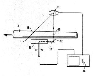
with reference to the appended drawing, where
figure lA is a side-view illustration of an embodiment of
the method of the invention,
figure lB is a top-view illustration of the embodiment of
figure lA,
figure lC illustrates the various images, obtained from
different directions, of one element in the
W092/06367 2 0 9 3 3 '~ 7 PCT/F191/00304
embodiment of figure lB,
figures 2A and 2s illustrate another embodiment of the
method of the invention, seen from the side and
from the top,
figure 3 is a side-view illustration of a third embodiment
of the method of the invention, and
figures 4A and 4B illustrate a fourth embodiment of the
method of the invention, seen from the side and
from the top.
The principle of an imaging method according to the inven-
tion is illustrated in figures lA, lB and lC, where the
imaging of the internal structure of a log is shown as an
example. The radiation source is an X-ray tube transmitting
X-ray radiation at a wide scanning angle. The log to be
inspected is passed through the conical X-ray beam. Radia-
tion is measured by means of linescan detectors 4, 5, 6, 7
and 8. The detector lines proceed transversally underneath
the log. A linescan detector may be composed of several
(e.g. lO0 - 500) parallel, separate X-ray detectors, or of
one long-detector, which is sensitive to location, i.e. both
detects and defines the location of the de~ected point in
relation to its length. Figure lC shows the images of one
element obtained from different angles, when the X-ray tube
has relatively passed over the log during scanning, and the
numbers 4, 5, 6, 7 and 8 below refer to the respective line
detectors which were used in scanning these directions. The
said elements may be for instance 0.5 cm in diameter. The
densities of wood elements are defined on the principle that
each element is imaged from dif,erent angles, and their
su~ned absorptions are always obtained from the respective
detector of the linescan detector, along with the time when
the log passes through scanning. In principle, at least 2
detector lines are always needed for scanning; the number in
the example is 5. The number of detector lines may vary in
different applications, and a larger number of detectors
generally produces a sharper image. The images are
W092/06367 ~0~ 3 317 PCT/FI91/003~4
computed, like in normal tomography, as cross-sectional
images of a given plane, for instance of the cross-sectional
image 1-6. When all cross-sectional images are processed,
we obtain a three-dimensional density image of the log in
question, with indication to knots, rot, cracks and often
height distribution, too. In addition, the apparatus can be
provided with a profile reader for defining the external
shape of the log. This is a useful aid in calculation,
when constructing sawing images. All elements of the log
are imaged from several directions, and therefore the loca-
tion of knots in the sawn goods produced of the log can also
be directly defined.
In the embodiments of figures 2 4, the radiation source
and the detector form a pair, one of which remains still and
the other moves along a circular path, so that the radiation
surface under measurement obtains a conical form. The ob-
ject under inspection passes through this conical surface,
in which case the image is normally constructed by~means of
a computer. In practical applications, it is possible to
use either several detectors or several radiation sources.
Different modifications can naturally be combined; for ex-
ample perpendicular imaging is carried out by means of a
stationary detector line, and inclined images from both
sides are obtained by using a rotating plate.
In the description below, we give -hree detailed examples of
the use of the method:
Example 1.
Figure 2 illustrates a log tomograph comprising a radiation
source 11 (X-ray machine) transmitting continuous X-ray
radiation, and a rotating plate 17 provided with a radiation
detector 12 for measurlng the amount of X-ray radiation at
any given moment. The object, i.e. log 13, under inspec-
tion, is conducted, for example on a conveyor belt, at an
even velocity through the tomograph. When the log enters
2~33 ~7 ~
W092~06367 P~T/FI~1/00304
the conical X-ray beam, the absorption curve of the cross-
section is measured on the spot 15, and a cross-sectional
density pro~ile is obtained. When the log passes through
the second surface 16 of the x-ray beam, lateral density
profiles are obtained. From these cross-images, the density
distributions in the internal structure of the log can be
defined, and on the basis of the said distributions, the
location of knots, cracks and rot in the log can be calcu-
lated, as was explained in the specification above. In
practice, the imaging rate can be for instance 2 m/s, and if
the sections are made at 1 cm intervals, the imaging rate
is 200 sections per second. If two X-ray detectors are
arranged on the circular path (with a phase difference of
90), the speed of rotation is 6000 rpm, which is achieved
with conventional technique. The protective screens seen in
figure 2 are provided in order to prevent the detector from
being incessantly subjected to radiation. The system can
incorporate onè or several datectors, and the data is read
along a line onto a computer 14, so that only one detector
at a time is exposed to radiation. An optional laser pro-
filator, for instance, can be provided in the apparatus in
order to define the cross-sectional contour image of the log
prior to inspecting the internal structure. This inrorma-
tion helps create the cross-sectional image more rapidly and
accurately.
Example 2.
Figure 3 illustrates a stone detector. The detection of
stone for instance in peat to be burned as fuel, or in a
flow of lumber to be chipped, ls an important task, but a
fully satisfactory method has ea~lier been lacking. Because
the resolution needed here is nol nearly as high as in the
previous case, the radiation source 21 is a radio isotope
source (e.g. 100 mCi Am-241). The detector 22 can now be
installed in a stationary fasnicn to measure tne radiation
intensity. Density differences -n the conveyed material 23
are defined from the absorptions on the perpendicular cone
W092/06367 2 ~ 9 3 ~ '1 7 pCT/FI91/00304
surface 25 and on the inc ned cone surface 26 by means of
the computer 24.
Example 3.
Figure 4 illustrates the definition of the size of chip
particles. ~n interesting object of measurement in the pulp
industry are the dimensions of chip particles: their length,
width and height distribution. Because it is not necessary
to observe the internal structure, an ordinary light suf-
fices as the radiation source. In the measuring arrangement
of figure 4, the employed sources are light emitting diodes
30, and the detectors are two photomultiplier tubes 31 and
32. The light sources, 20 or more in number, are attached
to a rotating plate 36, which in this case is rotated at the
speed of about 10,000 rpm. On a transparent conveyor belt
35, the chips 33 are scattered so that they do not fall on
top of each other. The belt is conveyed at the pace of for
example 1 m/s, so that each light source successively pro-
ceeds above the imaging aperture 37, underneath the trans-
parent conveyor belt 35. If the number of light sources is
20, and the speed is 10,000 rpm, the belt is scanned at the
intervals of 1/3 of a second. On the basis of the black-out
time of the photomultiplier located vertically above, the
width of the chip is obtained at the scanning point in
question. From the appearance time difference of the edge,
which is detected by means of the photomultiplier tube 32
looking from an inclined direction, the height of the chip
at the respective scanning point is obtained. On the basis
of these readings, the length, width and height of the chip
are calculated in a computer.
Other types of variation can also be used in the imaging
method, and the velocity of the objects of inspection, as
well as the imaging rate, can vary in different applica-
tions. In the preferred embodiments described above, the
objects of inspection are moved with respect to the radla-
tion source and the detector, b_t in other embodiments the
.
20'~33ll7
W092/06367 PCT/F191/00304
radiation source and/or the detector can be moved, while the
object under inspection remains in place. In addition to
stationary detector arrays, one or several rotatable plates
or platforms can be used. In another preferred embodiment,
on a rotatable platform there are located radiation detec-
tors for different radiation intensities, in which case more
than two inclined sections can be measured.
When using the method of the invention, the employed radia-
tion can be visible light, X-ray radiation, radar, gamma,
neutron, infrared or microwave radiation, pulsated magnetic
field or other such radiation or combinations thereo~, which
radiation or radiations interact with the object under in-
spection. An interesting branch of tomography is neutron
tomography, where a large, rotatable plate is provided with
a neutron source, and above it there is placed a neutron
detector. If the dimensions are 3 - 5 meters, this method
can be used for scanning vehicles, containers and the like
when searching possible organic materials, such as explo-
sives or drugs.
The invention is not restricted to the preferred embodiments
above, but it may vary within the scope of the inventional
idea specified in the appended patent claims.
