Note: Descriptions are shown in the official language in which they were submitted.
2093415
WO 92/11537 PCT/EP91/02519
- 1 -
TEST METHOD AND REAGENT KIT THEREFOR
This invention :relates to a method for the
.qualitative or quantitative determination of the
presence of an analyte in an aqueous medium.
The detection and/or assay of analytes using
immunoassay techniques is well established, particularly
.in relation to proteins such as antigens and antibodies,
as well as sugars, lectins and nucleic acids. However,
many current techniques, while being of great
sensitivity, are often laborious in requiring a number
of steps each of which may be of long duration. It has
proved possible to simplify some of such assays,
however, by immobilising one of the components of the
assay system on a solid support, since this facilitates
removal of excess reagents. Such assays will normally
involve the use of a labelled macromolecule, which may
be the analyte itself or a binding partner for the
analyte, carrying a suitable label such as a
radioisotope, a fluorophore or an enzyme producing a
characteristic reaction.
One simplification which has been proposed is to
use a coloured substance attached to one of the
immunoassay reactants as a visible marker. However, very
few coloured substances are able to produce a
sufficiently intense signal. US 4313734 of Akzona Inc.
describes the use of, inter alia, colloidal gold as such
a coloured material, specifying that the gold particles
should have a particle size of at least 5 nanometres,
preferably 10 to 1r~0 rm.
An improved immunoassay system is described in
WO89/06801 in which at least 75% of the gold particles
have a mean diameter of less than 5 nanometres. This is
CA 02093415 2002-02-15
20208-1507
-2-
said to give more rapid reaction of the gold reagent with the
immobilised reactant together with an increase in colour
intensity. We have now found that a yet further increase in
colour intensity may be obtained when the very small gold
particles of W089/06801 are formed into larger particles
(superaggregated gold-protein colloids) by a novel aggregation
process. The superaggregated particles are very different from
the monolithic gold particles used in immunoassays to date and
allow for analysis of substances at even lower concentrations
than the already impressively low levels made possible by the
systems of W089/06801.
Small gold particles are also used as markers in a
blotting system, as described in US 4775636 of Janssen
Pharmaceutica N.V. However, there is no suggestion that the
particles are aggregated, rather they are simply bound to the
component which it is desired to visualise.
According to the present invention we provide a
method for the qualitative or quantitative determination of an
analyte in a test sample wherein a labelled reagent comprising
a gold sol bound to a substance capable of specifically binding
to said analyte or to a specific binding partner therefor, is
caused to be immobilised in bound form on a solid phase to
provide an indication of the presence or quantity of the
analyte in the sample, characterized in that the labelled
reagent comprises a superaggregated complex of said substance
or specific binding partner therefor and a gold sol wherein at
least 75% by weight of the gold particles of the gold sol have
a mean diameter of less than 20 nanometres.
According to one aspect of the present invention,
there is provided a method for the qualitative or quantitative
determination of an analyte in a test sample comprising
immobilizing a labelled reagent on a solid phase
CA 02093415 2001-08-27
20208-1507
-2a-
to provide an indication of the presence or quantity of the
analyte in the sample, the labelled reagent comprising a gold
sol bound to a substance capable of specifically binding to
said analyte, or bound to a specific binding partner for said
substance, and wherein the labelled reagent comprises a
superaggregated complex of said substance or specific binding
partner therefor and the gold sol wherein at least 75% by
weight of the gold particles of the gold sol have a mean
diameter of less than 20 nanometres.
According to another aspect of the present invention,
there is provided a method as described herein comprising
contacting said sample, in an aqueous assay medium, with (i) an
analyte analogue or a specific binding partner for said analyte
immobilised on a solid phase and (ii) a labelled reagent
comprising a superaggregated complex as defined herein, whereby
a quantity of said labelled reagent is immobilised on said
phase to provide directly or indirectly a colour change
indicating the presence or quantity of the said analyte in the
sample.
According to still another aspect of the present
invention, there is provided a kit for the qualitative or
quantitative determination of an analyte in a test sample
comprising (a) a solid phase onto which a labelled reagent is
caused to be immobilised to provide an indication of the
presence or quantity of the analyte in the sample and (b) a
labelled reagent, characterized in that the labelled reagent
comprises a superaggregated complex of a substance capable of
specifically binding to said analyte or to a specific binding
partner for the substance and a gold sol wherein at least 75%
by weight of the gold particles of the gold sol have a mean
diameter of less than 20 nanometres.
CA 02093415 2001-08-27
20208-1507
-2b-
According to yet another aspect of the present
invention, there is provided a process for preparing a
superaggregated complex of a protein and a gold sol wherein at
least 75% by weight of the gold particles have a mean diameter
of less than 20 nanometres comprising: (i) mixing the protein
and gold sol at a pH such that the protein molecule exhibits at
least two positively charged groups to form macroscopic
aggregates; (ii) collecting the macroscopic aggregates so
formed; (iii) resuspending the macroscopic aggregates in a pH-
neutral medium, optionally with ultrasonic treatment, to form a
suspension of stable superaggregated complexes.
According to a further aspect of the present
invention, there is provided a superaggregated complex of a
protein and a gold sol wherein at least 75~ by weight of the
gold particles have a mean diameter of less than 20 nanometres,
obtained by a process disclosed herein.
In many types of solid phase assay it is advantageous
to couple an analyte analogue or a specific binding partner for
said analyte to a solid support to provide the solid phase onto
which the labelled reagent
203415
WO 92/11537 PCT/EP91/02519
- 3 -
is immobilised. As a further aspect of the invention
'therefore, we provide a method for the qualitative or
quantitative determination of an analyte in a liquid
sample, wherein said sample is contacted in an aqueous
assay medium with (i) an analyte analogue or a specific
binding partner for said analyte immobilised on a solid
support and (ii) a labelled reagent comprising a gold
sol attached to a molecule capable of specifically
binding said analyte or a specific binding partner
therefor, and optionally an enzyme capable of generating
.a characteristic reaction, whereby a quantity of said
labelled reagent is immobilised on said support,
inspection or determination of the colour of which
and/or the colour generated by said enzyme when exposed
to a substrate therefor is used to indicate the presence
or quantity of the said analyte in the sample, wherein
the labelled reagent comprises a superaggregated complex
of said substance or specific binding partner therefor
and optionally said enzyme, and a gold sol wherein at
least 75% by weight of the gold particles of the gold
sol have a mean diameter of less than 20 nanometres.
The solid phase onto which the labelled reagent is
immobilised may alternatively be inert and immobilise
the bound form of the labelled reagent by trapping the
latter physically, e.g. by not allowing the bound form
of the labelled reagent to pass through pores in the
solid phase, while allowing the unbound labelled reagent
to pass through such pores.
The term "analyte analogue" as used herein will be
'understood to refer to any species capable of
specifically binding to a specific binding partner for
the analyte under assay and thus includes within its
scope a further quantity of that analyte.
The mean diameter of a particle, which may not be
WO 92/ 11537
2 0 9 3 415 pC'f/EP91/02519
- 4 -
completely spherical, is the mean of the largest and
smallest diameters of that particle. It is preferred
that at least 75% by weight of the gold particles
forming the superaggregated complex have a mean diameter
less than 5 nm and particularly preferred that at least
80% of the gold particles are below; this limit. A lower
limit for the mean diameter of the,particles is
conveniently 1 nm. Certain batches of the product
Colloidal Gold Sol G5 of Janssen Life Sciences Products,
sold for use as a histological stain, have proved to be
useful. In one specific batch, 85% of the particles
were less than 5 nm in diameter, the average diameter
being 4.5 nm with a Gaussian distribution between 1.1
and 7.6 nm. Gold sols with average diameters in the
range 2-4 nm may also conveniently be made by slight
modifications of known methodology, e.g. variation of
tannic acid concentration in the procedure of Slot and
Geuze (Eur. J. Cell. Biol. 38, 87-93, 1985). We have
found that particles having a mean diameter of 4-4.5 nm
are preferable.
The superaggregated complexes may be formed from
the gold sol and the reagent to be labelled (protein) by
mixing the desired quantities of both in solution,
adjusting the pH to 1-5, preferably 3-4 and more
preferably 3.5, by addition of acid, for example, acetic
acid to a final concentration of about 10 mmol/1, and
collecting the macroscopic aggregates so formed by
filtration with washing, or alternatively by repeated
centrifugation and resuspension. The macroscopic
aggregates are resuspended in a pH-neutral medium, for
example containing 2% bovine serum albumin (BSA) by
weight, with optional ultrasonic treatment. The
macroscopic aggregates surprisingly disappear rapidly
and leave a suspension of stable superaggregated gold-
protein complexes.
WO 92/11537 2 0 9 3 41 5 p~/gp91/02519
- 5 -
Superaggregated colloids may also be formed with
some proteins at a neutral pH provided that the colloids
are in molar excess to protein, and that the protein
used exhibit a certain number of positively charged
groups (>2) at the actual pH used. However, an acidic
pH normally produces the best results.
The superaggregated complexes used in the methods
according to the invention are conveniently 50-5000 nm
nanometres in size, preferably 50-500 nm and most
preferably 100-200 nm. The number of gold sol particles
per complex will obviously depend on the particle size
and complex size, but an example may be given where 5 nm
particles are spaced 10 nm apart by intervening protein
molecules. Under such circumstances a 50 nm complex
will contain about 15 particles and a 5000 nm complex
about 20 million particles. A 200 nm complex was
observed to contain about 1000 particles which is
consistent with the 10 nm inter-particle spacing.
The superaggregated gold complexes obtainable by
the above described processes, and the processes for
forming them, are novel and as such form yet further
aspects of the invention.
By contrast with the above described processes, the
normal way of performing protein-gold conjugation is to
transfer the protein to a low-salt medium with a pH
close to the pI for the protein. Normally the pH is
recommend to be one pH-unit above the pI. In this
situation the protein possesses a minimum of positively
charged chemical groups. When this solution is mixed
together with gold colloids which are believed to have a
massive surface-localization of electrons, only a few
bonds between the protein and the colloid are
established. In this situation, the formation of
bridges between a multiple of proteins and colloids is
WO 92/11537 2 0 9 3 415 PCT/EP91/0251Q
- 6 -
avoided, and the colloids will be kept in solution as
single particles covered by protein molecules.
When the pH in the protein solution is lowered, the
number of positively charged groups on each protein
molecule increases. Thus, the number of possible ionic
bonds between protein and colloid increases, leading to
formation of multiple bridges and formation of
macroscopic aggregates. This is normally regarded as a
highly unfavourable situation which should be avoided.
However, the present invention takes advantage of this
effect. When a further addition of protein is made, the
macroscopic aggregates surprisingly dissolve and leave a
solution of uniformly sized superaggregates of protein
and gold colloids. Since the colloids are spaced by
protein molecules, we believe the surface to be greatly
increased in each superaggregate. Since it is believed
that the colour formed by the metal colloids is a
physical phenomenon related to the surface of the
colloids, the massive increase in the signal is probably
caused by a correspondingly massive increase in total
surface per superaggregated complex.
By way of illustration of the improvements realised
by the present invention, the colour intensity using
superaggregated complexes containing a binding partner
for an analyte immobilised on a solid matrix can be 5-30
times greater than the colour observed using a 4 nm
gold-antibody conjugate according to W089/06801,
depending on the precise system used.
It is possible to form superaggregated gold
complexes containing two or more types of protein
molecule thus giving a number of options for increasing
the flexibility and sensitivity of the assay methods
according to the invention. One possibility is to
aggregate the substance capable of binding the analyte
WO 92/11537 ~ p 9 3 ~ 15 PCT/EP91/02519
(or specific binding partner therefor) and an enzyme
capable of generating a characteristic reaction into a
superaggregated complex. This gives the possibility of
determining the presence or quantity of the analyte by
inspecting or determining the colour of the gold sol
and/or by exposing the enzyme to a substrate and
inspecting or determining the colour generated by the
enzyme. When the colour of the gold sol is below the
measurable detection limit, the enzyme may give a
l0 detectable colour upon prolonged incubation with a
suitable substrate. Examples of suitable enzymes are
alkaline phosphatase and peroxidases such as horseradish
peroxidase. It will be appreciated that when the colour
~of the gold sol is below the detectable limit for
~analyte determination then the gold sol superaggregate
is acting as a particularly mild form of protein-protein
cross linking which will have advantages in certain
circumstances compared to conventional covalent cross
linking.
Alternatively a superaggregated complex containing
'two substances capable of binding to different target
molecules may be formed, for example on the one hand an
antibody (Abl) for the analyte and on the other hand an
antibody (Ab2) for a different antigen. Once the
complex is bound via Abl to the analyte, itself bound
directly or indirectly to a solid support, then exposing
the whole to a further superaggregated complex
containing the antigen (Ag2) for antibody Ab2 will cause
~~ cluster of second complexes around the first complex
and an increase in the total gold sol colour. The
process could be continued for further stages if
desired, for example the second complex could contain
two antigens Ag2 and Ag3, the latter serving as an
attachment point for a yet further complex containing an
antibody therefor (Ab3).
WO 92/ 11537 2 0 9 3 415 PCT/EP91 /0251 ''
- g _
Other receptor-ligand pairs can of course be
envisaged in such,an amplification system such as
(strept)avidin and biotin, enzymes and enzyme
inhibitors, lectins and glycoproteins, protein A and
immunoglobulins, and so on.
The system may also be brought to form growing
complexes of aggregates by simultaneous addition of two
hybrid aggregates one of which can be bound to an
immobilized analyte receptor, and both carrying multiple
reacting groups of at least two types each, one of which
interacts with the other aggregate. The result will be
the formation of a network of aggregates which can be
formed in a dose-dependent way if the material is added
to a flow-through system carrying the immobilized
analyte.
Gold colloids aggregated with an antibody reacting
with an analyte antigen may agglutinate upon addition of
the analyte. This reaction may be slow. An
amplification may be achieved by forming a hybrid first
aggregate based on gold colloids and a first antibody
Abl reacting against the analyte antigen Agl, and a
second antibody Ab2. A second aggregate carrying
multiple antigens Ag2 reacting with Ab2 is added and
will speed up the agglutination reaction.
The methods according to the present invention can
be applied to any solid phase system for detection or
assay of analytes. The following types of assay are
typical:
1. A sandwich assay in which component A is bound to a
solid support. Test solution with analyte B is
added whereby B binds to A. Gold-labelled
component C is added and since C binds to B the
colloidal gold is immobilised and colours the solid
WO 92/11537 2 0 9 3 4 1 5 p~/Ep91/02519
_ g _
support.
Components A, B and C are all of receptor-ligand
types in which both A and C interact with B,
whereas A and C do not directly bind to each other.
2. A sandwich assay as in 1 except that the test
solution with analyte B and gold-labelled component
C are mixed and the mixture is added to the solid
support to which component A is bound.
:3. A competitive assay in which component A is bound
to a solid support. Test solution with analyte B
is mixed with a known amount of gold-labelled
analyte B and added to the solid support. B and
gold-labelled B will compete in binding to A and a
reduction of the colour of colloidal gold on the
solid support indicates increasing amounts of
analyte B in the test solution.
4't. A competitive assay as in 3, but sequential
addition of test solution and gold-labelled B.
5. Excess component A is labelled with colloidal gold
and mixed with test-solution containing unknown
amount of analyte B. A and B then couple. The
mixture is added t:o a porous support onto which
component B is immobilized. Remaining, unbound
labelled A will couple to the immobilized B on
solid support.
6. Analyte B is reacted with gold labelled component
C, optionally togeaher with one or more other
binding partners for analyte B to form a complex
aggregate. The reaction mixture is caused to
diffuse through an inert filter medium, the pores
of which are too small to allow the complex
WO 92/11537 2 ~ 9 3 415 pCf/Ep91/0251'~
- 10 -
aggregate to pass through but large enough to
permit excess gold labelled component C to pass
through.
The solid phase or support on to which the labelled
reagent is caused to be immobilised can take a number of
forms, of which the following are illustrative:
- A plastic stick, optionally covered with pads of
l0 any porous material. The stick may be dipped in
the reaction solutions in order to conduct the
various steps of an assay.
- The wall of a test tube, a well in a microtitre
plate or the wall of any other suitable reaction
chamber.
- A porous material, conveniently a membrane, in
which the reaction solutions may diffuse
transversely through or laterally. In the case
using the filtration principle, such materials
advantageously permit excess reagents to pass
through and may conveniently be combined with an
absorbent for such excess liquids.
- Beads (including microspheres) which may be
isolated by centrifugation, filtration or, where
the beads contain ferromagnetic compounds,
magnetism.
The coupling of the analyte analogue or specific
binding partner for the analyte under assay to the
support may be by covalent, electrostatic or hydrophobic
means or a combination of these methods. Such methods
are well established in the art.
The method of the invention may be used to detect
WO 92/11537 2 0 9 3 ~ 15 p~/Ep91/02519
- 11 -
or assay a wide range of analytes which may be selected,
for example, from the following ligand-receptor pairs:
antigen/antibody, hapten/antibody, hormone/hormone
receptor, sugar/lectin, biotin/avidin- (streptavidin),
protein A/immunoglobulin, enzyme/enzyme cofactor,
enzyme/enzyme inhibitor and nucleic acid pairs (DNA-DNA,
DNA-RNA or RNA-DNA). At least one of such reaction
partners may be bound or complexed with other molecules.
Thus, biotin or avidin or a wide range of antibodies may
be coupled to other molecules to provide a means of
assaying the latter. ror example, a specific nucleic
acid probe can be labelled via the introduction of
biotinylated nucleoside triphosphates. Such a probe,
after binding to analyte DNA or RNA, can then be
detected or assayed by the use of avidin or streptavidin
labelled with gold sol.
In general, where the analyte is one of those
listed above, a binding partner for use in the method of
the invention will be the other component of the pair.
In sandwich systems wherein the analyte binds both to an
immobilised binding partner and a binding partner
labelled with gold sol, the binding partners may be the
same or different. Preferably the binding partners will
each be an antibody reagent directed against different,
well spaced determinants of the analyte.
It will be understood that the term "antibody" as
used herein includes within its scope
(a) any of the various classes or sub-classes of
immunoglobin, e.g. IgG, IgM, derived from any of
the animals conventionally used;
(b) monoclonal antibodies; and
(c) fragments of antibodies, monoclonal or polyclonal,
WO 92/11537 2 0 9 3 415 p~ ('/Ep91/025~
- 12 -
which retain an antigen-binding site, i.e.
fragments devoid of the Fc portion (e. g. Fab, Fab',
F(ab'))2) or the so-called "half- molecule"
fragments obtained by reductive cleavage of the
disulphide bonds connecting the heavy chain
components in the intact antibody.
Below is a non-exhaustive list of the types of
immunogens which can be detected or quantified by the
method of the present invention.
proteins glycoproteins
nucleoproteins peptide hormones
serum proteins complement proteins
coagulation factors microbiocidal products
viral products bacterial products
fungal products
specific Immunogens
albumin angiotensin
bradykinin calcitonin
carcinoembryonic antigen creatinine kinase
isoenzymes
chloriomamotropin chorogonadotropin
cortiocotropin erythropoietin
Factor VIII fibrinogen
alpha-2-H-globulin fibrin degradation
follitropin products
Gastrin gastrin sulfate
glucagon gonadotropin
haptoglobin Hepatitis B surface
immunoglobulins antigen
(A,D,E,G,M) human C-reactive
insulin protein
kallidin lipotropin
melanotropin myoglobin
oxytocin pancreozymin
placental lactogen prathryin
proangiotensin prolactin
WO 92/11537 2 ~ 9 3 ~ 15 p~/EP91/02519
- 13 -
somatotropin relaxin
secretin somatomadin
somatostatin thryrotropin
thymopoietin vasotocin
vasopressin
alpha-1-fetoprotein alpha-2-H globulin
Particularly interesting analytes for assay by the
method of the invention are blood proteins such as
fibrin degradation products e.g. D2, which are bound by
immunoglobulins such as IgG; human c-reactive protein;
creatinine kinase isoenzymes; and myoglobin.
The analyte solution may be used directly or may be
diluted, e.g. with a suitable buffer solution. The gold
sol preparation may also be prepared at varying
dilutions using an appropriate buffer solution, the
dilutions being selected to give a colour of desired
intensity (i.e. optical. density or reflection) on
completion of the assay procedure. It may be desirable
to wash the support to remove excess reagents, e.g.
with a buffer solution prior to assay, in order to
reduce background colour.
Where the assay is based on the total amount of
gold sol retained on the immobilised support, the colour
may be estimated by a reflectometer, densitometer or
similar device.
The support used to immobilise one of the binding
partners in the assay or an analyte analogue may, for
example, be nitrocellulose, paper or cellulose acetate
activated with reagents such as cyanogen bromide and
nylon modified by introduction of tertiary amino groups.
Such supports are conveniently used in the form of
porous membranes.
20 934 1 5
In a particularly preferred method according to the
invention, the inert support; is a membrane, for example a
nylon membrane such as lIybond N* (sold by Amersham
International) which readilyr adsorbs proteins and which has
pores which permit passage of liquid. An absorbent pad such
as cellulose blotting paper is advantageously placed on one
side of the membrane and a __iquid impermeable sheet,
preferably white, placed aver the pad. A similar liquid
impermeable sheet is placed over the other side of the
membrane, a hole e.g. abatrt 3.5mm wide, being pravided in this
sheet to permit application of analyte solution and assay
liquids to the membrane. Initially, the membrane is activated
by application of a smal.l_ vralume, e.g. about 2u1, of an
aqueous solution containing a known quantity of binding
partnE'r for the analyte, fo7.lowed by drying e.g. by leaving to
dry ii: room temperature. A known volume of the aqueous
solution containing the ana7.yte, e.g. about 25u1, is then
applied to the membrane and allowed to pass through into the
absorptive pad beneath. An aqueous solution, e.g. 25u1,
containing a known quantity of colloidal Bald sol particles
label:Led with a binding partner for the analyte, which may be
the same as or different; from that initially applied to the
membrane, is then appl ed and allowed to pass through the
membrane.
A small volume of water or buffer may optionally be
appliE~d to wash through the gold sol reagent and thus minimise
*Trade-mark
20208-1507
20 934 1 5
background colour. The duantity of gold sol immobilised on
the membrane is then determ:lned by a reflectometer or by the
naked eye by comparison with a colour-scale.
In the operat: ion method ( 6 ) set out above, the
membrane may be sheet mat:er:lal of the desired porosity which
may be inert insofar as its only function is to act as a
filter. The aggregation of the analyte with the component C
may be enhanced by includi.nc~ two or more different binding
partners for the analyte to effect a form of cross-linking
leading to larger aggregate:. Alternatively, the component C
may comprise the binding partner for the analyte immobilised
on beads, for example mono- disperse beads such as
Dynospheres* (Dynal A/~>, Oslo, Norway).
The invention also includes kits for carrying out
the method of the invention comprising (a) a solid phase onto
which a labelled reagent is caused to be immobilised to
provide an indicat ion of them presence or quant ity of the
analyte in the sample and (b) a labelled reagent,
characterized in that the labelled reagent comprises a
superaggregated complex of ~3 substance capable of specifically
binding to said analyte or to a specific binding partner
therefor and a gold sol. whe;~ein at least 75~ by weight of the
gold part icles of the Bald :~ol have a mean diameter of less
than 20 nanomet res . A preff~rred form of kit comprises ( a ) a
solid support far immobilisF~tion of an analyte analogue or a
specif. is binding partner for the analyte or a complex of the
*Trade-mark
20208-1507
209415
- 15a -
analyte with one or more other reagents, (b) said analyte
analogue or binding partner and (c) a reagent comprising a
superaggregated complex of r~ molecule capable of specifically
binding to the analyte or a specif is binding partner therefor
and a gold sol wherein at lE~ast 75~ of the particles of the
gold sol have a mean di.ametc~r less than 20 nm. When an enzyme
capable of generating a characteristic reaction is included in
the superaggregated complex then a supply of the enzyme
substrate can also be included in the kit.
Optionally, the solid phase contained in the kit may
be a solid support ready for. contacting with the analyte by
the user, by preliminary coupling of an analyte analogue or a
specific binding partner for the
20Z08-1507
WO 92/11537 2 p ~ 3 41 ~ PCT/EP91/0251'
- 16 -
analyte to the support. For some assays, such a kit may
include a standard amount of the analyte, a standard
amount of a specific binding partner therefor and the
gold sol reagent. Standard amounts of analyte or
specific binding partner or reagent may be in the form
of aqueous solutions ar, more usually, lyophilised
preparations adapted for dissolution at the time of use.
In one form of assay, the solid support may be an inert
porous membrane which serves to retain a complex of the
analyte and a binding partner in aggregated form but
permits diffusion of the gold sol reagent, as in method
6 above. In such a system, the size of the analyte
complex may be increased by providing said binding
partner or analyte analogues attached to relatively
large particles e.g. aynospheres as mentioned above.
While the foregoing discussion and the following
examples concentrate on receptor-ligand assays, it will
be appreciated that the novel superaggregated complexes
will have a wide variety of other uses in areas where
gold colloids have found application. Thus the
aggregates may be used in blotting techniques when
linked to an antibody or other binding material directed
against a compound suspected to be present in a sample;
they will also be useful in staining tissue sections for
:light or electron microscopy and for other staining
techniques; in centrifugation they will be useful as
markers; and in agglutination assays they may replace
the latex particles currently used. Other applications
may readily be envisaged by those skilled in the art
where the superaggregated complexes may replace other
types of particle currently used.
The following Examples are given by way of
illustration only:-
2093415
WO 92/11537 PCT/EP91/02519
- 17 -
EXAMPLE 1
Colloidal gold with an average diameter of 4 nm was
produced as described in Mulphfordt (1982), Experientia
38, pp 1127-1128).
A mouse monoclonal antibody S4H9 directed against
the fibrin degration product D-dimer was developed by
ordinary hybridoma technology, the antibody was produced
in mouse ascites, and finally purified. Before use, the
l0 purified antibody was dialyzed against distilled water
and adjusted to a concentration of 1.5 mg/ml.
A suspension of gold colloids having an optical
density of 40 at 540 nm (estimated from dilution of the
colloid-suspension) was used. The suspension was
prepared by centrifugation, removal of 90-95% of the
supernatant, and resuspension is distilled water, prior
to use. To 25 ml of this solution was added acetic acid
to a final concentration of 10 mmol/1 giving a pH of
about 3.5, immediately followed by the addition of 15 ml
of the dialyzed solution of antibody S4H9. The mixture
was stirred for 20 minutes. Macroscopic aggregates were
immediately visible. .After 20 minutes the suspension
was centrifuged at 5000xg for 10 minutes. The
supernatant was removed and the sedimented aggregates
were resuspended in distilled water to a final volume of
40 ml. 10 ml of a solution of 10% bovine serum albumin
(BSA) was added. The suspension was cooled on ice and
then subjected to gentle ultrasonication for about 15
seconds. Larger volumes can preferentially be sonicated
in a flow-system. The suspension was diluted four times
to a final volume of 200 ml, subjected to sterile
filtration in a 0.22 micrometer filter, and finally
adjusted to 20 mmol/1 ?JaCl. Electron microscopy
revealed that the colloids were present as clusters with
diameters 100-200 nm (Fig. 1).
WO 92/ I 1537 2 0 9 3 41 ~ PCT/EP91 /0251 ~'°-
- 18 -
The suspension passed the 0.22 micrometer filter,
(=220 nm), whereas filters with diameters 100 nm and 50
nm completely arrested the coloured colloids.
A standard conjugate was made using the standard
labelling method for antibodies as described by Slot and
Geuze (Eur. J. Cell. 13io1. 38, pp 87-93, 1985). In this
procedure, no aggregates were formed since the pH was
kept close to the pI of the antibody under the
conjugation procedure.. Estimated ratio between gold and
antibody in the resulting conjugates was 1:1, and
electron microscopy vE:rified that the gold particles
were randomly distributed in solution (Fig. 2).
The conjugates were tested in the following test
device:
A 1 x 1 cm piece of nitrocellulose membrane with pore
size 0.6 um was placed under a strip of white polyvinyl
chloride (PVC), 0.28 mm thick, and with a 3.5 mm hole
centred over the membrane. The membrane was attached to
the plastic using double sided tape. The PVC-strip with
the attached membrane was then attached to a 1 mm thick
pad of cellulose paper to the tape area not covered by
the membrane. The device was closed underneath by
another strip of PVC, 0.40 mm thick, fixed to the pad
using double sided tape. This construction makes it
possible for liquid to pass through the hole in the
upper PVC-strip, through the membrane, and accumulate
into the pad. The membrane was activated by adding 2~.1
of a 3.0 mg/ml solution of antibody S4H9 and the
membrane was allawed to dry before further use.
25 ~1 of plasma sample known to contain D-dimer
were applied to the membrane surface in parallel holes
in the test device. After about 20 seconds the plasma
had passed through the membrane and into the pad. 25 ~cl
of gold conjugate of the aggregated form, and the 25u1
of gold conjugate of the non-aggregated form were added
2~9~41.~
_. 1 g _
to each of the two para:l:lel holes. When the liquid had passed
the membrane, a drop of 0.15 mol/1. NaCl was added to wash out
excess of conjugate. As a r_ontrol, plasma known to contain
normal, low levels of D-dimer was subjected to the same
procedure using the two conjugates. The results were
instrumentally read by employing a reflectometer (Color Eye*,
Macbeth), attached to an IBM PC and using Macbeth's software
program.
The results obtained showed that the colour formed
with the normal plasma with low level of D-dimer, produced an
equal low reflectance <0.18 at 540 nm. The colour formed with
the non-aggregated conjugate was generally weak, whereas the
colour formed with the aggregated conjugate was about ten
times stronger as judged from the reflectance values. The
results show that the new form of conjugates produced a net
result which was marked:l~y stronger than with the ordinary
conjugates. The background signal was not affected by the
aggregation.
Furthermore, t: he filtration test show that the size
of the aggregated gold colloids was in the range 100-200 nm.
This was verified by electron microscopy.
Example 2
In this example it is demonstrated that aggregated
conjugates also may be i_ormed using a simpler method without
sonication.
Gold colloids,, test device and antibodies were made
*Trade-mark
20208-1507
.. rr:
20 934 1 5
- 19a
as described in example 7... The gold colloid suspension was
prepared as described tc> the level of adjusting the pH to 3.5
using acetic acid. 25 ml of the pH-adjusted colloid-
suspension was added to 12.5 ml of a solution of 1.5 mg/ml of
S4H9. After 25 minutes at 20oC, the suspension was added to a
solution of 10~ BSA ad~~.~sted to
20208-1507
WO 92/11537 2 0 9 3 415 PCT/EP91/0251~
- 20 -
pH 9Ø The final concentration of BSA was 0.2%. The
suspension was then adjusted to 40 mmol/1 Tris-HC1.(pH
7.3). When the suspension was left stirring overnight,
the visible aggregates disappeared, and the suspension
passed a sterile filter with pore size 0.22 Nm, but not
a 100 am filter. Electron microscopy revealed that
aggregates were formed, although not as tightly packed
as in the procedure described in example 1. Testing
according to the scheme in example 1 revealed that the
signal was increased by about five times compared to
ordinary, non-aggregated conjugates.
Example 3
The procedure in example 1 was repeated with the
only exception that the gold colloid suspension was
added NaCl to 5 mmol/1 and kept at 4°C for 14 days
before use. This procedure makes the gold colloids form
aggregates in a more prominent way. When the conjugates
were tested according to the scheme in example 1, the
colour signals were about thirty times as strong as the
signals obtained using non-aggregated gold. The
background level was slightly increased indicating that
the aggregates foamed exhibited larger diameters.
Example 4
In this example, it is demonstrated that certain
antibodies may form aggregates with gold colloids at pH
close to neutral. The size and signal intensity that
arise from the use of such aggregates may be adjusted by
varying the ratio between protein and gold in the
conjugation procedure.
A mouse monoclonal antibody termed 2D2, specific
towards human serum albumin, was developed using the
well-known hybridoma technology. The antibody
production was scaled up using mouse ascites fluid
techniques, the antibody was purified, and dialyzed
WO 92/11537 2 Q 9 3 415 PCT/EP91/02519
- 21 -
against 2 mmol/1 sodium phosphate buffer (pH 6.8). The
final concentration of antibody was 1.0 mg/ml.
Gold colloids were made as described in Example 1.
The colloidal suspension was adjusted to optical density
40 at 540 nm.
Antibody solution and gold colloid suspension were
mixed in the following ratios: 1+2, 1+2.2, 1+2.5, 1+3,
1+3.5.
After 25 minutes at 20°C, the solutions were added
an equal volume of 1% BSA-solution.
The conjugates were tested in devices as described
in example 1. The membranes were activated by addition
of 2 u1 of a 3.0 mg/ml solution of another mouse
monoclonal antibody (2D3) directed against human serum
albumin. The membranes were allowed to dry before use.
Human urine samples with albumin added to
0(control), 10, 25, 50, 100 and 200 mg/1 respectively
were diluted in 0.15 mol/1 of NaCl in the ratio 1+20.
u1 of each dilution was added to five parallel holes
in the test devices. When the diluted samples had
25 sucked into the membrane, one drop of each of the
conjugates was added to each hole in each of the
parallel series. Finally, one drop of 0.15 mol/1 NaCl
was added to wash the membrane, and each hole was read
reflectometrically as described in example 1.
The results are shown in the Fig. 3, showing that
there is a marked increase in the resulting signal when
the amount of antibody increases. In none of the cases
could free antibodies (not conjugated to gold particles)
be found in the solutions. Because of this, and because
of the relative increase in reflectance values, the
increase in signal cannot be explained by an increased
209~4~15
WO 92/11537 PCT/EP91/0251''~
- 22 -
number of reacting antibodies as such. Experiments with
50 nm filters also showed that the 1+2 ratio conjugate
was mainly arrested by the filter, whereas the 1+3.5
ratio conjugate passed freely. Thus, aggregates with
increased ability to raise the signal had been formed
with antibody 2D2 at pH: close to neutral.
Example 5
The procedure of example 1 was repeated, with the
only exception that the antibody was replaced with a
monoclonal antibody T11.G8 directed against C-reactive
protein. The concentration of antibody, the solvent for
the antibody, the colloidal gold, and the entire
procedure was the same as in example 1. The filtration
test showed passage through 200 nm filters, but not in
50 or 100 nm filters.
For comparison, a standard conjugate was made
keeping the pH at 6.5 which is close to the pI of T11G8.
This conjugate readily passes a 50 nm filter indicating
that aggregates were not formed. Electron microscopy
verified this, showing only minor clustering of gold
colloids in the final preparation.
The two conjugates were tested in a device similar
to that described in example 1. The membrane was
activated by addition of 2 microliters of a 3.0 mg/ml
solution of monoclonal antibody 6405 directed against C-
reactive protein and dried before use. 25 ~,1 of diluted
plasma samples known t« contain C-reactive protein was
applied to each piece of membrane through the aperture
in the plastic cover, followed by 25 ~,1 of conjugate
solution in either of the two forms prepared, and
further one drop of 0.15 mol/1 NaCl to remove background
colour.
WO 92/ 11537 2 0 9 3 415 PCT/E P91 /02519
- 23 -
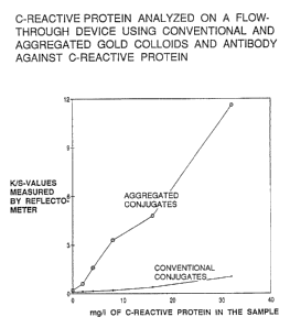
Fig. 4 show the results obtained by serial
dilutions from 1:50 to 1:800 of a plasma containing 32
mg/1 of CRP. The diluent was 0.15 mol/1 NaCl. As the
figure shows, the two types of conjugates demonstrate
considerably different staining intensity on the
membrane, the aggregated conjugate resulting in more
than ten times increased signals.
Both conjugates were kept at 4°C for a period of
nine months and tested regularly. None of the
conjugates showed altered properties after this time of
storage, indicating that the aggregated conjugates are
extremely stable.
Example 6
The experiment above was repeated by replacing
antibody T11G8 with a purified rabbit anti-human serum
albumin (anti-HSA). The resulting conjugates were
tested in a device containing membranes coated with a
monoclonal anti-HSA antibody using the same procedure as
in examples 1 and 5. The results showed that when
applying diluted urine known to contain albumin, the
aggregated conjugate resulted in about eight times
increased colour signal compared to the conventional
non-aggregated conjugate.
Example 7
Aggregated conjugates can also be formed using
proteins different from antibodies. The procedure of
preparation of gold colloids and aggregated conjugates
from example 1 was repeated, this time replacing the
antibody with bovine serum albumin. The albumin
concentration used was 1.5 mg/ml, as with the
antibodies. Aggregates with properties in filters
similar to the antibody-aggregates were formed. The
albumin-aggregated colloidal gold was examined by
electron microscopy and demonstrated a random
WO 92/11537 2 p 9 3 ~ 15 PCT/EP91/0251n
- 24 -
distribution of clustered colloids similar to those
formed in example 1.
Example 8
Another protein different from antibodies which can
be conjugated to colloidal gold in an aggregated form,
is Protein A. The same procedure as in example 1 was
used, using protein A concentration of 1.5 mg/ml as with
the other proteins. The resulting aggregated conjugate
as well as a conventional, non-aggregated was tested in
a device similar to that described in example 1. The
membrane was coated with a monoclonal IgG antibody
directed against human serum albumin. Since protein A
reacts directly with immunoglobulins, the addition of
various dilutions of the two conjugates showed that upon
addition of an equal amount of conjugated protein A, the
aggregated conjugate produced a signal which was about
twelve times stronger than the signal obtained with the
conventional conjugate.
Example 9
In this example is demonstrated the formation of a
hybrid aggregate containing two different proteins which
can be used for binding and signal formation,
respectively. The enzyme alkaline phosphatase (ALP) is
co-conjugated to gold particles together with rabbit
anti-mouse IgG. Such a conjugate allows detection of
mouse IgG's either by directly observing the gold stain,
or by making use of the enzyme in an ELISA-manner.
The enzyme (ALP) and the antibody were desalted by
gel filtration on a PD-10 column equilibrated with 10
mmol/1 acetic acid, and subsequently mixed in a mass
ratio of 1.5:1 (enzyme: antibody). The protein mixture
was then conjugated to gold colloids in 10 mmol/1 acetic
acid, the sum of the masses of the two proteins
amounting to the same protein:gold ratio described in
PCl'/EP91 /02519
WO 92/ 11537 2 ~ 9 ~ ~ ~. 5
- 25 -
example 1. The superaggregated, precipitated conjugates
were allowed to sediment passively, sonicated and
blocked with bovine serum albumin (BSA), and finally
sterile filtered, the whole procedure taking about one
hour.
Fig. 5 demonstrates the detection potential of the
resulting hybrid conjugate. Dilutions of the conjugate
were dotted directly onto nitrocellulose with an even
distribution over circles with diameter 3.5 mm. The
gold stains were measured by means of a reflectometer.
The nitrocellulose was subsequently transferred to an
alkaline phosphatase substrate solution containing
bromochloroindolylphosphate (BLIP). After incubation
with gentle shaking for 30 minutes, a dark, purple
product from the ALP's action on BLIP precipitated on
the nitrocellulose. Fig. 5 shows that a further
increase in sensitivity of ten times over that obtained
with the gold stain was achieved by utilizing this
optional enzyme activity.
Fig. 6 shows the detection of a mouse monoclonal
IgG dotted onto nitrocellulose, using the two optional
tags of the hybrid conjugate. A total of 0.1 - 100 ng
of antibody diluted in 100 ~g/ml BSA was dotted on the
nitrocellulose in duplicate. The nitrocellulose was
dried, blocked with BSA and incubated with the conjugate
in the presence of 0.1~ Tween 20 for 20 minutes. One of
the blots was transferred to the enzyme substrate (BCIP)
solution and incubated as described above. The
developed blots shows that 1 ng of antibody could be
detected using the gold stain, while 0.1 ng was detected
using the enzyme. The enzyme stain appears to give an
overall increased sensitivity of about 15 times compared
to the gold stain.
WO 92/ 11537 2 0 9 3 41 ~ p~'/Ep91 /0251
- 26 -
Example 10
In this example is demonstrated the formation of a
hybrid superaggregate containing colloidal gold and two
different proteins, one of which is a specific binding
partner for the analyte and the second protein not
participating in the reaction, or not being present in
the sample or the reagents in other ways. This hybrid
superaggregate can be used to further increase the
sensitivity of the assay by addition of a second
superaggregate containing colloidal gold and a specific
binding partner for said second protein.
Nitrocellulose membranes were coated with
monoclonal antibody S4H9 (lmg/ml), specific to the
fibrin degradation product D-dimer, and the nitro-
cellulose was placed in a test-device as described in
example 1. Preparations containing D-dimer in the
concentration range 0--8 mg/L, dissolved in 0.1 mol/L
Tris-HC1-buffer (pH7.4) containing 50 mg/ml BSA were
applied (50 ~,1) and allowed to soak through the
membrane. The D-dimer molecules caught by the
immobilised antibody were subsequently visualised by
application of 50 ~,1 of a solution containing a
superaggregate of 4 nm colloidal gold, S4H9, and human
serum albumin (HSA). This superaggregate was prepared
as in example 1 with a 1:1 (w/w) ratio between S4H9 and
HSA. The membrane was washed and the signal strength of
the colloidal gold retained on the membrane was measured
using a reflectometer. A second superaggregated complex
of colloidal gold and monoclonal antibody 2D2 was
prepared as described in example 4. This second
superaggregate was applied to the nitrocellulose, and
the aggregate was immobilised by linkages between the
HSA present in the first superaggregate and antibody 2D2
in the second aggregate. The membrane was washed, and
the resulting signal was measured using a reflectometer.
It could be demonstrated that the signal obtained after
209315
WO 92/11537 PCT/EP91/02519
- 27 -
the second step was four times the strength of the
signal obtained after the first step.
Fig. 7 illustrates the dose-response curves
obtained with increasing amounts of D-dimer in the
sample.