Note: Descriptions are shown in the official language in which they were submitted.
. , ,, 209~68~
~ .
8 llRG I CAL 8 UTURE IN~ T:RUMENT
FIELD OF THE INVENTION
The present invention relates generally to
surgical instruments. The present invention is
specifically directed to a suture instrument for sut~ring
tissue at a surgical site having limited dimensions.
DESCRIPTION OF THE PRIOR ART
For purposes of the present specification, the
term "surgery" applies to a medical operation involving
an incision to subcutaneous body tissue. Therefore, the
surgical incision includes cutting the patient's skin,
the fascia, i.e., the tough fibrous tissue which
envelopes the body beneath the skin, and/or the
peritoneum, i.e., the internal layer of thin connective
tissue that lines the abdominal cavity and covers most of
the viscera contained therein. As used herein, the term
"patient" is directed toward humans, but can also include
animals.
Surgical procedures can be "open" or "closed."
The term "open" surgery usually describes a surgical
procedure in which the surgeon accesses the surgical site
b~ making a relatively large incision in the pa~ient's
body. For example, laparoscopic surgery involves use of
a laparoscope, an illuminated optical instrument for
examining internal organs. In such surgery, access must
be gained to the desired body cavity. For "open" access,
a relatively large incision is made at the umbilicus, the
fascia is visualized, sutures are placed, and the
peritoneum is opened under direct vision allowing a blunt
trocar or port to be placed in the incision site. The
trocar can also be fixed in position by inflatable
balloons or threaded sleeves rather than suture. The
trocar has a system of channels to allow the passage of
various tools and carbon dioxide used to expand the
abdominal cavity, i.e., "pneumoperitoneum," to provide a
working space and to provide a sufficient opening to view
the working space by a laparoscope.
Alternatively, for "closed" access, a small
incision is made and a Verres needle inserted. A Verres
2~94685
~ .
needle is a special needle having a spring-loaded safety
tip that is designed to pierce skin, fat, fascia, and the
peritoneum, without causing unwanted damage to the
internal visceral organs. Carbon dioxide can then be
insufflated to provide the protective pneumoperitoneum,
which allows a trocar to be safely inserted. This trocar
can be of the sharp pointed variety, well known to the
art, or it may have a safety spring-loaded shield to
protect the viscera.
The surgeon places the laparoscope through the
trocar and attaches it to the camera to display the
internal view on a television monitor. Cnce a scope is
in place, other trocars or ports can be safely inserted
under direct laparoscopic view at different locations to
act as channels for scopes and instruments.
For example, in laparoscopic surgery directed
to the gallbladder, ports, typically 10-llmm in size, are
placed at the umbilicus and in the epigastric reyion high
in the mid-line just under the rib cage. Smaller ports
(5mm) are placed as necessary for additional instruments
to accomplish the procedure. For other procedures such
as appendectomies, hysterectomies, gastric, colonic or
other surgeries, several ports, typically 12mm in size,
may be placed at ~arious locations.
At the end of the procedure, the instruments
and trocars are removed. If the umbilical port was
placed in "open" fashion, the surgeon may have adequate
room to manipulate the fascia in order to close the
incision with sutures. I~, however, the umbilical or
other ports were placed in "closed" fashion, the skin and
fascial incisions are both relatively small. There is no
extra room to manipulate the fascia in order to place a
closing suture, and efforts to do so are often
rudimentary at best and often simply abandoned to the
possible detriment of the patient.
20~46~5
--3--
S~I~MARY OF T~-~E I~VENTION
It is therefore an object of the present
invention to provlde a surgical suture instrument, which
can place a stitch in an incision in a restricted or
"closed" surgical site.
This object is accomplished by the
presently claimed invention, which in one
embodiment provides a surgical suture device
0 comprising a casing with a slot for housing a suture
needle. The needle has a puncture end and a manipulation
end and is pivotally positioned within the slot such that
the puncture end of the needle may be exposed or
retracted within the casin~. The surgical suture
instrument also includes a means to manipulate the needle
within the casing. Preferably, the needle is manipulated
by companion manipulation rods, which are slidably
positioned within channels in the casing. The
manipulation rods have finger-activated ends and needle
manipulation ends.
The instrument of the present invention
simplifies surgical suturing processes, especially in
"closed" situations, by providing a device for suturing
body tissue in areas where the incision opening is tiny.
The present invention advantageously provides a
simple instrument, ~hich is easy to assemble or
disassemble, can be readily sterilized and comprises few
working parts. The instrument can also be conveniently
formed of disposable materials. The casing also
advantageously serves as an obturator to occlude the
fascial and skin openings, to retain pneumoperitoneum and
thus maintain direct laparoscopic vision for safe
operation of the instrument. Although the instrument is
specifically designed for laparoscopic surgery, it may
also ~e used for other surgical procedures, including
endoscopic surgical procedures such as arthroscopy,
gastroentroscopy, and laryngobronchoscopy.
2~9~6~
- 3A -
According to another aspect of this invention,
there is provided a surgical suture instrument,
comprising a casing comprising a pocket for housing a
suture needle; a suture needing having a puncture end and
a manipulation end, the suture needles being pivotally
positioned within the pocket, such that the puncture end
is alternately exposed or retracted within the casing;
and means associated with the manipulation end of the
suture needle and operable from a proximal end of the
lo instrument casing for manipulating the suture needle
between a retracted position and an exposed position
whereby the needle is pivoted outwardl~v away from the
casing.
A further embodiment of the present invention
provides a surgical instrument for closing a trocar
incision which comprises an elongated housing having at
least one longitudinal pocket adjacent a distal end
thereof; at least one needle deployably mounted on the
elongated housing; and movable means associated with the
housing, independent of at least one needle, for operably
deploying at least one needle between a first position
substantially within the pocket and a second position
outwardly spaced from the elongated housing means such
that the needle is moved outwardly away from the
elongated housing means.
A still further embodiment of the present
invention provides a surgical instrument for closing a
body cavity incision which comprises: an elongated
housing having a proximal end and a distal end; at least
one needle having a puncture end; means associated with
the elongated housing for deployably mounting at least
one needle, such that upon deployment o~ the needle, the
puncture end is directed toward the proximal end of the
elongated housing; and means disposed within the housing
for operably deploying at least one needle from a first
posltion to a second position spaced from the elongated
housing.
2Q~68~
- 3B -
A further feature of yet another embodiment of
the present invention provides an apparatus for closing a
trocar incision which comprises: an elongated housing
having at least one longitudinal pocket adjacent a distal
end thereof such that at least one longit.udinal pocket
de~ines an opening having a plane normal to a radial
plane of the elongated housing; at least one needle
deployably mounted on the elongated housing portion; and
actuating means for operably deplo~ing at least one
lo needle transverse to the normal plane of the pocket such
that the needle is moved outwardly away from the
elongated housing.
A still further embodiment of the present
invention provides a surgical instrument for closing a
trocar incision which comprises: an elongated housing
having a distal end; at least one needle deployably
mounted on the elongated housing, at least one needle
having a puncture end portion such that at least one
needle is oriented with the puncture end portion directed
toward a proximal end o~ the elongated housing; and means
associated with the housing, distally movable for
operably deploying at least one needle between a first
position and a second position outwardly spaced from the
elongated housing such that the needle is moved outwardly
away from the elongated housing.
Yet another feature of a still further
embodiment provides a laparoscopic surgical instrument
for closing a trocar incision which comprises: an
elongated housing having a proximal end portion and
distal end; at least one needle deployably mounted
adjacent the distal end; and means associated with the
elongated housing, detachably connected to at least one
needle, for operably deploying at least one needle
between a first position and a second position outwardly
spaced from the elongated housing such that the needle is
moved outwardly away from the elongated housing.
A further embodiment of the present invention
provides a laparoscopic surgical instrument comprising:
20~6~5
- 3C ~
an elongated housing having a proximal end and a distal
end; at least one needle; means for mounting at least one
need adjacent the distal end of the elongated housing for
movement between first and second positions; and means
for engaging the mounting means to move at least one
needle to the second position.
Yet another feature of one embodiment of the
present invention provides a surgical suture instrument,
comprising: a housing having needle protecting means
lo therein for retaining and protecting at least a needle
puncture end of a suture needle adapted to be mounted in
the instrument; and means for displacing the needle from
the housing to expose the needle puncture end when the
needle is displaced from the housing.
A still further feature of another embodiment
of the present invention provides a surgical suture
instrument, comprising~ a housing; a needle protecting
means in the housing adapted to receive a needle puncture
tip therein; and means for displacing the needle and the
needle puncture tip from the housing.
Yet another feature of one embodiment of the
present invention provides a surgical suture instrument
comprising a suture needle, and a housing having an
elongated body, the suture needle comprising a
substantially rigid needle capable of substantially
retaining its shape without deformation thereof during
manipulation of the needle, the needle having a puncture
end and being pivotally mounted in the elongated body,
the body having an interference-free suture deployment
and retraction path in which the deployment path extends
outwardly of the body, and in which the deployment path
terminates in a deployment position for the puncture end,
the deployment path being adapted to permit the puncture
end to be deployed exteriorly of the body for surgical
use, and the needle when in the retraction position is at
least partially stored in the body.
2~6~
, ~
BRIEF DESCRIPTION OF THE DRAWINGS
In the drawings:
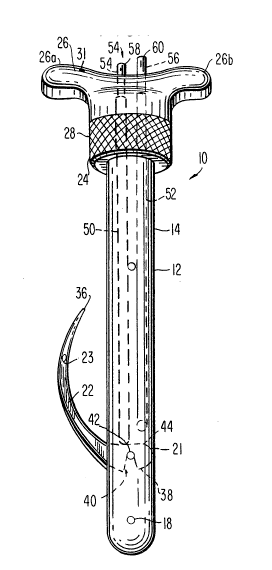
Fig. 1 is a perspective view of the surgical
suture instrument of the present invention.
Fig. 2 is a si~e plan view of the surgical
suture instrument o~ the present invention.
Fig. 2a is a cross-sectional view of the
surgical suture instrument of Fig. 2 taken along lines
2a-2a of Fig. Z.
Fig. 2b is a cross~sectional view of the
surgical suture instrument of Fig. 2 taken along lines
2b-2b of Fig. 2.
Fig. 2c is a cross-sectional view of the
surgical suture instrument of Fig. 2 taken along lines
2c-2c of Fig. 2.
Fig. 2d is a cross-sectional view of the
surgical suture instrument of Fig. 2 taken along lines
2d-2d.
Fig. 3 is a cross-sectional view of the
surgical suture instrument of Fig. 2 taken along lines 3-
3 of Fig. 2.
Fig. 4 is a side view of the suture needle of
the present invention.
Fig. 4a is a cross-sectlonal view of the suture
needle of Fig. 4 taken along lines 4a-4a.
Fig. 4b is a cross-sectional view of the suture
needle of Fig. 4 taken along lines 4b-4b.
Fig. 4c is a cross-sectional view of the suture
needle of Fig. 4 taken along lines 4c-4c.
Fig. 5 is a top view of the cap of the
instrument of Fig. 1.
Fig. 6 is a partially exploded side view of the
surgical suture instrument of the present invention.
Fig. 7 is a side view of the surgical suture
instrument of the present invention illustrating the
needle in retracted position.
2~9~6~
Fig. 8 is a side view of a needle manipulation
rod of the present invention.
Fig. 8a is a cross-sectional view of the needle
manipulation rod of Fig. 8 taken along lines 8a-8a.
Fig. 8b is a cross-sectional view of the needle
manipulation rod of Fig. 8 taken along lines 8b-8b.
Fig. 9 is a front view of a needle manipulation
rod of the present invention.
Fig. 9a is a cross-sectional view of the needle0 manipulation rod of Fig. 9 taken along lines 9a-9a.
DETAILED DESCRIPTIOM OF THE DRAWINGS
Referring now to the drawings, in which like
reference numerals refer to similar embodiments,
reference is initially made to Fig. 1, which illustrates
the surgical suture instrument of the present invention,
at reference numeral 10.
Casinq
The instrument 10 includes a generally tubular~
shaped casing 12 of a size and configuration suitable for
placement in a body opening. For purposes of the present
invention, the term "body opening" is intended to include
both surgically-manipulated and natural or non-surgically
manipulated openings in a body cavity of a patient.
Aside from its primary purpose as a suture instrument,
the tubular shape of the casing also serves as an
obturator to occlude the body opening. The blockage
prevents the escape of any gases and enables the body
opening to retain the pneumoperitoneum and to maintain
direct laparoscopic visionO
The casing 12 can be made of any material known
to the art and suitable for surgical applications. For
example, the casing 12 may be formed of a material
designed for re-use, such as stainless steel. The casing
12 can also be designed for single use and made o~
disposable plastics or aluminum.
Referring now to Fig. 2, the casing 12 is
preferably formed of two symmetrical pieces 14, 16 which
2~946~
", ~ ,
, .
are joined together by pins, screws or the like,
identified at reference numeral 18. In the case of a
disposable surgical instrument 10, the pins 18 may be
permanently positioned such that the pieces 14, 16 are
not capable of separating. If a re-usable instrument is
contemplated, th~ pins 18 will preferably be in the form
of screws in order to allow easy separation of the pieces
14, 16 for cleaning, sterilization and repairs.
Fig. 2 illustrates pieces 14, 16 in the casing
12, which are formed to provide a pocket 19 for slidably
receiving a surgical needle 22. A second pocket 21 on
the opposite side of the casing 12 is provided to allow
complete manipulation of the needle 22 as will be
described in more detail later in the specification.
The casing 12 may also be characterized by a
cut-away portion 20 as illustrated on Fig. 7. The cut-
away portion ~0 is adjacent the surgical suture needle
22, and is designed to provide a space for body tissue
between the needle 22 and the shaft of the casing 12 in
order to give the surgeon some "traction" to expose a
suture opening 23 in the needle.
Cap and CouPler
The casing 12 also preferably includes a collar
24, as illustrated in Figs. 1 and 6, for positioning an
instrument cap 26 onto the surgical instrument 10. The
collar 24 is designed to releasably mount a connecting
coupler 28 onto the casing 12. The coupler 28 is
provided with internal threads 30, which are designed to
cooperate with external threads 32 on the cap 26. The
assembling coupler 28 is provided to connect the cap 26
to the casing 12. The collar 24 is integrated with the
casing 12 to maintain the cap 26 in position on the
casing 12. Preferably, the casing 12 may be providPd
with a positioning button 25 on the shaft of t~e casing
35 12 above the collar 24, as illustrated in Fig. 6, to
coact with a channel 27, illustrated in phantom in Fig.
6, to properly position the cap 26 on the casi~g 12.
. .
.
209~685
As illustrated in Fig. 1, the cap 26 is
preferably provided with finger grips 26a, 26b to assist
the surgeon in manipulating the instrument 10. The
finger grips 26a or 26b may be provided with a marker 31,
e.g., a notice or other marking on one o~ the finger
grips, to identify the position of the cut-away portion
20 and the needle 22 when the casing 12 is within a body
opening.
Suture Needle
Referring now to Figs. 1 and 3, there is
illustrated the suture needle 22 of the instrument of the
present invention. Preferably, the suture needle 22 is
designed for single use and is therefore disposable. The
needle 22 may be made of any material known to the art
for use with surgical needles. Stainless steel is a
pre~erred material, especially for reusable needles.
Howe~er, disposable needles may be made of other surgical
steels as tarnishing is not a problem with disposable
needles. The suture needle 22 is also preferably
designed in a bowed configuration, although other
configurations, known to the art for surgical needles,
are contemplated. The suture needle 22 is characterized
by a suture opening 23 for positioning suture material.
The suture opening 23 is preferably located near the
puncture end 36 of the surgical needle 22. As
illustrated in Figs. 4a, 4b and 4c, the width of the
needle 22 increases as the from the puncture end 36 to
the needle connection end 38 opposite the puncture end
36.
3C The needle connection end 38 is characterized
by a notch 40, which engages with an axle 42 in the
casing 12 of the assembled instrument 10 to allow the -
suture needle 22 to pivotally rotate about the axle 42.
As illustrated in the fi~ures, the axle 42 may serve a
3S dual purpose of providing an axle for the suture needle
22 and providing an additional pin, e.g., pins 18, for
securing the pieces 14 and 16 of the casing 12. The
2~94,6~
.
suture needle 22 can pivotally rotate such that the
puncture end 36 is exposed, as illustrated in Figs. 1 and
3, or the puncture end 36 can be retracted within the
casing 12, as illustrated ln Fig. 7. The pocket 21 in
the casing 12 is provided to allow full mobility to the
needle 22. In either the retracted or extended position,
the needle connection end 38 extends into the pocket 21,
as illustrated in Fig. 7, and the rounded surface 44
remains flush with the surface of the casing 12.
The needle connection end 38 is further defined
by an internal edge 43 culminating at surface 44 that is
used for rotating the suture needle 22. As illustrated
in Figs. 3 and 4, the edge 43 is configured in a
substantially straight-line pattern, the purpose of which
will be explained later.
Needle Mani~ulation Rods
Positioned within the casing 12 are two
parallel disposed channels 50, 52, which may be of like
si~e and length. The channels 50, 52 are designed to
retain needle manipulation rods 54, 56, which are
illustrated in Figs. 8 and 9. Referring to Fig. 5, the
cap 26 is likewise provided with two channels 50a, 52a
which align with the channels 50, 52, respectively when
the cap 26 is placed in position on the casing 12.
As illustrated in Fig. 1, the needle
manipulation rod 54 is defined by a finger-activated end
58, which extends from the channel 50. Li~ewise, the
needle manipulation rod 56 is defined by a finger-
activated end 60 protruding from channel 5Za.
Figs. 8 and 9 illustrate one of the
manipulation rods, i. e., manipulation rod 54. The
manipulation rods 54, 56 are further defined by a body
62, which is preferably square. It is within the scope
of the present invention to provide a body 62, of any
shape. However, a rounded shape is not desired as it
will allow the manipulation rods 54, 56 to spin within
the channels 50, 52.
2Q9~85
_g _
The lower end of the body 62 is defined by a
slotted portion 64. The slotted portion 64 provides a
chamber 66 fo~ receiving the needle 22 when the needle is
in the retracted position as illustrated in Fig. 7. It
is within the scope of the present invention to provide
chambers 66 of the same size in each manipulation rod 54,
56. Alternatively, the chamber 66 of the manipulation
rod 56 may be shorter as it only needs to accommodate the
needle 22 at the area near the puncture end 36, while the
chamber 66 of the manipulation rod 54 must accommodate
substantially more of the needle 22 when the needle 22
retracts within the casing 12.
Located at the opposite end of the finger
activated ends 58, 60 are the needle manipulation ends
67, 68 respectively. Needle manipulation ends 67, 68 are
designed to coact with the edge 43 of the needle 22 to
expose or retract the needle 22 according to the finger
manipulations of the surgeon. In this manner the edge 43
provides a piroting surface for the rods 54, 56. For
example, by fully depressing the finger-activated end 58
of the manipulation rod 54 and simultaneously releasing
the finger-activated end 60 of the manipulation rod 56,
the needle manipulation end 67 of the manipulation rod 54
will coact with the edge 43 of the needle 22 moving the
needle 22 to the position shown in Figs. 1 and 3 and
causing the needle 22 to be exposed.
Alternatively, by depressing the finger-
activated end 60 of the manipulation rod 56 and releasing
the finger-activated end 58 of the manipulation rod 54,
the needle manipulation end 68 of the manipulation rod 56
will coact with the edge 43 of the needle 22 to retract
the needle 22 into the pocket 19 of the casing 12 and
into the slots 66 of the manipulation rods 54, 56, as
shown in Fig. 7.
Assemblv and DisassemblY
The instrument lO is designed to be easily
assembled or disassembled. To assemble the instrument
2 ~
. ~ ^\
--10~
10, the pieces 14, 15 of the casing 12 are position and
attached together by ~eans of the pins 18 and the axle
42. The needle 22 is placed through the pocket 19 and
positioned on the axle 42 as illustrated in Fig. 3. The
manipulation rods 54, 56 are then placed in the channels
50, 52 such that the slots 66 are in proper placement and
alignment with respect to the needle. The coupler 28 is
slipped over the casing 12 and placed in alignment with
the collar 24. The cap 26 is fitted over the
manipulation rods 54, 56 such that the channels 50a, 52a
in the cap coact with the channels 50, 52 in the casing
12. The coupler 28 is then thxeadably tightened onto the
cap. Disassembly follows the opposite procedure.
Preferred Mode of Use
A preferred method of closing an umbilical
incision with the surgical suture instrument 10 of the
present invention will no~ be described. Following
completion of the operative portion of the surgical
procedure, the umbilical trocar is removed. A finger or
blunt trocar may then be inserted into the incision site
to prevent carbon dioxide gas from leaking out of the
abdominal cavity.
When the suture is to be made at the umbilical
body opening, the finger or blunt trocar is removed and
the surgical instrument 10 is positioned in the body
opening. At this point, the surgeon fully depresses the
finger-activated end 60 of the manipulation rod 56 to
retract the needle 22 such that the puncture end 36 is
within the casing 12 as illustrated in Fig. 7~ The
surgical instrument 10 is then advanced into the body
cavity through the body opening. When the surgeon
observes that the needle 22 has passed beyond the body
cavity wall, the surgeon fully depresses the finger~
activated end 58 of the manipulation rod 54 to expose the
puncture end 36 of the needle 22. The needle 22 is then
ready to pierce the tissue wall. The instrument 10 is
then retracted from the body opening. Retracting the
2~9~68~
instrument 10 from the body opening enables the puncture
end 36 of the needle to penetrate the surgically-cut body
tissue a~jacent the body opening. The surgeon can
manipulate the needle 22 by adjustably depressing the
finger-activated ends 58 and 60 of the manipulation rods
. 54, 56. At this point, the puncture end 36 of the needle
has successfully punctured the tissue.
Gentle retraction of the surgical instrument 10
allows the needle 22 exit the body opening and expose the
suture opening 23. At this point, the body tissue is
s~ewered on the needle 22.
When the suture~opening 23 of the needle 22 is
exposed at skin level, a suture is passed through the
suture opening 23. Suture material can be any of a
variety of surgical suture thread-like material known to
the art~ The instrument 10 is then gentl~ reinserted in
the body opening, with minimal pressure, on the
manipulation rods 54, 56, allowing the needle 22 to
naturally pull through its track in the tissue until the
surgeon can see the full needle in the body cavity via
the endoscopic camera in the other body opening. The
instrument lO is then rotated approximately 1800. The
surgeon fully depresses the manipulation rod 54 to expose
the needle, and the instrument 10 is withdrawn from the
25 body opening. As before, the surgeon can balance the .
pressure between the manipulation rods 54, 56 in order to
"sheath the tip" of the needle 22 after it has passed the
body tissue and before it breaks the skin.
When the suture can be seen from outside the
body opening, the thread is grasped and extracted from
the suture opening 23 in the needle. The stitch is
placed, but not tied. The instrument 10 is then ready
for re-insertion in the body opening, in order to release
the needle 22 from the tissue. once the instrument has
been reinserted ln the body opening, the needle 22 is
then fully sheathed by pressing the ~inger-activated end
60 of the manipulation rod 56 and the instrument is
-- . .
2 ~ 9 4 6 8 ~
.
-12-
completely and finally removed from the body opening.
The suture i5 tied to close the body opening.
The instrument 10 allows a safe, secure and
expeditious tissue closure mechanism for small trocar
incisions while maintaining adequate pneumoperitoneum and
direct laparoscopic visualization.
It is understood that the invention is not
confined to the particular construction and arrangement
herein illustrated and described, but embraces such
modified forms thereof as come within the scope of the
following claims. For example, activators such as rocker
switches, handles and buttons are contemplated to
manipulate the needle 22.