Note: Descriptions are shown in the official language in which they were submitted.
'~92/10131 -l- 2 n 9 7 ~ 0 2 PCT/USg~ 16
NON-INVASIVE BLOOD GLUCOSE MF~R~ENT ~l~M
P~ OUND OF THE I~V~NTION
A. Field of the Tn~ention
The present invention relates to a non-invasive method
5 and apparatus for measuring the concentration of D-glucose
in the ocular aqueous humor. More particularly, the
present invention is a non-invasive technique for the
n vivo measurement of the glucose concentration in the
ocular aqueous humor employing the stimulated Raman
l0 effect.
B. Bac~ground of the Invention
Diabetes Mellitus is a major health problem in the
world today because of the physical complications which
arise from living many years with above-normal blood
15 glucose levels. Currently, over ll million people suffer
from diabetes in the United States alone. The two most
common forms of diabetes are Type I, juvenile-onset, and
Type II, adult-onset. Type I diabetes destroys the vast
majority of the insulin-producing beta cells in the
20 pancreas, forcing its sufferers to take multiple daily
insulin injections. Type II diabetes is usually less
severe than Type I as some endogenous insulin production
still occurs and, as a result, Type II diabetes can often
be controlled by diet alone.
The body requires insulin for many metabolic
processes; it is particularly important to the metabolism
of glucose. It is believed that many of the physical
complications associated with diabetes could be avoided if
normal blood glucose levels were maintained throughout
30 each day. A diabetic's blood glucose level can fluctuate
widely around each meal. Maintaining normal blood glucose
levels and reducing these fluctuations requires using some
form of feedback to regulate the multiple daily insulin
shots of Type I diabetics or the diet of Type II
35 diabetics.
Currently, the blood glucose level can be determined
WO92/10131 2 0 9 7 6 0 2 2 PCT/US91/0~ ~6
by a chemical reaction performed on a blood sample.
Although the state of the art glucose measurement devices
are very accurate, the need for a blood sample for each
measurement limits their utility. The most dedicated
5 diabetic patient may take only 4 or 5 measurements per
day, and many diabetics perform even fewer. Because a
diabetic's blood glucose level can fluctuate by a factor
of two or more in a period of an hour, this method cannot
provide the feedback necessary to maintain a normal blood
lO glucose level throughout the day.
A non-invasive blood glucose measurement technique
would allow a large number of daily measurements to be
taken without the problems associated with taking blood
samples. Various schemes have been attempted to
15 non-invasively measure blood glucose level. Many
promising techniques attempt to measure the glucose level
in the ocular aqueous humor because it has been shown that
the ocular glucose level directly correlates to the blood
glucose level and because the ocular aqueous humor
20 provides a much simpler spectroscopic environment than the
blood.
D-glucose occurs normally and in abundance in both the
blood and the ocular aqueous humor. There are two anomers
of D-glucose found in nature: a-D-glucose and
25 ~-D-glucose, which differ only in the orientation of the
groups attached to the C-l carbon. Physically, these two
anomers of D-glucose can be distinguished by their optical
activity; i.e. based upon their ability to rotate the
plane of polarization when illuminated with plane
30 polarized light. In general, the specific rotation, [a],
is defined as
(1) [a] = a
Id
'092/10131 PCT/US91/0~16
~3~ 2097~a2
where a is the total optical rotation of the plane of
polarization measured in degrees, ~ is the length of the
sample in decimeters, and d is the density in g/cm3. The
specific rotations of a-D-glucose and ~-D-glucose are 112
5 and lg degrees, respectively. In solution, one anomer is
converted into the other as necessary to achieve an
equilibrium solution which has a specific rotation of 52.7
degrees.
Since the specific rotation of D-glucose in solution
10 is known, from Equation (1) one can infer the
concentration of D-glucose in a given sample by measuring
the total optical rotation. The accuracy and linearity
observed at very low D-glucose concentrations led March et
al. to attempt non-invasive measurements in the eyes of
15 rabbits. See Rabinovitch, March and Adams, Non-invasive
Glucose Monitoring of the Aqueol~s H-~m~r of the F,ye:
Part I. Measurements of Very Small Optical Rotations,
5 Diabetes Care 1254 (May-June 1982); March, Rabinovitch
and Adams, Non-invasive Glucose Monitoring of the A~leous
20 Humor of the EYe: Part II. Animal Studies and the Scleral
Lens, 5 Diabetes Care 259 (May-June 1982). Unfortunately,
March and his colleagues experienced great difficulty in
measuring the concentration of D-glucose in the ocular
aqueous humor. Many compounds in the ocular aqueous humor
25 other than D-glucose are optically active and contribute
to the rotation of the plane of polarization. In
addition, the cornea has birefringence, which causes a
further rotation of the plane of polarization of the
incident light. See generally, Gough, The Composition of
30 and Optical Rotary Dispersion of Bovine Aqueous Humour, 5
Diabetes Care 266 (May-June 1982).
Both spontaneous and stimulated Raman spectroscopy are
potentially useful to measure the concentration of an
Raman active molecule in a medium. With spontaneous Raman
35 spectroscopy a monochromatic laser beam is directed into a
WO92/10131 2 0 ~ 7 ~ O ~ 4 PCT/US91/0~ ~
Raman-active medium. Some of the incident beam is
transmitted, some of it is absorbed, and some of it is
scattered. A small fraction of the radiation scattered is
shifted in frequency from the incident beam. The amount
5 of this relative frequency shift is related to the
vibrational states of the Raman active molecules in the
medium. The problem with spontaneous Raman scattering is
that the Raman power is scattered in all directions. This
makes the detection of the scattered radiation difficult
l0 for in v vo measurements.
Stimulated Raman spectroscopy (SRS) directs two
monochromatic laser beams, a pump laser beam and a probe
laser beam, into a Raman active medium. If the power of
the pump laser is modulated, then the spontaneous Raman
15 scattered power will also be modulated, which will induce
a signal on the probe laser beam. Thus, rather than
measuring the spontaneous Raman scattered power directly,
a measurement of an intensity fluctuation of the probe
laser beam can be made.
Stimulated Raman spectroscopy has been successfully
used to measure very low concentrations of certain
selected organic liquids diiuted by water and other
solvents. Owyoung and Jones performed a series of
experiments with benzene using stimulated Raman scattering
25 techniques. ~ Owyoung, Sensitivity Timitations for CW
Stimulated Raman Spectroscopy, 22 Optics Communications
323 (Sept. 1977); Owyoung and Jones, Stiml~lated Raman
Spectroscopy Using Low-Power CW Lasers, l Optics Letters
152 (November 1977). Their experimental set-up consisted
30 of two lasers, a tunable pump laser and a fixed frequency
probe laser. The pump laser power was modulated while the
probe laser power was held constant. The two laser beams
were combined and focused through a benzene cell. In the
cell the stimulated Raman effect caused a very small
35 fraction of the power at the pump wavelength to be shifted
-'O9~/10131 PCT/US91/0~16
~ ~5~ ~976~
to the probe wavelength. Thus, at the output of the
benzene cell the probe laser beam carried a small
modulation signal whose amplitude was directly
proportional to the concentration of the benzene in the
5 cell. The probe wavelength was separated from the pump
and converted to an electrical signal by a photodiode.
Both the probe signal and the input pump modulation signal
were fed into a synchronous detector which greatly
improved the signal-to-noise ratio. The pump laser is
10 then repeatedly tuned to new wavelengths to scan a range
of wavelengths, thus, obtaining a Raman spectra for the
Raman-active liquid or gas. This is the same type of
spectrum obtainable by using a commercially available
Raman spectrometer.
Until the present invention, no one has developed a
technique which would allow for non-invasive n yivo
measurement of the glucose concentration in the ocular
aqueous humor. March attempted a non-invasive technique
employing an energy wave transmitter, such as an infrared
20 source located on one side of the cornea and an associated
detector on the opposite side of the cornea. See U.S.
Patent 3,958,650. The wave source is aimed to cause the
radiation to pass throuqh the cornea and the aqueous humor
to the detector. A transmitter is mounted adjacent to the
25 detector and coupled thereto for transmitting a signal
that is a function of the radiation level detected. This
technique is seriously flawed. The radiation detected
will be a function of the concentration of all
substituents in the humor, not just glucose. The later
30 optical rotation technique of March, Rabinovitch and Adams
suffers from a similar flaw. Further, no one, until now,
has determined whether stimulated Raman spectroscopy may
be successfully used to measure concentrations of glucose
in the ocular aqueous humor.
CA 02097602 l998-03-06
WO 92/10131 -6- PCT/US91/08416
SUMMARY OF TXE lNv~NllON
In accordance with an aspect of the present invention
there is provided a method of non-invasively measuring the
concentration of a Raman active molecule in ocular aqueous
humor using stimulated Raman spectroscopy comprising the steps
of: generating a modulation signal; emitting a probe laser
beam having a first wavelength; emitting a pump laser beam
having a second wavelength differing from said first
wavelength by a third wavelength selected to be within a
characteristic ~Aman shift spectrum for the Raman active
molecule; modulating said pump laser beam using said
modulation signal; directing said probe laser beam and said
modulated pump laser beam into the ocular aqueous humor
thereby st;~lating Raman scattered radiation, said Raman
scattered radiation inducing fluctuations in said probe laser
beam, said fluctuations being related to the concentration of
the Raman active molecule in the ocular aqueous humor, said
probe laser beam exiting the ocular aqueous humor; detecting
said probe laser beam after it exits the ocular aqueous humor;
converting said detected probe laser beam into a Raman
electrical sign; and producing a signal representative of the
concentration of the Raman active molecule in the ocular
aqueous humor from said Raman electrical signal and said
modulation signal.
In accordance with another aspect of the present
invention there is provided an apparatus for non-invasively
measuring the concentration of an Ra~n active molecule in
ocular aqueous humor using stimulated Raman spectroscopy,
comprising~ n~ for generating a modulation signal; means
for emitting a probe laser beam having a first wavelen~h !
means for emitting a pump laser beam having a second
wavelength differing from said first wavelength by a third
wavelength selected to be within a characteristic Raman shift
spectrum for the Raman active molecule; modulating means for
modulating said pump laser beam using said modulation signal;
CA 02097602 l998-03-06
WO 92/10131 -7- PCT~US91/08416
means for directing said probe laser beam and said modulated
pump laser beam into the ocular aqueous h = or thereby
st;~--lAting RAmAn scattered radiation, said RA~-n scattered
radiation inducing fluctuations in said probe laser beam said
fluctuation being related to the concentration of the Raman
active molecule in the ocular aqueous humor; said probe laser
beam exiting the ocular aqueous humor; means for detecting the
probe laser beam after it exits the ocular aqueous humor;
means for converting said detected probe laser beam into a
Raman electrical signal; and means for producing a signal
representative of the concentration of the RAmAn active
molecule in the ocular aqueous humor from said modulation
signal and said Raman electrical signal.
In accordance with a further aspect of the present
lS invention there is provided an apparatus for non-in~asively
measuring the concentration of a R~m~n active molecule in the
ocular aqueous humor, comprising: a probe laser emitting a
probe laser beam, having a first wa~elength; a pump laser
emitting a pump laser beam, having a second wavelength which
second wavelength differs from said fist wavelength by an
amount selected to be within a characteristic RAmAn shift
spectrum for the RAmAn active molecule; a modulator means for
producing a modulation signal and for modulating said pn~r
laser beam; a fiber optic coupler receiving said probe laser
beam and said modulated pump laser beam and directing said
laser beams into the ocular aqueous humor thereby stimulating
Raman scattered radiation, said Raman radiation inducing
fluctuations in said probe laser beam, said fluctuations being
related to the concentration of the Raman active molecule in
the ocular aqueous humor, said probe laser beam exiting-
~ocular aqueous humor; a photodetector receiving said probe
laser beam and producing an electrical signal; and an
amplifier developing a dc voltage representative of the
concentration of the Raman active molecule in the ocular
aqueous humor from said electrical signal and said mo~nlAtion
signal.
CA 02097602 1998-03-06
W0 92/10131 -8- PCT/~S91/08416
A non-invasive blood glucose measurement technique
would allow more Crequent measurement of blood glucose
concentration5 without the pro~lems associated with taking
~lood samples. The present ~nvention achieves this goal
5 by providing an apparatus and a ~ethod for non-invasively
measuring the ~n v vo concentration of an Raman active
molecule in the ocular aqueous humor ~y using stimulated
Raman spectroscopy. The apparatus of the present
invention include,s a means for emittinq a probe laser beam
lO and a mea~s for eml~; nn ~ ~um~ laser beam. 30th means
emit monochromatic laser light and are separated in
wavelength by a wavelength chosen to be within a
characteristic Raman shift spectrum for the Raman active
molecule. ~y setting the separation in wavelength between
lS the pump and probe lasers, one may select which one of a
num~er of Raman active molecules will be measured. rn th~
preferred embodiment~~the selec~ed ~aman active molecule is
D-glucose and the separation between the pro~e ~avelength
and pump wavelength is chosen to ~e 518 cm~l in accordance
20 with the characteristic Raman shift spect_um for
D-glucose.
The apparatus also i~ludes a modulating ~eans 'or
modulati~a the output power ~1-' tn~_ Pumo laser ~eam. A
power source may be pro~ided ~or the probe~laser for for
25 maintainina its po~Pr output suostant1ally constant. The
modùlated pump laser beam is then combined with the probe
laser beam by a means for directinq the combined laser
beams into the-ocular aqueous humor.
The introduction of light into the ocular aqueous
30 humor stimulates Raman radiation which shifts energy from
the pùmp frequency to the probe frequency, thereby
inducing fluctuations in the probe laser beam dire~tly
related to the concentration of the selected Raman acti~e
molecule in the ocular aqueous humor. After the probe
-
CA 02097602 1998-03-06
WO 92/10131 -8a- PCT/US91/08416
laser beam e~its the ocular aqueous humor, means are
provided for detecting the probe laser beam and converting
it into a Raman electrical signal. The Raman electrical
signal is then compared to the modulation ~ignal preferably by
a synchronous detector, a dynamic signal analyzer, or a
computer based synchronous detection system, to produce a
voltage representative of the concentration of the Raman
active molecule in the ocular aqueous humor.
The method of the present invention non-invasively
lO measures the in vivo concentration of an Raman active
molecule in the ocular aqueous humor using stimulated
Raman spectroscopy. Preferably, two monochromatic laser beams
are provided, a probe laser beam and a pump laser beam. The
probe laser beam and the pump laser beam wavelengths are
15 separated from each other by a wavelength within a
characteristic Raman spectrum for the Raman active
molecule being measured. In the preferred embodiment, the
Raman active molecule is D-glucose and the separation in
fre~uency preferably chosen to be 518 cm~l. The output
20 power of the probe laser beam may be maintained
substantially constant, while the output power of the pump
laser beam is modulated by a modulation signal. The
probe laser beam and the modulated pump laser beam are
combined and directed into the ocular aqueous humor,
25 thereby stimulating Raman scattered radiation. The probe
laser beam is detected after it e~its the ocular aqueous
humor and converted into an electrical signal. The
electrical signal is then compared with the modulat~on
signal to produce a voltage representative concentration
30 of the Raman active molecule.
It is, therefore, an object of the present i~vention
to provide a system for measuring an Raman active molecule
in the ocular aqueous humor.
It is a preferred object of the present invention to
35 provide a system for measuring very small D-glucose
concentrations.
CA 02097602 1998-03-06
Wo 92/10131 -8b- PCT/US91/08416
It is yet another preferred object to t~e present
in~e~tion to make non-invasive in vivo measurements of ~-
glucose concentration.
It is a further preferred object of the present in~ention
to allow multiple daily measurements of D-glucose to be msde
non-in~asively.
It i~ a still further preferred object of the prese~t
invention to provide a non-in~a~ive glucose meaQurement system
which is inexpensive to manufacture, durable in construction,
lO and ef~lc~ent ln opera~lon.
~ fiese and other advantages will become apparent in the
discussion below.
~RI~F D~.~CRIPTION OF TH~ DRAWING~
Fig. l is a graph of the spontaneous Raman spectrum
15 for D-glucose.
Fig. 2 illustrates the~ stimulated Raman spectroscopy
(SRS) wavelength selection for the preferred embodiment.
Fig. 3 is a block diagram of one embodiment of the
present invention.
Fig. 4 depicts the preferred amplitude modulation of
the pump laser power versuS time.
Fig. 5 is a top view of an eye showing entering and
e~iting laser beams.
Fig. 6 illustrates the modulation of the detected
25 probe laser power after estraction verSuS time.
Fig. 7 illustrates the apparatus of the present
invention for L~ vivo me~surement.
Fig. 8 is a bloc~ diagram an alternative embodiment of
the present invention.
- 30Fig. 9 is a block diagram of another alternate
embodiment of the present invention using bulk optics.
n~T~Tr.~n n~ RT~Io2~
When a monochromatic laser beam is incident on a
Raman-active medium some of the incident beam is
35 transmitted, some of it is absorbed, and some of it is
Wo92/10131 2 ~ 9 16Q~ ~T~S 9 1 / 0 8 4 6
- -9- 03 Re~'d PCT/PTO t O FE3 1993
scattered. A small fraction of the radiation scattered is
shifted in frequency from the incident beam. The amount of
this relative frequency shift is related to the vibrational
states of the molecules in the medium. The D-glucose
molecule has several possible Raman active vibrational
states so that the Raman scattered power forms a spectrum
which is characteristic of D-glucose alone.
Turning now to the drawings in which like numerals
denote corresponding parts the preferred embodiment of the
present invention is shown. Fig. 1 illustrates the
characteristic spectrum for D-glucose dissolved in water
showing the relative intensity of spontaneous Raman power
versus the frequency shift. Each of the peaks in the
spectrum corresponds to a particular vibration of the
D-glucose molecule, the largest peak occurring at a
frequency shift of 518 cm~1. The absolute intensities of
the peaks are directly related to the conecntration of the
Raman-active molecule, in this case D-glucose.
Use of a single monochromatic laser beam to generate
spontaneous Raman scattering is difficult for ln vivo
measurements since the Raman power is scattered in all
directions. This problem can be solved by having two
monochromatic laser beams (a pump laser and a probe lase)
incident on the chosen sample. Thus, in the preferred
embodiment, the probe laser is at the same frequency as
the Raman scattered power from a large peak in the
D-gl~cose spectrum, as illustrated in Fig. 2. The pump
laser is at a frequency whose difference from the probe
frequency is equal to the frequency shift of the large
peak selected for the probe laser. In order to use
stimulated Raman spectroscopy to measure the concentration
of D-glucose in the ocular aqueous humor, the frequency
difference between the pump laser beam and the probe laser
~ beam must be chosen to coincide with one of the peaks in
the Raman spectrum for D-glucose. If the power output of
SUBSTITUTE SHEET
WOg2/10131 2ag7 602 10 PCT/US91/0~16
the pump laser is modulated, then the spontaneous Raman
scattered power will also be modulated which will induce a
signal on the probe laser beam. Rather than measuring the
spontaneous Raman scattered power directly, a measurement
5 of an intensity fluctuation on the probe laser beam is
made. This method is called stimulated Raman spectroscopy.
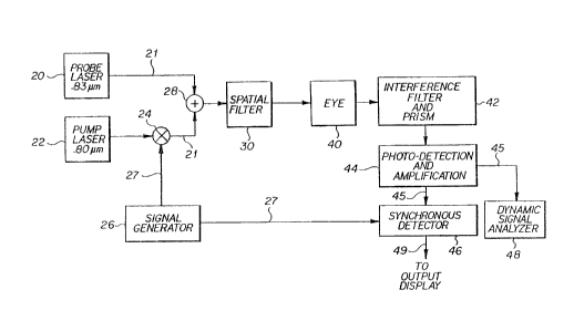
As shown in Fig. 3, the present invention involves two
lasers, a probe laser 20 and a pump laser 22. Both lasers
20 and 22 emit monochromatic laser beams. The relative
10 wavelength difference between these two laser beams is
adjusted to be the same as the wavelength shift of one of
the largest spontaneous Raman peaks for D-glucose, 518
cm~l. Other peaks unique to the D-glucose spectrum may be
chosen, for example, 400 cm~l.
The probe laser 20 chosen operates at a wavelength of
approximately 0.83 micrometers with an output power of
20 mW. The probe laser power output should remain
substantially constant over time in order to minimize
errors in measurement. Thus, the sensitivity of the
20 system is directly related to the ability to maintain the
probe laser power output constant. The laser diode
chosen, an SDL-1401-H2, is slightly tunable with
temperature.
The pump laser 22 chosen emits light with a wavelength
25 of approximately 0.8 micrometers and an output power of
100 mW. The power output of the pump laser should
ordinarily be greater than that of the probe laser by
about 5X depending upon the threshold conditions for the
medium. The pump laser wavelength is also slightly
30 tunable with temperature to allow fine adjustment for
optimal signal level.
The actual wavelengths chosen for the pump and probe
laser beams are not as important as the separation between
them. That separation should correspond to a vibrational
35 state of the Raman active molecule being measured. For
WO92/10131 ~G ~ S PCT/~S9l/0~4l6
-1l- 03 Rec'd PCT/P~O ~ O FE~ 199~
example, should it be desirable to concentrate on the
D-glucose peak at 400cm~1~ a different separation would be
chosen for the wavelengths output by the probe and pump
laws. The particular wavelengths of 0.8 and 0.83 were
selected because of the availability of commercial diode
lasers of such wavelenghts.
The pump laser beam is amplitude modulated by a biased
square wave signal from signal generator 26. The output of
signal generator 26 is used to modulate the current to the
10 diode of the pump laser 22 thereby modulating the amplitude
of the output of the laser beam. The output of pump laser
22 is a biased square wave, with a maximum amplitude of
approximately 100 mW and a minimum of approximately 0 mW.
An example of amplitude modulated pump laser power over time
15 is illustrated in Fig. 4. t
It is possible to use other types of modulation to
measure the concentration of Raman active modules using
stimulated Raman spectroscopy. Amplitude modulation was
chosen over other types of modulation, such as pulse width
20 modulation, because amplitude modulation is easier to
generate. Further, the type of modulation chosen affects
the complexity of the detection scheme that must be used.
The choice of modulation technique also affects the power
incident upon the eye. Any technique which reduces this
25 power reduces possible damage to the eye and is to be
preferred.
The probe laser beam and the amplitude modulated pump
laser beam are fed to a fiber optic coupler 28 via fiber
optic pigtails 21. Only 50~ of the power in the probe and
30 pump laser beams is coupled into the fiber optic pigtails 21
due to their coupling efficiency. Another 50~ of the power
in the probe and pump laser beams is lost when the fiber
optic pigtails 21 are joined together by optic coupler 28.
Thus, the maximum power which could reach eye 40 is
35 approximately 25 mW when starting with a laser power
SUBSTITUTE SHEET
WO92/10131 2 0 9 7 ~ 0 2 -12- PCT/US91/0~16
output of 100 mW.
Fiber optic coupler 28 combines the probe and
modulated pump laser beams and directs them into an
optional spatial filter 30. The spatial filter 30
5 converts the cross-section intensity to a Guassian
Distribution which allows the beam to be focused more
precisely and insures the complete combination of the two
wavelengths before the laser beams are directed into the
eye 40.
The combined laser beams travel through fiber optic
cables associated with means for delivering the laser
beams to the eye, preferably in the form of a handset (not
shown). The handset is held up against eye 40 and directs
the probe and modulated pump laser beams into the ocular
15 aqueous humor. The trajectory of incident laser beams 60
can be seen in Fig. 5. The laser beams 60 are passed
through the cornea 36 and the aqueous humor 38 in such a
manner as to bypass the lens 32 and the iris 34. Pump and
probe beams 60 excite stimulated Raman radiation while
20 inside the ocular aqueous humor 38. The scattered Raman
radiation causes a small amount of the energy at the pump
frequency to be shifted to the probe frequency, thereby
inducing fluctuations in the previously constant power
level of the probe laser beam, as illustrated in Fig. 6.
25 These fluctuations in the probe laser beam power are
directly related to the concentration of D-glucose in the
ocular aqueous humor 38. The now modulated probe laser
beam and the pump laser beam exit the eye and are coupled
into an optical fiber in the handset-cable assembly.
The coupled detected laser beams are passed through a
set of cascaded narrow band interference filters 42.
These optional filters are centered at the probe
wavelength plus or minus about 5 nm, thereby filtering the
pump laser beam away from the probe laser beam. Thus, if
35 the probe wavelength is set at 0.83 microns, the band
WO92/10131 -13- ~ 9 ~ ~ U ~
width (BW) for the filters would be BW e 825 nm c ~ < 835
nm. The filters are cascaded because the desired
reduction in the pump laser power cannot be achieved with
a single filter. Further reduction in the power of the
5 pump wavelength may be accomplished with a prism or
grating.
Thereafter, the modulated probe laser beam is applied
to a photo-voltaic diode in photodetector/amplifier 44,
which outputs an electrical signal. The photo-voltaic
10 diode provides a current output in relation to the amount
of light input. The photodetector/amplifier 44 includes a
low-noise amplifier which amplifies the low current level
output of the photo-voltaic diode to achieve a voltage
level compatible with the circuitry of the synchronous
15 detector 46. A transresistance gain of greater than 108
is achieved by the low noise amplifier, which also filters
off the large DC bias.
The operating band of the low noise amplifier is
determined by the noise spectrum of the probe laser. The
20 noise spectrum of the probe laser is relatively constant
between 1-10 kHZ, and increases below this frequency
range. Restricting the passband of the low noise
amplifier to this frequency range helps eliminate
undesired noise from the probe laser. Those skilled in
25 the art will understand that the passband chosen depends
upon the noise spectrum particular to the laser used as
the probe laser. Different lasers may be espected to
require different low noise amplifiers.
The output 45 of the photodetector/amplifier 44 is fed
30 to synchronous detector 46, such as a Princeton HR-8 PAR
lock-in amplifier. The synchronous detector 46 compares
output 45 to the output 27 of signal generator 26, and
generates siqnal 49, which is representative of the
concentration of D-glucose in the ocular aqueous humor.
35 The synchronous detector output 49 may then be fed to any
WO92/10131 2 0 9 7 6 0 2 -14- PCT/US91/0~16
standard display element.
Use of the lock-in amplifier involves using the pump
laser modulation signal as an external reference signal
and feeding the SRS signal into the signal input of the
5 amplifier. The bandpass filter for the external reference
may need to be tuned to the pump modulation frequency
depending on the model of the lock-in amp. A phase offset
between the reference and the SRS signal should be zeroed
as indicated in the installation manual. Then the
10 appropriate gain setting for the SRS input signal should
be set. The time constant or integration time setting may
vary depending upon the noise present on the signal. The
DC output signal may be read from the meter in the unit or
directed to an external display.
An alternative, and more costly, method of generating
a representative electrical signal from the
photodetector/amplifier 44 output is to feed output 45 to
a dynamic signal analyzer 48, such as an HP 3561 made by
Hewlett-Packard. Analyzer 48 measures the power contained
20 in the detected probe laser beam at the modulation
frequency. This power is likewise related to the
D-glucose concentration. Thus, it is possible to use at
least two independent methods to calculate the D-glucose
concentration.
The procedure for the dynamic signal analyzer invovles
connecting the SRS signal to the input jack of the
analyzer. The soft key programming is generally discussed
in the user's manual for the analyzer. The frequency span
should be set to a center frequency equal the pump
30 modulation frequency and a span of about 100 Hz which may
vary depending upon signal noise. Preferably, the unit
should also be programmed to RMS average 50 samples.
There is a setting for peak tracking which will display a
numerical value for the frequency peak in the local
35 portion of the signal spectrum which is currently
~092/10131 -l5- 2 0 9 ~ 6 ~ ~
displayed. The vertical scale units should be set to
"linear" and thus the value for the SRS signal
corresponding to the glucose concentration will be the
value of the peak frequency component. The numerical
5 value is displayed on the screen of the analyzer.
Before using the apparatus of the present invention
background measurements should be made to establish a
signal reference point and to be certain that there are no
spurious signals in the passband of the low noise
l0 amplifier which forms part of the photodetector/amplifier
44. A noise spectrum measured by the synchronous detector
46 with only the probe laser turned on should be made.
Another important background measurement is pump laser
power "leakthrough". Although the two narrow band
15 interference filters filter the pump wavelength, some
small amount of pump power still reaches the detector 46.
Since this signal is present whenever the pump laser is
on, it will offset other measurements. In addition to the
presence of pump leakthrough, stimulated Raman scattering
20 takes place in the fiber optic cables 21 which carry the
input power to the eye 40 and this SRS signal offsets the
SRS signal from D-glucose in the ocular aqueous humor.
These offset signals do not directly limit the sensitivity
of the D-glucose measurement since the SRS signal from the
25 D-glucose adds to the offset signals and thus the offset
signals can be subtracted out in the calculation of the
D-glucose concentration. But the relative amplitude of
these offset signals does limit the detector gain which
ultimately limits the system sensitivity.
Occurring naturally in the ocular aqueous humor, water
is also a Raman-active molecule. There is no peak present
in the Raman spectrum for water at the frequency shift of
518 cm~l; even so, some broad features of the water
spectrum will produce an SRS signal at a shift of
35 518 cm~l. This SRS signal from water contributes to the
WO92/10131 ~9 7 6 ~ ~ -16- PCT/US91/0~16
offset signal and should be subtracted out in the
calculation of D-glucose concentration.
A schematic of the present invention for n vitro
measurement can be seen in Fig. 7. The entire apparatus
5 is mounted on a wheeled cart 68. The pump laser
(SDL-2412-H2) and its power supply 72 are mounted
vertically on one side of the cart, while the probe laser
( SDL-24 12-H2 ) and its power supply 70 are mounted
vertically on the opposite side of the cart. The
l0 preferred power supply for the probe laser is an LDX 3620
by ILX lightwave, Montana, because of its low noise
current to the laser diode and constant power mode having
an automatic compensation feature. Outputs from each of
the pump and probe lasers are fed by optical fiber
15 pigtails 73 to a fiber optic coupler 75. The pigtails are
multi-mode fiber optic cables to match those supplied with
the lasers. The combined probe and pump laser beams are
then fed by fiber optic cables 76 from the fiber optic
coupler 75 up to the spatial filter 77 and from there into
20 an eye, or alternatively, into a glucose test cell 78,
shown in place on a table behind the cart. The spatial
filter 77 is preferably a Model 900 from Newport Optics.
Glucose test cells 78 are used for in vitro measurement.
The test cell 78 is machined plastic with special windows
25 of a high quality, low impurity glass with a special
coating to reduce reflections of optical wavelengths
selected for the lasers. Preferred coatings include a
magnesium fluoride or a coating broadband near infrared
coating like Newport #AR.16.
A set of interference filters 79, previously
described, receive the pump and probe laser beams as they
exit the glucose cell 78. Fiber optic cable 81 passes the
detected laser beams from filters 79 to detector unit 74,
which houses both the photo-voltaic diode and the low
35 noise amplifier. The output from the detector uni'~ 74 is
~092/10131 PCT/US91/0~16
2 ~ 2
then fed to the synchronous detection unit. A switchable
laser temperature readout 82 may be provided to monitor
the output of lasers 70, 72.
A block diagram of an alternative embodiment of the
5 present invention is shown in Fig. 8. The alternative
apparatus incorporates both a probe laser 120 and a pump
laser 122. These lasers are both monochromatic and both
operate at the same wavelengths discussed hereinabove with
respect to the preferred embodiment of Fig. 3. The pump
10 laser 122 is modulated using an AM modulation source 126,
such as HP 3314A.
The output of probe laser 120 is connected to an
optical coupler 150 which is reversed to split the probe
laser output into two beams, one containing 5% and the
15 other containing 95% of the probe laser power.
Another fiber optic coupler 128 combines ninety-five
percent of the probe laser power with the modulated pump
laser beam and outputs the combined beams to spatial
filter 130. Using a handset (not shown) the combined
20 beams are then directed into the eye 140. Inside the
ocular aqueous humor of the eye 140 stimulated Raman
radiation causes a portion of the power at the pump
frequency to be shifted to the probe frequency, thereby
modulating the probe laser beam. As the pump probe beams
25 exit the eye they are coupled into a fiber optic cable to
transport the SRS optical signal to the photo detector.
The pump laser beam is filtered from the probe laser beam
using a series of narrow band filters incorporated into
the photodetector/amplifier 144. The modulated pump probe
30 laser beam is then transduced from an optical signal into
an electrical signal using a photo-voltaic diode. The
electrical output from this diode is thereafter amplified
using an extremely large gain, low noise amplifier
incorporated in photodetector/amplifier 144 to achieve the
35 signal levels compatible with a detection scheme. The
WO92~10131 ~ O 9 7 6 0 2 PCT/US91/0~'~
amplifier's gain is on the order of 108. The amplifier's
passband corresponds to a frequency range in which the
probe laser's noise spectrum is substantially constant.
The output of the photodetector/amplifier 144 is then fed
5 into a computer-based synchronous detector 156.
Five percent of the probe laser power is applied to
the photodetector/amplifier 152, which is substantially
similar to photodetector and amplifier 144 and which
generates an electrical signal representative of the probe
10 laser beam power. This output may then be fed to an
optional time delay 154 to compensate for the delay of the
laser beam through the spatial filter, fiber optic coupler
and photodetector amplifier path. However, since this
time delay is small and is physically hard to realize,
15 time delay 154 may also be eliminated without
substantially effecting the accuracy of the D-glucose
measurement.
The computer-based synchronous detector 156 compares
the photodetector/amplifier output 144 with the output of
20 the AM modulator 126 and photodetector/amplifier 152
output, allowing the computer-based synchronous detector
156 to compensate for amplitude variations in the probe
laser beam caused by internal noise and thermal drift of
the probe laser. A data acquisition/interface board to
25 the computer connects the signals from
photodetector/amplifiers 144, 152 to the computer. The
data acquisition/interface board preferably consists of 3
primary A/D channels. Two of these channels are 16-bit
resolution and the third channel is only 8-bit
30 resolution. The 16-bit channels are used to convert the
SRS signal and the PROBE Noise signal, while the 8-bit
channel converts the modulation signal. The
specifications should meet or exceed the following:
16-bit Converters - Analog Devices (1376A)
16-bit Track-Hold - Analog Devices (389KD)
~092/10131 PCT/US91/0~16
--19--
~097~
Amplifiers
8-bit Converter - Analog Devices (574A)
8-bit Track-Hold -Analog Devices (HTC-0300)
A sampling rate of l0 KHz is used so the Nyquist frequency
5 is 5 KHz. Appropriate anti-aliasing filters should be
used prior to conversion which limit the bandwidth of the
input signals to <5KHz. Also there are four secondary A/D
inputs to monitor various background activities like laser
temperature. Once the analog signals are converted to
l0 digital values they are converted to floating point
numbers and stored in memory arrays by low level
programming. Currently the data is processed by
algorithms to yield a stable SRS value for a given glucose
concentration. These algorithms include the following:
l) The use of infinite impulse response (IIR)
filters, such as a Butterworth filter, to further narrow
the bandwidth of the signal.
2) The subtraction of amplitude noise originating on
the PROBE Laser.
3) A conventional cross-correlation algorithm to
yield a final result.
An alternate approach to the optical fiber band system
described above would be to use entirely bulk optics in
the system, thus eliminating the optical fibers
25 completely. This bulk optics implementation, although
more costly, will yield a higher system sensitivity than
the optical fiber based system. This arises from the
elimination of one of the sources of "leak through"
signals and the conversion to single-mode laser diodes.
30 Current technology limits the use of these high power
single-mode laser diodes to a bulk optic system. These
single-mode laser diodes concentrate their optical power
into a much narrower spectral line which will greatly
improve the SRS signal to noise ratio.
As illustrated in Fig. 9, the output of each laser
WO92/10131 ~ 7 ~ ~ 2 PCT/US91/08'-~
-20-
220,222 of the bulk optic system is delivered to beam
collimating optics 221, 223, which preferably consist of a
pair of cylindrical lenses for each laser, to focus the
laser beam along their orthagonal axes to make a
5 collimated beam from the asymetrical cone shaped output of
the lasers. Optical isolators 224, 22S are provided to
prevent reflection of the beams back into the lasers. The
isolators can be broad band isolators covering the band
width of 750 to 950 nm, such as the Newport ISO-7885
l0 optical isolator. The output of pump laser 222 passing
through optical isolator 224 is delivered to an optical
chopper 226 or electric optic modulator which preferably
operates at lkHz, though may be operated at different
frequencies depending on the modulation desired for the
15 pump laser output. Beam samplers 227, 260 are provided to
reflect a small sample portion of the beam or beams, the
amount of the sample depending upon the angle of placement
of the beam sampler in the path of the beam. Beam sampler
227 is used to provide a small sample of the probe laser
20 output to the photodetector and amplifier 252, the output
of which 254 provides a probe laser noise signal to the
computer based synchronous detection system 256. Beam
sampler 260 provides a small sample of the beams provided
to the spatial filter 230. The output of the
25 photodetector and amplifier 262 coupled with the beam
sampler 260 provides a synchronous detection reference
signal 266 to the computer based synchronous system 256.
An optical spectrum analyzer 264 is used to monitor the
beams provided to the spatial filter 230 in the laboratory
30 setting because the single mode lasers have a tendency to
"mode hop" and change the wavelength of their output.
The rest of the path in Fig. 9 beginning with the
spatial filter 230, the glucose sample 40 through to the
computer based synchronous detection system 256 is the
35 same as the embodiments discussed above with the
"092/10131 PCT/US9l/0~16
~ -21- 2~97602
exception of the addition of the prism or grading 241. If
a prism is used, a standard dispersing prism is
preferred.
From the bulk optic system block diagram of Fig. 9 one
5 can see that this system is functionally equivalent to the
optical fiber based system. The major differences between
the two implementations are as follows:
1) Now the polarization of the optical fields must be
carefully controlled. It is vitally important that both
10 the pump laser wavelength and the probe laser wavelength
have nearly the same polarization of their optical
fields. Here linear polarization is maintained throughout
the system.
2) Due to technological limitations, the pump
15 single-mode laser diode's optical power cannot be
modulated by the modulation of its current. This is due
to the fact that the wavelength of the laser diode's
output power does depend upon its current. Thus, both
laser diodes are operated with a constant current source.
20 Now the pump laser's optical power is modulated by -an
optical chopper 226 which yields an equivalent optical
power modulation to the optical fiber based system.
3) A key addition to this system is an optical
isolator 224, 225 which is used with each laser diode to
25 minimize reflections of the optical power back into the
laser cavity. This is very important since the reflected
power can cause the laser diode to change its wavelength
during operation. This phenomenon has been observed in
the optical fiber based system.
4) The spatial filter 230 is now required to insure
that the optical power from each laser is collinearly
focused through the glucose solution 240. The glucose
solution may be in an optical test cell or in the ocular
aqueous humor.
The addition of the optical spectrum analyzer 264 is
WO92/10131 PCT/US91/08~ ~
2~9~ 602 -22-
optional. Its purpose is to monitor a portion of the
optical power to insure the proper optical wavelengths are
present. Also the wavelengths for the single-mode laser
diode have been changed slightly due to the availability
5 from the manufacturer. The difference between these two
wavelengths still coresponds to a frequency difference of
518 cm~l as previously discussed.
It will be obvious to those skilled in the art that
many variations may be made in the embodiment chosen for
lO the purpose of illustrating the best mode of making and
operating the present invention, without departing from
the scope thereof as defined by the appended claims.