Note: Descriptions are shown in the official language in which they were submitted.
WO92/10974 PCT/US91/0918~
2098182
Safety Penetrating Instrument
BACKGROUND OF THE INVENTION
Field of the Invention
The present invention pertains to surgical penetrating
instruments and, more particularly, to safety surgical
penetrating instruments having tubular needles for penetrating
tissue and safety probes for protecting the sharp tips of the
needles.
Discussion of the Prior Art
Surgical penetrating instruments are widely used in
surgical procedures to gain access to anatomical cavities
ranging in size from the abdomen to small blood vessels, such
as veins and arteries, epidural, pleural and subarachnoid
spaces, heart ventricles and spinal and synovial cavities.
Such surgical penetrating instruments include a penetrating
member or implement having a sharp tip or point to pierce or
penetrate the tissue forming the cavity wall, and the force
required to penetrate the cavity wall is dependent upon the
type and thickness of the tissue of the wall. Once the wall
is penetrated, it is desirable to protect the sharp tip of the
penetrating member to prevent inadvertent contact with tissue
in or forming the cavity, and a particular problem exists
where substantial force is required to penetrate the cavity
wall in that, once penetration is achieved, the lack of tissue
resistance can result in the sharp tip travelling too far into
the cavity and injuring adjacent tissue.
Safety penetrating instruments including a safety probe
biased to extend beyond the sharp tip of a penetrating member
have become widely accepted for use in penetrating anatomical
cavities. For example, the Verres needle, commonly used to
create a pneumoperitoneum, has a spring-loaded inner member
disposed within a tubular needle. U.S. Patents No. 1,527,291
to Zorraquin, No. 2,623,521 to Shaw and No. 2,630,803 to Baran
are exemplary of safety penetrating instruments with a
spring-loaded inner member disposed in a needle, while U.S.
WO92/10974 PCT/US91/091~
2098182 2-
Patent No. 4,254,762 to Yoon shows an endoscope spring-biased
in a hollow needle. Safety trocars having a spring-biased
protective shield disposed between an outer sleeve and an
inner trocar are marketed by Ethicon, Inc. as the Endopath and
by United States Surgical Corp. as the Surgiport. U.S. Patents
No. 4,535,773 to Yoon and No. 4,654,030 to Moll et al are
exemplary of such safety trocars. German Offenlegungsschrift
2,544,262 discloses an intrauterine catheter including a tube
having a distal sharp point, a spring-biased blunt member in
the tube distal end and a hose or catheter slidable over the
tube.
While the prior art safety penetrating instruments are
widely used, they suffer from many disadvantages when used in
the procedures for which they are presently recommended; and,
additionally, prior art safety penetrating instruments cannot
be used in many procedures for which safety of penetration is
highly desirable. One of the disadvantages of prior art
surgical penetrating instruments is that, when the penetrating
member is a tubular needle with an acutely angled distal end,
the sharp tip is not well protected and is still at least
partially exposed when the safety probe is extended to the
protective position such that use in penetrating small of
narrow anatomical cavities is not safe, while another
disadvantage is that the safety probe cannot be locked in an
extended, needle tip protecting position. A further
disadvantage is that the sharp tip of the needle cannot be
selectively exposed for effective use in further penetration
of tissue after a cavity wall is initially penetrated.
SUMMARY OF THE INVENTION
Accordingly, it is a primary object of the present
invention to overcome the above mentioned disadvantages of the
prior art by utilizing a tubular needle with a safety probe
movable therein in a safety penetrating instrument including
a portal sleeve thereby increasing safety and efficac~y in a
wide range of surgical procedures.
WO92/10974 PCT/US91/09188
3 20981~2
Another object of the present invention is to position
the distal edge of a portal sleeve in alignment with or
forward of the trailing edge of an angled open end of a needle
in a safety penetrating instrument having a safety probe
movable in the needle such that the needle and portal sleeve
present a substantially smooth profile for tissue penetration.
A further object is to optionally allow locking of a
safety probe in an extended position in a needle of a safety
penetrating instrument including a portal sleeve or to allow
the safety probe to move proximally from the extended
position.
Another object of the present invention is to utilize a
tubular needle to position a portal sleeve through a cavity
wall with the sharp tip of the needle received in a protective
recess in a safety probe to minimize exposure of the sharp tip
both after penetration of tissue and during handling by
medical personnel thereby reducing the opportunity for contact
and/or piercing of tissue inadvertently.
A further object of the present invention is to align an
angled distal end surface of a safety probe with an angled
sharp distal end of a tubular needle within a portal sleeve
such that the distal ends of the safety probe and the needle
are in substantially the same plane during tissue penetration
to facilitate placement of the portal sleeve.
An additional object of the present invention is to
utilize a safety probe in a tubular needle having an open
distal end with a portion curving toward the longitudinal axis
of the needle to terminate at a sharp tip such that, in an
extended position, the distal end of the safety probe
protrudes over the sharp tip of the needle.
The present invention has another object in that a safety
probe movable relative to an elongate penetrating needle
within a portal sleeve is biased toward an extended position
and can be selectively, releasably locked in a retracted
position such that the safety probe can be disabled prior to
or after penetration of a cavity wall.
WO92/10974 PCT/US91/09188
2098i~
Yet another object of the present invention is to
construct a safety penetrating instrument such that a distally
biased safety probe can be manually pulled proximally toward
a retracted position to expose the sharp distal end of a
tubular needle without requiring a force applied to the distal
end of the safety probe from tissue contact.
A further object of the present invention is to utilize
a safety probe having an expandable distal end in a safety
penetrating instrument including a portal sleeve such that in
an extended position the distal end of the safety probe is in
an expanded state protecting the sharp tip of a tubular needle
while in a retracted position the distal end of the safety
probe is in a contracted state substantially filling the
needle. The distal end of the safety probe can be slotted or
split to permit further use for grasping tissue, such as for
biopsy.
The present invention has an additional object in the use
of a pin and slot mechanism to provide selective locking of
a safety probe distally biased relative to a penetrating
needle, the pin extending externally of a hub to form a handle
graspable by a surgeon to selectively move the safety probe.
When the pin is in a longitudinal portion of the slot, the
safety probe can be moved against the bias; and, when the pin
is in proximal or distal transverse portions of the slot at
opposite ends of the longitudinal portion, the safety probe
is releasably locked in retracted and extended positions,
respectively.
Some of the advantages of the present invention over the
prior art are that very small cavities, such as veins,
arteries, pleural spaces, spinal canals and subarachnoid and
epidural spaces, can be safely penetrated to establish a
portal in communication therewith, the chance of developing
a hematoma during penetration of a vein or artery is
substantially reduced, second puncture endoscopic procedures
are facilitated, safe penetration is achieved while permitting
injection or evacuation of fluids, penetration into additional
tissue after penetration of a cavity wall can be accomplished
WO92/10974 PCT/US91/09188
2098182
with a single instrument, such as into a cystic cavity or soft
organ structure (e.g., ovarian cyst penetration or liver
tissue biopsy), a single puncture can be used for both
insufflation and forming an endoscopic portal thereby
simplifying procedures such as laparoscopies, and safety
penetrating instruments according to the present invention can
be inexpensively manufactured to permit universal use in place
of presently used penetrating members, such as trocars and
tubular needles.
The present invention is generally characterized in a
safety penetrating instrument including a tubular needle
having a distal end with a sharp tip, a safety probe movable
within the needle between an extended position protecting the
needle tip and a retracted position exposing the needle tip
and biased toward the extended position, a hub receiving the
proximal ends of the needle and the safety probe, and a portal
sleeve surrounding the needle to establish communication with
an anatomical cavity with penetration by the needle. The
needle distal end has a leading edge defining the sharp tip
and a trailing edge, and the distal edge of the portal sleeve
is disposed in substantial alignment with or forward of the
trailing edge of the needle distal end. A locking mechanism
automatically locks the safety probe in the extended position
after the safety probe is returned thereto or can optionally
allow locking of the safety probe or allow proximal movement
of the safety probe after return to the extended position.
The locking mechanism also provides selective locking of the
safety probe in the retracted position. The distal end of the
safety probe has an angled end surface such that, in the
retracted position, the angled end surface of the safety probe
is in substantially the same plane and the angled distal
peripheral edge of the needle.
Other objects and advantages of the present invention
will become apparent from the following description of the
preferred embodiments taken in conjunction with the
accompanying drawings wherein like parts in each of the
WO92/10974 PCT/US91/09188
2 U ~
~everal figures are identified by the same reference
characters.
BRTEF DESCRIPTION OF THE DRAWINGS
Fig. l is a broken side view, partly in section, of a
safety penetrating instrument according to the present
invention.
Fig. 2 is a view section taken along line 2-2 of Fig. l
showing the hub and locking mechanism.
Fig. 3 is a broken perspective view of the distal end of
the safety penetrating instrument of Fig. l in a retracted
position.
Fig. 4 is a broken perspective view of a modification of
the distal end of the safety according to the present
invention.
Fig. 5 is a broken side view, partly in section, of
another modification of the distal end of the safety
penetrating instrument according to the present invention.
Figs. 6, 7 and 8 are broken top, bottom and end views,
respectively, of the safety penetrating instrument of Fig. 5.
Figs. 9 and l0 are broken views of a further modification
of the distal end of the safety penetrating instrument
according to the present invention in extended and retracted
positions respectively.
Fig. ll is a broken, exploded view of another embodiment
of the safety penetrating instrument according to the present
invention.
Fig. 12 is a broken side view of the safety penetrating
instrument of Fig. ll with the safety probe in an extended
position.
Fig. 13 is a broken, exploded view of another embodiment
of the safety penetrating instrument according to the present
invention.
Fig. 14 is a broken side view of the modified safety
penetrating instrument of Fig. 13 with the safety probe in an
extended position.
WO92/10974 PCT/US9l/09188
7 2098182
Fig. 15 is a broken, exploded view of another embodiment
of the safety penetrating instrument according to the present
invention.
Fig. 16 is a broken side view of the safety penetrating
instrument of Fig. 15 with the safety probe in an extended
position.
Figs. 17, 18 and 19 are broken views illustrating use of
the safety penetrating instrument of Fig. 1.
Fig. 20 is a broken view of a modification of the portal
sleeve of the safety penetrating instrument according to the
present invention.
Fig. 21 is a broken sectional view of a modification of
the safety penetrating instrument according to the present
invention.
DESCRIPTION OF THE PREFERRED EMBODIMENTS
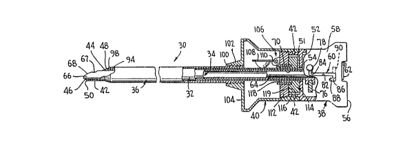
A safety penetrating instrument 30 according to the
present invention is illustrated in Fig. 1 and includes an
elongate, tubular needle 32, a safety probe 34 movably
disposed concentri~cally within needle 32, an elongate, tubular
portal sleeve 36 concentrically disposed around needle 32, a
hub 38 mounting needle 32 and safety probe 34 and a housing
40 mounting portal sleeve 36. The hub 38 can be latched to
housing 40 with the use of any suitable releasable mechanism,
such as ball detents 42, allowing the hub to be removed from
the housing withdrawing the needle and safety probe from the
portal sleeve. Accordingly, the safety penetrating instrument
30 may be considered to be formed of a portal unit and a
penetrating unit, the portal unit including portal sleeve 36
and housing 40 and the penetrating unit including needle 32,
safety probe 34 and hub 38.
Needle 32 is preferably made of a cylindrical length of
stainless steel having a diameter and wall thickness dependent
upon the procedure to be formed and the anatomical cavity to
be penetrated. The needle has a distal end 42 terminating at
a peripheral edge 44 disposed in a plane positioned at an
acute angle relative to the longitudinal axis of the needle
WO92/10974 PCT/US9l/09l~
20~8182 8 ~
to define a sharp, tissue penetrating tip or point 46 at a
leading or front edge and, proximally spaced from the leading
edge, a trailing or rear edge 48. At the leading edge, the
wall of the needle is ground at an angle, as shown at 50, to
terminate at sharp tip 46 such that tip 46 is aligned with the
inner surface of the cylindrical needle wall. Needle 32 has
a proximal end 51 secured to a front wall 52 of hub 38 by any
suitable means, such as threads 54 or cement.
Hub 38 is preferably made of plastic to reduce cost and
has an external configuration to cooperate with housing 40 to
be easily grasped with one hand for use in penetrating tissue.
Hub 38 is substantially rectangular in cross-section and
includes, as best shown in Figs. 1 and 2, four side walls
extending from front wall 52 to a rear wall 56 to provide a
rearwardly flared outer profile with one side wall, indicated
at 58, having a slot 60 therein. Rear wall 56 has a recess
therein receiving and protecting a valve 62 which is shown as
a flap valve but could be any type of valve, such as a stop
cock. A tube 64 has a flange 65 threadedly secured to rear
wall 56 adjacent valve 62 such that valve 62 controls flow
therethrough, and tube 64 extends through hub 38 to be
slidably received in safety probe 34.
Safety probe 34 is preferably made from a cylindrical
length of a rigid or flexible material, such as stainless
steel or plastic dependent upon use of the safety penetrating
instrument, and has a distal end 66 with a configuration to
protect sharp tip 46 of needle 32 in an extended position as
shown in Fig. 1. The distal end 66 has a hole 67 therein and
an end sur~ace 68 disposed at an acute angle to the
longitudinal axis of needle 32 substantially the same as the
acute angle of the needle peripheral edge 44. In this manner,
the peripheral edge 44 of the distal end of needle 32 and the
distal end surface 68 of safety probe 34 will be maintained
in substantially parallel relation when the safety probe is
in the extended position and will be positioned in
substantially the same plane when the safety probe is in the
retracted position as illustrated in Fig. 3. End surface 68
WO92/10974 PCT/US91/091~
9 2098182
has a rounded, partially spherical or bulging, dome-shaped
configuration; however, dependent upon use of the safety
penetrating instrument, the end surface can be substantially
flat so long as sharp surfaces are avoided. By grinding the
distal end of the needle, the sharp tip 46 is positioned in
abutment with the lateral wall of the safety probe in the
extended position to be well protected and unexposed. As best
shown in Fig. 2, the safety probe has a proximal end 70
disposed within hub 38 carrying spaced annular ribs 72 and 74
between which is rotatably mounted a member or plate 76 having
a pin 78 threadedly secured thereto and extending through slot
60, the pin 78 having a spherical end or knob to be easily
grasped. A helical spring 80 is mounted in compression
between plate 76 and the end wall of hub 38, and the spring
80 has longitudinally extending ends received in apertures in
plate 76 and flange 65 such that the spring can be wound in
torsion to bias the plate and the pin 78 carried thereby in
a clockwise direction looking at the proximal end of the
safety penetration instrument or upwardly looking at Fig. l.
The slot 60 formed in hub 38 has a longitudinal portion 82
aligned in parallel relation with the longitudinal axis of the
needle, a distal transverse portion 84 and a proximal
transverse portion 86 having a recess 88 at the end thereof
extending parallel with the longitudinal slot portion 82. If
desired, for purposes to be explained hereinafter, the
proximal slot portion can extend in the same direction as the
distal slot portion as shown in dashed lines at 90.
Portal sleeve 36 is preferably made of a cylindrical
length of stainless steel or other suitable, medically
acceptable, plastic or metal material and can be rigid or
flexible and transparent or opaque. The portal sleeve has a
distal end 94 terminating at a peripheral edge 96 disposed in
substantial alignment with or forward of the trailing edge 48
of the needle such that the distal ends of the needle and the
portal sleeve present a substantially smooth profile to
facilitate tissue penetration. The distal end 94 has a
tapered, conical portion 98 disposed at the same acute angle
WO92/10974 PCT/US9l/09188
209~182 10 w
to the longitudinal axis of needle 32 as the angle of the
peripheral edge 44 of the needle such that the portal sleeve
36 smoothly follows the needle during tissue penetration. The
portal sleeve 36 has a threaded proximal end 100 removably
received in a threaded nipple 102 extending from a front wall
104 of housing 40. Housing 40 is preferably made of plastic
and has a rectangular configuration in cross-section
corresponding to the cross-sectional configuration of hub 38
with a flared external profile adjacent front wall 104 to
facilitate grasping during use. A valve assembly 106 is
mounted in housing 40 to control flow through the portal
sleeve once the penetrating unit is removed therefrom. The
valve assembly can have any acceptable configuration and, as
shown, includes a flapper valve member 108 spring-biased to
a closed position against an annular valve seat formed by the
distal end of a cylindrical tube 110 secured in a threaded
plate 112 pivotally mounting the valve member and threadedly
secured in an annular threaded wall portion of housing 40.
An adapter plug 114 is mounted on the proximal end of tube 110
and is integrally constructed of a flexible resilient
material, such as Teflon, silicone rubber or plastic. The
adapter plug 114, as also shown in Fig. 21, has a thick flange
portion 116 with an annular recess 117 therein for receiving
the proximal end of tube 110 and a cylindrical inner wall 118
extending from an inner edge of a central aperture 119 in the
flange 116 to be snugly received within tube 110. The
thickness of wall 118 is dependent upon the size of the
instrument to be inserted through the portal sleeve such that,
if instruments smaller than the penetrating unit are to be
introduced into the body after the penetrating unit is
withdrawn from the portal sleeve, the adapter plug 114 is
removed from the proximal end of tube 110 and another adapter
sleeve is inserted therefor having an inner cylindrical wall
of greater thickness to engage the smaller instruments along
the length of the wall. That is, the diameter of the inner
wall 118 and the thickness thereof will vary dependent upon
the diameter of instruments passing through the portal unit,
WO92/10974 PCT/US91/09188
ll ' ~O9S182
and the inner diameter will be substantially the same as or
slightly less than the outer diameter of the instrument
passing therethrough to produce a seal there- around extending
along the length of the wall. By utilizing various size
adapter plugs with inner sealing walls to accommodate various
size instruments, an effective seal can be produced for
instruments varying greatly in size, for example from 2mm to
12mm. In order to assemble the safety penetrating
instrument 30, the safety probe 34 can be inserted in the
proximal end of the hub and through the needle along with the
tube 64 and spring 64. With the plate 76 in place, the pin
78 is passed through the slot 60 and screwed into the plate
such that the plate cannot rotate within the hub. The tube
64 is twisted clockwise to create torsion in the spring to
rotationally bias plate 76 and pin 78 clockwise as noted
above. The tube flange 65 is then screwed into the end wall
56 of the hub to place the safety penetrating instrument 30
in the condition shown in Fig. 2 with spring 80 biasing the
safety probe distally such that the distal end 66 is normally
in the extended position shown in Fig. 1 protecting the sharp
tip 46 of the needle. The pin 78 will be, accordingly, biased
distally along longitudinal portion 82 of slot 60 to a
position adjacent distal transverse portion 84 and be biased
into transverse slot portion 84 due to the torsional bias from
the spring to releasably lock the safety probe in the extended
position. The penetrating unit formed by the needle 32, the
safety probe 34 and the hub 38 is then combined with the
portal unit by passing needle 32 through the central aperture
in adapter plug 114 and through housing 40 and portal sleeve
36 moving valve member 108 away from the valve seat on the
distal end of tube 110. With the hub 38 abutting the housing
40, a cylindrical skirt 119 extending from hub front wall 52
will be disposed within the open proximal end of the housing,
and the detents 42 will hold the hub in position with respect
to the housing. In this position, the distal peripheral edge
96 will be disposed substantially in alignment with the
trailing edge 48 of the needle distal end to facilitate tissue
WO92/10974 PCT/US9l/09188
2~9~1~ 2 12
penetration by the safety penetrating instrument 30. It is
important for smooth tissue penetration that the distal end
of the portal sleeve be positioned at least in alignment with
the trailing edge of the needle or forward thereof. If the
distal end of the portal sleeve is proximally spaced from the
trailing edge of the needle, tissue penetration will be more
difficult with increased trauma. Accordingly, the positioning
of the distal ends of the needle, the safety probe and the
portal sleeve are extremely important in facilitating tissue
penetration with minimal tissue resistance and trauma.
The distal end of the safety penetrating instrument 30
is illustrated in Fig. 3 with the safety probe in the
retracted position, and it can be seen therefrom that the end
surface 68 of the distal end of the safety probe is in
substantially the same plane as the peripheral edge 44 at the
distal end 42 of the needle 32 with the distal end of the
safety probe substantially filling the open distal end of the
needle to reduce gaps between the distal ends of the needle
and the safety probe and the trapping or jamming of tissue
therebetween.
A modification of the safety probe 34 is illustrated in
Fig. 4 wherein the safety probe has a distal end 120 having
an end surface 122 angled similar to end surface 68 in Fig.
3. A protruding peripheral rim 124 extends from end surface
122 to expand laterally in all directions when the safety
probe is in the extended position and contract to completely
fill the open distal end of the needle when the safety probe
is in the retracted position. In this manner, the reduced
gaps between the distal ends of the needle and safety in the
embodiment of Fig. 3 are prevented to eliminate trapping or
jamming of tissue.
A modification of safety penetrating instrument 30 is
illustrated in Figs. 5, 6, 7 and 8 with the primary
differences being that the sharp point 46 at the distal end
of the needle 32 is not angularly ground such that the sharp
tip is aligned with the outer surface of the wall of the
needle as in conventional hypodermic needles and that the
WO92/10974 PCT/US9l/09188
13 2 0~ 81 8 2
safety probe 34 has a groove or recess 126 formed therein such
that the distal end is closed with the exception of a lateral
hole 128. The distal end of the safety needle has a specific
configuration to protect the sharp needle tip 46 due to the
needle tip being received in groove 126 when the safety probe
is in the extended position and further due to the distal end
being bent or angled, as best shown in ~ig. 5, relative to the
longitudinal axis of the needle such that, in the extended
position, the bent portion forms a second recess 130 for
receiving the sharp needle tip 46. By constructing the safety
probe of a flexible material, such as plastic, the safety
probe will straighten or realign when moved to the retracted
position and, when moved to the extended position, will return
to the bent or angled configuration forming recess 130 for
receiving the needle tip.
In a further embodiment illustrated in Fig. 9, the distal
end of the safety probe is angled or bent as described with
respect to the previous embodiment and, additionally, the
distal end is split or slotted to form opposing parts 132 and
134. The split or slot 136 terminates at radiused ends 138
such that the parts 132 and 134 are biased away from each
other to produce an expanded state when the safety probe is
in the extended position. Use of the safety penetrating
instrument of Fig. 9 for grasping and/or cutting tissue, for
example for biopsy, is illustrated in Fig. 10 wherein it can
be seen that tissue 140 can be grasped by placing the safety
probe adjacent the tissue in the expanded state, as shown in
Fig. 9, and, thereafter, pulling the safety probe proximally
into the needle by manually grasping pin 78 and moving the pin
along the longitudinal slot portion 82 against the bias of
spring 80. The safety probe can be releasably locked in the
retracted position by moving the pin 78 into transverse slot
portion 86 and, into the recess 88 formed therein to prevent
the pin from returning to its normally biased position. When
the safety penetrating instrument is used for biopsy, the
safety probe is preferably made of stainless steel and the
WO92/10974 PCT/US9l/091~
209~182 14 _
distal edges are inwardly sharp to cut and grasp the tissue
to be taken for biopsy.
Another embodiment of a safety penetrating instrument
according to the present invention is illustrated in Figs. 11
and 12 wherein an elongate tubular needle 142 has a distal end
143 with an open, acutely angled end defining a peripheral
edge 144 with a portion 146 of the distal end curving toward
the longitudinal axis of the needle to terminate at a sharp
distal tip 148 laterally or radially displaced from the wall
of the needle. A safety probe 150 has a distal end 152 with
a curvature to protrude over sharp tip 148 when the safety
probe is in the extended position as illustrated in Fig. 12.
The distal end 152 has a bulbus configuration with an aperture
154 therein to permit communication through the safety
penetrating instrument for injection or evacuation of fluid.
While a smoothly curving configuration can provide protection
for the sharp needle tip 148, a recess in the form of a
concave portion 155 shown in dashed lines can be provided in
the safety probe distal end to further prevent exposure of the
sharp needle tip.
Another embodiment of a safety penetrating instrument
according to the present invention is illustrated in Figs. 13
and 14 wherein an elongate tubular needle 156 has a curved
distal portion 158 extending less then portion 146 shown in
Fig. 11 to terminate at a sharp tip 160. A safety probe 162
has a longitudinal groove 164 therein to form a recess
receiving the sharp needle tip 160 at the distal end of the
needle as illustrated in Fig. 14. The distal end of the
safety probe has a large hole 166 and a plurality of small
holes 168 therein. By providing a plurality of holes in the
distal end of the safety probe, communication can be
maintained even if one of the holes becomes blocked.
The embodiment of a safety penetrating instrument
according to the present invention illustrated Figs. 15 and
16 is similar to that illustrated in Figs. 13 and 14 with the
exception that the safety probe 170 has an annular open distal
end 172 allowing communication directly therethrough. The
2098 1 82
round peripheral edge of distal end 172 is interrupted to form
a recess in the form of a slot or groove 174 for receiving the
sharp needle tip 160.
Only the distal ends of the safety penetrating instrument
have been illustrated in Figs. 5 - 16; however, it will be
appreciated that the pro~;m~l portions can be formed by any
suitable cooperating structure and are particularly advantageous
when utilized with the locking and spring biasing mechanism
illustrated in Figs. 1 and 2. Similarly, various features from
the disclosed embodiments can be combined dependent upon the
procedure to be performed with a particular safety penetrating
instrument.
In use, it will be appreciated that with the pin 78
disposed in distal transverse slot portion 84, the safety probe
cannot move pro~;m~lly within needle 32 thereby assuring that
the sharp tip 46 is protected to prevent inadvertent contact.
When it is desired to penetrate an anatomical cavity, the blunt
end surface 68 is positioned in abutment with the tissue T to be
penetrated as illustrated in Fig. 17, and a finger is utilized
to manually move pin 78 counter-clockwise against the torsional
bias of spring 80 to be aligned with the longitn~;n~l portion 82
of slot 60. With the pin 78 in this position, when the end
surface 68 of the distal end of the safety probe is forced
against the tissue to be penetrated, the safety probe 34 will
move proximally against the bias of spring 80 to the retracted
position as illustrated in Figs. 3 and 18, it being noted that
when the distal end of the safety probe includes a bent or
angled portion, the distal end of the safety probe will
straighten out as the safety probe is withdrawn into the needle
and additionally that when the distal end has an expanded state
in the extended position the distal end will be in a contracted
state when the safety probe distal end is mo~ed to the retracted
position. With the safety probe in the retracted position,
penetration through the tissue with m;n;m~l tissue jamming and
trapping is accomplished due to the smooth profile of the distal
end of the safety penetrating instrument. Pin 78 will not enter
' ~ ~J
WO92/10974 PCT/US9l/~l~
2098~82
16
proximal transverse slot portion 86 when the pin is aligned
therewith due to the torsional or rotational bias of spring
80 in the opposite direction; and, thus, once the anatomical
wall has been completely penetrated, spring 80 will bias the
safety probe distally such that the safety probe returns to
the extended position as illustrated in Fig. 19 to protect the
sharp tip 46 such that the sharp tip is exposed only during
the tissue penetrating step. When the safety probe includes
a protective recess such as the groove 126 in the embodiment
of Fig. 5, the bent or angled portions 130 in the embodiments
of Figs. 5 and 9, the slot 136 in the embodiment of Fig. 9,
the concavity 154 in the embodiment of Fig. 11 and the grooves
or slots 162 and 174 in the embodiments of Figs. 13 and 15,
the sharp needle tip is protected by being received in the
recess. As previously noted, when the angle of the end
surface of the safety probe distal end corresponds with the
angle of the needle distal end, the distal ends of the needle
and the safety probe will be disposed in substantially the
same plane to reduce tissue resistance during penetrating and
produce a minimum gap between the safety probe and the needle
to minimize trapping or jamming of tissue therebetween. The
reduction in tissue resistance is further enhanced by the
positioning of the peripheral distal edge of the portal sleeve
substantially in alignment with the trailing edge of the
needle distal end. Protection of the sharp needle tip is
particularly effective where the distal end of the needle is
ground as shown at 50 in that the sharp needle tip will abut
the distal end of the safety probe when the safety probe is
in the extended position.
If, after penetration into the anatomical cavity, it is
desired to penetrate additional tissue, the surgeon can
selectively release the safety probe from the extended
position by moving pin 78 counter-clockwise to be aligned with
longitudinal slot portion 82. If the protection provided by
the safety probe is not required or desired for further
procedures, the pin 78 can be moved into recess 88 of proximal
transverse slot portion 86 to releasably lock the safety probe
- 17 2 n98l ~2
in the retracted position or a position further withdrawn into
needle 32 dependent upon the longitl~;n~l position of transverse
slot portion 86. If pro~;m~l transverse slot portion 90 is
provided, the safety probe will automatically lock in the
retracted position when pin 78 is aligned with slot portion 90
due to the rotational bias from spring 80. If pin 78 is
positioned distally of slot portion 90 when the safety probe is
in the retracted position, the safety probe will not
automatically lock during use but rather the pin will have to be
manually pulled back to alignment with slot portion 90 to lock
the safety probe within the needle.
While plate 76 and pin 78 can rotate slightly within hub
38, safety probe 34 does not rotate therewith to maintain
alignment with needle 32, and various index configurations to
maintain rotational alignment between the safety probe and the
needle can be used, if desired. For example, the safety probe
can be splined with the needle or with tube 64.
Once the safety penetrating instrument has penetrated into
an anatomical cavity, the safety penetrating instrument can be
used to permit flow of fluids into or out of the cavity since
comm~]n;cation is established through hole 67 and the body of the
safety probe 34 and through tube 64 and valve 62. In this
manner, the safety penetrating instrument 30 can be used, for
example, to create a pneumoperitoneum.
Once the needle has been used to penetrate tissue to the
extent desired by the surgeon, the portal sleeve 36 can be left
in place by withdrawing the penetrating unit from the portal
unit; and, once the penetrating unit is withdrawn, valve member
108 will engage the valve seat formed by the distal edge of tube
110 to prevent fluid flow through the portal formed by the
portal sleeve 36 and housing 40. If it is desired to then
insert an instrument having a diameter less than the diameter of
needle 32, adapter plug 112 is removed from the proximal end of
tube 110 and replaced with an adapter plug having a central
aperture and tubular passage formed by the inner wall 118 of a
diameter corresponding to the smaller instrument to be inserted.
20981 8~
18
For procedures where it is desired to gain access to an
area of an anatomical cavity substantially offset from the
longitudinal axis of the safety penetrating instrument, the
portal sleeve can have a normal, non-linear configuration such
that, once penetration is completed and the penetrating unit is
withdrawn from the portal unit, the portal sleeve will return to
the non-linear configuration. For example, the portal sleeve is
shown in dashed lines in Fig. 19 as having a curved
configuration 181 to which the portal sleeve returns when the
needle and safety probe are withdrawn.
A modification of the portal sleeve of the safety
penetrating instrument is shown in Fig. 20 wherein the portal
sleeve 35 has a distal end with a peripheral edge 176 obliquely
arranged at an acute angle relative to the longitudinal axis of
the needle with a rounded leading edge 178 disposed in
longitudinal alignment with sharp needle tip 46 and a trailing
edge 180 in alignment with the trailing edge 48 of the
peripheral edge at the needle distal end. The modification of
Fig. 20 has the advantages of assuring that at least the leading
edge of the portal sleeve has penetrated the anatomical wall
when the safety probe returns to the extended position and of
facilitating tissue penetration since the distal end of the
portal sleeve gradually enters the tissue following the needle.
Another modification of the safety penetrating instrument
of the present invention is shown in Fig. 21 wherein the valve
assembly 106 is formed of a member 182 having an annular base
184 sealingly engaged with the distal end of tube 110 and a
plurality of spaced, spreadable, legs 186 extending distally
from base 184 to produce a normally conical configuration. The
member 182 is preferably made of unitary construction of a
material facilitating movement of medical instruments
therethrough and allowing legs 186 to flex, such as a plastic
like nylon. The legs 186 are concentrically disposed around
annular base 184 and the central opening therethrough and are
slightly spaced from each other and terminate at curved relief
recesses (not shown). A conical, stretchable sleeve or
t.~ ~;
~.; ,.-
WO92/10974 PCT/US91/09188
.~.
19 209~182
membrane 188, preferably made of a rubber-like material such
as silicone, has a configuration to tightly fit over legs 186
such that the sleeve and legs are normally biased to a closed
position at the apex to prevent passage of fluids through the
valve assembly. The valve assembly is normally biased to a
closed position due to the elastic force of conical sleeve 188
around legs 186; and, when a surgical instrument is inserted
through the portal unit, the surgical instrument will cause
legs 186 to flex outwardly stretching conical sleeve 188 such
that conical sleeve acts as a seal to prevent passage of
fluids thereby and tightly engages the surgical instrument in
the gaps where the legs are spread apart. The valve assembly
provides a passage of variable size to accommodate surgical
instruments of various sizes passing therethrough in that the
legs and conical sleeve contract to sealingly engage the
surgical instrument regardless of the diameter thereof, and,
in combination with adapter plug 114, an excellent seal
through the portal unit is established.
In the modification of Fig. 21, portal sleeve 36 is
resiliently, movably secured to the housing 40 to allow the
portal sleeve to be slightly moved with respect to the housing
and the penetrating unit against a spring bias. The proximal
end 190 of the portal sleeve slidably received in a nipple 192
which is threadedly secured to a front wall 193 of housing 40,
and a seal is provided by one or more O-rings 194. The
proximal end 190 carries an annular end collar 196 limiting
distal movement of the portal sleeve by abutment with the
front wall portion of housing 40 formed by nipple 192. A
helical spring 198 is mounted in compression between collar
196 and an annular shoulder 200 fixed in housing 40 such that
the portal sleeve is distally biased. During penetration of
tissue, the portal sleeve can move proximally against the bias
of spring 198 to facilitate penetration during difficult
procedures.
If automatic locking of the safety probe 34 in the
extended position is not desired, the spring 80 is not wound
during assembly such that pin 78 is not rotationaily biased.
WO92/10974 PCT/US91/091~
2098182 20 '~ ~~
Accordingly, after spring 80 returns the safety probe 34 to
the extended position when the force on the safety probe
distal end from tissue resistance is removed, the pin will not
enter transverse slot portion 84, and the safety probe will
not be locked in the extended position. Thus, the surgeon has
the option of locking the safety probe in the extended
position by manually moving pin 78 into transverse slot
portion 84 or allowing the safety probe to be free to move
proximally by not moving pin 78 into transverse slot portion
84.
Safety penetrating instruments formed of an elongate
tubular needle with a safety probe having a distal end
configured to define a protective recess for receiving the
sharp tip of the needle in the extended position are
particularly advantageous for safely introducing a portal
sleeve into veins or arteries with reduction of the chances
of developing hematoma. Where the safety penetrating
instrument utilizes an elongate tubular needle with an acutely
angled distal end including a portion curving toward the
longitudinal axis of the needle to terminate at a sharp tip
radially inwardly spaced from the wall of the needle and a
safety probe having a distal end configured to protrude over
the sharp tip in the extended position, rotational penetration
can be safely performed to introduce a portal sleeve into the
body. The distal end portion of the needle can be curved
inwardly to any desired extent from a slight curve, as shown
in Figs. 11, 13 and 15, to a curve through almost the entire
diameter of the needle to produce a sharp, spoon-like shape
with the distal end of the safety probe configured to protrude
over the sharp tip by providing a protective recess therein
or by bending in the same direction as the curvature of the
curved position.
The safety penetrating instrument of the present
invention can be used for penetrating cavity walls in a manner
similar to presently used trocars and can have various
diameters ranging from 2mm to 12mm. The use of a tubular
needle in the safety penetrating instrument of the present
WO92/10974 PCT/US91/091
21 2 09818 2
invention has the advantages of permitting placement of the
portal sleeve or cannula in small or large cavities to provide
a portal communicating therewith, of permitting a scooping
movement during penetration to get through the wall without
compressing the cavity when the needles of Figs. ll, 13 and
15 are used, of being useful for first, second or other
puncture techniques, of permitting penetration of a second
cavity wall after penetration of a first cavity wall and of
allowing the performance of additional procedural steps, such
as suction, cutting, biopsy and aspiration. Dependent upon
the configuration of the safety probe to be used and the
procedure to be performed, the safety probe can be made of
metal, such as stainless steel, rubber or plastic, with
materials having good sliding characteristics, such as Teflon,
being preferred. While the safety probes are advantageously
hollow or tubular to allow passage of fluids therethrough, the
safety probes can be solid if desired.
From the above, it will be appreciated that safety
penetrating instruments according to the present invention
provide increased protection of the sharp tip of a tubular
needle while also facilitating us~ and tip exposure after
penetration of an anatomical cavity by providing selective,
releasable locking positions for the safety probe. It will
be appreciated that the safety penetrating instrument of the
present invention is useful to penetrate tissue in various
portions of the body, and "anatomical cavity" as used herein
refers to any area of the body requiring penetration of tissue
for access.
Inasmuch as the present invention is subject to many
variations, modifications and changes in detail, it is
intended that all subject matter discussed above or shown in
the accompanying drawings be interpreted as illustrative only
and not to be taken in a limiting sense.