Note: Descriptions are shown in the official language in which they were submitted.
~ n ~ ~ ~ g ~
PSEUDOMONAS PEPTIDE COMPOSITION AND METHOD
1. Field of the Invention
The present invention relates to Pseudomonas-derived
polypeptide antigens, to methods of producing such anti-
gens, and to antibodies immunoreactive against the anti-
gens.
2. References
Adams, M.H. Methods of study of bacterial viruses,
p. 443-452. In M.H. Adams (ed.), Bacteriophages. Inter-
science Publishers, Inc., New York (1959).
Ausubel, F.M., et al., Current Protocols in Molecu-
lar Biology, John Wiley and Sons Inc., Media, PA (1990).
Beachy, E.H. 1981 J. Infect. Dis. 143:325-345
(1981).
Boulianne, G.L. et al., Nature 312:643-646 (1984).
Carr, B., et al., Gerontology 35:127-129 (1989).
Cwirla, S.E. et al., Proc. Natl. Acad. Sci. USA
87:6378-6382 (1990).
Devlin, J.J., et al., Science 249: 404 (1990).
~92/12169 PCT/CA91/O~F
2og8296
Doig, P., et al., Infect. Immun. 56:1641-1646
~1988).
Doig, P., et al., Infect. Immun. 58:124-130 (1990).
Franklin, A.L., et al., Infect. Immun. 55:1523-1525
~1987).
Geyson, H.M. et al., in Synthetic Peptides as Anti-
gens; Ciba Foundation Symposium 119:131-149 (1986).
Irvin, R.T. and Ceri, H., Can. J. Microbiol. 31:268-
275 (1985).
Irvin, R.T., et al., Microbial Ecology Health
Disease, 3:39-47 (1990).
Lee, K.K., et al., Infect. Immun. 57:520-526 (1989).
Lee, K.K., et al, Inf Immunol, 58:2727-32 (1990).
Marrs, C.F., et al., Am. J. Med. 88 (Suppl 5A):
36S-40S (1990).
McBride, L.J., et al., Clin Chem, 35:2196-2201
(1989).
McEachran, D.W., et al, Can. J. Microbiol.
31:563-569 (lg85).
McEachran, D.W., et al., J. Microbiol. Meth. 5:99-
111 (1986).
Morrison, S.L., et al., Proc. Natl. Acad. Sci. USA
81:6851-6855 (1984).
Nieto, A., et al., Mol. Immunol. 21:537-543 (1984).
Pasloske, B.L., et al., J Bacteriol, 170:3738-3741
(1988).
Paranchych, W., et al., Can. J. Microbiol. 25:1175-
1181.
Paranchych, W. et al., Advan. Microbiol. Phys.
29:53-114 (1988).
Parmley, S.F. and Smith, G.P. Gene 73:305-318
(1988).
Rabbitts, T.H. et al., Nucleic Acids Res. 9:4509-
4524 (1981).
., , , ~ , ,
i , . . . . .
~ 92/12169 PCT/CA91/OW59
20982~6
Sanger, S., et al., PNAS (USA), 74:5463-5467 (1977).
Sato, H. and Okinaga, K., Infect. Immun. 55: 1774-
1778 (1987).
Scott, J.K. and Smith, G.P., Science 249:386-390
(1990).
Sastry, L., et al., Proc. Natl. Acad. Sci. USA
86:5728-5732 (1989).
Sastry, P.A., et al., Can. J. Cell Biol. 63:284-291
(1985).
Staddon, W., et al., Can. J. Microbiol. 36:336-340
(1990) .
Todd, T., et al., Am. Rev. Respir. Dis.
140:1585-1589 (1989).
Tsai, C., et al., Anal. Biochem. 119:115-119 (1982).
Worobec, E.A., et al., J. Biol. Chem. 260:938-943
(1985).
zu Putlitz, J., et al., Bio/Technology 8:651-654
(1990 ) .
3. Background of the Invention
During the past two decades, Pseudomonas aeruginosa
has been recognized as a pathogen which causes between
10% and 20% of infections in most hospitals. Pseudomonas
infection is especially prevalent among patients with
burn wounds, cystic fibrosis, acute leukemia, organ
transplants, and intravenous-drug addiction. P. aerugi-
nosa is a common nosocomial contaminant, and epidemics
have been traced to many items in the hospital environ-
ment. Patients who are hospitalized for extended periods
are frequently affected by this organism and are at
increased risk of developing infection. The most serious
infections include malignant-external otitis, endophthal-
mitis, endoconditis, meningitis, pneumonia, and septice-
mia. The likelihood of recovery from Pseudomonas infec-
WO92/12169 2 0 9 ~ ~ g 6 PCT/CA91/~
tion is related to the severity of the patient's underly-
ing disease process. The reported mortality for P. aeru-
ginosa pneumonia is as high as 50-80%. Even with the
development of newer antibiotics, resistance remains a
problem necessitating combined antibiotic treatment for
severe P. aeruginosa infections.
Various therapies for the management of severe P.
aeruginosa infections have been evaluated for many years,
with particular attention focused on virulence factors.
As with most bacterial pathogens, virulence of P. aerugi-
nosa is multifactorial and is the product of many inter-
acting variables, involving both the bacterium and the
host. Evidence suggests that the initial event in infec-
tion is the a &erence of microorganisms to epithelial
cells of mucosal surfaces tBleachy). Organisms that are
unable to adhere to mucosal surfaces fail to colonize
because they are removed by the secretions that bathe the
mucosal surfaces (Bleachy). The adherence process is
dependent upon the specific recognition between bacteria
and epithelial cells.
For a number of gram-negative bacteria, including P.
aeruginosa, attention has been directed to surface appen-
dages as mediations of adherence. The surface of many
gram-negative bacteria, e.g., Escherichia coli, P. aeru-
ginosa, Moraxella bovis, Neisseria gonorrhea, are coveredwith filamentous structures called pili or fimbriae.
Pili are composed primarily of protein (pilin) and have
been found to act as antigenic determ;n~nts when injected
into test ~nt~l S. In P. aeruginosa, strain-specific
pili, such as those designated PAO, PAK, and CD4, mediate
the colonization of the bacteria in humans (Doig,88).
Some P. aeruginosa bacteria lacking these pili,
either through mutation or loss of the plasmid carrying
the pilus gene, are incapable of colonizing mucosa.
CA 02098296 1998-04-14
Apparently, the pili on the surface of the bacterium adhere
to the lining of the throat and trachea through specific
interactions with epithelial cell receptors. P. aeruginosa
can utilize both pili and alginate (the principle component
of the P. aeruginosa capsule) as adhesions to mediate
attachment to human respiratory epithelial cells (Doig).
Equilibrium analysis of P. aeruginosa binding to human
respiratory epithelial cells indicates that the Pseudomonas
pilin adhesion has a considerably higher apparent affinity
or binding constant than does the alginate adhesion
10(McEachran, 1985; 1986). These observations suggest that
the pilus adhesion is likely the dominant Pseudomonas
adhesion in the initiation of an infection (Irvin).
Adhesion-medicated anchorage is a prerequisite for the
induction of disease by P. aeruginosa.
15PCT Application Publication No. WO 90/13563 discloses
a P. aeruginosa peptide derived from the C-terminal region
of the P. aeruginosa pilin protein, and specifically, the
C-terminal region which includes two Cys residues and the
intervening amino acid residues. The derived region of
representative peptides vary in length between 14 and 19
amino acid residues, including the two Cys residues, and
are prepared in both oxidized (disulfide-linked) and
reduced (non-cyclized) form. The peptides (in both reduced
and oxidized form) were shown to have the following
properties:
(a) ability to bind to human tracheal epithelial cells
(TECs) and human buccal epithelial cells (sEcs)i
(b) ability to inhibit binding of Pseudomonas pilin
peptide to tracheal epithelial cells (TECs) and buccal
epithelial cells (BECs);
(c) ability to elicit serum antibodies which are
immunoreactive with Pseudomonas pilin peptide; and
CA 02098296 1998-04-14
5a
(d) ability to elicit serum antibodies which block
binding to Pseudomonas pilin peptide to BECs.
Watts et al (Infect. Immun. 42(1): 113-121, 1983)
described a peptide fragment TCIV comprising Pseudomonas
aeruginosa strain PAK pilin residues 121-144 of the pilin
protein and a subfragment of TCIV, comprising pilin
residues 128-144. Whereas the two cysteines present in
TCIV(121-144) were oxidized to form disulfide bridges, the
cysteines in TCIV(128-144) were carboxymethylated and
therefore unable to form disulfide linkages. These
peptides were found to have similar reactivities with anti-
PAK pilus antiserum. Based on these results, it was
suggested that the disulfide bridge between residues 129
and 142 and resulting cyclic structure are not required for
antigenicity and recognition of the region by anti-pilus
antibodies.
Lee et al. (Infect. Immun. 57(2): 520-526, 1989)
described production of polyclonal antibodies in response
to injection of conjugated C-terminal peptide-BSA
conjugates into rabbits. The C-terminal peptides comprised
amino acid residues 128-144 of Pseudomonas aeruginosa
strain PAK pilin. Antibodies formed in response to both
oxidized and reduced forms of the peptide were shown to
react similarly with strain PAK pilus structures (pili).
However, only one of the antibodies (raised against the
oxidized peptide-conjugate) was found to cross-react with
P.aeruginosa strain PAO pili. Based on these results, Lee
concluded that the disulfide bridge is important in the
immunogenicity of antisera reactive with pili from
different strains of Pseudomonas.
9 ~
It has now been discovered that the Pseudomonas-
derived peptide is able to inhibit binding of unrelated
bacterial and fungal organisms to human TECs and/or
BECs. Thus, the epithelial cell receptor site(s) which
bind the Pseudomonas-derived peptide, and thereby
inhibit binding of Pseudomonas pilin (and Pseudomonas
bacteria) to TECs andtor BECs is also involved in
binding of other bacterial and fungal organisms to
these target cells. It has further been shown, in
studies conducted in support of the present invention,
that monoclonal antibodies prepared against the
Pseudomonas-derived peptide are effective in blocking
fungal cell adherence to BECs.
These combined findings show that the Pseudomonas-
derived peptide, and antibodies produced in response tothe peptides, are capable of inhibiting bacterial and
fungal infections in which the infecting microorganism
has surface proteins which are antigenically
crossreactive with antibodies produced against the C-
terminal, disulfide-linked peptide region of P.
aeruqinosa pilin protein.
4. summarY of the Invention
The invention includes, in one aspect, a peptide
having a sequence corresponding to a'C-terminal region
of a P. aeruqinosa pilin protein, and more
specifically, to one of the sequences:
(i) K C T S D Q D E Q F I P K G C S K,
(ii) K C T S D Q D E Q F I P K G C S R,
(iii) A C K S T Q D P M F T P K G C D N,
(iv) T C T S T Q E E M F I P K G C N K P,
(v) S C A T T V D A K F R P N G C T D,
(vi) A C T S N A D N K T L P K T C Q T A T T T T P,
~ ,:
92/12169 PCI/CA91/00459
-- 20982~
(vii) N C K I T K T P T A W K P N Y A P A N C P K S,
(viii) T C G I T G S P T N W K A N Y A P A N C P K S,
~ix) T C G I T G S P T N W K T N Y A P A N C P K S, and
(x) G C S I S S T P A N W K P N Y A P S N C P K S,
including amino acid variations which are internally
consistent among sequences (i)-(vi) and among sequences
(vii) - (x) .
The peptide of the invention is further characte-
rized by (a) a disulfide linkage between the two Cys (C)
residues; (b) immunospecific binding to PK99H or PK34C
monoclonal antibody; (c) specific binding to tracheal or
buccal epithelial cells; and (d) absence of specific
binding to P. aeruginosa pilin protein.
In a related aspect, the invention includes a com-
position for use as a vaccine against infection by bac-
terial and fungal organisms which have surface proteins
which are antigenically crossreactive with antibodies
produced against the C-terminal, disulfide-linked peptide
region of P. aeruginosa pilin protein. The composition
includes the above peptide and an immunogenic carrier to
which the peptide is attached. In one embodiment, for
use as a vaccine against Pseudomonas infection, the pep-
tide is ;mmllnoreactive with PK34C monoclonal antibody.
In another embodiment, for use as a vaccine against
Candida infection, the peptide is immunoreactive with
PK99H antibody.
In another aspect, the peptide has an amino acid
sequence selected from the group consisting of:
(i') D F Q F I P K,
(ii') D E Q F I P K,
~iii') D P M F T P K,
(iv') E E M F I P K,
(v') D A K F R P N, and
(vi') D N K Y L P K,
WO92/12169 PCT/CA91/O~'
i~098~
including amino acid variations which are internally con-
sistent among sequences (i')-(vi') and characterized by:
(a) immunospecific binding to a PK99H or a PK34C mono-
clonal antibo~y; (b) specific binding to human buccal or
human tracheal epithelial cells; and (c) absence of spe-
cific binding to P. aeruginosa pilin protein.
The peptide composition is employed in a vaccination
method of protecting an individual against infection by
bacterial and fungal organisms which have surface pro-
teins which are antigenically crossreactive with anti-
bodies produced against the C-terminal, disulfide-linked
peptide region of P. aeruginosa pilin protein.
Also forming part of the invention is a method of
producing a composition for use as a vaccine against
infection by bacterial and fungal organisms which have
surface proteins which are antigenically crossreactive
with antibodies produced against the C-terminal, disul-
fide-linked peptide region of P. aeruginosa pilin pro-
tein. The method relies on the selection of random-
sequence peptides produced by a vector library of random-
sequence polynucleotides, typically corresponding to a
random sequence of 5-10 codons. Selection is by peptide
binding to a monoclonal antibody immunoreactive with the
C-ter~;n~l, disulfide-linked peptide region of P. aerugi-
nosa pilin protein, and preferably to the PK34C or PK99Hmonoclonal antibody. The sequence of the selected bind-
ing polypeptide can be determined from the polynucleotide
coding sequence of the corresponding library vector.
From this sequence, the desired polypeptide can be made
by synthetic or recombinant means.
Also disclosed is a chimeric monoclonal antibody
composed of the variable regions of mouse PK34C or PK99H
monoclonal antibody, and the constant regions of a human
immunoglobulin G antibody. The antibody can be used to
~ 92/12169 PCT/CA91/00459
2~9~9e
treat infection by bacterial and fungal organisms having
surface proteins which are antigenically crossreactive
with antibodies produced against the C-term;n~l, disul-
fide-linked peptide region of P. aeruginosa pilin pro-
tein.
In still another aspect, the invention includes a
method of treating an infection of the lung caused by a
bacterial or fungal organism which have surface proteins
which are antigenically crossreactive with antibodies
produced against the C-terminal, disulfide-linked peptide
region of P. aeruginosa pilin protein. The method in-
cludes forming an aerosol of the peptide and administe-
ring the peptide by inhalation.
These and other objects and features of the present
invention will be come more fully apparent when the fol-
lowing detailed description of the invention is read in
conjunction with the accompanying drawings.
Brief Description of the Drawings
Figure l shows the amino acid sequences of Pseudomo-
_ peptides of the invention;
Figure 2 shows the relative binding affinity of na-
tive PAK peptide and PAK peptides with modified C-
terminus and N-terminus and peptides containing Ala
substitutions at each of the 17 peptide residues except
for the Cys residues for monoclonal antibody PK99H;
Figure 3 is a plot showing the binding of synthetic
peptide PAKr~d (solid squares) and PAKoX to human BECs;
Figure 4 is a modified Lineweaver-Burk plot of the
- 30 binding of PAK pili to human BECs, showing inhibition of
binding by increasing concentrations of PAK peptide;
Figure 5 is a bar graph showing the percent of PAK
pili binding to BECs, after preincubation of BECs with
Fab fragments of the antibodies indicated;
A ,s s'~
Figure 6 is a plot showing the inhibition of Pseudo-
monas bacteria binding to BECs by Fab fragments of mono-
clonal antibodies PK99H (open squares) and PK34C (open
triangles);
Figure 7 shows fractionated _. catarrhalis proteins
stained by immunoblot with polyclonal anti-PAK pili anti-
body (lane 1), and with protein stain (lane 2) and
standards (lane 3);
Figures 8A and 8B are immunoelectron micrographs
showing M. catarrhalis cells after binding with
polyclonal anti-PAK pili antibody (8A) and a control
antibody (8B);
Figures 9A and 9B are electron micrographs showing
M. catarrhalis bacteria with PK99H antibody localized by
colloidal gold;
Figure 10 shows a Western blot of several Bacteroi-
des strains immunoblotted with PK99H monoclonal antibody;
Figure 11 is a phase contrast micrograph of Candida
cells bound to BECs;
Figure 12 shows SDS-PAGE gel patterns of purified
Candida fimbrial protein (lane 3);
Figure 13 is a bar graph showing the inhibition by
Candida fimbriae on Candida adherence to BECs;
Figure 14 is a bar graph showing the inhibition by
Pseudomonas pili on Candida adherence to BECs;
Figure 15 is a bar graph showing the inhibition by
Candida fimbriae on Pseudomonas pili binding to BECs;
Figure 16 is a bar graph showing the inhibition by
anti-PAK pili antibodies on Candida binding to BECs;
Figure 17 illustrates recombinant methods for produ-
cing and selecting random-sequence peptides, in accord-
ance with the invention; and
'' ' ~B
. .
WO92/12169 PCT/CA91/~59
Figure 18 illustrates recombinant methods suitable
for producing a chimeric monoclonal antibody in accord-
ance with the present invention.
Detailed Description of the Invention
I. Definitions
The terms "epitope" and "epitopic," as used herein,
designate the structural component of a molecule that
is responsible for specific interaction with correspon-
ding antibody (immunoglobulin) molecules elicited by the
same or related antigen.
The term "antigen," as used herein, means an entity
that is recognized by an antibody.
The term "immunogen," as used herein, describes an
entity that induces antibody production in the host
~nim~l. In some instances the antigen and the immunogen
are the same entity, while in other instances the two
entities are different.
The term "immunologically mimics" is used herein to
mean that an immunogenic polypeptide of this invention is
not a natural protein or a cleaved fragment of a natural
protein, but a manufactured polypeptide, as by solid
phase synthesis or genetic engineering techniques, which
polypeptide induces production of antibodies that bind to
the inducing polypeptide and also to a corresponding
pilin or pilin polypeptide portion.
All amino acid residues identified herein are in thenatural or L-configuration unless otherwise specified.
In keeping with standard peptide nomenclature, abbrevia-
tions for amino acid residues that have been used herein
are as follows:
~ . ,
B
9 ~
12
Symbol Amino Acid
1 Letter 3 Letter
Y TYR -L-tyrosine
G GLY -glycine
F PH~ -L-phenylalanine
M MET -L-methionine
A ALA -L-alanine
S SER -L-serine
I ILE -L-isoleucine
L LEU -L-leucine
T THR -L-threonine
V VAL -L-valine
P PRO -L-proline
K LYS -L-lysine
N ASN -L-asparagine
H HIS -L-histidine
Q GLN -L-glutamine
E GLU -L-glutamic acid
W TRP -L-tryptophan
R ARG -L-arginine
D ASP -L-aspartic acid
C CYS -L-cysteine
II. P. aeruginosa Pilin Peptide
Figure 1 shows the C-terminal amino acid sequences,
and corresponding polynucleotide coding sequences, of the
pilin protein from ten P. aeruginosa strains which have
been sequenced to date. The P. aeruginosa strains or
isolates from which the sequences were obtained are given
at the left in the figure, and are used herein to desig-
nate the particular pilin peptide sequence. Strains PAK,
PAO, and 492c have been reported ~Doig, 1990), as have
strains CD4, KB7, K122, GAl, and TBOU1 ~Posloske). The
strain designated PAK~R) is muànt PAK strain containing a
C-ter~ Lys-to-Arg substitution (Sastry). The C-ter-
minal sequences of strains PAK(R), PAO, CD4, K122, KB7,
13
P1, 492C, and TBOU1 were reported in WO 90/13563.
The corresponding polynucleotide coding sequences
for the various strains were determined from published
reports, or by sequencing isolated P. aeruginosa genomic
material obtained from the individual strains. The
genomic material was amplified by polymerase chain
reaction (PCR) methods, using degenerate probes corre-
sponding to N-terminal and C-terminal regions of the
amino acids sequences shown in the figure, according to
conventional procedures (McBride). Sequencing of the
amplified genomic material was by dideoxy sequencing,
according to standard procedures (Sanger).
With continued reference to Figure 1, the preferred
peptide sequences include (a) the Cys-to-Cys residues,
(b) a residue immediately N-terminal to the Cys-to-Cys
residues and (c) the residue immediately C-terminal to
the Cys-to-Cys residues, and preferably all of the resi-
dues C-terminal to the Cys-to-Cys residues. These pre-
ferred sequences are indicated as sequences (i)-(x)
below, using the conventional single-letter amino acid
codes given above:
(i) K C T S D Q D E Q F I P K G C S K,
(ii) K C T S D Q D E Q F I P K G C S R,
(iii) A C K S T Q D P M F T P K G C D N,
(iv) T C T S T Q E E M F I P K G C N K P,
(v) S C A T T V D A K F R P N G C T D,
(vi) A C T S N A D N K Y L P K T C Q T A T T T T P,
(vii) N C K I T K T P T A W K P N Y A P A N C P K S,
(viii T C G I T G S P T N W K A N Y A P A N C P K S,
(ix) T C G I T G S P T N W K T N Y A P A N C P K S, and
(x) G C S I S S T P A N W K P N Y A P S N C P K S,
As seen, the ten sequences can be classed into two
groups: one group (sequences i-vi) containing 14 Cys-to-
-JO 92/12169 PCT/CA91/0~
2~9~2~
14
Cys residues, and a second group (sequences vii-x) csn-
taining l9 Cys-to-Cys residues. The P. aeruginosa
strains from which each sequence is derived are~
PAK; (ii), PAK ~Lys-to-Arg mutation); ~iii), PAO; ~iv),
CD4; (v), KB7; (vi), Kl22; (vii), Pl; (viii), GAl; (ix),
492C; and (x), TBOUl. The peptides are referred to here-
in by their strain designation. For example, a peptide
containing the sequence (i) is referred to herein as the
"PAK peptide", m~n; ng the C-terminal, disulfide-linked
peptide region of P. aeruginosa K pilin protein, having
the additional constraints noted below.
Preferred disulfide-linked peptides also include
amino acid variations of the given sequences which are
internally consistent among sequences (i)-(vi) and among
sequences (vii)-(x). Thus, for example, the first (left-
hand) residue into the group of peptide (i)-(vi) contains
possible variations K, A, T, and S, and the third posi-
tion in this group contains possible variations T, K, and
A.
It is noted that the internal-variation substitu-
tions are those substitutions found ir, nature, and thus
are apparently compatible with requisite antigenic pro-
perties of the peptide. Further, the substitutions are
generally within groups of amino acids having similar
properties related to one or more of the following: (l)
hydrophobicity; (2) polarity; (3) size of side chain; (4)
charge; t5) preference for turns; (6) preference for beta
strand secondary structure; and (7) preference for
helical secondary structure. For example, at several
positions, the allowed substitution variations is between
T (Thr) and S (Ser), or between Y (Tyr) and F (Phe). The
amino acid variation is also supported by the Ala-
substitution effects discussed below.
In addition, the peptides are characterized by:
~ ~,
~ ~'
-
(a) a disulfide linkage between the two Cys (C) residues;
(b) immunospecific binding to PK99H or PK34C monoclonal
antibody (Mab);
(c) specific binding to human buccal or human tracheal
epithelial cells; and
(d) absence of specific binding to P. aeruginosa pilin
protein.
The disulfide link between the two Cys residues
effectively cyclizes the peptide. This link has been
found to be important in determining cross-immunogenicity
of the peptide. Section IIIA, below, describes studies
published by Lee, et al. and by WO 90/13563. These
studies showed that the presence of the disulfide link
(oxidized form of peptide) in the peptide immunogen was
crucial to preparing anti-sera that were cross-reactive
to pili from other strains of Pseudomonas as well as to
pili from the Pseudomonas strain from which the peptide
was derived. In contrast, animals immunized with the
reduced form of the peptide (lacking a disulfide linkage)
produced antibodies that reacted only with the same
strain of Pseudomonas from which the peptide was derived.
The PK99H and PK34C Mab's are prepared against the
PAK peptide (in oxidized form) and are cross-reactive
with various other P. aeruqinosa strain pili, as will be
seen below. The requirement that the peptide of the
invention have crossreactivity with at least one of these
antibodies ensures that the peptide has requisite epito-
pic similarity to the PAK peptide.
Also as will be seen below, the peptide of the in-
vention has the ability to bind to a receptor site onhuman buccal epithelial cells (BECs) and human tracheal
epithelial cells (TECs), and this binding is effective to
inhibit P. aeruqinosa binding to these epithelial cells.
The requirement for peptide binding to these cells
B
15A
ensures that the peptide has the requisite receptor
binding activity.
The absence of specific binding to P. aeruginosa
pilin protein distinguishes the peptide from earlier
reported C-terminal P. aeruqinosa fragments (Paranchych)
which contain the C-terminal sequences of the PAK strain
peptide, but in addition, contain N-terminal residues
which cause peptide binding to pilin protein. Such
r
B
~ 92/12169 PCT/CA9l/004'
~09829~
16
binding is presumably related to the self-aggregating
property of the pilin protein. Such binding represents
unwanted epitope(s) for the purposes of the present
invention.
The peptides of the invention may contain additional
N-term;nAl or C-terminal residues, consistent with the
above constraints.
The effect of Ala substitutions at each of the 15
residue positions in the PAK peptide other than the two
Cys residues was ex~m;~ed, to identify the region of the
peptide most sensitive to amino acid variations. Brief-
ly, peptides with specific Ala substitutions were com-
pared with unsubstituted PAK peptide for binding affinity
to anti-PAK monoclonal antibody PK99H (described below).
The unsubstituted and substituted peptides were prepared
by solid-phase synthesis, substantially as described in
Example 1. Relative binding affinities of the unsub-
stituted and each of the substituted peptides was deter-
mined by competitive enzyme-linked immunosorbent assay
(ELISA), according to standard procedures. Relative
binding was expressed as logIC50 (substituted)-logIC50
(native), where IC50 is the peptide concentration needed
to displace 50% of an enzyme-linked peptide from
immobilized antibody.
The results are shown in Figure 2. A positive value
of logIC50 (substituted)-logIC50 (native) indicates loss of
binding affinity in the substituted peptide. As seen,
the residue positions most sensitive to Ala substitution
are positions 7 (Asp), 8 (Glu), 10 (Phe), 11 (Ile), 12
(Pro), and 13 tLys). As seen in Figure l, these
positions are highly conserved in the peptide sequences
(i)-(vi), particularly at positions 7 (Asp and Glu), 10
(Phe and Tyr), 12 (Pro), and 13 (Lys and Asn).
~ 92/12169 PCT/CA91/0~59
~9829~
17
Since residues 7-13 are most critical for binding
activity, and substitution on either side of this region
has relatively little effect on binding activity, it is
seen that a peptide containing this 7-mer, and
optionally, N-term;n~l or C-terminal flanking residues,
may also be used for the peptide applications described
below, including its use as a vaccine against P. aerugi-
nosa and against bacterial and fungal bacterial and
fungal organisms which have surface proteins which are
antigenically crossreactive with antibodies produced
against the C-terminal, disulfide-linked peptide region
of Pseudomonas aeruginosa pilin protein.
The sequences of the 7mer peptide, and corresponding
17mer peptides derived from sequences (i)-(vi) above are
shown at (i')-(vi') below, respectively. As with sequen-
ces (i)-(vi), the 7mer sequences include internal varia-
tions among the six sequences, and the peptide is further
characterized by (a) immunospecific binding to PK99H or
PK34C monoclonal antibody; and (b) absence of specific
binding to P. aeruginosa pilin protein. The peptide may
be flanked by disulfide-linked Cys groups, preferably
spaced 1-5 residues from the N-terminal D or E residue of
the 7mer, and 1-2 residues from the C-terminal Lys or Asn
residue of the 7mer.
(i') D E Q F I P K,
(ii') D E Q F I P K,
(iii') D P M F T P K,
(iv') E E M F I P K,
(v') D A K F R P N, and
- 30 (vi') D N K Y L P K.
The P. aeruginosa strains from which each sequence
is derived are: (i'), PAK; (ii'), PAK (Lys-to-Arg muta-
tion); (iii'), PA0; (iv'), CD4; (v'), KB7; and (vi'),
K122. These "core" peptides are referred to herein by
v
q ~
9 ~
18
their strain designation. For example, a peptide
containing the sequence (i) is referred to herein as the
"PAK core peptide", meaning the C-terminal, disulfide-
linked peptide region of P. aeruginosa K pilin protein,
having the 7 core residues and additional constraints
noted above.
The binding of the PAK peptide (in both reduced and
oxidized form) to BECs and TECs, and the ability of the
peptide to inhibit pilin protein binding to TECs and BECs
has been described in application Wo 90/13563. Briefly,
BEC and TEC preparations were made as described in
Example 2. Binding of the PAK peptide to BECs was
carried out by first successively contacting BECs with
(a) the PAK peptide, (b) PK99H mouse monoclonal antibody
(which binds immunospecifically to PAK peptide), and (c)
enzyme-labeled goat anti-mouse antibody. The amount of
peptide bound (expressed as measured enzyme activity) as
a function of peptide concentration is shown in Figure 3
for reduced (solid squares) and oxidized (+) peptide.
Binding to the PAK peptide to TECs was similarly shown.
The ability of the PAK peptide to inhibit pilin pro-
tein binding to BECs was measured by competitive binding
in which BECs were first incubated with one of a series
of increasing concentrations of reduced-form PAK peptide,
then with pilin protein, at one of a series of increasing
concentrations. The amount of pilin protein bound to the
cells was measured by contacting the cells successively
with a PK3B mouse monoclonal antibody (which is immuno-
specific against pilin protein, but does not recognize
the PAK peptide) and enzyme-labeled goat anti-mouse anti-
bodies, then assaying enzyme activity bound to the cells.
The results are plotted as a Lineweaver-Burke plot in
Figure 4, which shows the inverse of measured enzyme
8 ~ 9 6
19
activity plotted as a function of the inverse of pili
protein concentration, at 0 (X), 40 (X), 80 (open
triangles), and 120 (open diamonds) nmoles/ml of peptide.
As seen, the peptide produced a concentration-dependent
inhibition of pilin protein binding to BECs.
III. Anti-Peptide Antibodies
This section summarizes methods of production, and
antibody binding characteristics of polyclonal and mono-
clonal antibodies which are immunoreactive with the pep-
tides of the invention. The antibodies are useful in
selecting random-sequence peptides having cross-reac-
tivity with P. aeruginosa pilin C-terminal peptide, as
detailed in Section V, and in preparing chimeric thera-
peutic antibodies, as detailed in Section VII.
IIIA. Polyclonal Antibodies
Polyclonal antibodies specific against reduced and
oxidized forms of PAK peptide were prepared as described
in application W0 90/13563, and as published (Lee).
Briefly, PAK peptides were conjugated to keyhole limpet
hemocyanin (KLH), and the conjugate was used to immunize
female Flemish rabbits. The peptides include the PAK
peptide in reduced (PAK~) and oxidized form (PAKoX) form,
and PAK with an Ala substitution at the N-terminal Cys
residue (PAKA~). Rabbits were given an initial
immunization, two weeks later given a booster
immunization, and then bled two weeks later. An immuno-
globulin fraction was purified by Protein A affinity
chromatography. Antibody binding to native PAK pilin
protein, PAK peptide, and PA0 peptide was examined by
standard ELISA procedures (Worobec). Antibody specifici-
ties were as follows:
~92/12169 PCT/CA91/004~
- X0982~6
(a) The antisera produced by both PAKoX and PAKrQd
was able to bind native PAK pili, and the titres raised
against both peptides were similar;
(b) The antisera raised against the PAKox peptide was
strongly crossreactive with native PA0 pili;
(c) The antisera raised against the PAKrod peptide
was only weakly crossreactive with native PA0 pili; and
(d) antisera prepared against the PAKA1~ peptide did
not bind to either PAK or PA0 pili protein.
The results show that, although both oxidized and
reduced forms of the peptide are effective to induce
antibodies which are reactive with same-species pilin
protein, the oxidized (disulfide-linked) form of the pep-
tide is important for stimulating production of anti-
bodies which are cross-reactive with pilin proteins from
other _. aeruginosa strains.
The ability of the polyclonal antibodies to inhibit
PAK pilin binding to BECs was ~X~ml ned, as detailed in
Example 3. Briefly polyclonal antibodies were prepared
against several peptide regions corresponding to the PAK
peptide and from these, Fab fragments were prepared. The
Fab fragment designations are ("rl, n "r2") and ("ol" and
"o2"), against the reduced (r) and oxidized (o) forms of
PAK peptide (residues 128-144 of the PAK pilin protein);
"22," against residues 22-33 (of the PAK pilin protein);
"41," against residues 41-49; "58," against residues
58-70; "75," against residues 75-84; "89," against resi-
dues 89-99; "107, n against residues 107-116; and "117,"
against residues 117-125. "Pre" refers to preimmune
sera; and "99H" to monoclonal antibody PK99H. The Fab
fragments were preincubated with PAK pili before the
addition of BECs, and the amount of pilin protein bound
to the BECs was detected, as above, by successive binding
of mouse monoclonal antibody PK3B (which is specific
~92/12169 PCT/CA91/00459
2~9~%g6
21
against pili protein, but not the PAK peptide), and
enzyme-linked goat anti-mouse antibody. The results,
expressed as percent inhibition of pili binding with
respect to preimmune antibody Fab fragments, are shown in
the bar graph of Figure 5.
The bar graph demonstrates that Fab fragments pro-
duced against regions other than the C-terminal of PAK
pilin are ineffective at preventing pilin binding to
BECs. The most effective fragments are rl, r2, ol and
02, directed against residues 128-144, reducing pili
binding to 40% to 70% of the control and preimmune serum.
This is similar to the effect shown by Fab 99H which is
made from anti-PAK pilin monoclonal antibody PK99H (de-
scribed below) which is also directed at this C-terminal
region.
The studies with monoclonal antibodies, presented
below, confirm that antibody inhibition of pilus binding
to TEC or BEC cells also inhibits P. aeruginosa binding
to these cells.
IIIB. Monoclonal Antibodies
Monoclonal antibodies against native PAK pili pro-
tein were prepared according to methods described else-
where by the inventors (Doig). Briefly, BALB/c mice were
immunized with weekly injections of PAK pili. Spleen
cells from the ~n;m~l S were fused with mouse myeloma cell
line NS1 (Irvin), and successful fusions were screened by
an ELISA method for ability to secrete anti-pilin anti-
body . A library of 262 hybridoma clones that secreted
antibodies immunoreactive with PAK pili were obtained.
Protein A purified Mabs were then screened against pilin
peptide fragments (Doig), to determine specificities of
these antibodies. Four hybridoma cell lines were selec-
ted for further specificity studies: cell lines PK99H,
:
2 ~ ~
PH34C, PK3B, and PK4lC.
Immunoblots of purified PAK and PAO pili revealed
that PK99H and PK3B Mabs were specific for PAK pilin
protein, while PH34C and PH41C Mabs were immunoreactive
S with both PAK and PAO pilin peptide. PK99H and PK34C
Mabs were both immunoreactive with a C-terminal
fragment of PAK pilin, comprising residues 128-144.
Fab fragments prepared from PK99H and PH34C were
~x~;ned for their ability to inhibit Pseudomonas pili
binding to BEC's, as detailed in Example 4B. Briefly,
Fab fragments of PK99H, PK34C, and non-immune IgG were
preincubated with PAK pili at the concentrations
indicated in Table 1, followed by addition of BEC's and
further incubation at 37~C for 2 hours. Binding to the
15 BEC's was detected by an ELISA method, with the results
shown in Table 1.
TABLE 1
Fab Concentration ~ of
Fragment (~g/ml) Control
PK99H 100 53.5 + 3.3
PK99H 200 7.5 + 0.6
PK34C 100 44.5 + 0.1
PK34C 200 4.5 + 0.7
IgG 100 95.6 + 0.9
IqG 200 94.9 + 2.2
Final Concentration of Fab fragments. The final con-
centration of PAK pilii used was 5 I~g/ml.
Fab fragments prepared from normal mouse IgG.
As seen, both PK99H and PH34C Fab fragments produced a
concentration-dependent inhibition of pili binding to
BEC's. Non-immune IgG Fab fragments produced only a slight
decrease in pili binding.
The ability of Fab fragments of PK99H and PH34C to block
binding of a number of diffe~ent P. aeruqinosa strains
(Table 2) to BEC's was also investigated. The bacterial
strains were first incubated with the Fab
2 ~ ~
-23-
fragments, then mixed with BECs. Binding of the
bacteria to the cells was performed as
described (McEarchran). Figure 6 shows the
inhibition of P. aeruginosa K binding
to BECs, as a function of antibody Fab concentration.
The Fab fragments were prepared from the monoclonal anti-
bodies PK99H (open squares) and PK34C (open triangles).
As seen, both antibody fragments are effective in inhi-
biting P. aeruginosa binding to target epithelial cells.
The effect of the PK99H, PK34C, and non-i D une control
IgG Fab fragments binding of the different Pseudomonas
strains is given in Table 2.
Table 2
Bacteria bound/BEC9
Strain
Controlb PK99H PK34C
PAK 352 + 1.4 25.6 (72.7) + 1.0 23.8 (67.5) + 1.70
PA0 50.5 + 2.0 55.5 (110) + 11.1 45.9 (91) + 0.7
HD1 38.0 + 3.6 31.0 (81.6) + 3.5 31.3 (82.5) + 1.1
492c 30.9 + 0.2 23.8 (77.1) + 0.5c 26.2 (84.4) + 1.4
P1 36.3 + 2.5 34.7 (95.6) + 5.9 29.5 (81.3) + 0.2
K122-4 41.8 + 1.5 38.3 (91.8) + 2.8 28.0 (67.1) + 0.4
PAK/3 13.1 + 1.4 12.0 (91.9) + 0.7 12.1 (93.3) + 0.6
~ The concentration of PK99H and PK34C Fab used in the
inhibition assay was 100 ~g/ml and had a titer of 105 by
ELISA, using PAK pili as the antigen (coated at 1 ~g per
well). Given is the mean + the standard deviation. The
percent of control is given in parentheses.
b Control value when 100 ~g of Fab fragments per ml
produced from normal mouse IgG was added. No difference
was noted between these values and those from tubes to
which no Fab fragments were added.
c The significant difference (P cO.05) was determined by
using the Student t test.
~,.
~'~92/12169 PCT/CA91/004'r
~g~3~9~;
2~
The data show that the PK99H antibody produces bind-
ing inhibition of binding of strains PAK, HDl, and 492c.
The PK34C antibody, by contrast, produces a significant
inhibition of b; n~ ng of all of the strains tested except
PAK/3. The results indicate that the PK34C antibody is
more crossreactive, among Pse~o~o~s strains, than the
PK99H antibody. The data also ~mo~ctrate that anti-
bodies effective in inhibiting Pseudomonas binding to
BECs are also effective in inhibiting Pseudomonas
bacterial attachment to BECs.
IV. Inhibiting Bacterial and Fungal Infections
As ~emo~strated above, antibodies produced against
the C-terminal disulfide-linked peptide region of P.
aeruginosa X pilin protein, such as monoclonal antibody
PH34C, are immunoreactive with pilin protein from a
variety of P. aeruginosa strains, as e~idenced by the
ability of the antibody to block binding of different
PsuedomonaS strains to BECs.
In accordance with one aspect of the invention, it
has been disco~ered that antibodies produced against the
C-t~rm~ n~ 1 ~ disulfide-linked peptide region of P. aerugi-
nosa are crossreactive with surface proteins on a variety
of bacterial and fungal microorganisms. Antibody binding
to bacterial and fungal proteins and/or cells are pre-
sented below. Additional studies on the ability of the
antibodies to inhibit Candida albicans b; n~; ng to BECs,
also presented below, demonstrate that such binding is
effecti~e to inhibit cell binding to target epithelial
cells, such as BECs.
The invention thus includes, in another aspect, a
method of blocking att~rh~ent to target epithelial cells,
of bacterial and fungal organisms which have surface
~r ~ ~ j hich are antigenically crossreactive with anti-
~, .
SU~T~l~lJiTI~ 5~i~ET
'-~92/12169 PCT/CA91/O~59
~8~
bodies produced against the C-ter~;n~l, disulfide-linked
peptide region of Pseudomonas aeruginosa pilin protein.
The method includes contacting the bacterial or fungal
microorganism with such antibodies produced against the
C-ter~ , disulfide-linked peptide region of P. aerugi-
nosa, to bind to the crossreactive surface protein. This
binding is then effective to block binding of the micro-
organism with target epithelial cells, such as TECs and
BECs.
The peptide used to produce the antibody is prefer-
ably selected from the peptide disclosed in Section II,
including a peptide selected from the group of peptides
identified as (i)-(vi), (vii-x), and (i')-(vi'). Alter-
natively, the peptide used to produce the antibody is one
selected for its cross-reactivity with P. aeruginosa, as
described in Section V.
The antibody, either a polyclonal or monoclonal, may
be generated by standard methods, such as those outlined
in Section III. One antibody useful in the method is the
PK99H or the PK34C monoclonal antibody described in Sec-
tion III. For therapeutic purposes, i.e., where the
antibody is administered parenterally, the antibody is
preferably a ch;m~riC antibody containing the variable
region of a mouse monoclonal antibody, such as antibody
PK99H or PK34C, and the constant region of a human immu-
noglobulin gene. Details of preparing such an chimeric
antibody of this type are given in Section VII below.
Alternatively, the antibody may be produced by vaccina-
tion with a C-term; n~ 1 P. aeruginosa peptide, as de-
scribed in Section VI.
IVA. Moraxella catarrhalis
Moraxella catarrhalis produces two morphologicalforms of pili ~Marrs) and binds to human respiratory epi-
SU13STll~l~TE SHEET.
WO92/12169 PCT/CA91/0~'
2~9~
26
thelial cells (Carr). It has been suggested (Marrs) thatat least one of the pili produced is an N-methylphenyl
alanine (N-MePhe) pilus, the same class of pili that is
produced by Neisseria, P. aeruginosa, Moraxella bovis,
Bacteroides nodosus, and Vibrio cholerae (Paranchych,
1988) based on agar corrosion, twitching motility, and
probing with a M. bovis pilin gene probe.
Experiments conducted in support of the present
invention have confirmed the earlier observations (Marrs)
that M. catarrhalis produces two morphological forms of
pili, designated alpha and beta pili. Further studies in
support of the invention have shown that a l.2 kb HlndIII
P. aeruginosa PAK pilin gene probe hybridizes with rea-
sonable stringency to restriction endonuclease frayments
of a number of M. catarrhalis clinical isolates. Addi-
tional studies have established that rabbit polyclonal
anti-Psel~o~onAs PAK pili antisera (Section III above)
reacts specifically with an 18 kD protein ~lane 2 in
Figure 7) in immunoblots. This 18 kD protein constitutes
the structural subunit of the beta pili.
The beta pilus type is significantly associated with
virulence in M. catarrhalis, being found with high fre-
quency in virulent strains in a retrospective epidemiolo-
gical study. In one study, the distribution of alpha and
beta pili types among 43 clinical isolated of colonized
and infected patients showed, for alpha pili, 67% and 87%
in colonized and infected patients, respectively, and for
beta pili, 42~ and 81% in colonized and infected pili,
respectively. The immunolocalization study described in
Example 5 shows that polyclonal anti-PAO pili antisera
binds with high affinity to the surface and surface
appendages of M. catarrhalis.
IVB. Porphyromon~s gingivalis
SU~T~T~TE SHEET
~~~92/12169 PCT/CA91/00459
~98~96
Monoclonal antibody ~K99H was found to cross-react
with a Porphyromon~s (previously referred to as Bacterio-
ides) gingivalis 40 kD cellular protein in a number of
isolates tobt~;ne~ from Dr. R. Ellen, Faculty of Den-
tistry, University of Toronto, Toronto, Ontario) on thebasis of an immunoblot. Briefly, total cellular protein
of P. gingivalis colonies cultured anaerobicallY in an
anaerobe jar on BHI agar was solubilized, separated by
SDS-PAGE, electrophoretically transferred onto nitrocel-
lulose, and ;mm11~oblotted with monoclonal antibodies
PK99H and PK34C as previously described. PK99H was
observed to bind specifically to a 40-50 kD protein
depending on the isolate, as seen in Figure l0. The
legend for the Western blot is as follows:
l. BacteroideS intermedius, ATCC 25611
2. Bacteroides intermedius, NTCC 9336
3. Porphyromonas (Bacteroides) gingivalis 381
4. Porphyromonas gingivalis 9-14k-l
5. Bacteroides melaninogenicus 20/30
6. Porphyromonas gingivalis 33277
7. Bacteroides melaninogenicus VPI 2381
The lack of reaction with bacteria other than P.
gingivalis, and the weak reaction with B. intermedius
NTCC 9336 may be due to low pilus expression by the other
strains, rather than lack of reactivity with the PK99H
antibody.
IVC. Candida albicans
The binding of C. albicans to TECs and BECs was
ex~m;ned in studies on inhibition of fungal cell attach-
ment to target epithelial cells. Several C. albicans
strains identified in ~x~mple 6A were used. BECs and
TECs were obtained as described in Example 6B and 6C,
respectively. The binding to C. albicans cells to TECs
SU~T~ IEET
W092/12169 PCT/CA91/004C~
~09~5~ '
.
28
and BECs can be shown by microscopic methods, such as
described in Example 6D. Figure 11 is a phase-contrast
photomicrograph showing C. albicans cells (light cells)
bound to TECs (dark cells). Quantitative binding of C.
albicans cells to BECs and TECs was also demonstrated by
the adhesion assay detailed in Example 6E.
The adhesion protein in C. albicans is a fimbrial
protein which forms surface self-polymerized fimbriae on
the cell surface of the yeast cells. Fimbriae were ob-
tained in substantially purified form by the isolationmethod detailed in ~x~mple 6F. Fractionation of purified
fimbriae by sodium dodecylsulfate polyacrylamide gel
electrophoresis ~SDS-PAGE) ga~e the single 64 kD protein
shown by silver s~n;ng in lane 3 of Figure 12. The
purified fimbriae consists of about 15% (w/w) protein and
85% carbohydrate (w/w) on the basis of colorometric
assays.
The ability of purified fimbriae to inhibit Candida
attachment to BECs was studied by a direct competition
method, as outlined in ~x~mple 6H. The results, given in
the bar ~graph in Figure 13, show that increasing concen-
trations of fimbriae produce increasing inhibition of
Candida binding to the epithelial cells. These findings
are consistent with the role of Candida fimbrial protein
in fungal cell attachment to target epithelial cells.
S; ~ r competitive inhibition studies were carried
out with purified P. aeruginosa K pili, with the results
shown in the bar graph of Figure 14. As seen, relatively
high concentrations of PAK pilin protein inhibited Can-
dida binding to BECs in a concentration-dependent manner.
Results of the reciprocal study, showing inhibition by
Candida fimbriae protein of PseudomonaS pilin protein
binding to BECs, are shown in Figure 15.
SU~T~TlJ~ S~iEET
WO92/12169 ~ a ~ ~ 2 ~ ~ PCT/CA91/~59
The inhibition results just discussed indicate that
attachment of both Pse~-~o-cn~.~ cells through pili, and
Candida cells through fimbriae, occur at a co~on epithe-
lial cell receptor which is at least partially blocked by
either the pilin or fimbrial protein.
These results, like the results obtained above for
surface proteins from Pse~o~on~c, Moraxella, and Bacte-
roides bacterial cells, suggest a conser~ation of anti-
genic epitopes between Pseudomonas C-ter~ pili pep-
tide and fungal cell proteins. This conservation ofsites is ~on~trated by reciprocal competiti~e ELISA
studies on pilin and fimbrial proteins for b; n~; ng to
antibodies specific against the C-ter~; n~l pilin peptide
(P~99H and P~34C Mabs) and an antibody specific against
fimbrial protein. The latter antibody is a polyclonal
antibody prepared against purified fimbriae.
Details of the binding method are given in Examples
7A and 7B. Briefly, fimbrial or pilin protein were
immobilized on a solid support. The competitor antigen
and antibody are mixed together, then added to the solid
support, at an antibody concentration such that about 50%
of the immobilized antigen would be bound to the anti-
body, in the absence of the competitor. The amount of
antibody actually bound to the solid support was deter-
mined by a st~n~rd ELISA method. The results of thestudy are given in Table 3 below. The similar binding
affinity values of each of the three antibodies for the
two different antigens indicates a strong conser~ation of
epitopic sites between the two antigens.
,30 ~ ~ ~ 8 2 9 ~
Table 3
Fim FimPili Pili
Competitor Fim Pili Fim Pili
Antibody
PK99H 1.21X10' 2.04X10' 1.84X10~ 3.0X10~
PK34C 1.50XlOs 3.16X10² 2.82X10² 3.8X10²
Anti-Fim 4.74X101 2.50X101 l.24X102 3.9X101
In a related study, rabbit polyclonal antibodies
prepared against the PAK peptide in o~;~;7ed or rP~l-ce~
form were examined for h~ n~ ng affinity to the fibrial
and pili proteins. Antibody was added to the immobilized
antigen, either pilin or fimbrial protein, at one of the
4 antibody dilutions shown at the left in Table 4. The
amount of antibody bound to the support was assayed by
the above ELISA method, with the results given in Table
4. Each polyclonal antibody against the C-terminal,
disulfide-l;nke~ pili proteins showed high affinity for
- both pilin and fimbrial protein, again demonstrating a
high conservation of epitopes between the two proteins.
Table 4
Anti-Oxidlzed Ab Anti-Reduced
Ab
Antigen T -bl 1 i zed
25 Dilution Fim Pili Flm Pili
10-' ~2.oll ~2.0 ~2.0 ,2.0
lo-2 ,2.0 ~2.0 ,2.0 ~2.0
10~ 0.450 ~2.0 0.598 ~2.0
10' 0.430 0.496 0.480 1.28
1. The values are EI.ISA A~o~ v~lues.
The ability of the monoclonal antibodies PK99H and
PK34C to cross react with a variety of C. albicans
strains was ~Y~m;ned by dot blotting, according to the
method described in Example 7C. Briefly, cells of a
selected Candida strain were immoh~ ed on a nitrocel-
lulose filter, and exposed successively to the PK99H or
PK34C antibody, and goat anti-mouse antibody conjugated
' ~ 92/12169 PCr/CA91/OW59
2as~96
31
to ~ 1 tne phosphatase. The amount of antibody bound
was measured by color change of a nitro blue tetrazolium
substrate. The antibody levels measured are given in
Table 5 below. It is evident from the results that all
5 of the Candida strains were highly immunoreactive with
the two anti-pili antibodies.
Table 5
Candida
Strain No. PK99H PK34C
104 lo2
2 104 104
3 5X10⁴ 103
4 5X10⁴ 103
5Xl04 1 o3
6 5X10⁴ 10'
7 104 104
8 103 105
9 102 104
11 5X10' 10'
12 5X10³ 10'
13 5X10⁴ 105
14 5X10⁴ 105
5X10⁴ lo2
16 104 105
17 104 lo6
18 104 103
19 5X10⁴ 103
#10 103 103
#30 5X10⁴ 103
ImmunospecifiC bl n~l~ ng of PK99H, PK34C, and of polyclonal
35 anti-fimbrial antibody has also been demonstrated by in-
direct immuofluoresecence, after antibody binding to Can-
dida cells.
The apparent common binding receptors for Pseudomo-
nas pili and Candida fimbria and the conser~ation of epi-
40 topes between the two proteins indicate that the anti-
SU~ E HEET
WO92/12169 PCT/CA91/~4'
2~9~2~
Psel1~omon~s antibodies would be effective in blockingfungal cell b~ n~ n~ to target epithelial cells. This
effect has in fact been observed with the PK34C and PK99H
antibodies. Figure 16 shows the percent inhibition of
Candida b; n~; n~ to BECs after initial exposure of the
fungal cells to the antibody indicated. Details of the
inhibition method are gi~en in Example 7D. Significant
; nh ~ h~ tion was seen with both antibodies which are spe-
cific against Pseudomonas pilin protein C-term~n~l se-
~uence.
It will be appreciated from the foregoing that avariety of bacterial and fungal cells have surface pro-
teins which are immunoreactive with antibodies prepared
against the Pseudomonas C-term;nA1 pili peptide of the
invention. Since binding of these cells to target epi-
thel; Al cells is inhibited by antibodies prepared against
the C-terminal pili peptide, such antibodies and vaccines
for their production can be used to prevent and treat
infection by the crossreactive microorganisms.
Bacteria and fungi which are responsive to such
treatment can be readily identified by the methods de-
scribed abo~e, for example, by showing binding of anti-
bodies prepared against Pseudomonas pili C-terminal
peptide to the microorganism~ or by showing crossreacti-
vity of isolated adhesins to the antibodies.
V. ~An~nm-Se~uence Antigens
The studies described above ~o~strate that an
antibody produced against the C-ter~;nA1 pili peptide of
the invention is specific against an epitope present in
Pseo~omo~A-~ pili as well as surface protein present in
unrelated bacterial and fungal microorganisms. This
f; n~; ng can be exploited, in accordanCe with another
aspect of the invention, for producing a generalized,
SU~ST~ E ~ItliEET
~ ~92/12169 PCT/CA91/0~59
~0~2~q~
33
random-sequence peptide which contains the epitope common
to the different surface proteins. Such a generalized
peptide has use in a vaccine composition, for provoking
antibod~es agaisnt the co = on epitope ~Section VI), for
preparing ~hi~riC antibodies ~Section VII), and for
the,a~euLic use in a peptide aerosol method of treatment
(Section VIII).
Methods for generating and identifying generalized,
random-sequence peptides having a selected epitopic site
have been reported recently ~Scott; Cwirla). Both
studies ~~monstrate that a large population of random-
sequence peptides containing random-sequence peptides of
5-10 residues in length can be successfully screened, by
immunospecific binding to a selected antibody (or other
receptor) for the presence of peptides having a selected
binding activity with respect to the receptor molecule.
This method is thus effective to generate and identify
novel sequences which are predicted to be alternate
immunogens for generation of ;mmtln;ty against P. aerugi-
nosa and other microbial species having a common ;mmllno_
reactive site.
Example 8 describes a method by which random se-
quence peptides can be prepared and selected for useful-
ness as ~mllnogens according to the invention. In the
preferred method, approximately 107 - 10~ novel heptapep-
-tides are generated through construction of an epitope
library using the filamentous phage fUSE5 as a vector.
Other filamentous phage vectors are considered to be
equally efficacious in developing such a library. Alter-
natively, similar epitopic libraries can be generated in
bacterial expression systems or in mechanically generated
peptide systems ~Geyson et al. CI8A Foundation Symp. 119:
131-149).
SUBS~ SB~ET
WO92/12169 PCT/CA91/004~
209~9~
34
Figure 17 shows schematically the sequence of steps
necessary to generate and screen a fUSE5 filamentous
phage epitopic library. Briefly, fUSE5 RF DNA iS subjec-
ted to digestion with restriction endonuclease SfiI to
create an insertion site for insertion of foreign DNA. A
synthetic tl5+3m) base pair (bp) BglI DNA fragment is
prepared which contains a degenerate se~uence of the form
(NNK)m, where N represents A, G, C, or T; K represents G
or T; and m can vary from 2 to l5. In the preferred em-
bodiment of the invention, m ranges from 5-lO, typically
6-7, and the bases are r~n~omly added in single addition
events to the template primer. An alternative method of
achieving r~n~nm addition of codons coding for the twenty
amino acids is to randomly attach trinucleotide codons
representing each amino acid to the template primer.
Following ligation of the insert to the cloning
vector, amplification of the f;lAmPntous phage vector is
achieved by transfection of E. coli cells. Successful
transfection is measured by the presence of vector borne
markers. In the preferred embodiment of the invention,
this marker is tetracycline resistance. Recombinant
phage are then isolated from bacterial cells. Phage
bearing sequences of interest are isolated by an antibody
panning method in which phage are incubated with the
antibody of interest, e.g., PK34C or PK99H. Biotinylated
second antibody (goat anti-mouse IgG) is then added, and
complexes cont~; n' ng biotinylated second antibody, anti-
body PK34C or PK99H, and immunoreactive peptide bearing
phage are separated from unreacted antibodies and phage
by adhesion onto a streptavidin coated plate. After
eluting phage-bearing ;mml~noreactive sequences, the
correspon~;ng DNA coding sequences are determined.
The coding sequences corresponding to the selected-
epitope peptide~s) are exploited using conventional pep-
S~ 5~EE~
'-~92/12169 PCT/CA91/00459
~098~9~
tide synthesis methods to produce the epitopic peptide.
This may involve solid-phase synthesis, as described in
Example l, or recombinant peptide expressiion according
to known methods.
Foreign DNA sequences present in the filamentous
phage fusion protein pIII determine the sequence of the
immunoreactive peptide. Peptides discovered to be immu-
noreactive through this procedure can then be synthesized
by standard peptide synthetic methods and prepared as
immunogens by conjugation to an appropriate peptide
carrier.
VI. Vaccine Compositions
Also included in the invention is a vaccine composi-
lS tion cont~i n; ng a C-term;nAl Psel1~om~ pili peptide and
an immunogenic peptide carrier to which the peptide is
bound. The composition is used as a vaccine against
infection by bacterial and fungal organisms which have
surface proteins which are antigenically crossreactive
with antibodies produced against the C-tPrm;n~l~ disul-
fide-linked peptide region of P. aeruginosa pilin pro-
tein.
In one embodiment, the peptide includes the se-
quence:
~i) K C T S D Q D E Q F I P K G C S K,
(ii) K C T S D Q D E Q F I P K G C S R,
(iii) A C K S T Q D P M F T P K G C D N,
(iv) T C T S T Q E E M F I P K G C N K P,
(v) S C A T T V D A K F R P N G C T D,
(vi A C T S N A D N K Y L P K T C Q T A T T T T P,
(vii) N C K I T K T P T A W K P N Y A P A N C P K S,
(viii T C G I T G S P T N W K A N Y A P A N C P K S,
(ix) T C G I T G S P T N W K T N Y A P A N C P K S, and
(x) G C S I S S T P A N W K P N Y A P S N C P K S,
SUBSTITl~T~ SHEET
WO92/12169 PCT/CA91/0045~
~9~2~
36
including amino acid variations which are internally
consistent among sequences (i)-(~i) and among sequences
(vii)-(x). The peptide is further and characterized by:
(a) a disulfide linkage between the Cys (C) residues and
S (b) i~ ospecific binding to a PK99H or a PK34C mono-
clonal antibody.
In another embodiment, the peptide includes the
sequence:
(i') D E Q F I P K,
(ii') D E Q F I P K,
~iii') D P M F T P K,
(iv') E E M F I P K,
(v') D A K F R P N, and
(vi') D N K Y L P K,
including amino acid variations which are internally
consistent among sequences (i')-(vi'). The peptlde is
further characterized by: (a) immunospecific binding to a
PK99H or a PX34C monoclonal antibody; (b) specific bind-
ing to human buccal or h~n tracheal epithelial cells;
and (c) absence of specific binding to P. aeruginosa pili
adhesin.~
Particularly useful immunogenic carriers include
keyhole limpet hemocyanin (KLH), tetanus toxoid, poly-
L-(LYS:GLU), peanut agglutinin, poly-D-Lysine, diphtheria
toxoid, ovalbumin, soybean agglutinin, bovine serum albu-
min (BSA), human serum albumin, and the like.
The peptide may be conjugated to the carrier by a
variety of known methods, including chemical derivatiza-
tion or by st~n~rd genetic engineering techniques (e.g.,
Ausubel).
Vaccines and inocula of the present invention may be
administered by injection, usually intramuscularly or
subcutaneouslY, orally by m~s of an enteric capsule or
SUBST~ TE ~EET
~ ~92/12169 PCT/CA91/00459
2~9~9g
37
tablet, as a suppository, as a nasal spray, and by other
suitable routes of administration. For a hl1m~ patient,
a suitable dose of the polypeptide depends, in part, upon
the chosen route of ~m; n; stration and a number of other
factors. Included among those factors are the body
weight of the subject to be ;mm~ zed, the carrier used,
the adjuvant used, and the number of inoculations desired
to be used.
Individual inoculations for a human patient typi-
cally contain unit doses of about lO micrograms to aboutlO0 milligrams of polypeptide, exclusive of any carrier
to which the polypeptide may be linked. If desired, a
series of doses may be ~m; n;stered over a period of time
for optimum immunity. Unit dosage forms of the vaccine
can also be provided, if desired, containing the afore-
mentioned amounts of the polypeptide.
VII. Chimeric Antibodies
The present invention also contemplates a chimeric
antibody having variable (antigen-reactive) regions which
are immunospecific for the C-terminal region of P. aeru-
ginosa pilin protein, preferably derived from the vari-
able regions of the above mouse monoclonal antibodies
PK9gH or PK34C, and constant antibody regions from human
immunoglobulin constant regions. The ~h;meriC antibody
described here is an IgG antibody, it being recognized
that other ;~m11nogluobulin types, such as IgM antibodies
are also suitable.
Antibody molecules of the IgG class consist of two
heavy (H) ~h~;ns and two light ~L) rh~nS~ linked toge-
ther by disulfide bonds as indicated. As shown in Figure
18, the ~ariable regions of the antibody molecule consist
of portions of both the heavy and light chain polypep-
tides ~VN and VL). Likewise the constant regions of the
SUlBSTllr~TE SllEET
WO92/12169 PCT/CA91/004C
~9~2~
3B
molecule consist of portions of both the heavy and light
chain polypeptides ~C~ and CL)~ Therefore, in order to
construct a chimeric molecule comprising variable regions
derived from mouse monoclonal antibodies and constant
S regions derived from human IgG, partial genes coding for
the appropriate portions of the polypeptides must be
joined prior to expression of the polypeptide.
Methods for construction of ch; ~~ric mouse-human
antibodies by recombinant methods are known in the art
(~oulianne; Morrison). Suitable expression systems
include, but are not limited to prokaryotic and
eukaryotic expression systems known in the art. Prefer-
ably, the expression system is an insect cell (Spodoptera
frugiperda) system, which is infected by a recombinant
baculoviral vector. Likewise, it will be understood that
the recombinant DNA sequences coding for the polypeptide
rh~; n.s of the chimeric antibody can be inserted into
separate vectors which are then co-transfected into
cells, or they can be inserted into the same expression
vector. Preferably, the recombinant DNA sequences are
sequentially inserted into a coexpression baculovirus-
derived vector (pACVC3) which contains two polyhedrin
promoters in opposite orientation which drive the tran-
scription of the inserted gene sequences (zu Pulitz).
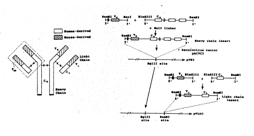
Figure l8 illustrates a preferred construction
-suitable for use in the baculoviral expression system
illustrated. As indicated in the figure, mRNA isolated
from a suitable hybridoma cell line, such as cell line
PK34C or PK99C, is incubated with a 3' primer to a con-
stant region fl ~nk; ng the variable region of interest and
incubated with reverse transcriptase. Gene amplification
by polymerase chain reaction (PCR) is carried out using a
5' primer selected from a constant flanking region. This
procedure, including appropriate primers for the variable
SUBST~ L SHEET
' ~ 92/12169 PCr/CA91/00459
.
2~98~
39
region of the mouse heavy chain tVH), is described by
Sastry et al.
The gene coding for VH is preferably subjected to
digestion by restriction endonucleases BamHI and SacI to
5 produce a BamHI/SacI (S'-3') fragment. A similar proce-
dure is carried out to obtain the gene fragment coding
for the mouse VL region, which is preferably digested
with restriction endonucleases BamHI and HlndIII to
produce a BamHI-HlndIII (5'-3') fragment.
Likewise, fragments of genes coding for human heavy
and light chain constant regions ~CH and CL, respective-
ly) are obtained by methods known in the art (Rabbitts).
The CH COnt~n~ng gene is preferably subjected to
digestion by restriction endonucleases HindIII and BamHI
to produce a Hind III-Bam HI (5'-3') fragment. The CL
containing gene is preferably subjected to digestion by
HindIII and B~m~I to produce a HindIII-BamHI ((5'-3')
fragment.
A gene coding for a rhlme~ic heavy chain is then
obtained by joining the BamHI-SacIDNA fragment coding for
VH with the HindIII~BamHI fragment cont~;n;ng human IgG1
or IgG2 heavy chain constant region (CN) using a SalI
l;nkPr. A chimeric kappa light chain gene is constructed
by joining the BamHI/HlndIII fragment cont~ ;ng mouse
PK99H or PK34C VL to the HlndIII site of a HindIII/BamH1
fragment cont:~ning ht-m~n CL (Boulianne et al. (Nature
312: 6g3-646; 1984); Morrison et al tPNAS 81: 6851-68SS;
1984)).
The resulting recombinant DNAs are then preferably
inserted sequentially into the coexpression baculovirus
vector pACVC3 sequentially. First the BamHI fragment
coding for the chimeric heavy chain (VH_CH) is inserted
into the BglII site of the vector to yield pVH3. The
SlJB~ SHE~T
WO92/12169 PCT/CA91/0045'
~ ~ 9 ~
BamHI chimeric light chain gene fragment (VL_CL) is then
inserted at a BamHI site of pVH3 to yield pVIH3.
Spodoptera frugiperda cells are infected with recom-
binant baculovirus pVL~3 (Putlitz et al.). Binding capa-
city of secreted antibodies is analyzed by ELISA as hasbeen described.
The chimeric antibodies produced in accordance with
the in~ention are useful in the treatment or prevention
of mam~l~An infections of Pseu~omon~s and crossreacti~e
infectious agents, by parenteral ~m; ni stration of the
antibodies.
VIII. Peptide Treatment
In one preferred mode of ~mi ~; stration, peptides of
the invention are delivered by nasal insufflation of pow-
ders or atomized solutions cont~; n; ng the peptide. This
mode of administration has the advantage that deli~ery of
the peptide is made directly to the pulmonary mucosal
epithel;~l surface.
Yet another use of the peptides of the invention is
as target molecules for drug delivery to pulmo~ry epi-
thelial cells. Since the peptides bind specifically to
plllmo~ry epithel t ~1 cells, they are construed to be use-
ful as therapeutic adjuvants in pathological conditions
involving the lungs. One such condition is carcinoma of
the lung. In one preferred use, the peptides of the in-
vention are conjugated to a photoactivatable chemothera-
peutic agent useful in the treatment of lung carcinoma.
The drug-peptide con~ugate is then ~mt nt stered by nasal
insufflation, and the drug is activated by high intensity
light delivered through a bronchoscope.
The following examples illustrate methods for prepa-
ring and using the peptide and antibody of the in~ention.
SUBSTITUTE SHEET
~'~92/12169 PCT/CA91/~459
2~829~
41
The examples are intended to illustrate, but not limit,
the scope of the invention.
Example 1
Solid-Phase Synthesis of Pilin PAK Peptide
Abbreviations used in this example are BOC, tertiary
butoxycarbonyl; DCM, dichloromethane; TFA, trifluoroace-
tic acid; and BOC-AA-OH, amino acids protected at the
alpha amino group by BOC group.
Commercially available phenylacetamidomethyl resin
for polypeptide synthesis was obtained from Applied
Biosystems (Foster City, CA). BOC-AA-OH were obtained
from Institute Armand Frappier (Laval, Quebec, Canada).
Side-chain protecting groups on the residues are as
follows: o-(p-bromobenzoyloxycarbonyl) for tyrosine,
o-benzyl for threonine, serine, aspartic acid and gluta-
mic acid; S-methoxy-benzyl for cysteine, 2-chloroben-
zyloxycarbonyl for lysine and formyl tryptophane.
A. Solid-phase Synthesis
In preparing a synthetic polypeptide of this inven-
tion by the above solid-phase method, the amino acid
residues are linked to a resin ~solid-phase) through an
ester linkage from the carboxy-terminal residue.
Reactive amino acid side ch~~ns are also protected
during synthesis of the polypeptide. Couplings are typi-
cally carried out using a 2-fold molar excess of protec-
ted amino acid and one equivalent of dicyclohexyl car-
bodiimide over the number of milliequivalents of initial
N-terminal amino acid. For asparagine (N) and glutamine
(Q), 2 molar equivalents of N-hydroxy-benzotriazole and
dicyclohexyl carbodiimide were used. Coupling reactions
are monitored by the ninhydrin test of Sarin (1981) and
are typically more than 99% complete.
WO92/12169 PCT/CA91/004'
~3~
42
B. Oxidation and Purification of the Peptide
The peptide is cleaved from the resin and subse-
quently cyclized to form a disulfide bond. The cleavage
of the peptide and the complete removal of the side-chain
protecting groups is accomplished using using anhydrous
hydrogen fluoride. The resin is suspended in a mixture
containing hydrogen fluoride and anisole (9:1, v/v) and
the reaction is allowed to proceed in vacuo for 45
minutes at 5~C. The hydrogen fluoride is then
evaporated. The resin is removed and washed with e~her (
3 x 10 ml) and the peptide is extracted with 30% acetic
acid (3 x 10 ml). The combined filtrates are diluted to
give a 5~ aqueous acetic acid solution and lyophilized.
The crude peptide can be purified on an analytical
reversed-phase HPLC column (250 x 4.6 mm internal diame-
ter) using a shallow gradient. The crude peptide was
dissolved in the smallest volume of starting buffer
possible (about 5 ml). The highly concentrated peptide
was centrifuged to sediment undissolved material. An
analytical sample, 5-10 ~1, was chromatographed using a
linear gradient (solvent A is 0.05% aqueous TFA and
solvent B is 0.05% TFA in acetonitrile) to determine the
total amount of peptide present. When the crude peptide
contained hydrophilic and hydrophobic impurities with
retention times close to that of the peptide of interest
in the analytical run (1~ B/min gradient rate), a shallow
gradient of 0.2% B/min with a flow rate of 1 ml/min was
employed.
The whole stock solution of 30-50 mg was injected
onto the column and the run was monitored at 210 nm.
Fractions (1 ml) were collected and analysed. Every
third or fifth fraction was analysed to identify the
region on the chromatogram with the peak of interest.
Further analysis of the fractions within this region
~ '92/12169 PCT/CA91/00459
~J~9~2~
43
would then be carried out. The chromatogram of each run
could be compared with the initial analytical run prior
to purification to ascertain the peak of interest. In
this way, the shoulders of the neighboring peaks were
eliminated, while fractions of interest were pooled and
freeze dried. Dried peptides were stored in glass vial
in a dessicator.
Mass spectrometry and HPLC anlysis were used to
confirm the PAK peptide structure.
Example 2
Preparation of Epithelial Cells
A. Buccal Epithelial Cell (BEC) Preparation
BECs were collected from ten healthy non-smoking
male volunteers via wooden application sticks rubbed
gently on the inside of cheeks, three wooden application
sticks per cheek. These sticks were rubbed gently to-
gether in 30 mL phosphate buffered saline to suspend the
BECs. These cells were washed three times with 30 mL
phosphate buffered saline by successive centrifugation
(650 x g) and resuspended. The final pellet was
suspended in 5 mL phosphate buffered saline at pH 7.2.
This suspension was filtered (prewetted 70 ~m nylon mesh)
and the cells were diluted to a final concentration of 2
x lOs cells/mL in phosphate buffered saline at pH 7.2.
This suspension was stored at 4~C until ready for use.
B. Tracheal Epithelial Cell Preparation
Human ciliated tracheal epithelial cells (TECs) were
obtained from patients in the Surgical Intensi~e Care
unit at Toronto General Hospital by bronchoscopic brush-
ing of the bronchial mucosa as described by Franklin et
al. (1987). TECs were obtained by bronchoscopy from sur-
gical patients (under general anesthetic), intubated in-
W092/12169 PCT/CA91/0~5'
2 ~ ~
44
tensive care unit (ICU) patients, and health volunteers.For the surgical and ICU patients, bronchoscopy was per-
formed with a flexible Olympus Type 2 BF bronchoscope
inserted through an endotracheal tube. A cytology brush
was used to abrade the tracheal-bronchial mucosa, and
TECs were collected in high-glucose Dulbecco modified
Eagle medium containing 1% (w.v) sodium citrate.
The cell suspension obtained by bronchoscopy con-
tained both ciliated and nonciliated cuboidal and colum-
nar epithelial cells in addition to various amounts of
mucus, erythrocytes, granulocytes, and cell debris and
was not suitable for direct use in an adhesion assay.
The cell suspension was vortexed briefly, sequentially
passed through 70- and 30-micron pore size mesh nylon
screens, washed twice (500 x g for 15 min at 4~c) with lO
ml of O.Ol M phosphate-buffered saline (pH 7.2) (PBS),
and then resuspended in l ml of PBS. The cell suspension
was then fractionated by density gradient centrifugation
(500 x g for 15 min at 4~C in a swinging bucket rotor) on
a PBS-preformed (48,000 x g for 40 min at 4~C) 65~ (vol~-
vol) percoll gradient.
The TEC band was collected and applied to a second
percoll gradient. The ciliated TEC band was collected
from the second gradient, and the cells were washed once
in PBS and then resuspended in l.5 ml of PBS. A direct
cell count was performed with a hemacytometer; cell
viability was determined by trypan blue dye exclusion.
The cell fractionation procedure typically yielded (2.08
+ 0.3~) x 105 cells (mean t standard error), of which
32.8 + 6.5~ were ciliated TECs. The vast majority of
these cells were viable, and in many cases the cilia were
still beating. The fractionated TECs contained only epi-
thelial cells, were essentially free of contaminating
mucus, and were used directly for adhesion assays.
' ~ 92/12169 PCI/CA91/00459
45 ~9~296
Example 3
Polyclonal Antibody Inhibition of Pilus Binding to BECs
5 A. Preparation of Fab Fragments
Polyclonal antibodies were prepared against the fol-
lowing peptide regions of PA~C pilin protein: "rl," :r2,"
"ol" and "o2," against the reduced (r) or oxidized (o)
PAK peptide composed of residues 128-144 of native PAK
pilin protein; "22," against residues 22-33; "41,"
against residues 41-49; "58," against residues 58-70;
"75," against residues 75-84; "89," against residues
89-99; "107," against residues 107-116; and "117,"
against residues 117-125. "Pre" refers to preimmune
15 sera; and "99H" to monoclonal antibody PK99H.
Fab fragments of the above polyclonal sera derived
from each peptide antigen were prepared using immobilized
papain ~Pierce Chemical Co., Rockford, IL). Briefly,
affinity purified polyclonal antibody was dialyzed
20 against 20 mM cysteine HCl, lO mM tetrasodium ethylenedi-
aminetetraacetic acid (EDTA) in 20 mM sodium phosphate
buffer pH 6.2. Antibody (l ml containing approximately 2
mg antibody) was added to 0.5 ml immobilized papain and
incubated at 37~C for 20 h with shaking at 150 rpm. The
25 immobilized papain was removed by centrifugation and the
supernatant containing the Fab fragments was diluted with
l ml of PBS.
The Fab fragments were purified by HPLC using a
Protein G column eluted with PBS. Fab fragments were
30 collected in the flowthrough, and Fc fragments were
eluted from the column with 10 mM glycine pH 2.75. Fab
fragments were concentrated by placing the Fab effluent
in dialysis tubing (molecular weight cutoff of < 8000)
and extracting liquid from the dialysis sack using poly-
WO 92/12169 PCr/CA91/0045~
~3~ 46
ethylene glycol (molecular weight of 15,000 - 20,000).
The fragments were then dialyzed against PBS. Activity
of Fab fragments was checked by ELISA and production of
Fab fragments was confirmed by SDS-PAGE.
B. Inhibition of PAK pilin Binding to 8ECs by Fab
Fragments
PAK pilin protein was isolated according to pub-
lished methods (Paranchych et al, 1979). Fab fragments,
prepared as above, were preincubated with PAK pili before
the addition of BECs (l x 105 cells/mL final concentra-
tion) and pili binding was detected using monoclonal
antibody PK38B (which is specific against pilin protein,
but not the PAK peptide). All Fabs were diluted such
that their final titre as measured by ELISA to PAK pili
was 10-3.
Example 4
Monoclonal Antibody Inhibition of
P. aeruginosa and Pili Binding to BECs
A. Monoclonal Antibodies
Hybridoma cell lines PK99H and PK34C (Doig) were
deposited in the cell depository of the Department of
25 Medical and Infectious Diseases of the University of
Alberta, Alberta, Canada, and are identified by cell line
Nos. PK99H and PK34C. Fab fragment of the PK99H and
PK34C Mabs, and non-immune IgG were prepared as described
in Example 3A.
B. Inhibition of Pili Binding to BECs
BECs were prepared as described in Example 2. PAK
pili were isolated according to published procedures
(Paranchych et al., 1979). PAK pilin protein was iso-
~92/12169 PCT/CA91/0~59
~a~82~
lated according to published methods (Paranchych). Fabfragments of PX99H, PK34C, and non-immune IgG were prein-
cubated with PAK pili at the concentrations indicated in
Table l above (Doig). After incubation, BECs were added
to a final concentration of l x lOs cells/mL. Pili bind-
ing was detected using monoclonal antibody PK3B, followed
by reaction with enzyme-labeled goat anti-mouse antibody,
as above. Pili binding, as measured by enzyme activity
associated with BECs, is expressed as percent control (no
Fab fragments added) in Table l.
C. Inhibition of P. aeruginosa binding to BECs
P. aeruginosa strains PAK, PA0, HDl, 492c, Pl, Kl22-
4, and PAK/3 are as reported (Doig). Fab fragments of
PK99H, P~34C, and normal mouse non-immune IgG were pre-
pared as above. The Fab fragments (0.5 ml of 0 to 3.2
mg/ml) were added to O.l ml of bacteria containing 0.7 x
lO~ CFU/ml in PBS, and the mixture was incubated for 30
minutes at room temperature. To this mixture was then
added 0.4 ml PBS and either l ml of BECs containing 2 x
105 cells or l ml PBS ~to assess non-specific binding of
bacteria to filters. After incubation at 37~C for 2
hours with s~Ak~ng, the cells were washed, filtered, and
the filters were assayed for the presence of bound
bacteria by an ELISA method employing an anti-pilin mono-
clonal antibody and an enzyme-linked antibody, as
described above. The results are shown in Table 2 above.
Example 5
Monoclonal Antibody Immunolocalization on Moraxella Cells
Immunolocalization studies employing the polyclonal
anti-PA0 pili antisera utilized M. catarrhalis cells
which were absorbed onto the surface of carbon coated,
glow discharged electron microscope grids, blocked with
WO92/12169 PCT/CA91/~4'
~O9~2~
48
1% (w/v) BSA in PBS pH 7.4 for 2 x 5 min, and reacted
with rabbit anti-PAO pili in PBS pH 7.4 containing 0.5%
(w/v) BSA for 35 min at 37~ C. The grids were then
washed on 5 drops of PBS pH 7.4 containing 0.5% (w/v)
BSA, blocked with 1%(w/v) BSA for 2 x 5 min, reacted with
goat anti-mouse IgG-20 nm colloidal gold conjugate (E.Y.
Labs Inc., San Mateo, California) in PBS pH 7.4 contain-
ing 0.5% (w/v) BSA for 30 min, washed on 5 drops of PBS
pH 7.4 containing 0.5% (w/v) BSA, then washed on 5 drops
of H2O and finally blotted dry. The grids were then
stained with 1%(w/v) phosphotungstic acid before ex~m~na-
tion in a Philips EM 401 transmission electron microscope
operating at an accelerating potential of 80 kV. Con-
trols included no first antibody and normal mouse IgG
(Jackson Laboratories). The polyclonal anti-PAO pili
antisera was observed to bind with high affinity to the
cell surface and surface appendages of the Moraxella
catarrhalis.
Immunolocalization studies with monoclonal antibody
PK99H were carried out with thin sections of M. catar-
rhalis cells that had previously been embedded in LXl2,
sectioned, thin sections collected on the surface of 3mm
copper EM grids, and the sections etched with saturated
sodium metaperiodate to remove osmium and regain antige-
nic activity. The thin sections were then treated asdescribed above except that monoclonal antibody PK99H was
used instead of the polyclonal antibody. Immunospecific
binding of monoclonal antibody PK99H was observed, with
the antibody binding both to cell surface components and
to cytoplasmic components in the M. catarrhalis cell.
Example 6
Inhibition of Candida Binding to BECs
~92/12169 PCT/CA91/~459
2~8~
49
A. C. albicans culture conditions
The C. albicans strains used were isolated from the
trachea of intubated intensive care unit patients at
Toronto General Hospital, Toronto, Ontario (for fimbrial
purification and characterization) or obtained from the
Department of Microbiology, University of Alberta Hospi-
tal, Edmonton, Alberta.
A loopful of culture from Sabouraud-dextrose agar
(Gibco) was used as a source of inoculum for 10 ml of M9
medium (A~ms, 1959) supplemented with 0.4% (wt/v) glu-
cose. Two incubation protocols were used. Cultures sha-
ken at 150 rpm were either incubated at 25~ C for 19
hours or for 16 hours at 25~ C followed by a 3-hour in-
cubation at 37~ C. Cultures to be used in the radio-
adhesion assay were supplemented with 55 Ci/ml of [35S]-
L-methionine ~New England Nuclear, Boston Mass.) after 17
hours of incubation. Cells were harvested by centrifuga-
tion (12,000 x g for 10 min) and washed 3 times with PBS
pH 7.2 to remove unincorporated methionine. Washed cells
were resuspended in PBS pH 7.2 at varying concentrations.
Cells which had been incubated at 37~ C were forcibly
passed twice through an 18 gauge needle to break up
clumps. No clumping was observed during the adhesion
assays.
B. Buccal epithelial cells
BECs were collected with wooden applicator sticks
from healthy, non-smoking, male volunteers (n=10). BECs
were removed from the applicator sticks by gentle agita-
tion in PBS pH 7.2. The BECs were washed 3 times (2 000
X g for 10 min at 40C) with PBS pH 7.2, then passed
through a 70 ~m nylon mesh. The cell concentration for
the BECs was determined with a hemocytometer and the BEC
concentration was adjusted. The viability of BECs
WO92/12169 PCT/CA91/~4C
~g8 ~ 50
obtained by this procedure was generally about 5%, as
determined by trypan blue dye exclusion.
C. C. albicans Adherence to Human Ciliated Tracheal Epi-
thelial Cells
Human ciliated tracheal epithelial cells ~TECs) were
obtained as described by Franklin et al. (1987) and by
Todd et al. (1989). Briefly, cells were obtained by
bronchoscopy following administration of 5 ml of 1% (w/v)
xylocaine to the nasal and oral pharyngeal passages with
a further 5 ml of 1% (w/v) xylocaine being administered
via the suction port of the bronchoscope at the level of
the glottis. An additional 5 ml was further administered
within the trachea before repeated (n=10) brushing of the
trachea with a disposable cytology brush. Cells were
eluted from the brush by agitation in 30 ml of serum free
Dubecco's Modified Eagle's Media (high glucose formu-
lation) containing 1~ (w/v) sodium citrate and stored at
40~C before use.
TECs were fractionated from mucus, blood cells,
microbial contaminants, and debris by gentle filtration
through a 70 ~m and a 30 ~m nylon mesh, washing the cells
three times with 10 ml of PBS pH 7.2 (500 X g, at 40C for
10 min) concentrating the cells following centrifugation
(500 X g, at 40C for 10 min) in 1 ml of PBS pH 7.2, two
sequential density gradient centrifugations on preformed
65% (v/v) Percoll gradients in PBS pH 7.2 (preformed by
centrifugation at 48,000 X g for 40 min at 40~C) for 20
minutes at 500 X g for 20 min at 40~C, washed twice with
5 ml of PBS pH 7.2, and resuspended in 1 ml of PBS pH
7.2. TECs were quantitated by direct counting employing
a hemacytometer.
~8~9~
WO92/12169 PCT/CA91/0~59
51
D. Immunofluorescence of Candida Fimbriae
Yeast were grown àt 25~ C or shifted to 37~ C as
described above. Yeast were harvested by centrifugation.
Cells were fixed with l.0% formaldehyde in PBS for 30 min
and washed twice with PBS. Primary antibody was added
and the mixture was incubated at 37~ C for l hour,
sh~k~ ~g at 300 rpm. Yeast were then collected by
centrifugation (12 000 x g for l min at room temperature)
and washed 3 times with PBS pH 7.2. Rabbit anti-mouse
IgG (H+L) affinity purified IgG conjugated to fluorescein
isothiocyanate (Jackson Laboratories) in PBS pH 7.2
(l/500 dilution) was added to the washed yeast
preparations and ;~c1~h~ted for 30 min at 37~C, agitating
at 300 rpm. The yeast were washed 3 times as described
above and resuspended in O.l mL of PBS pH 7.2. Wet
mounts were prepared, and e~A~ined by epifluorescence and
phase contrast microscopy using a Lietz Laborlux equipped
with a MPS4 camera system. Photographs were recorded
with Kodak T-Max fi1m.
E. Adhesion assay
The adhesion assay of McEachran as modified as
described by Staddon was used to determine the number of
bacteria bound per epithelial cell. BECs (l mL of 2 X 10⁵
cells per mL) were mixed with an equal volume of
radio-labelled yeast suspended in P8S pH 7.2 and
incubated at 37~C for 2 h, sh~king at 300 rpm. Epitheli-
al cells with bound yeast were then collected by
filtration on 5 micron polycarbonate filters (Nuclepore)
pretreated with 2~ (w/v) bovine serum albumin ~BSA) in
PBS pH 7.2 to reduce nonspecific binding, washed with 15
mL PBS pH 7.2 and then placed in scintillation vials.
Aquasol (5 mL) was added to each vial and the amount of
radioactivity was deterr;ned by scintillation counting in
~Trademark
n
~92/12169 PCT/CA91/0045r
~g8 2g~ 52
a Beckman LS-150 liquid scintillation counter.
Triplicate aliquots were filtered for each sample.
Binding of yeast to epithelial cells was corrected for
nonspecific binding of yeast to the 12 ~m filter
5 (nonspecific binding was generally less than 15% of the
experimental value). The epithelial cell concentration
was determined at the end of the assay to correct for
cells lost during incubation.
Total and viable cell counts were performed before
and after the adhesion assay. Total cell counts were
determined using a hemacytometer. Viable counts were
determined by serially diluting C. albicans in PBS pH 7.2
and plating appropriate dilutions on Sabouraud-dextrose
agar (Gibco) which were incubated at 37~ C until visible
and countable colonies formed (usually 24 to 48 hours).
F. Purification of Candida Fimbria
C. albicans were cultured in Mg medium (Adams, 1959)
supplemented with 0.4% (wt/v) glucose overnight at 37~ C
at 150 rpm. This culture was used to inoculate Sabouraud-
-dextrose agar (Gibco) in aluminum trays (approximately 2
ml/tray). The trays were incubated at 37~ C for 5 days.
Yeast cells were then scraped from the surface of the
agar with a bent glass rod and suspended in PBS pH 7.2
containing 1 mM phenylmethylsulfonyl fluoride (Sigma) as
a protease inhibitor. Fimbriae were then sheared from the
cell surface by blending (4 x 15 second cycles using a
Waring blender). Cells were examined by phase microscopy
and appeared to be intact.
Cells were removed by centrifugation (12,000 x g for
20 min.) and by subsequent filtration of the supernatant
through a 0.45 ~m polycarbonate filter (Nuclepore Corp.,
Pleasanton, Calif.). The supernatant was placed in dialy-
sis tubing ~Spectrum, Los Angeles, Calif.; M.W. cut off
, ~. . : . .
-Y' it
~n ~ r ~
- 7 ~ ~ ~ 2 9 6
WO92/12169 PCT/CA91/~59
6000-8000 Da) and concentrated with polyethylene glycol
~M.W. 15,000-20,000, Sigma) ~PEG). Finally the sample was
dialyzed against PBS pH 7.4. The final preparation was
termed crude fimbriae ~CF) and was stored at -70~ C.
Crude fimbriae were purified by HPLC size exclusion
chromatography on a Protein-p~k 300 SW column (Millipore
Inc.) having a size exclusion limit of 300,000 daltons
operating at 0.5 ml/min flow rate and previously equi-
librated with PBS pH 7.2 buffer containing lmM CaClz and
eluted with the same buffer. Purified fimbriae were
obtained by re-chromatographing the material that ini-
tially eluted in the void volume of the column and col-
lecting the material that still eluted within the void
volume from the second chromatographic run. Purified
fimbriae consisted of -15% (w/w) protein and -85% car-
bohydrate (w/w) on the basis of colorimetric assays and
consisted of a single polypeptide of -64,000 daltons on
the basis of SDS-polyacrylamide gel electrophoretic
analysis utilizing silver staining (see figures).
G. Effect of Candida fimbriae on PAK pilus binding to
BECs
An immunoassay was performed to assess the effect
of Candida fimbriae binding of pili from PAK to BECs.
BECs (l mL at 2.0 X lOs BECs/mL), PAK pili (0.5 mL of 80
ug/mL), and fimbriae (0.5 mL of 400 or 800 ug/mL) in PBS
pH 7.2 were mixed and incubated at 37~C, sh~ktng at 300
rpm in a New Brunswick gyroshaker. After l h BECs were
collected by centrifugation (12 000 X g for lO min at
4~C) and washed twice with PBS pH 7.2. Anti-PAK pilus
monoclonal antibody PK3B (Doig et al. l990) was added to
the BEC pellet (l mL of a lO-~ dilution) and incubated as
described above for l h. The BECs were then collected by
centrifugation and washed twice with PBS pH 7.2. Goat
~ m~rk
-, . .
WO92/12169 PCT/CA91/0~C~
~ ,2~ 54
anti-mouse IgG(H~L) peroxidase conjugate ~Jackson Labora-
tories) was added to the BEC pellet (l mL diluted per
instructions for use) and the mixture was incubated as
described above for l h. The BECs were collected by
centrifugation, transferred to a clean test tube, and
washed twice with PBS pH 7.2. The pellet was resuspended
in l mL of a solution containing lmM 2,2'-azinobis[3--
ethylbenzothiazoline-6-sulfonic acid), 0.03% (vol/vol) in
lO mM citrate buffer pH 4.2. The reaction was stopped by
the addition of l mL of 4 mM NaN3 and the optical density
at 405 nm was determined after removal of the BECs by
centrifugation. The BEC concentration in each tube was
determined with a hemocytometer at the end of the assay
prior to the removal of BECs by centrifugation.
H. Assessment of PAK pilus or Candida fimbriae inhibi-
tion of Candida binding
Pili and fimbriae inhibition assays were performed
using a direct competition method. Direct competition of
pili/fimbriae and yeast was achieved by the simultaneous
addition of pili/fimbriae, yeast and BECs at the com-
mencement of the yeast binding assay. The number of
yeast bound per BEC was determined as described above.
Example 7
Pseudomonas/Candida Antibody Crossreactivities
A. Enzyme-linked immunosorbant assay (ELISA)
Antigens were coated on NUNC 96-well polystyrene
wells. Antigen (lO ug/mL in O.Ol M carbonate buffer, pH
9.5) was added to each well (lO0 ~l/well) and left for 6
h at room temperature. Wells were then washed 3 times
with 250 ~l of PBS pH 7.4 supplemented with 0.02%
(wt/vol) BSA (buffer A). Wells were blocked with 5%
f~ ! ' . f
9 ~
W092/12169 PCT/CA91/OW~9
(wt/vol) BSA in PBS pH 7.4 overnight at 4~C. Wells were
washed three times and 100 ~1 of primary antibody was
added for 2 h. Each well was then washed 3 times with
250 ~1 buffer A using aspiration. A goat anti-mouse IgG
(H+L) immunoglobulin-horse radish peroxidase conjugate
~Jackson Laboratories) in buffer A (100 ~l/well) was
added and incubated for 2 h at room temperature. The
wells were washed 3 times with buffer A and 250 51/well
substrate solution (lmM 2,2'-azino-di-(3-ethylbenzthi-
azoline sulfonic acid), 0.03% (vol/vol) hydrogen peroxidein lOmM sodium citrate buffer pH 4.2) added. The reac-
tion was stopped by the addition of 250 ~l/well of 4mM
sodium azide and absorbance at 405 nm determined using an
EL-407 plate reader.
B. Competitive ELISA
Competitor and antibody were mixed together in 10 mM
PBS pH 7.2 buffer cont~;n;ng 0.05% (w/v) BSA and incuba-
ted for 30 min at room temperature. The conditions of
the assay were such that ~50% of the antigen immobilized
on the ELISA plate surface would be bound with antibody
if there was no competitor present. The mixture of anti-
body and competitor was then added to wells (100 ~l/well)
coated with PAK pili or Candida fimbriae and blocked with
BSA as described above. The ELISA was then performed as
described above. The apparent affinity of the antibody
for the competitor was determined as described by Nieto
et al. (1984) following deterr;n~tion of the concentra-
tion of competitor that would give 50% inhibition of
antibody binding to antigen.
C. Whole Cell C. albicans Dot Blots
Dot blots were performed using a Bio-Rad dot blot-
ting manifold. Whole cells of ~arious clinical isolates
~radem~k
.
J~
~ ~ 9 8 ~ 9 6
WO92/12169 PCT/CA91/0~59
of Candida albicans were initially washed 3 times with
PBS pH 7.2 buffer and lO0 ~l of cell suspension contain-
ing 2 X lO' CFU was added per well on a pre-wetted nitro-
cellulose membrane. The cells were collected on the
surface of the filter by vacuum filtration. The wells
were washed 4 times with 0.1% (vol/vol) Tween 20,50 m M
Tris buffered saline pH 7.5 ~TTBS) ~200 ~l/well) and
blocked with lO0 ~l of 3% (wt/vol) BSA in TTBS for l h.
The wells were then washed 4 times with TTBS and mono-
clonal antibody PK99H and PK34C at various dilutions inTTBS were added (lO0 ~l/well) and incubated for l h. The
blot was washed 4 times with TTBS and lO0 51 of a goat
anti-mouse IgG (H+L) immunoglobulin-alkaline phosphatase
conjugate in TTBS was added to each well for l h. After
washing the blot 6 times with TTBS, a substrate solution
consisting of 0.33 mg/mI nitro blue tetrazolium chloride,
0.165 mg/mL 5-bromo-4-chloro-3-indolyl-phosphate, lO0 mM
- sodium chloride, 5 mM magnesium chloride, lO0 mM Tris
buffer pH 9.5 was added. Color development was stopped
by aspiration and rinsing the membrane in distilled
water.
D. Assessment of antibody Fab fragments on yeast binding:
Effect of Fab fragments on binding was performed as
follows. Fab fragments (0.5 mL of 800 ~g/mL in PBS pH
7.2) were added to O.l mL of yeast in PBS pH 7.2 and
incubated for 30 min at room temperature. To this 0.4 mL
of PBS pH 7.2 and either l.0 mL of BECs (2 X lOs
cells/mL) or l.0 mL of PBS pH 7.2 was added. The mixture
was then incubated at 37~C sh~king at 300 rpm for 2 h.
The r~r~n~r of the adhesion assay was performed as
described above.
rademark
B
'~l~92/12169 PCT/CA91/00459
57 ~oC~8296
Example 8
Preparation of Random-Sequence Peptide
A. Construction of epitope library for peptide ligands
An epitope library which approximately 108 novel
heptapeptide sequences is constructed as described by
Scott and Smith. Alternatively a similar epitope library
can be constructed as described by Cwirla et al or Devlin
et al. Figure 17 shows a schematic representation of the
construction of the library as described by Scott and
Smith and summarized here.
Filamentous phage fUSE5 is constructed as a vector
for the epitope library as described by Parmley and Smith
(1988; Gene 73: 305-318) and by Scott and Smith. This
phage contains a tetracycline resistance gene, and is
designed to have cloning sites, insertion into which
result in addition of peptide sequence at the exposed N-
terminus of the minor capsid protein pIII. Addition of
foreign peptide sequence at this site does not substan-
tially inhibit infectivity of the phage. However itslocation does make it exposed on the surface of the phage
and thus amenable to recognition by antibodies.
In preparation for ligation with a foreign DNA
fragment, fUSE5 is digested with Sfi I ~BRL; 120 units/30
ug fUSE5 RF DNA). Following extraction with phenol and
chloroform, the volume is ad~usted to approximately 0.8
ml with TE buffer (lO mM Tris pH 8, lmM EDTA), and the
DNA is precipitated by additions of sodium acetate buffer
~3M; pH 6) and isopropanol. Following incubation (20 min
at 0 degrees) and pelleting, the pellet is washed with
70% (v/v) ethanol, redissolved in TE buffer, ethanol
precipitated and redissolved in TE.
An insert containing a -random heptapeptide sequence
is prepared by polymerase chain reaction (PCR) amplifica-
WO92/12169 PCT/CA91/~9
58
tion of a 73 base degenerate template shown in Figure 17with 5' biotinylated primers corresponding to the first
20 bases at the S'ends of both strands shown in Figure
17_(top). The template is then cleaved at the two BglI
sites shown and then adsorbed onto streptavidin-agarose
to remove the biotinylated terminal fragments along wlth
the undigested and partially digested by-products. The
PCR mixture contains l ~g of template, 5 ~g of each of
the biotinylated primers, and 25 units of AmpliTaq DNA
polymerase (Perkin-Elmer/Cetus).
The mixture is subjected to five temperature cycles
(2.5 min. at 95 degrees, 4 min. at 42 degrees, 4.4 min.
at 72 degrees, 5 min. at 72 degrees),then the reaction is
stopped by addition of ~DTA solution (final
concentration, l mM), pH 8. Following precipitation with
ethanol and dissolution in O.l ml TE buffer, a portion of
the product is digested with Bgl I (Promega; 6.4 units/ul
final concPntration) for 2 hours at 37 degrees to produce
a 36 bp degenerate fragment. The randomly selected
codons in this fragment are represented by (NNK) 7~ where
N stands for an equal mixture of deoxynucleotides G, A,
T, and C, and K stands for an equal mixture of G and T. M
stands for an equal mixture of C and A in the
complementary strand. NNK therefore represents an equal
mixture of 32 triplets, including codons for the 20 amino
acids plus the amber stop codon. The digestion by Bgl I
is stopped by addition of EDTA solution (lO mM, final
concentration). Pre-washed streptavidin beads are then
added to the solution and mixed for 30 minutes, and
removed by centrifugation. This step is repeated with
fresh stepta~idin beads. The final product is extracted
(phenol plus chloroform) and evaporated to a volume of
O.l ml.
~dem~k
B
" ~92/12169 PCT/CA91/0~59
~9823~
59
Ligation of the insert to the SfiI digested fUSE5 RF
is carried out in a volume of 2 ml. The reaction mixture
consists of 36 ~l of the degenerate 36 bp insert and lO
ug of the Sfi I digest of fUSE5. The product is extrac-
ted with phenol and chloroform, ethanol precipitated, anddissolved in 0.2 ml TE buffer.
The ligation product is electroporated into E. coli
MCl061 cells for amplification of phage carrying degene-
rate peptide sequences. Following electroporation, the
bacteria are diluted into growth medium containing tetra-
cycline. Tetracycline resistance of cells indicates
successful transfection.
B. Selection of Antibody-Binding Peptide
Phage are isolated from plate stocks by scraping
from the agar surface, resuspending in L broth and clear-
ing the supernatant twice by centrifugation (8000 rpm for
lO minutes in a Beckman JAlO rotor at 4 degrees). Phage
particles are precipitated with polyethylene glycol
(3.3%) in 0.4 M NaCl, followed by centrifugation. Phage
pellets are resuspended in TBS buffer (50 mM Tris-HCl, pH
7.5, 150 mM NaCl) and stored at 4 degrees.
Phage are affinity purified using the monoclonal
antibodies PK34C or PK99H in a panning procedure.
Alternatively, FAb fragments of these antibodies are used
in this procedure. Briefly, after incubation of phage
~lO'l-lOl2 infectious particles) overnight with l ug of
purified antibody, phage expressing peptides with af-
finity for the antibodies or their Fab fragments are
isolated using the panning method of Parmley and Smith.
Biotinylated goat anti-mouse antibodies are added to the
mixture, and the mixture is then added to a streptavidiin
coated plate. This procedure can also be carried out
without use of the second (goat) antibodies, by directly
WO92/12169 PCT/CA91/0045~
s~
biotinylating the primary antibodies of Fab fragments.
Following incubation ~10-30 minutes) the streptavidin-
coated plate is washed, and adherent phage are eluted for
10 minutes in a buffer containing 0.1 M HCl (pH 2.2,
S adjusted with glycine) and bovine serum albumin (1
mg/ml). Neutralization of the eluate is achieved using
an aliquot of 2 M Tris. Eluted phage are then amplified
by infection of E.coli, followed by incubation on agar
plates containing tetracycline. The resulting amplified
phage pool is repurified twice by the same panning proce-
dure described above.
Phage selected through 2-3 rounds of panning are
cloned and propagated, and their DNA's are sequenced
using standard techniques to determine the amino acid
sequences of their peptide epitopes.
Although preferred embodiments of the invention are
described herein in detail, it will be understood by
those skilled in the art that variations may be made
thereto without departing from the spirit of the inven-
tion or the scope of the appended claims.