Note: Descriptions are shown in the official language in which they were submitted.
WO 93/07819 PCI`~US92/08860
2~92~7
A DEVICr FOR THE CON~ROLLED EXCISION O
TISSUE FROM A LIUING ~ODY
Technic_l Fiel~ -
The invention relates ~o a device for the controlled excision of 2
piece of tissue from a living body. The term "tissue," as used herein,
refers, but is not limited to dense structures such as bone and other
calcified structures and to soft structures such as solid Drgans and
tumors. The tissue removed could be for the purpose of diagnosing a
suspected pathologic condition that is involving the tissue, and/or for the
purpose of completely removing all diseased tissue, thus effecting
treatment of the disease. Another purpose of removing a piece of tissu2
would be to provide an access port to fluid or tissue belo~ the surface cf
an organ or structure; the aspiration of marrow from the cavity of a bone
is an example of this application of the invention.
Back~ound Art
In the practice of clinical and research medicine, the
practitioner is frequently required to remove a piece of tissue from an
organ or structure that is suspected to be involued with a disease process.
The tissue is then sent for microscopic examination and/or culture so that
a diagnosis of the sxact pathologic process can be made. At times, the
entire area of suspacted involvement is removed so that the disease process
is not only diagnosed but is also definitively treated. Excision of
diseased tissue may be performed through a surgical incision; the tissue
then is excised under direct vision. Alternatively, the tissue may be
excised by an instrument that is passed through a puncture wound in the
skin and directad to the araa of interest. This percutaneous technique
eliminatss the recovery period required after an open procedure. This in
turn reduces the costs of the procedure as well as morbidity.
Three essential conditions must be satisfied by a device used to
excise tissue: 1) The device must be controllable so that it does not
deviate from the area of interest during the cutting process; 2) The depth
o~ penetration must be displayed to the operator or controlled by a
,
' , ~ ' ' , ' :
W O 93/0781~ PCT/US92/08~60
2~99%~7 2
mechanical stop so that underlying stru~tures deep to the area of in~eres~
are no~ damage~; 3) The sample of tissue cannot be crushed or other~ise
damaged by th= excision process which could result in an inaccurate
pathologic diagnosis. A forth condition, required in specifio
applications, is continued access to the area where tissue has be removed.
This would allow removal of additional tissue either by further excision
or by aspiration. It would also permit application of a plug in the void at
the excision site, thereby preventing hemorrhage or leakage of other bodily
fluids.
There have been many prior art devices developed to re~ove tissue
from a living body, some used during a surgical operation, others used
percutaneously. Many of these devices utilize a narrow gauge hollow needle
which is plunged into the area of interest and then withdrawn; a very small
core of tissue is thus harvested. There are many variations to the
"plunge type" instruments, but since none use a moving cutting edge, there
use is limited to very soft tissue. A potential problem with these devices
is that the architecture of the sxcised tissue may not be preserved due to
compression of the specimen, even when used in soft tissue. Qlso, the
depth of the tissue core cannot be monitored effectively.
" ~
U.S. Patent 4,262,676 describes a bone biopsy device developed by
Jamshidi that uses a hollo~ shaft cutter which is manually driven into the
tissue using an oscillating rotational motion. A significant amount of
pressure has to be maintained on the instrument to advance into the bone;
when performing a biopsy on an irregularly shaped bone, the device may
deviate laterally, and possibly completely slip off the surface. This can
result in damage to surrounding structures or, minimally, not excising
tissue from the precise area of interest. The device can only be used to ~
excise a core from relatiuely soft cancellous bone. It does not . ~-
incorporate a depth control feature. ;~ ;
.
There have many prior art devices developed to provide a more
controlled excision of a core of hard tissue such as bone, some of ~hich
use a po~:Fed cutting cylinder. U.S. Patent 4,306,570 describes a device
-:
: ~ .'.- '. ':
.. ...
. ~ . .
WO 93/0781 9 PCI /US9~08860
20~9287
used to exoise har~ tissue ~hich features t~o counterrotatins concentr~c
cutting edges tha' are said to eliminate the p~oble~ of lateral migration
as the cutting eoges engage the tissue. The device neither has a depth
control feature nor a means to allow for continued access to the area of
excision after the core of tissue has been removed.
U.S. Patent 3,993,445 describes a device for the biopsy of bone marrou
which consists of a sheath device and a central cutting needle. The sheath
is first positioned on the surface of the bone; a cutting cannula is then
introduced through the sheath to excise a core of bone. The sheath does
not substantially penetrate the bone and, therefore, one must maintain its
position manually throughout the procedure in order to provide continued
access to the tissue follo~ing removal of a core sample. No depth control
feature is provided in this device. ;~
. .
U.S. Patent 4,142,517 describes another instrument uhich uses a sheath
that minimally penetrates the bone surface to prevent lateral deviation of
a cutting shaft which is subsequently introduced. After the core is
excised, the sheath may be driven into the bone as a separate operation,
thus giving the operator Fontinued access to the underlying tissue.
.
SUMMARY OF THE INVENTION
The present invention provides a means of excising a core of
tissue from a living `body in a safe and controlled manner either
percutaneously or during a surgical operation. The core of tissue is not
altered by the excision mathod, thereby allowing for accurate examination
and diagnosis. The device can be positioned to a specific area to be
excised and, by virtue of a lateral control msans, this position can be
maintained as the cutting edge engages the tissue surfsce. Tha chanca of
the device slipping off the surface of the tissue to be excisad and
damaging adjacant structures is greatly reouced compared to prior art
devices.
'
~' :
WO 93/07819 PCI/US92/08860
2~99287
Tne dep~h of ~issue cor~ to be excise is d~spl2y~d to the operator
anc, if besirec, can ba pre-set se that a predett~rmined oepth cannol be
exceeded. This oesign feature makes the procedure safer, as underlyins
structures deep to the area to be excised ~ould not be injured.
The presant invention also, if desired, providas for continued access
to the site of excision after the core of tissue has been removed. This is
effected by incorporating an external, penetrating sheath which is
temporarily affixed to or embedded into the tissue surface. The operator
does not have to manually hold the sheath in position after it is placed,
thereby freeing up both hands for the remainder of the procedure. Placement
of the penetrating sheath occurs as the core of tissue is excised, thereby
elimlnating additional steps in the procedure.
The sheath is especially useful ~hen the operator desires to aspirate
fluid from the void left in the tissue (e.g. in the case of bons marro~
aspiration) or excise additional tissue adjacent or deep to the previous
core. Additionally, the clinician may wi~h to placa a plug in the void
remaining in the tissue after specimen excision in order to prevent the
escape of fluid. It would be impossible to percutaneously locate the area
of excision after removal of the cutting cylinoer uithout an external
sheath which remains attached to the tissue surface. To maintain the
position of a non-penetrating or minimally penetrating, manually held
sheath is difficult after removing the central cutting cylinder.
A first novel feature of the invention is the use of a hollow
cutting shaft ~ith a lateral location means that prevents deviation of the
device thereby allowing for retrieval of a tissue specimen from a specific
location.
. . .
A second novel feature is the use of a lateral location means
that does not substantially alter the structure of the excised tissue
` specimen, thereby allo~ing for precise microscopic examination.
' ' .'
1,,
W O 93/07~19 PCTtUS92/08860
20992~
A thir~ novel feature of the invention is the use of an external
sheath tha~ is affixed to the tissue anc which may be temporarily lef~ in
place, thereby providing the operatnr ~ith continued access to the exac~
area of excision for further removal of tissue, aspiration of fluid, and/or
placement of a plug to seal the hole created by the procedure.
A forth novel feature of the invention is the use of an external
sheath that is affixed to the surrounding tissue simultaneously with
penetration by a central coring cylinder thus lessening the number of
individual steps in the procedure.
A fifth novel feature of the invention is to provide access to
fluid contained within a solid structure by first removing a plug of tissue
~ith the cutter shaft, then aspirating the fluid through a sheath affixed
to the structure.
A sixth novel fsature of the invention is the use of the movement
of a slidable central guide pin as a means to monitar the depth of core
excision in addition to providing a means of Lateral location of the
cutting cylinder.
A seventh novel feature of the invention is the use of the contact
of a slidable central guide pin with a mechanical stop to prevent excision
of a core of tissue beyond a pre-set depth.
An eighth novel feature of the invention is the improved safety
that results from the prevention of lateral movement of the cutting shaft
over the surface of the tissue and possible damage to adjacent structures.
A ninth novel featùre of the invention is the positive control
over the depth of core excised, thereby preventing possible damage to
structures that lay deep to the area.
tenth novel feature of the invention is the elimination of
operator technique as a factor in excising tissue, thereby praducing
' ' , .
: ~ '
: ~ :
W O 93/078]9 PCT/US92tO8860
~ ~rl 6
specimans that are more consistent. The above-mentioned features, as
~ell as o~her features of the present inventior, all become readily
apparent from the following non-limiting description and the accompanying
drawings.
~RIEF DESCRIPTION OF THE DRA~INGS
The above-enumerated objects and features of the present invention
~ill more fully appear from the following detailed dsscription ~hen read in
conjunction uith the accompanying drawings. It is to be understood that
the drawings are for the purpose of illustration only and are not intended
as a definition of the limits of the invention.
:',
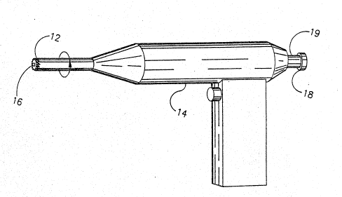
Figure 1 is a Vi9W of an embodiment of the tissue coring device
with central guide pin locked in place.
Figure 2 is a cross sectional view of the distal snd o~ the
device being used for percutaneous tissue excision, the tip of the guide
pin seen slightly impressed into the tissue surface.
Figure 3 is a cross sectional view of the distal end of the ;
device penetrating a tissue structure by using a rotational motion applied
to the cutting edge, the central guide pin seen freely sliding through the
cutting sha~t so as not to further penetFats the core of tissue.
Figure 4 is an enlarged view of the proximal end of the coring
device detailing one embodiment of a self releasing connection bstween the
cutting shaft and the central guide pin, shown in this figure as the two
begin to uncouple.
'
Figure 5 details the same embodiment of the selF releasing ~ -
connection shown in Fig 4 after the t~o elements become uncoupled, th~
: '
; ~ '' ','
,:
W O 93/07819 PCT/VS~2/08860
7 20~287
central locating pin seen to be moving distal ~ith r-speo~ t3 the cuttins
shaft as the core is taken.
Figure 6 illustrates use of the central locating pin as a means
for positive control of depth of the core specimen.
Figure 7 is a view of the sheath-cutter embodiment of the
invention.
Figure 8 is a cross sectional view of the sheath-cutter embodiment
of the invention, the two simultaneously being rotationally driven into the
tissue.
Figure 9 illustrates the embedded external sheath in Fig. 8 after
removal of the central cutter with its core of tissue, the sheath thus
giving continued access to the void in the tissue.
Figure 1û is an alternate embodiment of the sheath-cutter design
in cross section, the sheath being driven with the rotating cutting shaft
through a friction coupling until the sheath's penetration is halted by a ;-;
step fashioned in its outside diameter, the central cutting shaft
continuing into the depths of the tissue for an additional distance.
Figure 11 illustrates the sheath--cutter embodiment shown in
Figure 10 with the cutting shaft's distal end having penetrated the tissue
for a distance beyond the distal end of the shaath.
.~
Figure 12 illustratss the extsrnal sheath remaining embedded in
the tissue after removal of the cutting shaft with its core of tissue, thus
giving access to a large surface area inside the cavity remaining.
Flgure 13 shows an alternate embodiment of the invention where a
penetrating sheath provides both lateral location of the cutting edge and
for~ continued access to the cavity, shown in this illustration with the
distal end of the sheath embedded into the tissue surface before contact is
; ~ . ~ ,'
~:
' '
WO 93/07819 PCT/US92/08861)
2099287
made by a centrally placed cutting snaft.
Figure 14 is the embodimenl in Figure 10 ~ith tne central cuttins
shaft having propelled both itself and the external sheatn in~o the tissue
depths, thus giving continued access to t~e void in the tissue after
removal of the cutting shaft with its tissue core.
Figure 15 is the distal aspect of an alternate embodiment of the
sheath incorporating perforations which allow communication between the
lumen and the outside of the sheath, thus giving access to additional
surface area to the tissue surrounding it.
DETAILED DESCRIPTION ûF THE PREFERRED EMaODIMENT
In Figure 1, a preferred embodiment of the invention is shown.
The hollow cutting shaft 10 with its saw tooth cutting edge 12 is driven ; -
by a power unit 14. Although this particular embodiment uses rotational
motion applied to tha cutting edge, it should be understood that other
embodiments of the invention could e~ploy an oscillating motion or a
percussive force to propel tha cutting edge. It should also be understood
that different cutting edges may be employed, including smooth and
serrated, depending on the nature of the tissue to be excised.
The distal end of the central guide pin 16 is shnwn in the locked
position with its tip protruding just beyond the cutting edge 12. The
proximal tip of the guide pin 1a employs a simple threaded coupling to
secure itself to the proximal end of the cutting shaft 19. This allows the
tip of the guide pin 16 to be firmly pressed against the surface whèn a
linear force is applied through the cutting shaft 10.
Figure 2 is a cross sectional view of the invention being used
percutaneously to excise a core of tissue. The cutting shaft 20, with its
guide pin 22 locked in place,~ has been introduced through the skin 24 and
. ':,. ...
,:
:' ,,.", ' '
: , .
` ' . ,'.~: '
.. .... ~. , . : . . .
W O 93/0781g PCT/~IS92/08860
2~9928'7
placss against the 'issue 2a. The pratruding tip 28 at the distal end cr~
guide pin 22 is sho~n having been impresse~ inta the surface of the tissu~
26. Tha illustrated embodiment of the auide pin 22 has a single poin~ec
protrusion 2~, although alternate embodiments may have several protrusions,
a prc~rusion that is threaded like a self-tapping scre~, a drill bit
protrusion, or other methods of gaining purchase of the tissue to be corec.
The guide pin tip 28 may be impressed into the tissue 26 by a
separate percussive or linear force or rotational motion applied to thP
guide pin 22. Alternatively, the guide pin tip 28 may be driven into the
tissue 26 by the force which is used to propel the cutting shaft 20 into
the tissue 26, this force being transmitted from the cutting shaft 20 t3
the guide pin 22 through a coupling. Other embodiments of the central guide
pin may use non-penetrating means to affix itself to the tissue surface,
such as a vacuum applied through an orlfice in its distal end. In any case,
the tip o~ the guide pin 28 must penetrate ar affix itself to the surface
of the tissue 26 or adherent structure covering the tissue bafore the
cutting edge 30 engages these tissues, thus preventing any lateral
deviation of the cutting edge 30 from the exact location to be cored.
In the preferred embodiment of the invention, the depth of penetration of
the guide pin tip 28 is very shallow so as not to significantly alter the
structure of the core to be excised. In this case it is necessary to
maintain some linear force transmitted from the cutting shaft 20 to the
guide pin tip 28 through a guide pin 22--cutting shaft 20 coupling as the
coring procedure commences. The linear force along the guide pin 22 is
released ~hen the cutting edge 30 firmly engages the tissue 26.
Figure 3 is a cross sectional vieu of the cutting shaft--guide pin
combination. The cutting shaft 40 is penetrating the tissue 42 by a
rotational movement, producing a core of tissue 44 inside the cutting shaft -
40. The guide pin 46 slidably moves within the cutting shaft 40 as the
core 44 is taken, thus ~amage to the specimen by further penetration of the
guide pin 46 is prevented. When the desired depth of core has been reached,
the core 44 is snapped free of the surrounding tissue 42 by applying a
. .
,
' '
.
.
W O 93/078l9 PCT/US92/08860
~992~7 1C
g~ntle side-to-sibe motion of the ~utting sha~ 4C. The aevic~ is then
~itndra~n ~ th the core of tissue being retaine~ in the cut'ing shaft 40.
In Figure 4 an eMbodiment of tha coupling bet~een the proximal end
of the cutting shaft 50 and the proximal end of the guide pin is
illustrated. This particular embodiment employs a threadsd coupling
between a male fittirig 52 which is rigidly attached to the cutting shaft 50
and a female fitting 54 ~hich is rigidly attached to the guide pin 53.
The proximal end of the drive unit 51 is alse illustrated. As the cutting
shaft 50 rotates and begins to core into the tissue~ its rotational
velocity becomes greater than that of the guide pin 53, thus unlocking the
threaded coupling bet~een the cutting shaft fitting 52 and the guide pin -
fitting 54. '
, ~ ..
Figure 5 illustrates the relative movement of the guida pin 60 in
relation to the cutting shaft 62 and its rigidly attached fitting 64.
Index markings 66 on the guide pin 60 show thc operator exactly ho~ deep
the cutting shaft 62 has penetrated the tissue.
Other embodiments of a coupling between the guide pin and the
cutting shaft may be employed to allow for the two to release as the core
is taken, so that no additional penetration of the specimen by the guide
pin takes place. It should also be understood that a guide pin ~ay be used
that is driven into the specimen for a sufficient distance that no cutting
shaft-~guide pin coupling is necessary to maintain contact between the tip
of the guide pin and the surface of the tissue to be cored. Such a guide
pin would freely slide through the cutting shaft during all phases of the
coring operation.
Figure 6 illustrates the use of the sliding central guide pin 70
and a stationary mechanical stop 72 attached to the proximal end of the
drive unit 73 as a positive depth control mechanism. During the coring
process, the guide pin 70 will move with respect to the cutting shaft 74
until it contacts the mechanical stop 72. The operator is thus prevented ':
from taking a core beyond the pre-set depth.
:,
' ~,..
. :,
.. ,:
W O 93/07819 PCT~VS92/08860
1 1 2 ~ 9 9 2 8 7
Alternate embodiments of the positive depth ~on~rol feature may be
used includino ones that are fully adjustable. The o~era~or would se the
depth before taking the core of tissue.
Figure 7 illustrates the sheath--cutting sha~~ embooiment of the
invention. The cutting shaft 80 and the sheath ~2 are rotated together,
the rotational motion being transmitted from the cutting shaft ~0 to the
sheath 82 through the drive pin 84. The sheath--cutting shaft combination
are unlocked by rotating the cutting shaft 80 opposite to the illustrated
cutting direction; the cutting shaft 80 may then be withdrawn from the
sheath 82. In this embodiment, the cutting teeth of the cutting shaf~ 88
lay distal to the teeth of the sheath 86. For purposes of clarity, the
central guide pin is not shown.
Figure 8 is a cross sectional view of the sheath--cutting shaft
embodiment as the two are being driven into tissue 93 through a puncture in
the skin 95. The rotational rnotion is being supplied to the sheath 92 by
the cutting shaft 90 through the drive pin 91. A core of tissue 94 has
displaced the guide pin 96 proximally with respect to the cutting shaft 90.
In Figure 9 the coring motion has been halted and the cutting shaft 100
with its core of tissue 102 is being slidably withdrawn after having been
unlocked from the sheath 104. The sheath 104, which remains firmly
embedded in the surrounding tissue 106, allows for continued access to the
resulting tissue cavity 108. Fluid may be aspirated or injected through
the sheath 104, and/or additional tissue may be subsequently excised deep
to the prsvious specimen. At the end of the procedure, the sheath 104
allo~s for placement of a hemostatic agent or other type of material to
plug the cavity 10B, thereby preventing escape of ~luid from the tissue
106. The sheath 104 also can be used as way of implanting a tissue dwelling
device, either acutely or chronically. When access to the site is no longer
needed, the sheath 104 is removed from the surrounding tissue 106 by
applying a rocking or twisting motion to it while simultaneously pulling on
its pro~imal end.
WO 93/07819 PC~/US92/OB860
2099287 1~
Figur- 1C illustrates another embodiment o, the sheath--cuttinc
shaft combination, in cross section. The combination is introduced through
the skin 128 to perform a percutaneous excision of tissue 122. In this
illustration, the sheath 124 is being driven into the tissue 122 using the
rotational motion from the cutting shaft 120 through a friction coupling
consisting of several protrusions on the cutting shaft 128 in contact with
protrusions on the sheath 130. As the step 126 in the sheath 124 makes
contact with the surface of the tissue structure 122, the friction
coupling bet~een th~ cutting shaft 120 and the sheath 124 releases,
stopping progression of the sheath 124 while allo~ino for continued forward
advancement of the cutting shaft 120. The guide pin 132 is slidably moving
up the cutter ehaft 120 as the core 134 is taken.
....
In Figure 11, the friction coupling has released and the cutting
shaft 146 has been driven deep into tissue 142, producing a core 144. After
the desired depth of core has been reached, the operator gently rocks the
cutting shaft 146 from side to side to break the core 144 free from the
surrounding tissue 142. The cutting shaft 146 with its tissue core 144 is
then withdra~n, leaving the sheath 140 embedded in the tissue 142.
In Figure 12, the cutter shaft and its core have been removed,
leaving the sheath 150 firmly embedded in tissue 152. The sheath 150 gives
access to the large surface area 154 of the cavity after the core is
removed. Without the sheath 150, re-establishing contact ~ith the cavity
154 through the skin 156 would be very difficult. The sheath 150 is easily
removed from the tlssue 152 at the end of the procedure.
Figure 13 illustrates another embodiment of the invention where
an external sheath 160 directed percutaneously through the skin 161 serves
both as a means to prevent lateral deviation of the cutting edge 162 of a
centrally placed cutting shaft 164 and as a means to provide access to the
resulting tissue cavity after removal of a tissue core. The distal end 166 ;
of the sheath 160 is first embedded into the tissue 168 or removably
affixed to the tissue 168 before the cutting edge 162 engages the surface
of tissue 168. Placement of the sheath may be done by needle or guide uire
. ~.
.''
W(~ 93/07819 PCl`/US92/08860
2099287
direction, as ia commonly done in clinical percutaneous procedures.
In Figure 1~, the central cutting shaf~ 170 has been driven into
the tissue 174 producing a tissue cor~ 17~. In this embodiment, the
external sheath 172 is further driven into the tissue 174 by the coring
action of the cùtting shaft 170. The method of coupling the cutting shaft
170-and the sheath 172 could be similar to that illustrated in Figure 7, or
by the method sho~n in Figure 10 if a partially driven sheath is desired.
After remo~al of the cutting shaft 170 with its tissue core 173 the sheath
172 provides continued access to the cavity created by removing tissue core
173. At the end of the procedure, the sheath 172 is withdrawn through the
skin 176.
Figure 15 illustrates an alternate embodiment of the sheath 180 where
perforations 182 in the distal aspect allow communication between the l~men
1a6 and the outside tissue 1B4. This embodiment is especially useful when
one wishes to aspirate or inject through the sheath 180; the perforations
182 allow for access to a greater surface area of the tissue 184 in which
it is embedded, compared to the other embodiments of the sheath.
,'
-..K
'' . .; ":
'