Note: Descriptions are shown in the official language in which they were submitted.
CA 02099578 2000-OS-17
-1-
LASER DELIVERY SYSTEM
Background of the Invention
The present invention relates to laser delivery
systems and more particularly to such systems used in
ophthalmic surgery and the like.
It is known that lasers may be used in ophthalmic
surgery. Typically, the laser light is transmitted from a
laser source (which is disposed at some distance from the
patient) through an optical fiber cable (which can be
eight feet or so in length) to the patient. The optical
fiber cable terminates proximally in a laser connector
(for connection to the laser source) and terminates
distally in a handpiece which is manipulated by the
surgeon.
Although such systems perform their desired
function, they could be improved. For example, during
ophthalmic surgery it is often necessary to remove blood
and blood clots from the surface of the retina before
laser treatment can be applied. Currently this is done by
using a second probe (one in addition to the laser
handpiece) which has a vacuum or suction capability.
Given the small incision sizes used in eye surgery, it is
often difficult to place the suction probe in the eye
simultaneously with the laser probe and an illumination
probe because of size limitations, and because the
surgeon has only two hands. The laser handpiece must be
removed from the eye during suction and replaced when
laser treatment is required. This unnecessarily increases
the complexity and duration of the medical procedure.
During such medical procedures, the suction probe
occasionally draws in material (such as a portion of the
retina) which must remain in the eye. Reflux of these
materials from current suction probes is not always
simple.
20 9 95 78
-2-
These medical procedures presently require at least two
hands for operation of the laser handpiece and the suction
probe, but both hands are generally not available since one
hand is generally occupied with an illumination probe. As a
result, the procedures presently require sequential
replacement of laser handpiece and suction probe. Reflux of
material from the suction probe can require even more hands
and/or can significantly increase the complexity of the
medical procedure.
Present laser delivery devices could also be improved in
another way. Presently, the long optical cable is free to
move throughout its length (except at the two ends). This
can result in the cable contacting a non-sterile surface and
thereby contaminating the sterile field.
SUMMARY OF THE INVENTION
Among the several advantages and features of the present
invention may be noted the provision of an improved laser
delivery system which is especially suited for ophthalmic
surgery or the like; the provision of such a system which
provides laser and suction capability in a single device;
the provision of such a system which provides suction
capability precisely at the point where it is needed; the
provision of such a system which provides laser and suction
capability in a device operable by one hand; the provision
of such a system which holds the optical cable thereof such
that the possibility of contact with a non-sterile surface
is reduced, without significantly reducing the ability of
the surgeon using the system to move the handpiece freely;
and the provision of such a system which is reliable, yet
relatively simple to manufacture.
~~~ ~os95 ~s
-3-
Other advantages and features will be in part apparent
and in part pointed out hereinafter.
Briefly, a laser delivery system of the present
invention is especially suited for ophthalmic surgery and
the like. The system includes a handpiece, a laser
connector, and an optical cable. The handpiece has a
handpiece body and a hollow tip of a size suitable for
insertion into a human eye, which hollow tip extends
distally from the hand piece body. The laser connector is
suitably adapted for connection to a laser source. The
optical fiber (terminating proximally in the laser connector
and terminating distally in the handpiece) transmits laser
light from a laser source to an eye to be treated. The
optical fiber extends at least partially through the
handpiece tip. The tip also includes a fluid path from the
distal end thereof to the interior of the handpiece body.
The handpiece body has a fluid path in fluid communication
with the fluid path of the tip, which handpiece body fluid
path extends to the exterior of the handpiece. With this
construction fluid in the eye may flow through the tip and
the handpiece body while laser light from the laser source
is directed by the optical fiber into the eye.
In another aspect of the present invention, a laser
delivery system for ophthalmic surgery and the like includes
a handpiece having a handpiece body and a hollow tip of a
size suitable for insertion into a human eye. The hollow tip
extends distally from the handpiece body. A laser connector
is included for connection to a laser source. An optical
fiber terminating proximally in the laser connector and
terminating distally in the handpiece is provided for
transmitting laser light from a laser
W~~3/08742 ~ ~ ~ ~. ~ ~ ~ PCT/US92/09152
- 4 -
source to an eye to bs treated. A clamp is included for
removably securing an intermediate portion of the optical
fiber in a fixed position with.respect to an operating
field.
Brief Descrit~tion of the Drawiners
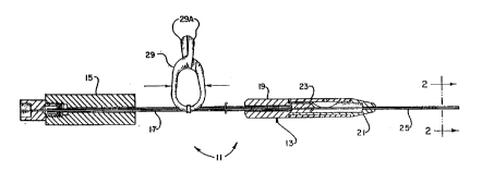
Fig. 1 is is a side view, with parts broken away for
clarity, of the laser delivery system of the present
invention:
Fig. 2 is an enlarged sectional view taken along
lines 2--2 of Fig. 1~
Fig. 3 is a rsectional view, on an enlarged scale, of
the distal end of the~handpiece body of the laser delivery
system of Fig'. 1; and
Fig. 4 is a sectional view, similar to Fig. 3, but on
a somewhat smeller scale, of an alternative embodiment of
the handpiece body of the present invention.
Similar reference characters indicate similar parts
throughout the several views of the drawings.
Descri tion of the Prqferred Embodiment
Turning to the drawings, a laser delivery system il
of the present invention includes a handpiece 13, a laser
connector 15, and an optical fiber cable 17. Iiandpiece 13
has a handpiece body made up of a handpiece proximal end
portion 19, a handpiece distal end portion 21, and a
reflex sleeve 23. A hollow metal tip 25 of a size
suitable for insertion into a human aye extends distally
from the handpiece body. Tip 25 is preferably a metal
tube approximately one and three-quarters inches long
which is suitably secured in the distal end of the
handpiece body with approximately 1.38 inches of the tube
exposed distally from the handpiece body. The outer
diameter of the metal tube is, for example, approximately
0.0355 inch, and its inner diameter is approximately 0.025
inch. These dimensions are illustrative of those for a
tip suitable for insertion in the hum$n eye.
WO 93/08742 _ ~ 2 0 9 9 5 7 8 . PGT/US92/09152
_ 5 _
Laser connector 15 may be of any desired construction
suitable for connection to a laser source (not shown).
The construction shown is illustrative only.
As can be readily seen in Fig. 1, optical fiber cable
17 terminates proximally in laser connector 15 in such a
manner that it is exposed to the laser light from the
laser source. The optical cable extends for any desired
length (such an eight feet or so) and terminates distally
in the tip 25 of handpiece 13. Optical fiber cable 17
thereby forms an optical path for the laser light from the
laser source to an eye being treated.
Also shown in Fig. 1 is a clamp 29 used to removably
secure cable 17 to any appropriate structure to hold the
cable in place without significantly restricting movement
of the handpiece by the surgeon. For example, clamp 29
may be readily secured to a surgical drape (not shown) in
the operative field by pressing both sides in the
directions indicated by the arrows in Fig. 1. This
.pressure opens the jaws 29A of the clamp so that the jaws
may be placed over a fold in the drape. Once the pressure
is removed, the jaws close on the drape, thereby holding
the optical cable fixed at that position in the surgical
field. This prevents the optical cable from moving
excessively during the medical procedure, which movement
could otherwise result in contamination of the sterile
field.
Turning to Fig. 2, there is shown on a greatly
enlarged scale the relationship between optical cable 17
and tip 25. The portion of optical cable 17 which is
disposed in tip 25 has an outer diameter of approximately
0.013'x, for example, while the inner diameter of the tip
is approximately 0.025. ThisJdiffarence in diameter
leaves a gap 31 disposed between the optical fiber and the
tig. This gap runs the entire length of the tip and forms
WO 93/08742 ~ ~ ~ ~ ~ 7 ~ ' PGT/US92/09152
- 6. -
a fluid path frown the distal end of tip 25 to the interior
of the handpiece body.
Note that if the optical fiber were secured to tip 25
by adhesive (~s has been done previously), the adhesive
Mould tend to block off gap 31, To prevent this, the
optical fiber is not secured directly to tip 25 at all.
Instead it is suitably-secured to proximal end portion 19
of the handpiece body. Note as wall that, although the
optical fiber I7 is shown centered in tip 25 in Fig. 2,
the .fiber can in fact be off-center in the tip without
closing off gap 3I.
The fluid path formed by gap 31 allows fluid and
other material to be withdrawn through the gap.
Significantly, the distal end of this fluid path is
disposed immediately adjacent the spot where the laser
light exits the tip, so that removal of fluid from the
operative site takes place almost exactly where needed.
The fluid Bath formed by gap 31 is in fluid
communication with a fluid path through handpiece 13.
That latter fluid path in formed by a cavity 33 (Fig. 3)
formed in handpfece distal end portion 21, which opens
into a cavity 3.5 between handpiece distal end portion 21
and reflex sleeve 23. Sleeve Z3 has a port 37 formed
therein above cavity 35, so that fluid in cavity 35 may
flow out port~37 to the exterior of handpiece 13.
As is known, the interior of.an eye being operated on
may be placed under pressure with a suitable solution so
as to mafntain its sphericity during the procedure. This
pressure causes fluid'to flow through gap 31 in handpiece
tip 25, through cavities 33 and 35, and out port 37. The
surgeon can stop this flow simply by covering port 37 with
a finger.
Note that with this construction, fluid in the eye
may flow through the tip and the handpiece body even while
WO 93/08742 ~ ~ ~ 9 ~ ~ 7 ~ PCT/US92/09152
....
7 _
laser light from the laser source is directed by the
optical fiber into the aye. The surgeon, with one-handed
control, can thus apply laser power to the surgical area
while. at the same time suction away fluid and other
material from exactly the same area.
On occasion, distal tip 25 can suction in undesired
material, such as a portion of the retina. With the .
present construction, this material can easily and rapidly
be refluxed back into the eye, again with a one-handed
operation. Reflux sleeve 23 is formed from a relatively '
soft, elastically deformable., resilient material such as
50 durometer silicone rubber. Note that port 37 is
located directly over cavity 35 (which cavity is formed by
cutting away the corresponding part of handpiece distal
end portion 21). By pressing downwardly on sleeve 23~
above cavity 35, the surgeon applies pressure on the fluid
path from port.37 through the distal end of tip 25. This
pressure forces any undesired material back out of the
distal end of tip 25. Note that because port 37 is
disposed directly above cavity 35, the mere act of
pressing downwardly on the sleeve in this location
automatically closes port 37. This ensures that the
effect of the downward motion is to force fluid and any
accompanying material distally out of tip 25.
It should be appreciated that the construction shown
in Fig. 3 at beet can expel a volume of material from the
tig approximately equal to the volume of cavity 35. If it
is desired to reflux a~larger volume of material, the
alternative construction of sleeve 23 shown in Fig. 4 may
be used.
In Fig. 4, the rsflux sleeve, labelled 23A, includes
a hollow protrusion or bulb 39 in which is disposed port
37. Tha pros~nca of bulb 39 increases the volume of
material which can be expelled from tip 25, while at the .
2099578
v~; f~R~/08742 PGT/US92f09152
_ g ..
same time providing a positive indication to the surgeon
of the location~of port 37. This positive. indication is
especially useful for locating the port while the surgeon
is in the dark.
The handpie~e of Fig. 4 also differs in having the
capability of applying suction from a separate suction
source (not shown) to the distal end of tip 25.
Specifically, proximal section 19A of the handpiece shown
in Fig. 4 has a first bore- 41 through which passes optical
fiber 1~, and a second bore 43 in fluid communication with
cavities 33 and 35. Bore 43 may be attached to any
suitable suction source by means of a conventional tube 45
fixedly secured in-bore 43.
Like the handpiece of Fig. 3, the one of Fig. 4 has
the property that the application of pressure on the
reflex sleeve above cavity 35 results in simultaneous
closure of the fluid path through the handpiece.
As another alternative, the reflex means may include
a tube (made of a suitable resilient material such~as
silicone or the like)~which exits the handpiece at a
suitable point at Which it can be manually pressed or
squeezed and then reenters the handpiece. -
The apparatus of the present invention may also be
used in other ways. For example, if there is a retinal
detachment, one method of flattening the retiz~a is to
infuse an air bubble or silicone oil into the eye and then
aspirate the subretinal fluid with a suction probe through
a retinal tear or through a hole in the retina
(retinotomy) made so the subretinal fluid can be
aspirated. Conventionally, the suction probe is then
removed and the laser probe introduced into the eye. The
laser probe is used the seal the retinal tear when the
retina is. reattached. This prior art procedure requires
replacing the suction probe with the laser probe. With
WO 93108742. ~ ~ 2 0 9 9 5 7 8 PL'1'/US92/09152
,...
- g
the apparatus of the present invention, this replacement
is unnecessary, thereby simplifying the procedure. In
addition., sometimes fluid re-accumulates under the hole
during the procedure, and with conventional equipment the
laser cannot be immediately applied. With the apparatus
of the present invention, the aspiration of the
re-accumulated fluid may take place immediately, With no
delay for switching probes.
The apparatus of the present invention is also
especially suited for other methods or applications. If
bleeding on the surface of the retina occurs during the
operation,~the bleeding vessel or tissue must be
coagulated so that visibility of the tissues will not be
obscured. Presently, the vacuuming needle or "'flute
needle"' is used to remove the blood and then a separate
diathermy probe (coagulating tip) is used to cauterize the
tissue. occasionally bleeding is so active that when
instruments are exchanged, blood covers the bleeding
tissue again. The apparatus of the present invention
allows simultaneous aspiration of blood and the use of
laser energy to coagulate surface bleeding, so visibility
is not lost.
In addition, the structure of the apparatus of the
present invention may be used to infuse small amounts of
medication precisely to specific areas of the retina
during surgery. For instance, if bleeding is encountered
during surgery a small syringe containing thrombin (or
other,suitable blood clotting factor) may be applied
locally over the bleeding site to clot the blood and
prevent-further bleeding. This is superior to the current
system in which thrombin must be infused throughout the
eye, exposing all the retinal tissue to thrombin, which
may cause inflammation.
W /08'142 ~ 2 0 9 9 5 7 8 p~/US92/09152
_ lp _
Moreover, the apparatus of the present invention may '
be used to deliver a small amount of laser activated
tissue glue or adhesive tp retinal tears or other target
tissue using the same system. Then the laser is applied
to activate the glue, forming the adhesive more precisely
than is possible with present systems.
In view of the above it will be seen that the various
objects and features of the above described invention are
achieved and other advantageous results obtained. The
description and drawings o~ the present invention
contained herein are illustrative only and are not to be
interpreted in a limiting sense.
