Note: Descriptions are shown in the official language in which they were submitted.
F a~86 ~
HEPATIC GROWTH FACTOR RECEPTOR IS
THE MET PROTO-ONCOGENE
Field of the Invention
The present invention relates to a complex
comprising hepatocyte growth factor (HGF) and met proto-
oncogene protein. The present invention also relates to
methods for detecting the presence of HGF ligand, met
proto-oncogene receptor and methods for isolating either
the ligand, receptor or complex comprising both.
The present invention further relates to
methods of diagnosing and treating conditions
proliferative disorders such as hepatitis,
hepatocarcinogenesis, carcinogenesis and wound healing.
In particular, the present methods involve detection of
the ligand-receptor pairs.
Background of the Invention
Hepatocyte growth factor (HGF) was first
purified from human and rabbit plasma and rat platelets
on the basis of its ability to stimulate mitogenesis of
rat hepatocytes (E. Gohoda et al., ~. Clin . Invest . 8 1 ,
414 (1988); R. Zarnegar and G. Michalopoulos, Cance~ Res.
49, 3314 (1989); T. Nakamura et al. FEBS Lett . 224, 311
(1987)). Thus, HGF may act
66956-20
2039~6S
W092/13097 PCT/US92/~071
as a humoral factor promoting liver regeneration
after partial hepatectomy or liver injury (G.K.
Michalopoulos, fASEBJ. 4, 176 (1990)). The same
factor was purified from human fibroblast culture
medium and shown to act on melanocytes and a variety
of epithelial and endothelial cells (J.S. Rubin et
al., Proc. Natl.Acad. Scl. USA 88, 415 (1990)). Together
with evidence of HGF expression in several organs
(J.S. Rubin et al., Proc. Natl. Acad. Sci. USA 88, 415
(1990); K. Tashiro et al. Proc. Natl.Acad. Sci. USA 87, 3200
(1990); R. Zarnegar et al., Pr~ Natl.Acad.Sci. USA 87,
1252 (1990); T. Kinoshita et al. rio~hei". Biop~. Res. Comm.
165, 1229 (1989)), these findings indicate that HGF
may also act as a paracrine mediator of
proliferation for a broad spectrum of cell types.
Molecular cloning of HGF revealed a remarkable
structural homology to plasminogen and related
serine proteases (J.S. Rubin et al., Proc Ns~.~d.Sci.
USA 88, 415 (1990); T. Nakamura et al., Narure 342,
440 (1989); K. Miyazawa et al., Biophys. Res. Com~m.
163, 967 (1989)). Recent evidence that HGF induces
rapid tyrosine phosphorylation of proteins in intact
target cells suggests that a tyrosine kinase
receptor might mediate its mitogenic signal (J.S.
Rubin et al., Pr~ Natl.Acad.Sci. USA ~, 415~990) ) .
HGF is structurally related to the family
of serine proteases that includes plasminogen,
prothrombin, urokinase, and tissue plasminogen
activator (J.S. Rubin et al., Proc. Natl. Acad. Sci. U.SA 88,
415 (199o)); T. Nakamura et al., Natvre 342, 440
(1989)). As defined in the present invention, HGF
includes a variant of HGF previously characterized
as a broad-spectrum mitogen called plasminogen like
209~6~
~092/13097 PCT/US92/~71
growth factor (PLGF). Several proteases, including
members of the serine protease family, stimulate DNA
synthesis presumably through a proteolytic mechanism
similar to tryptic activation of the insulin
receptor (S.E. Shoelson et al. J. Bi~. Chem. 263, 4852
(1988)). Only urokinase has been found to associate
with a specific cell-surface receptor, which itself
bears no homology to any known tyrosine ~inase
receptors (A.L. Roldan et al., EMBO J. 9, 467 (l990)).
It is clear that a need exists to identify
the receptor of HGF. The present invention
describes the complex comprising HGF and met proto-
oncogene protein and identifies the met proto-
oncogene as the receptor for HGF. The met proto-
oncogene protein is a member of the tyrosine kinase
growth factor receptor family. Knowledge of this
receptor/ligand relationship should facilitate the
study of proliferative disorders in which expression
of these molecules may pl~y an important role.
Additionally, identification of the metproto-
oncogene receptor HGF complex provides a means for
identifying tissues other than liver tissue affected
by factor binding.
SUMMARY OP THE lNV~. LlON
It is an object of the present invention
to provide a complex comprising a hepatocyte growth
factor (HGF) ligand and met proto-oncogene protein
receptor and methods of utilizing the complex.
various other objects and advantages of
the present invention will become apparent from the
drawings and the following description of the
invention.
r ~ ~J ~ ~ 8 6 5
In one embodiment, the present invention
relates to a complex of a HGF ligand and met proto-
oncogene receptor protein wherein said complex is
free of protein with which it is naturally
associated.
In another embodiment, the present
invention relates to a complex comprising a HGF
ligand and met proto-oncogene receptor protein
wherein one member of said complex is bound to a solid
support.
In yet another embodiment, the present
invention relates to a method of detecting a HGF:met
proto-oncogene receptor protein complex in a sample
comprising reacting said sample with an antibody
that binds specifically with either HGF or met
proto-oncogene receptor protein or the complex. A
positive immunological reaction is indicative of the
presence of the complex in the sample.
In a further embodiment, the present invention
relates to a method of diagnosing a proliferative
disorder in a-patient suspected of having the disorder
comprising reacting a biological sample from the patient
with an antibody that binds with a HGF-met proto-oncogene
receptor protein complex.
In yet another embodiment, the present
invention relates to a method of diagnosing a tissue
undergoing regeneration in a patient comprising, reacting
a biological sample from the patient with an antibody
that binds to a HGF-met proto-oncogene receptor protein
complex.
A further embodiment of the present
invention relates to a method of diagnosing a
66956-20
2099~,6~
092/13097 PCT/US92/00071
diseased state in a patient suspected of having the
stated disease comprising reacting a biological
sample from the patient ~ith an antibody that binds
with a HGF- met proto-oncogene receptor protein
complex.
In another embodiment, the present
invention relates to a method for detecting HGF in a
sample comprising contacting the sample with met
proto-oncogene receptor protein under conditions
such that binding of HGF present in the sample to
the receptor is effected and detecting the presence
of bound HGF.
In a further embodiment, the present
invention relates to a method for detecting met
proto-oncogene receptor protein in a sample
comprising the steps of contacting the sample with
HGF under conditions such that binding of said
receptor present in the sample to HGF is effected
and detecting the presence of bound receptor.
Another embodiment of the present
invention relates to a diagnoStic kits for measuring
would healing and proliferative disorders. One type
of kit comprises labeled HGF in one container and
ancillary reagents suitable for use in detecting the
presence or absence of met proto-oncogene receptor
in a biological sample.
A second type of kit comprises labeled met
proto-oncogene receptor protein in one container and
ancillary reagents s~itable for use in detecting the
presence or absence of HGF in a biological sample.
~The entire contents of all publications)
mentioned herein are incorporated by reference.
~' '' .1 ':
, .
209~86S PCTJUS91 /00071
IpEAus o 9 N~
BRIE~ DESCRIPTION OF THE DRAWING8
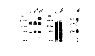
Figures 1~- - show the tyrosine phosphorylation of
pl45 in B5/589 hllma~ mm~ry epithelial cells in
response to HGF. Fig. lA is an immunoblot of
phosphotyrosyl p-~teins from untreated control cells
(C), cells treated with HGF, and with EGF
(Collaborative Research). HGF was purified as
described (J.S. Rubin et al., Proc. Natl. Acad. Sci U.S.A.
88, 415 1990)). Serum-starved cells were exposed to
growth factor (ln0 ng/ml) for 10 min at 37 C as
indicated, detergent-solubilized on ice, and
immunoprecipitated with monoclonal anti-pTyr
(Upstate Biotechnology). Immunoprecipitated
proteins were resolved by 7.5% SDS polyacrylamide
gel electrophoresis (SDS-PAGE) (U.K. Laemmli Nature
227, 680 (1970)), and immunoblotted with the same
antibody as described (D.P. Bottaro et al., J. Biol.
Chem. 265, 12767 (~990)). Fig. lB is an autoradio-
gram of 32P-labele,: phosphoproteins from control (C)
and HGF-treated c~lls. Serum-starved cells were
metabolically lab~led with 32P-orthophosphate (1.0
mCi/ml) as descril~ed (M.F. White and C.R. Kahn, in
Insulin Receptors, Part ~ Methods for the Study of Structure and
Functio~ C.R. Kahn ~nd L. Harrison, Eds. (Liss, New
York, 1988) pp. 1!5-147). The cells were treated
with HGF (100 ng/ll) for 10 min at 37 C as
indicated, and de~ergent-solubilized on ice.
Phosphotyrosyl pr~)teins were immunoprecipitated with
anti-pTyr and resolved by 7.5% SDS-PAGE. Fig. lC
shows a phosphoamino acid analysis of pl45 from lane
2 of Fig. lB which was performed as described (M.F.
White and C.R. Kahn, in Insulin Receptors, Part A: Methods for
the Study of Structure and Func~ion, C.R. Kahn and L. Harrison,
Eds. Liss, New York, 1988, pp. 125-147).
SU~STmJ~E S~EF~
2 0 9 ~ IP~S ~ 9 NOV 199~
The dotted circles indicate the migration of
unlabeled phosphoserine (pS), phosphsthreonine (pT),
and phosphotyrosine ~pY~.
Figures 2A-2B show the identification of pl45 as the
~-subunit of the c-mel pro~o-oncogene product. Fig.
2A is an anti c-met immunoblot of anti-pTyr
immunoprecipitates from control tC) and HGF-treated
B5/589 cells. Samples for immunoprecipitation (2 mg
protein) were prepared as described in Fig. lA,
resolved by 7.5% SDS-PAG~, transferred to Immobilon
(Millipore) membranes and detected with monoclonal
anti-c-mel and [l25I]-protein-A. To quantify the
percentage of c-met protein that was immuno-
precipitable with anti-pTyr, 200 ~g of B5/589 cell
lysate (LYSATE) was resolved by SDS-PAGE and immuno-
blotted directly with monoclonal antibody to c-met.
Fig. 2B is an autoradiogram of 32P-labeled phospho-
proteins from control (C) and H~F-treated B5/589
cells resolved by 7.5% SD'-PAGE under reduced (R)
and non-reduced (NR) cond-tions. Serum-starved
cells were metabolically abeled with 32P-ortho-
phosphate, left untreated (C) or treated with HGF,
and immunoprecipitated wi h anti-pTyr as described
in Fig. lB. Samples were reduced with 100 mM ~-
mercaptoethanol before el~ctrophoresis as indicated.
Figures 3A-3C demonstrate the covalent affinity
cross-linking of 125I-labeied HGFp28 to the c-met
protein-tyrosine kinase. Fig. 3A is an immunoblot
of lysates (200 ~g protein) prepared from M426 human
lung fibroblasts and B5/589 cells using monoclonal
antibody to the cytoplasmic domain of c-mel protein.
Fig. 3B shows cross-
SUBS~T~ S~E~
PC~IU391 /00071
2 0 9 9 ~ 6 ~ IPEA~us ~ 9 NOV 1992
linking of 125I-labeled HGFp28 to ~426 and B5/589
cells resolved by 6.5% SDS-PAGE under non-reduced
(NR) and reduced (R) cond tions. HGFp28 was
purified as described and radiolabeled with [~25I]-Na
by the chloramine-T method (W.M. Hunter and F.C.
Greenwood, Nature 19 4, 495 (1962)). Cells were
incubated with HEPES binding buffer (D.P. Bottaro et
al., J. Biol. Chem. 265, 12767 (1990) containing '25I-
labeled HGFp28 (5 x 10⁵ cpm) for 45 min at 25 C,
washed with cold HEPES-buffered saline (pH 7.4), and
treated with disuccinimidyl suberate (D.P. Bottaro
et al., J. Biol. Chem. 265, 12767 (1990). The cells were
then solubilized with SDS and boiled for 3 min in
the presence 100 mM ~-mercaptoethanol as indicated.
~25I-labeled proteins were resolved by 6.5~ SDS-PAGE
and autoradiography at -70 C. Fig. 3C shows the
immunoprecipitation of [125I]-HGFp28-cross-linked
complexes from B5/589 cells with c-met peptide
antiserum (A. Gonzatti-Haces et al., Proc. NatL Acad. Sci
U.S.A. 85, 21 (1988)). Sample prepcration and cross-
linking prior to immunoprecipitati~n, performed as
described in reference to Fig. 3B, yielded the
electrophoretic pattern shown in t~e left lane
(LYSATE) under reduced conditions. The adjacent
lanes show immunoprecipitation of the cross-linked
species with c-met peptide antiser~m (1:100) in the
absence (~-MET) or presence (+COMI) of competing
peptide (10 ~G/ml). I~munoprecipttated proteins
were absorbed to immobilized prot~in-G (Genex) and
eluted with SDS prior to electrop~oresis and
autoradiography as described in reference to Fig.
3B.
~IJBSTITUTE SHEEl~
,092/13097 2 0 9 9 ~ 6 ~ PCT/US92/00071
DETAILED DESCRlPTION OP ~ V~. llON
The present invention relates to a complex
comprising hepatocyte growth factor (HGF) and met
proto-oncogene protein. The present invention
further relates to method~ of utilizing the complex.
One embodiment of the present invention
relates to a complex formed by the interaction of
HGF with its receptor, the met proto-oncogene
protein. The complex is free of protein with which
it is naturally associated. The binding of HGF to
its receptor, the met-proto-oncogene protein,
regulates the intrinsic tyrosine kinase activity of
the receptor.
The direct interaction of HGF with the c-
met receptor tyrosine ~inase suggests a biochemical
mechanism of mitogenic signal transduction similar
to that of insulin, EGF and other peptide growth
factors. This interaction represents a significant
functional divergence from HGF's structurally
related family of serine protease homologs.
The present invention also relates to
detection and quantitation methods that may be used
in diagnostics to identify HGF (ligand), met-proto-
oncogene receptor or the ligand-receptor complex.
Since the met-proto-oncogene receptor is expressed
on many cell types and tissues including the liver,
the methods described herein provide a means for
identifying tissues other than liver affected by HGF
binding. The methods of the present invention also
aid in understanding the role of the interaction
between receptor and ligand in regulating
biochemical and physiological mechanisms in a broad
spectrum of tissues.
WO92/13097 2 0 9 9 8 6 ~ PCT/US92/~71
The present invention further relates to a
method of detecting and quantitating HGF receptor in
a biological sample using labeled HGF as a probe.
Suitable labels include, for example, radiolabels
S such as '"I, and flourescein.
Using standard methodologies well known in
the art, a biological sample can be extracted with a
non-ionic detergent and inc--h~ted with labeled HGF
in the presence or absence of unlabeled HGF. The
resulting complex can be separated from the
uncomplexed (or unbound) labeled material, for
example, by immunoprecipitating the complex with a
specific polyclonal or monoclonal antibody that
recognizes the met-proto-oncogene receptor protein
or the HGF-met proto oncogene receptor complex. The
overall signal resulting from the presence of label
associated with the resulting complex is compared
with the signal from a mock sample. The mock sample
is prepared ~sinq purified met-oncogene receptor
protein in a known quantity treated the same way as
the biological sample.
Alternatively, the complex may be
separated from uncomplexed material by precipitating
with polyethylene glycol. In both methodologies,
the amount of label that is immunoprecipitated or
precipitated is directly related to the amount of
complex in the biological sample.
The present invention also relates to a
method for detecting and quantitating HGF in a
30 biological sample using labeled HGF receptor as a
probe. The method is carried out as a reciprocal
binding assay following the methodology described
above except substituting as antibody, one that
/092/13~7 PCT/US92/~071
specifically recognizes HGF or the HGF-met proto-
oncogene receptor complex.
The present invention also relates to
further methods of detecting and quantitating HGF-
met proto-oncogene receptor complexes in a sample.
In one aspect, complexes are detected and
quantitated using antibodies. Antibodies utilized
in this embodiment can be directed against HGF, met-
proto-oncogene receptor protein or the HGF-receptor
complex. Antibodies can be either polyclonal or
monoclonal. A sample can be extracted with non-
ionic detergent and incubated with labeled HGF or
met - proto-oncogene receptor protein. After
incubation, the sample is covalently cross-linked
lS with a bifunctional reaqent such as a chemical
cross-linker, for example, disuccinimidil suberate
(DSS). After quenching the reaction with a
quenching agent, the sample is immunoprecipitated
with specific antibody or precipitated with
polyethylene glycol. Quantitation requires
chromatographic separation by, for example, gel
electrophoresis, followed by autoradiography.
ln another method for detecting HGF-met
proto-oncogene receptor complexes in a sample, the
simultaneous expression of HGF and met proto-
oncogene receptor mRNAs are determined.
Simultaneous co-expression of HGF and met proto-
oncogene receptor can be determined by Northern
analysis usinq oligo- or cDNA probes derived from
the sequence of either gene to identify mRNA or
using the polymerase chain reaction (PCR) or any
combination. Northern analysis and the PCR
2099~65
WO92/13097 PCT/US92/00071
technology are methods well known to those skilled
in the art.
The present invention further relates to
diagnostic methodologies using the methods described
above. The disorders which diagnosed by the methods
of the present invention include, for example,
proliferative disorders such as hepatocellular
carcinoma or other carcinomas of tissues that
normally express met proto-oncogene receptor. Such
tissues can be derived from epithelial cells such as
skin, lung, stomach, kidney or colon, liver or
endothelial cells, such as those contained in the
vascular lining or bone marrow, or hematopoietic
stem cells. The present diagnostic methods can also
be used to measure wound repair in tissues derived
from the cells described above, and in cells that
normally express HGF such as platelets, fibroblasts
(stromal tissue of skin and other organs) and
spleen.
Inactivation of the HGF/met mitogenic
pathway provides the basis for-therapeutic
methodologies designed to diminish or arrest normal
or pathological cell proliferation. These
methodologies include the production of genetically
engineered HGF species that lack or possess an
impaired met-binding domain, or that lack or possess
an impaired activating domain, but that otherwise
retain the structural and biochemical
characteristics of HGF. Similarly, production of
genetically engineered met species that lack or
possess an impaired HGF-binding domain, or lack or
possess an impaired tyrosine kinase domain, but
which otherwise retain the structural and
2~9986a
/092/13097 PCT/US92/~071
13
biochemical characteristics of the met protein.
These methodologies also include the production of a
water-soluble form of met protein consistinq of the
extracellular HGF-binding domain that can act as an
antagonist of normal met protein activation by HGF.
The delivery of the genetically engineered HGF or
met protein species described above to the selected
site of action may be achieved using conventional
methods of drug delivery, gene transfer, or any
combination thereof.
Artificial activation of the HGF/~et
mitogenic pathway provides the basis for therapeutic
methodologies designed to restore, replace, or
enhance naturally occurring wound repair mechanisms.
These methodologies include application to the wound
site of genetically engineered HGF or met species
that enhance the binding interaction between met and
HGF and thereby create an artificially sustained
HGF/met interacticn. For example, site-directed
mutagenesis of the HGF-binding domain of met, or the
met-binding domain of HGF (or both) may be used to
create a member of the HGF/met pair with higher
binding affinity for the other member of the pair
and thus affect accelerated growth or regeneration
of the wounded tissue. Similarly, conventional
recombinant DNA techniques could be used to enhance
or sustain the kinase activity of the met protein
normally regulated by HGF binding, including met
mutations possessing a constitutively activated
tyrosine kinase. The delivery of the genetically
engineered HGF or met protein species described
above to the selected site of action can be achieved
usinq conventional methods of drug delivery, gene
WO92/1~97 2 0 9 9 ~, 6 ~ PCT/US92/~071
transfer, or any combination thereof. Activation of
the HGF/met mitogenic pathway by means of
supplementinq the natural expression of met by
recombinant DNA techniques in combination with
exogenously administered HGF is also included.
Example~
Example l. Tyrosine phosphorylation of pl45 in
B5/589 human mammary e~ithelial cells
in response to HGF
The human mammary epithelial cell line
BS/589 is pa-rticularly sensitive to the mitogenic
effects of HGF (J.S. Rubin et al., Pr~.Na~.h~d. Sci. U.SA
88, 415 (l990)). Intact serum-starved B5/589
cells were treated with HGF (approximately lO0
lS ng/ml) for lO min at 37~C and solubilized on ice.
Phosphotyrosyl proteins were isolated from cell
lysates by immunoprecipitation with antibody to
phosphotyrosine (anti-pTyr). These proteins were
resolved by SDS polyacrylamide gel electrophoresis
(SDS-PAGE) and immunoblotted with the same antibody.
Several phosphotyrosyl proteins were detected in
untreated cells by this method (Fig. lA). Treatment
of intact cells with HGF induced phosphorylation of
a 145-kD protein (pl45) (Fig. lA, center lane).
B5/589 cells exposed to epidermal growth factor
(EFG) displayed tyrosine phosphorylation of the EGF
receptor, but not pl45 (Fig. la, right lane). When
lysates from control and HGF-treated cells that had
been labeled with ~2 P-orthophosphate were used for
~Q immunoprecipitation with anti-pTyr, phosphorylation
of pl45 was specifically detected in HGF-treated
~092/13~7 2 0 9 9 8 6 ~ PCT/US92/~071
cells (Fig. lB). Phosphoamino acid analysis of "P-
labeled pl45 confirmed the presence of
phosphotyrosine, and revealed the presence of
phosphoserine as well (Fig. lC). The HGF-stimulated
phosphorylation of pl45 on tyrosine and its apparent
molecular weight were consistent with the
possibility that pl45 represented the receptor
tyrosine k~n~se for HGF.
Example 2. Identification of pl45 as the B
subunit of the c-met proto oncogene
product.
A number of receptor-like molecules have
been described for which there are as yet no known
ligands. One of these is the c~ proto oncogene
product, which is a receptor-like tyrosine kinase
comprised of disulfide-linked subunits of 50-kD (~)
and 145-kD (B) (P.R. Tempest et al. Br.J.~c~ 58 3
(1988); S. Giordano et al. OI~c~ene 4 1383 (1989)).
In the fully prores~e~ c-met product, the ~ subunit is
extracellular, and the B subunit has extracellular,
transmembrane, and tyrosine kinase domains as well
as sites of tyrosine phosphorylation (S. Giordano et
al., Ol,cogene ~, 1383 (1989); A. A. Gonzatti-Haces et
al., Proc.Natl.h~d.Scl.U.SA 85, 21 (1988).
To test the hypothesis that pl45 might
represent the c-met protein B subunit, proteins
immunoprecipitated by anti-pTyr from control and
HGF-treated BS/589 cells were immunoblotted with a
monoclonal antibody directed against the cytoplasmic
domain of the c-met product. Specif~cally, a mouse
monoclonal IgG raised against recombinant human c-met
W092/13097 2 0 9 9 8 ~ ~ PCT/US92/~71
16
protein cytoplasmic domain was used. Recognition of
human c~et protein by immunoprecipitation or
immunoblotting can be specifically blocked by
incubating in the presence of the recombinant
protein fragment.
The prominent 145-kD protein observed
specifically in HGF-treated cells (Fig. 2A) provided
direct evidence that this mitogen induced
phosphorylation of the c~et protein on tyrosine
residues. When whole lysates prepared from
identically treated cells were blotted directly with
the c-mct antibody, the percentage of c~t protein
phosphorylated on tyrosine in response to HGF could
be quantitated (Fig. 2A). It is estimated that at
least 10% of the total cellular c-met protein content
was immunoprecipitated by anti-pTyr after HGF
stimulation. Analysis of the time course of HGF
action revealed that the c~et protein could be
recovered by i~munoprecipitation with anti-pTyr
within l min of treatment and that thi~ effect
persisted for at least 3 hours. Comparison of the
electrophoretic mobility of pl45 under reduced and
non-reduced conditions confirmed that it was the B
subunit of the c~et protein (Fig. 2C). Without
reduction, the 50-kD ~ subunit of the c-met protein
remains disulfide-linked to the B subunit and
substantially retards its migration in SDS-PAGE
(P.R. Tempest et al., 8r.J.Cancer 58, 3 (1988); S.
Giordano et al., Oncogene 4, 1383 (1989); P.R. Tempest
et al., F~BSLett. 209, 357 (1986); M. Park et al., Proc.
Natl. Acad. Sci. U.SA 84, 6379 (1987); A. Gonzatti-Haces et
al., Proc Natl. Acad. Sci. l,'SA 85, 21 (1988) ) . Similarly,
pl45 immunoprecipitated from "P-labeled B5/589 cells
2099865 ~CTIUS91 /00~71
~S ~ 9 NOY 1992
17
that had been treated with HGF displayed a shift in
mobility characteristic of the c-met proto oncogene
product when subjected to reduced or non-reduced
electrophoretic conditions (Fig. 2B). Together
these results identified pl45 as the c-met protein
subunit and established that HGF stimulated its
phosphorylation on tyrosine residues.
Example 3. 125I-HGFp28 is physically associated
with the c-met protein-tyrosine kinase.
The rapidity and extent of c-met protein
tyrosine phosphorylation in response to HGF
supported the possibility that c-met protein was the
cell-surface receptor for HGF. However, there is
evidence that receptor kinases can phosphorylate
other receptors (D.F. Stern and M.P. Kamps, EMBO J.
7, 995 (1988); C.R. King et al., EMBO J. 7, 1647
(1988)). Thus, conclusive identification of the ~-
met product as the HGF receptor required a
demonstration of their direct interaction. 125I-
labeled HGF was unsuitable for covalent affinity
cross-linking because it consisted of a mixture of
single chain and heterodimeric labeled species. A
smaller form of HGF with similar binding propertie;,
designated HGFp28, was '25I-labeled as a single
entity and used to characterize the HGF receptor.
HGFp28 was labeled with [~25I]Na by the
chloramine-T method as follows: HGFp28 (3 ~g in 5
~1 of 20 mM phosphate buffer containing 1.0 M NaCl,
pH 7.4) was reacted with chloramine-T (1.2 ~g in 4
~1 of phosphate buffer) and [125I]Na (1 ~Ci) at 24 C
for 1 min. The reaction was terminated by addition
STi~TE SH~E~
WO92/13097 ~ Og 9 ~, 6 PCT/US92/00071
of sodium metabisulfite (lO ~g in 8 ~l of phosphate
buffer). The mixture was diluted with phosphate
buffer containing 0.1% bovine serum albumin (200 ~l)
and applied to a column (300 ~l packed volume) of
heparin-Sepharose CL-6 ~that had been equilibrated
in phosphate-buffered saline containing 0.1% BSA
(PBS/BSA). The column waQ washed with 30 ml of
PBS/BSA and eluted with PBS/BSA containing l.0 M
NaCl (200 ~l/fraction), removing 98% of
trichloroacetic acid-precipitable radioactivity from
the column. Peak fractions (specific activity: 150
to 250 ~Ci/~g) were 99~ trichloroacetic acid-
precipitable, ~nd migrated as a single band on SDS-
PAGE.
Comparative cross-linking studies were
performed using 12~ I-labeled HGF p28 on B51589 cells
and M426 human fibroblasts, an HGF-insensitive cell
line which also lacks detectable amounts ~f c~t
protein (Fig. 3A). The '~I-labeled HGFp28~cross-
linked to its receptor on B5/589 cells migrated as a
2lO-kD protein complex under non-reduced conditions
(Fig. 3B). Under reduced conditions, a major 170-
kD complex was observed (Fig. 3B). These apparent
molecular sizes were consistent with a direct
interaction between the labeled HGFp28 and the 145-
kD B subunit of the c-met protein. Under reduced
conditions, two minor bands of l90-kD and about 300-
kD were also detected (Fig.3B). Cross-linking of
"'I-labeled HGFp28 to the species observed under
reduced conditions was blocked by addition of either
unlabeled HGFp28 or HGF-neutralizing antisera.
Under identical conditions, "'I-labeled HGFp28
r,~ r ~ f-,1
. ~ .. . .
, .,
209986i
1092/13097 PCT/US92/~071
failed to cross-link to any large proteins in M426
cells (Fig. 3B).
To establish that "I-labeled HGFp28 was
physically associated with the c~t protein, '~I-
labeled HGFp28 cross-linked complexes were
immunoprecipitated with a polyclonal antiserum (A.
Gonzatti-HaCeS et al., Proc. N~a.A~. Scl. USA 85, 21
(1988) specific to the carboxyl-terminal 28 amino
acids of the B subunit of the c~e~ protein. The
covalently cross-linked major 170-kD and minor 300-
kD species detected under reduced conditions were
immunoprecipitated by the antibody, and their
detection was specifically blocked by competing
peptide (Fig. 3C). These results demonstrate a
direct molecular interaction between 'Z'I-labeled
HGFp28 and the c-met B subunit. The composition of
the minor 300-kD cross-linked species remains to be
determined. All of these findings establish that
the c-met product i8 the cell surface receptor for
HGF.
While the foregoing invention has been
described in some detail for purposes of clarity and
understanding, it will be appreciated by one skilled
in the art from a reading of this disclosure that
various changes in form and detail can be made
without departing from the true scope of the
invention.