Note: Descriptions are shown in the official language in which they were submitted.
WO 92/13482 PCT/US91/09790
a:,~
.. Ni ~'~~~J
APPARATUS AND METHOD
FOR MEASURING A BLOOD PARAMETER
BACKGROUND OF THE INVENTION
It is sometimes necessary or desirable to
measure various parameters of blood, such as hematocrit,
oxygen saturation, carboxyhemoglobin, pH, etc. These
blood parameters can all be measured using optical
techniques.
For example, to measure oxygen saturation of
whole blood, red light at, for example, 660 nanometers
(nm) and infrared light at, for example, 805nm are
directed at the blood. The reflectance at both wave- ,
lengths is measured and appropriately ratioed to provide
a measurement of oxygen saturation.
The hematocrit in whole blood may be measured,
for example, by directing infrared light at the blood
and detecting the reflectance of the infrared light at
two spaced detectors. The hematocrit can then be
determined by using a ratio of the two detected light
levels or the difference between the two detected light
levels. Another way to measure hematocrit is to employ
a pair of spaced light sources and a single detector.
All of the techniques described above rely
upon measuring of different detected light levels.
Unfortunately, when the absorbance of the light directed
at the blood is substantial, very low light levels are
available for detection, and this results in a poor
signal-to-noise ratio. In addition, when a constant
level of light intensity is directed at the whole blood,
the detected light level rises to a maximum near the
middle of the physiologic range of hematocrit and then
falls off this peak with increasing hematocrit levels.
This curve creates an indeterminate condition in that i~
WO 92/13482 PCT/US91/09790
is not possible to determine which side of the peak is
being observed so that an accurate hematocrit
measurement is not obtainable.
Heinemann Patent No. 4.447.150 discloses a
technique for measurement of blood oxygen saturation
which compensates for variations of hematocrit levels by
assuring uniform depth of penetration of light into the
blood being sampled. In this system, red and infrared
light sources direct light toward a blood sample, and a
single detector detects the light which is reflected or
transmitted. Optical feedback from the detector is used
to control the light emitted by one of the sources so
that the light detected by the detector from such source
is constant. The intensity of the light emitted from
the second source is determined by a ratio between the
current needed to drive the first source and the current
needed to drive the second source. This latter
technique does not establish the intensity of the second
light source as accurately as may be desired. Also,
this technique either suffers from inaccuracy resulting
from inherent differences and drift between the light
sources or it requires a matched set, which is more
expensive to provide.
SUMMARY OF THE INVENTION
This invention provides a method and apparatus
for measuring a blood parameter which generally
overcomes the disadvantages noted above. According to
this invention, optical feedback is used from a signal
detector to each of the light sources being employed so
that each of these sources can be more accurately
controlled than in the prior art. In addition, this
invention compensates for errors in the intensity of the
light emitted by the light sources so as to provide '
improved accuracy without the need for matched sources.
WO 92/13482 PCT/US91/09790
One example of this invention is an apparatus
for measuring hematocrit of whole blood. This apparatus
includes a light source for ezaitting light toward a
blood receiving location, a signal detector for
receiving light from the light source after the emitted
light interacts with the blood at the blood-receiving
location, and a feedback loop. The feedback loop is
responsive to the intensity of the light received by the
signal detector to provide a feedback signal for
adjusting the intensity of the light source so that the
intensity of the light received by the signal detector
is substantially constant over a range of values of the
blood parameter. Means responsive to the feedback
signal provides an output signal which provides an
indication of the hematocrit. The feedback loop
provides for accurate control of the light source.
Although a single light source emitting in the
infrared range is suitable if the apparatus is only to
detect hematocrit, to adapt the apparatus to measure
other blood parameters, one or more additional light
sources which emit light having appropriate wavelength
characteristics, may also be employed. For example, to
measure oxygen saturation, first and second light
sources which emit light having first and second
wavelength characteristics, respectively, are employed.
A wavelength characteristic has reference to the
wavelength or wavelengths which are suitable for
measurement of the blood parameter of interest. For.
both hematocrit and oxygen saturation measurements, a
narrow band is suitable. For example, one of these
sources may emit red light of 660nm and another of the
sources may emit infrared light at al2nm. Both of these
light sources can be used for oxygen saturation
measurements, and only the infrared source is required
for hematocrit measurements.
WO 92!13482 PCT/US91/09790
2~.~~~~3 -
Although multiple signal detectors can be
employed, if desired, only a single detector is
necessary, and a single detector is preferred for
ratioing purposes. The signal detector receives light
from the first and second light sources after
interaction of the light with the blood at the
blood-receiving location. This interaction may include
transmission, reflection, diffusion, absorbance and/or
back scattering of the light in the blood. Preferably,
the signal detector receives substantially only light
that has been reflected or back scattered, and
optimally, only backscattered light is received by the
detector.
The intensity of the first light source is
adjusted so that the intensity of the light at the
signal detector from the first light source remains
substantially constant over a range of values of the
blood parameter. Similarly, the intensity of the light
emitted by the second light source is adjusted so that
the intensity of light at the signal detector from the
second light source is also substantially constant over
a range of values of the blood parameter. Thus, both
the. first and second light sources are controlled
directly by the light intensity at the signal detector,
and this provides greater control over the light sources
and improved accuracy.
The apparatus also provides a signal which is
related to the intensity of at least one of the first
and second light sources and which provides an
indication of the blood parameter of interest. For
example, for hematocrit the signal may be a function of
only one of the light intensities and not the other '
intensity. On the other hand, for oxygen saturation,
the signal may be a function of the ratio of both of the
light intensities.
WO 92/1342 PLT/US91/09790
~r~~' ~ ~ ~ .~ 3'~ l~ ~
The percent of oxygen saturation varies with
hematocrit. To compensate for this effect which
hematocrit has on oxygen saturation, the signal is
preferably corrected for hematocrit so that the true
oxygen saturation reading can be obtained.
The light intensity adjustments to maintain
the desired emitted light levels from the first and ,
second light sources can be accomplished in different
ways. Fox example, this could be accomplished by
appropriate attenuation of light intensity from strong
light sources. However, preferably, this adjustment is
accomplished by variably energizing the light sources.
With this arrangement, the hematocrit or other blood
parameter signal is related to the driving signal or
current signal applied to the Light source to generate
the light intensity of the source.
The signal detector may provide a detector
signal related to the intensity of the light received at
the detector. The intensity adjustment may include a
feedback loop responsive to the detector signal for
providing a feedback signal to adjust the intensity of
the light sources.
Ideally, the emitted light intensity varies or
tracks with the feedback signal in accordance with a
predetermined relationship. Preferably, this is a
linear relationship. However, variables, such as
nonlinearity of the light source with current input and
temperature and aging of the light source make the
emitted light intensity subject to deviating from, or
not accurately tracking with, the predetermined
relationship. Thus, there may be a difference between
the light intensity commanded by the feedback signal and
the light intensity actually emitted by the light
source.
Another feature of this invention is to make
the emitted light intensity more in accordance with the
WO 92/13482 PCT/US91 /09790
predetermined relationship, i.e., track more accurately
with the feedback signal. This can be accomplished, for
example, by producing a reference signal which is
related, preferably linearly, to the light intensity
actually emitted by the light source. The reference
signal and the feedback signal are then used to control
the light source to provide an emitted light intensity
which is more in accordance with the predetermined
relationship. Because this invention restores the
relationship between the feedback signal and emitted
light intensity, the feedback signal becomes an accurate
variable to use as an output signal to provide an
indication of the blood parameter of interest.
Although various techniques can be used for
making the emitted light intensity more in accordance
with the predetermined relationship, preferably the
apparatus includes a reference detector for receiving at
least some of the light emitted by the light source and
circuit means responsive to the intensity of the light
received by the reference detector for adjusting the
intensity of the light emitted by the light source,
i.e., to compensate the light source. The reference
detector provides a reference detector signal which is
related to the intensity of the light it receives. The
circuit means receives the feedback signal and the
reference detector signal and provides a driving signal
to drive the light source to make the emitted light
intensity more in accordance with the predetermined
relationship. If the reference detector is a silicon
diode, the relationship is linear.
The invention, together with additional
features and advantages thereof, may best be understood
by reference to the following description taken in
connection with the accompanying illustrative drawings.
WO 92/13482 PCT/US91/09790
- 7 _
r~~sy<~
2 ~. t: a;
BRIEF DESCRIPTION OF THE DRAWING
Fig. 1 is a schematic view illustrating one
preferred form of the invention.
Fig. 2 is a more detailed schematic of the
preferred form of the invention.
Fig. 3 is a diagram showing system clock
pulses and pulses generated in response to the clock
pulses.
Figs. 4a-4d show examples of signals occurring
at various points in the circuit of Fig. 2.
Fig. 5 shows one example of integrator output.
Fig. 6 is an exemplary plot of hematocrit
versus the feedback signal from the infrared light
source.
Fig. 7 is a family of plots showing how
hematocrit affects the percent oxygen saturation.
DESCRIPTION OF THE PREFERRED EMBODIMENT
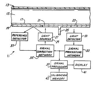
Fig. 1 shows an apparatus 11 which comprises
an in-line flow-through housing 13 having a passage 15
extending therethrough and defining a blood-receiving
location. The housing 13 is adapted to be coupled into
a circuit, such as an extracorporeal circuit (not shown)
of the type used in open-heart surgery. The apparatus
11 is adapted to measure hematocrit and oxygen
saturation of whole blood in real time. of course, the
apparatus 11 can also be used to measure these blood
parameters in a stationary blood sample.
The housing 13 includes a transparent window
17. An opaque cover 19 is suitably mounted, such as on
the housing 13, contiguous the window 17. The cover 19
has a sending aperture 21 and a receiving aperture 23
spaced from the sending aperture. Except for an optical
path through the window ,17 and the passage 15, the
apertures 21 and 23 are suitably optically isolated from
each other as by an optical wall 25.
WO 92/13482 PCf/US91/09790'
- 8 -
n.~cl.~7-~~
.,
~G~.~. e~
The apparatus 11 also includes a red light
source 27 and an infrared light source 29 arranged to
form point light sources on the axis of the sending
aperture 21. The light sources 27 and 29, which may be
light-emitting diodes, are arranged to be as close
together as physically possible and are preferably
pulsed using a short duty cycle to minimize self-heating
of the LED.
The apparatus 11 also includes a light
detector or signal detector 31 positioned on the axis of
the receiving aperture 23 and adapted to provide a
detected signal, such as a current signal, which is
proportional to the intensity of the light detected by
the detector. With this arrangement, the only light
path from the sources 27 and 29 to the detector 31 is
through the blood in the passage 15. The apertures 21
and 23 are preferably spaced so that substantially only
baekscattered light, which has been back scattered by
the blood in the passage 15, will reach the detector 31
from the sources 27 and 29. This spacing can be
adjusted by those skilled in the art and may be, for
example, from about 2.5 to about 3.5mm between the axes
of the apertures 21 and 23. Similarly, the spacing
between the sources 27 and 29 and the aperture 21 can be
varied depending upon the desired angle of emission of
the emitted light from the sources. Preferably, the
detector 31 and the light sources 27 and 29 are spaced
equally from the associated apertures 21 and 23.
It is desirable to adjust the intensity of the
light emitted by the light source 27 so that the
intensity of the light at the detector 31 from the light
source 27 remains substantially constant over a range of
values of the blood parameters being measured.
Similarly, the intensity of the light emitted by the
light source 29 is also adjusted so that the intensity
of the light at the detector 31 from the light source 29
WO 92/13482 PCT/US91/09790
9
~~,~a
is substantially constant over a range of values of the
blood parameters being measured. Preferably, although
not necessarily, the constant intensities of the sources
are also known, predetermined values.
To control the intensity of the light sources
27 and 29, the detected signal from the light detector
31 is applied to a signal processing network 33 which
provides a feedback signal which can be used to
controllably drive the light sources 27 and 29.
Although the feedback signal could be used directly to
control the light sources, in this embodiment, the
feedback signal is applied to a signal correction
network 35.
Various factors, such as inherent nonlinearity
of the light sources 27 and 29 and temperature and
aging, can cause the light sources 27 and 29 to emit a
light intensity different from the light intensity
commanded by the feedback signal from the network 33.
To make the emitted light intensity from the light
sources 27 and 29 more in accordance with the correct or
predetermined relationship between the feedback signal
from the network 33 and the emitted light intensity, a
reference detector 37 receives some of the light from
the sources 27 and 29, such light being reflected from
the cover 19. The reference detector 37 provides a
reference detector signal to the signal correction
network 35. The signal correction network 35 is
responsive to the feedback signal and the reference
detector signal to provide a driving signal to the light
sources 27 and 29.
By alternately pulsing the light sources 27
and 29, the signal processing described above can be
repeated until balance or equilibrium is reached for
each of the light sources, i.e., until the intensity of
the light detected at the detector 31 is constant at a
desired intensity for each of the light sources. The
WO 92/13482 PCT/US91/09790~
n 4~'~ - 10 -
~,f, ~ ~'s ~'
feedback signals from the signal processing network 33
form output signals which are processed in a signal
processor 39 to determine the values of the blood '
parameters being measured, and these values are
displayed by a display 41.
When using the apparatus 11 with blood flowing
through the passage 15, the process described above is
run continuously to provide a real time display of the
blood parameters being measured. In this embodiment,
hematocrit is calculated as a function of the intensity
of the infrared light source 29 after equilibrium has
been reached. More specifically, the feedback signal
from the signal processing network 33 resulting from
operation of the infrared light source 29 after
equilibrium has been reached is utilized by the signal
processor 39 to calculate hematocrit in that hematocrit
is proportional to that feedback signal.
More specifically, the feedback signal derived
from the infrared light source 29 is linearly related to
hematocrit. The slope and offset of the linear
relationship shown, by way of example in Fig. 6, is
established during calibration and stored in a
calibration memory 42 so that the curve of Fig. 6 can be
established by the signal processor 39. With the curve
of Fig. 6 established, the feedback signal derived from
the infrared light source 29 establishes a point on the
curve which represents the hematocrit. In terms of the
usual straight-line equation, Hct=m(infrared feedback
signal) + b where m is the slope of the curve shown in
Fig. 6 and b is the offset from the X axis.
Oxygen saturation is determined by the signal
processor 39 as 1 minus the ratio of the light
intensities of the sources 27 and 29 at equilibrium.
Specifically, the measured percent saturation equals 1
minus A/B where "A" is the intensity of the red light
source 27 and "B" is the intensity of the infrared
WO 92/13482 PCT/US91/09790
- 11 -
~~ZF
source 29. The feedback signals from the signal
processing network 33 are used to represent the light
intensities.
For more accurate oxygen saturation results,
the measured oxygen saturation should be corrected by a
correction factor which is a function of the hematocrit.
More specifically, the percent oxygen saturation as
determined from the oxygen saturation formula set forth
above is preferably corrected utilizing the family of
curves shown in Fig. 7. Thus, by knowing the hematocrit,
one curve of the family curves in Fig. 7 is selected so
that the measured oxygen saturation can be corrected to
yield a true oxygen saturation. Fig. 7 shows by way of
example, oxygen saturation correction curves for only
three values of hematocrit, but of course, a separate
curve can be provided for as many hematocrit values as
desired.
The curves of Fig. 7 can be established, for
example, by empirical derivation during calibration.
The correction factors represented by the family of
curves can be stored in the calibration memory 42 and
applied to the measured oxygen saturation by the signal
processor 39 to result in the display 41 displaying the
true oxygen saturation.
The signal processing network 33, the signal
correction network 35 and the signal processor 39 can be
implemented using a variety of analog and/or digital
techniques. Fig. 2 shows one preferred way o°
implementing this circuit.
Fig. 2 can best be understood by first
considering Fig. 3 which shows clock pulses 43 cf the
system clock. Derived from the clock pulses 43 are red
emission pulses 45, infrared emission pulses 47, red
switch pulses 49 and infrared switch pulses 51. By way
of example, the pulses 45 have a duration of about 610
microseconds and are spaced by an interval of about 19.5
WO 92/13482 PCT/US91/09790
- 12 -
3
~il iseconds. The infrared pulses 47 may have an
identical duration and interval. As shown in Fig. 3,
the pulses 45 and 47 occur alternately.
The switching pulses 49 and 51 are used to
control switches as described below. The red switching
pulse 49 occurs during the last half of each red
emission pulse 45, and similarly, each of the infrared
switching pulses 51 occurs during the last half of an
associated infrared emission pulse 47. In this
embodiment, intensity of the illumination of the sources
27 and 29 is controlled by changing the amplitude of the
associated pulses 45 and 47. The interval between
pulses remains fixed.
The light sources 27 and 29 emit light pulses
in response to each of the pulses 45 and 47,
respectively. Each of the light pulses is coextensive
in time with the duration of the associated energizing
emission pulse.
The detector 31 provides a detected signal
during each of the light pulses. The detected signal is
amplified by an amplifier 53 (Fig. 2), and this provides
a detected signal 55 at the output of the amplifier 53
as shown by way of example in Fig. 4a. The detected
signal 55 has a do level above a baseline 57 of zero
volts which represents some of the ambient light seen by
the detector 31 and a varying ac component 59 which
represents variations in ambient light as seen by the
detector. Superimposed on the ac component are detected
pulses 61 and 63 which represent light from the light
sources 27 and 29, respectively, resulting from one each
of the emission pulses 45 and 47.
The detected signal 55 is applied to a filter
and synchronous detector 65 which eliminates the do
component and noise and detects the signal component of
the signal 55 to provide the detected pulses 61 and 63
as shown in Fig. 4b. If desired, the filter 65 may
WO 92/13482 PCT/US91/09790
13 -
2~~~~.~
include an amplifier which amplifies the detected pulses
61 and 63 so that they may have an amplitude of, for
example, about several volts.
The detected pulses 61 and 63 are applied to
one input of a comparator 67, which may be a voltage
divider, and the other input of the comparator is
coupled to a negative do voltage reference. For each
detected pulse 61 or 63, the output of the comparator 67
is a pulse having an amplitude equal to the algebraic
sum of the positive detected pulse 61 or 63 and the
negative do reference. Assuming that the circuit is not
in equilibrium, then the output of the comparator 67 is
modified red pulses 69 and modified infrared pulses 71
(Fig. 4c) which correspond to the pulses 61 and 63,
respectively. The first half of each of the pulses 69
and 71 may be distorted as shown, by way of example, in
Fig. 4c. Of course, with the circuit in equilibrium,
there is a zero-volt output from the comparator 67.
The output of the comparator 67 is applied to
a red selector switch 73 and an infrared selector switch
75. The switches 73 and 75, which may be field effect
transistors, are normally open. However, the switches
73 and 75 are closed by an appropriate logic circuit
during the red switch pulse 49 and the infrared switch
pulse 51, respectively.
The effect of closing the switches 73 and 75
in this manner is twofold. First, because the switches
73 and 75 are closed only during the presence of
associated modified pulses 69 and 71, they serve as a
selector to apply the modified red pulse 69 to an
integrator 77 and the modified infrared pulse 71 to
another integrator 79. Second, because the switches 73
and 75 are closed only during the last half of the
associated modified pulses 69 and 71, they serve to
eliminate the distortion appearing in the first half of
WO 92/13482 PCT/US91/09790
- 14 -
such pulse and provide shaped red pulses 76 and shaped
infrared pulses 78, respectively, (Fig. 4d).
The number of integrators preferably equals
the number of the light sources, and in this embodiment,
two light sources 27 and 29 and two integrators 77 and
79 are provided. The integrators 77 and 79 are
conventional analog integrators which integrate the
shaped red pulses 76 and shaped infrared pulses 78,
respectively. For example, the integrator 77 integrates
a succession of the shaped red pulses 76 to provide an
output which gradually approaches a desired output in
stepwise fashion as shown by way of example in Fig. 5.
Fig. 5 shows by way of example a voltage that is
initially too low, thereby generating an illumination
level at the source 27 which is too dim, and this may
occur, for example, at startup. The desired
illumination level is represented by a reference voltage
level 81. In this example, each of the shaped red
pulses 76 is of progressively increasing amplitude
thereby resulting in a step-up in level of the output of
the integrator 77 from a Zero-volt baseline through a
series of intermediate levels 83 until the reference
voltage level 81 is reached. With each successive pulse
76.of increased amplitude, a new and higher intermediate
level 83 is provided by the integrator as shown in Fig.
5. Typically, the incremental increases in intermediate
voltage levels 83 are of progressively reducing
magnitude as the reference voltage level is approached.
The integrator 79 functions in the same manner with
respect of the shaped infrared pulses 78.
The output of each of the integrators 77 and
79 constitutes a feedback signal which can be used to
control the intensity of the light sources 27 and 29,
respectively. The outputs of the integrators 77 and 79
are processed by the signal processor 39 which, in this
embodiment, includes a multiplexer 82 for multiplexing
WO 92/13482 PCT/US91/09790
15 - 1
the feedback signals resulting from the light sources 27
and 29, an A to D converter 84 for digitizing the
multiplexed signals and a microprocessor 86 for
performing the calculations discussed above to ascertain
values for hematocrit and percent oxygen saturation.
The microprocessor 86 also corrects the measured oxygen
saturation for hematocrit to provide true oxygen
saturation levels which, together with the hematocrit,
are displayed by the display 41.
The outputs of the integrators 77 and 79 are
applied to selector switches 85 and 87, which may be
identical to the switches 73 and 75, respectively, and
which are closed by conventional logic circuits in
response to the pulses 45 and 47, respectively, in the
same manner as described above for the switches 73 and
75. Thus, the switches 85 and 87 are closed only during
the duration of the pulses 45 and 47, respectively.
The feedback signals from the integrators 77
and 79 are also applied through the switches 85 and 87,
respectively, to one input of an operational amplifier
89 through a resistor 91. A junction between the
resistor 91 and the amplifier 89 is coupled to ground
through a resistor 93. The resistors 91 and 93 form a
voltage divider. The other input of the operational
amplifier 89 is coupled to the reference detector 37
through a conventional current-to-voltage converter 95.
The reference detector 37 provides a reference detector
signal, which is a current signal, which is linearly
related to the intensity of the light emitted by the
light source 27. The converter 95 converts this
reference detector current signal to a reference
detector voltage signal and provides the reference
detector voltage signal to the other input of the
operational amplifier 89. Accordingly, the amplifier 89
has as its inputs the feedback signal, which is
commanding a particular light intensity, and the
WO 92113482 PCTf 0591 /09790'
- is -
reference detector signal which represents actual
intensity of the light source. Of course, the reference
detector 37 also provides the reference detector signal
in response to the light source 29 when that light
source is energized. ,
The output of the operational amplifier 89 is
a drive signal which the amplifier adjusts so as to
attempt to obtain a reference detector signal which is
equal to the feedback signal. The drive signal is
applied to the appropriate one of the light sources 27
and 29 by selector switches 85' and 87' and drivers 97
and 99. The switches 85' and 87' are, like the switches
85 and 87, operated by the pulses 45 and 47,
respectively so that the switches 85' and 87' are closed
only during the duration of the pulses 45 and 47,
respectively. In this manner, the drive signal from the
amplifier 89, which is derived from the integrator 77
and the reference detector signal from the light source
27, is directed to the light source 27. Similarly, the
appropriate drive signal for the light source 29 is
directed to ti~at light source.
With each pulse from the light source 27, the
integrator 77 provides a feedback signal (Fig. 5) which
is closer to the reference level 81. When the reference
level is reached, the system is balanced and in
equilibrium such that the feedback signal can be used
for determination of the blood parameter of interest,
such as hematocrit and the percent oxygen saturation.
Similarly, the reference detector 37 and the resulting
reference detector signal simultaneously correct for the
variables caused by the light sources 27 and 29 such
that the actual and commanded light intensities are
substantially equal.
Although an exemplary embodiment of the
invention has been shown and described, many changes,
modifications and substitutions may be made by one
v
WO 92/13482 PGT/US91/09790
- 17 -
.:
having ordinary skill in the art without necessarily
departing from the spirit and scope of this invention.
..
.. .. 4: ~. . .