Note: Descriptions are shown in the official language in which they were submitted.
WO 92/13946 , PCT/US92/00823
_ 21 00 586
HTLV-I AND HTLV-II PEPTIDE ANTIGENS AND METHODS
15
1. Field of the Invention
The present invention relates to an HTLV-I
specific antigen, and to methods of preparing and
using the antigen.
2. References
Cwirla, S.E., et al., .Proc Nat Acad. Sci, USA,
87:6378 (1990) .
Huynh, T.V., et al., in "DNA Cloning, Volume 1,"
ed. D.M.Glover, Washington, D.C.: IRL Press, 1985
(Chapter 2).
Laemmli, U.K., Nature, 22?:680 (1970).
Lipka, J. J. , et al . , J Infect Dis, 162 : 353 ( 1990 ) .
Lipka, J.J., et al., Proceedings of the 43 Meeting
(1990) .
~"~..
WO 92/13946 PGT/US92/00823
2~ oo ~.a$ _~
2
Maniatis, T., et al., Molecular Cloninq: A
Laboratory Manual, Cold Spring Harbor Laboratory
(1982) .
Matsushita, S., et al., Proc Natl Acad Sci (USA),
83:267? (1986) .
Miyoshi, I, et al., Nature, 294:770 (1981).
Poiesz, B.J., et al., Proc Natl Acad Sci (USA),
77:7415 (1980).
Popovic, M., et al., Science, 29:856 (1983).
Samuel, K.P., et al., Science, 226:1094 (1984).
Samuel, K.P., Gene Anal Tech, 2:60 (1985)
Scott, J.T.< et al., Science, 249:386 (1990).
Seiki, M., et al., Proc Natl Acad Sci (USA),
80:3618 (1983).
Shimotokno, K., et al, Proc Nat Acad Sci, USA,
82:3101 (1985).
3. Background of the Invention
The human T-cell leukemia viruses (HTLV) represent
a family of T-cell retroviruses with three known mem
bers. HTLV type I (HTLV-I) has transforming activity
in vitro and is etiologically linked to adult T-cell
leukemia, which is known to be endemic in several
parts of the world. HTLV-II is another retrovirus
having transforming capacity in vitro, and has been
isolated. from a patient with a T-cell variant of hairy
cell leukemia. HTLV-III, which has also been called
lymphadenopathy-associated virus and is now known as
the human immunodeficiency virus (HIV), is lytic for
certain kinds of T cells and has been linked to the
w
P
f
WO 92/13946 ~ 1 ~ ~ g s PCT/US92/00823
3
etiology of acquired immunodeficiency syndrome (AIDS).
Unlike the HTLV-I and -II viruses, HTLV-III is not
known to have in vitro transforming activity.
The diagnosis of HTLV-I infection is usually based
on serum antibody response to HTLV-I peptide antigens.
This usually involves an initial screening assay to
identify HTLV-I antibodies, based on an enzyme immuno
assay (EIA) with HThV-I virion peptides. The assays
presently used for blood.screening detect about 0.5 to
0.05% HTLV-I and HTLV-II positives; of these, about 4
out of 5 are false positives. Therefore, positive
sera must be further tested in a confirmatory assay,
using Western blot or radioimmunoprecipitation assays
which detect antibody reaction to specific HTLV-I pep
tide antigens.
Current blood testing procedures require confirma-
tion tests based on immunoreaction with HTLV-I p24 gag
protein and at least one of the envelop proteins gp46,
gp2l, or gp68. When the test antigens are prepared
from virion proteins, only gp46 gives a high rate of
antibody reaction with true HTLV-I seropositives.
Even then, the reaction with gp46 may be detected only
by additional antigen testing with a more sensitive
radioimmunoprecipitation assay. The above screening
and confirmation testing identifies HTLV-I and HTLV-II
positives, but does not distinguish between the two
HTLV viruses.
It would therefore be desirable to provide an
improved method for detecting HTLV-I positive sera.
In particular, the improved test should be capable of
21~~586 _
WO 92/13946 PCT/US92/00823
4
detecting all HTLV-I and HTLV-II positive sera, with a
minimum number of false positives, and also be able to
distinguish HTLV-I from HTLV-II positive sera.
4. Summary of the Invention
It is therefore one object of the invention to
provide an improved method and kit for detecting HTLV-
I and HTLV-II positive human sera.
Another object of the invention is to provide such
method and kit capable of distinguishing HTLV-I and
HTLV-II positive sera.
In the above-cited patent application for "HTLV-I
'Peptide Antigen and Assay," there is disclosed an
HTLV-I peptide composed of a region of the HTLV-I gp46
envelop protein which is immunoreactive with the .5a
monoclonal antibody (Mab) produced by ATCC cell line
HB8755 (Matsushita). The region is contained in a 42
amino acid sequence overlap of three gp46 peptide an
tigens, designated MTA-1, MTA-4, and MTA-5. The 42
amino acid sequence overlap region contains the
sequence Ser-Leu-Leu-Val-Asp-Ala-Pro-Gly-Tyr-Asp-Pro-
Ile-Trp-Phe-Leu-Asn-Thr-Glu-Pro-Ser-Gln-Leu-Pro-Pro-
Thr-Ala-Pro-Pro-Leu-Leu-Pro-His-Ser-Asn-Leu-Asp-His-
Ile-Leu-Glu-Pro-Ser, and may include the additional
residues Ile-Pro-Trp-Lys-Ser-Lys at the C-terminal Ser
residue of the 42 amino acid sequence. A common amino
acid sequence in recombinant and synthetic peptides
which is immunoreactive with the .5a Mab is the
sequence Thr-Ala-Pro-Pro-Leu-Leu-Pro-His-Ser-Asn-Leu-
Asp-His-Ile-Leu-Glu-Pro-Ser.
WO 92/13946 ~ ~ o ~ C~ ~ PCT/US92/00823
In another aspect, the invention includes a kit
for detecting the presence of HTLV-I infection in
human serum. The kit includes a solid support on
which the gp46 peptide antigen is carried, and a
5 reporter system for detecting the presence of human
antibodies bound to the peptide antigen.
In one embodiment, the kit is in an EIA format for
screening human sera for HTLV-I antibodies. In ano-
ther embodiment, the peptide antigen is immobilized on
a strip, along with one or more confirmatory HTLV-I
antigens, in a Western blot format for confirming
HTLV-I serum antibodies.
In still another embodiment, the kit includes an
HTLV-II specific antigen, defined below, capable of
reacting specifically with antibodies from HTLV-II
positive sera. The kit allows for specific detection
of HTLV-I and HTLV-II positive sera.
Also included in the invention is a method of
detecting HTLV-I positive human sera. In this method,
test sera is reacted with a peptide antigen which is
immunoreactive with anti-HTLV-I monoclonal antibody
(Mab) derived from ATCC cell line H88755, designated
.5a Mab. The presence of anti-HTLV-I antibodies bound
to the antigen is detected by a suitable reporter-
labeled anti-human antibody.
The .5a Mab-reactive peptide may be produced by a
random-sequence selection method in which a mixture of
random-sequence polynucleotides, preferably encoding
5-10 amino acid residues, is introduced into a suit-
able expression vector, to form a library of random-
zsoo~~s
WO 92/13946 PCT/US92/00823
6
sequence vectors. The expression products of the
library vectors are screened for the presence of an
amino acid sequence which is immunoreactive with the
.5a Mab. The library clone which expresses such an
immunor-'active amino acid sequence is then isolated
and used for producing the polypeptide encoded by the
inserted sequence.
Also disclosed herein is an HTLV-II peptide anti
gen comprising less than about SO amino acids derived
from HTLV-II envelope protein gp46, and including the
immunogenic region formed by the amino acid sequence
Met-Thr-Leu-Leu-Val-Asp-Ala-Pro-Gly-Tyr-Asp-Pro-Leu-
Trp-Phe-Ile-Thr-Ser-Glu-Pro-Thr-Gln-Pro-Pro-Pro-Thr-
Ser-Pro-Pro-Leu-Val-His-Asp-Ser-Asp-Leu-Glu-His-Val-
Leu-Thr-Pro-Ser-Thr-Ser-Trp-Thr-Thr-Lys. A common
amino acid sequence in recombinant and synthetic
peptides which is immunoreactive with HTLV-II antisera
has the sequence Ser-Pro-Pro-Leu-Val-His-Asp-Ser-Asp-
Leu-Glu-His-Val-Leu-Thr-Pro-Ser or the same sequence
extended at the Ser C-terminus by the amino acid
sequence Thr-Ser-Trp-Thr-Thr-Lys.
The peptide antigen is used in a test kit for
detecting the presence of HTLV-II infection in a human
serum. The kit includes a solid support which carries
the peptide antigen, and a reporter system for detec-
ting the presence of human antibodies bound to the
peptide antigen.
These and other objects and features of the pre-
sent invention will become more fully apparent when
WO 92/13946 ~ ~ O O g ~ PCT/US92/00823
7
the following detailed description of the invention is
read in conjunction with the accompanying drawings.
Brief Description of the Drawings
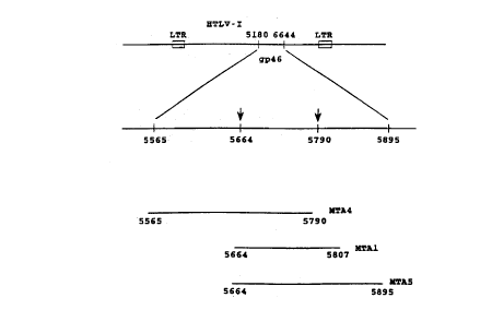
Figure lA shows, in the upper line, a portion of
the HTLV-I genome containing the gp46 envelop protein
coding sequence, and in the lower line, a portion of
the gp46 coding region containing the sequences which
encode overlapping HTLV-I peptide antigens formed in
accordance with the invention, and designated MTA-4,
MTA-1, and MTA-S in Figure 1B;
Figure 2 shows the HTLV-I coding sequences and
corresponding amino acid sequences for a portion of
the HTLV-I envelop protein:
Figure 3 shows amino acid sequences of homologous
regions of HTLV-I and HTLV-II gp46 in the region of
the peptide antigen of the invention, and peptide
sequences of several HTLV-I gp46 peptide antigens
(upper part of figure) and HTLV-II peptide antigens
(lower part of figure) in accordance with the
invention;
Figures 4A and 4B show antigenicity plots for the
MTA-1 peptide and corresponding HTLV-II gp46 peptide;
Figure 5 illustrates recombinant methods for
producing and selecting random-sequence peptides, in
accordance with the invention;
Figure 6 shows the HTLV-II coding sequence, and
corresponding amino acid sequence in the region of the
gp46 envelop protein from which HTLV-II peptides of
the invention are derived; and
X100586
WO 92/13946 PCT/US92/00823
8
Figure 7 shows modified Western blots containing
HTLV-I viral lysate and recombinant proteins p2lE and
MTA-4, where lanes A-F and G-R are HTLV-I and HTLV-II
antisera, respectively.
Detailed Description of the Invention
I. Preparing HTLV-I Peptide Antigens
This section describes the preparation of HTLV-I
peptide antigens which are immunoreactive with anti
HTLV-I antibodies found in individuals with HTLV-I
related T-cell leukemia. The antigens are prepared
using random HTLV-I gene sequences 100-300 base pairs
in length cloned in a suitable expression vector, then
selected with antibody for expression of
immunoreactive peptides.
A. HTLV-I Genomic Libraries
Genomic libraries of HTLV-I are prepared conven-
tionally from cellular DNA containing an HTLV-I provi-
ral genome. Duplex DNA may be prepared from HTLV-I
infected cells, including T-cells isolated from
patients known to be infected with HTLV-I virus, or
known cell lines, such as HUT 102-82 (Poiesz), MT-2
(Miyoshi), and MJ-tumor (Popovic) cells, all of which
have been shown to produce HTLV-I virus. The viral
genome is integrated into host DNA in these cells.
Methods for preparing cell lines containing the HTLV-I
genome are detailed in the above references.
The total host genomic DNA from the above cell
line is partially digested with a frequent cutter,
such as HaeIII or Alul, under conditions which produce
" WO 92/13946 ~ ~ ~ ~ PCT/US92/00823
9
partial digest fragments in the 15-20 kbase size
range, and the digested material is fractionated, for
example, by sucrose gradient centrifugation, to iso-
late the 15-20 kbase fragments. The fragments are
then cloned into a suitable cloning vector, preferably
a phage cloning vector which can efficiently incorpo-
rate a 15-20 kbase insert. In a preferred method, the
isolated fragments are treated with EcoRI methylase,
and EcoRI linkers are ligated to their ends under
standard conditions (Maniatis), and then cloned into a
phage vector, such as ~, Charon 4a, having a unique EcoRI
insertion site.
The cloned genomic fragments are screened with a
probe which is complementary to a selected sequence of
the full-copy HTLV-I genome. HTLV-I sequences are known
(Seiki), as are methods for producing radiolabeled syn-
thetic oligonucleotide probes for selected sequences. In
addition, synthetic oligonucleotides of specified sequen-
ces can be made by commercial services, such as provided
by Synthetic Genetics, Inc. (San Diego, CA). Using such
an oligonucleotide probe, molecular clones containing
HTLV-I sequences are isolated from the library by stan-
dard hybridization procedures (Maniatis, p. 322). The
clones can first be analyzed by restriction site analy-
sis, to confirm that the full viral genomic sequence is
present, as indicated by the presence of direct long
terminal repeats which flank the integrated viral genome.
The identified molecular clone is digested with a suit-
able endonuclease to release the full-copy viral genome.
A preferred -endonuclease for this purpose is SacI, which
cuts the viral genome in the long terminal repeats (LTR)
2~.~~5~~
WO 92/13946 PCT/US92/00823
at either end of the viral coding sequences, but does not
produce internal cleavage. If the clonal HTLV-I genome
is a variant with a third SacI site, an appropriate
restriction enzyme will be chosen to isolate the full-
y length qenome. The purified full-copy sequence is about
a 9.5 kilobase fragment. Alternatively, a fragment of
the genome representing the env gene sequences alone may
be purified for production of the expression library.
Alternatively, cloning vectors containing full-copy
10 HTLV-I duplex DNA have been reported (Seiki) and may be
obtained directly from the investigators, as indicated in
Example 1.
To produce the desired HTLV-I genomic library, the
full-copy HTLV-I insert is excised from the above cloning
vector, such as by complete digestion with SacI, and iso
lated as a 9.5 kilobase fragment, as described in Example
1. The isolated full-copy fragment is digested to pro-
duce DNA fragments, and preferably random fragments with
sizes predominantly between about 100-300 base pairs.
Example 1 describes the preparation of such fragments by
DNAase digestion. Because it is desired to obtain pep-
tide antigens of between about 30-100 amino acids, the
digest fragments are preferably size fractionated, for
example by gel electrophoresis, to select those in the
approximately 100-300 base pair size range.
The genomic digest fragments are inserted into a
suitable cloning vector, preferably an expression vector
which permits expression of the coded-for peptide in a
suitable host. One preferred expression vector is ~,gtll,
which contains a unique EcoRI insertion site 53 base
pairs upstream of the translation termination codon of
the ~-galactosidase gene. Thus, the inserted sequence
WO 92/13946 210 0 ~ 8 6 PCT/US92/00823
11
will be expressed as a ~-galactosidase gene. Thus, the
inserted sequence will be expressed as a ~-galactosidase
fusion protein which contains most of the N-terminal por-
tion of the ~-galactosidase gene, the heterologous pep-
s tide, and at least a portion of the C-terminal region of
the ~-galactosidase gene. This vector also produces a
temperature-sensitive repressor (cI857) which causes
viral lysogeny at permissive temperatures, e.g., 32°C,
and leads to viral lysis at elevated temperatures, e.g.,
42°C. Advantages of this vector include: (1) highly
efficient recombinant generation, (2) ability to select
lysogenized host cells on the basis of host-cell growth
at permissive, but not non-permissive temperatures, and
(3) high levels of recombinant fusion protein production.
Further, since phage containing a heterologous insert
produce an inactive ~-galactosidase enzyme, phage with
inserts can be readily identified by a ~-galactosidase
colored-substrate reaction.
The digest fragments inserted into the expression
vector may be modified, if needed, to contain selected
restriction-site linkers, such as EcoRI linkers,
according to conventional procedures. Example i illus
trates methods for cloning the digest fragments into
2,gt11, which includes the steps of blunt-ending the frag
ments, adding EcoRI linkers and ligating the fragments
with EcoRI cut ~.gtll. The resulting viral genome library
may be checked to confirm that a relatively large
(representative) library has been produced. This can be
done, in the case of the ~,gtll vector, by infecting a
suitable bacterial host, plating the bacteria, and exa-
mining the plaques for loss of ~-galactosidase activity.
2~.Ofl586
WO 92/13946 PCT/US92/00823
12
Using the procedures described in Example 1, about 60~ of
the plaques showed loss of enzyme activity, when compared
to the level of background phage showing loss of enzyme
activity, as seen in Example 1.
B. Peptide Antigen Expression
The genomic library formed above is screened for
production of peptide antigen (expressed as a fusion pro-
tein) which is immunoreactive with the human anti-HTLV-I
antibody of interest. One antibody of particular inte-
rest for diagnosing HTLV-I infection is the ~ 5a mono-
clonal antibody (Mab) which, as noted above, ~s has the
same specificity as antibodies present in patients with
T-cell leukemia related to HTLV-I infection. The antibo-
dy is produced by the EBV-transformed B-lymphocyte cell
line having ATCC Deposit No. HC8755 (See Example 2).
In a preferred screening method, host cells infected
with phage library vectors are plated, as above, and the
plate is blotted with a nitrocellulose filter, to trans-
fer recombinant antigens produced by the cells onto the
filter. The filter is then reacted with the anti-HTLV-I
antibody, washed to remove unbound antibody, and reacted
with reporter-labeled, anti-human antibody, which becomes
bound to the filter, in sandwich fashion, through the
anti-HTLV-I antibody.
Typically, phage plaques which are identified by
virtue of their production of recombinant antigen of
interest are re-examined at a relatively low density, for
production of antibody-reactive fusion protein. The
screening procedures described in Example 2 are illustra-
tive. Several recombinant phage clones which produced
immunoreactive recombinant antigen were identified in the
procedure.
WO 92/13946
21 D 0 5 8 6 I p~/US92/00823
13
The one or more library vectors identified as above
are preferably analyzed by nucleic acid sequencing, to
determine the positions of the peptide-coding regions
within the HTLV-I genome. Methods for excising the
heterologous insert (including adjacent coding sequences
of the fusion protein, if desired) from the selected
library vectors, and for purifying and sequencing the
excised fragments generally follow known procedures, as
outlined in Example 3. The coding sequences of three
peptides which were found to be immunoreactive with the
.Sa Mab are shown in the drawing. The three heterologous
sequences were matched with the known sequence of HTLV-I
(Seiki). As discussed more fully in Example 3, all of
the sequences fall within base pairs 5565 and 5895 of the
HTLV-I genome, within the gene coding for the HTLV-I
envelope protein gp46 (drawings, part A), and have an
overlapping coding sequence (defined by the two arrows in
the drawing) between base pairs 5664 and 5790 (drawing,
part B). As seen in the drawing, part C, the overlapping
sequence codes for a 42-amino-acid peptide antigen having
the following amino acid sequence:
Ser-Leu-Leu-Val-Asp-Ala-Pro-Gly-Tyr-Asp-Pro-Ile-Trp-Phe-
Leu-Asn-Thr-Glu-Pro-Ser-Gln-Leu-Pro-Pro-Thr-Ala-Pro-
Pro-Leu-Leu-Pro-His-Ser-Asn-Leu-Asp-His-Ile-Leu-Glu-
Pro-Ser. Screening studies conducted in support of the
invention indicate that the MTA-1 peptide picks up the
highest percentage of HTLV-I positive sera, particularly
among subjects of Japanese ancestry. As seen in Figure
3, the MTA-1 peptide includes the additional Ile-Pro-Trp-
Lys-Ser-Lys residues at the Ser C terminus of the above
sequence. In a preferred embodiment of the invention,
the HTLV-I specific peptide contains the immunogenic
21445~~
WO 92/13946 PCT/US92/0082?
14
region of the C-terminal 48 amino acid MTA-1 sequence
which is immunoreactive with the .5a Mab.
More generally, the HTLV-I peptides of the invention
include the immunogenic region of the above amino acid
sequence which is immunoreactive with the .5a Mab. As
defined herein, the specified sequence includes minor,
neutral amino substitutions which do not appreciably
decrease the immunoreactivity of the peptide antigen for
the .Sa Mab. Such amino substitutions may be selected on
the basis of similarities in hydrophobicity, size,
charge, hydrogen bonding ability, and effect on secondary
structure according to known amino acid substitution
principles.
The selected clones are used for scale-up produc
tion, for purposes of recombinant protein purification.
Scale-up production is carried out using one of a variety
of reported methods for (a) lysogenizing a suitable host,
such as E. coli, with a selected 7~gt11 recombinant, (b)
culturing the transduced cells under conditions that
yield high levels of the heterologous peptide, and (c)
purifying the recombinant antigen from the lysed cells.
In one preferred method involving the above ~,gtll
cloning vector, a high-producer E. cola host, BNN103, is
infected with the selected library phage, and replica
plated on two plates. One of the plates is grown at
32°C, at which viral lysogeny can occur, and the other at
42°C, at which the infecting phage is in a lytic stage
and therefore prevents cell growth. Cells which grow at
the lower, but not the higher temperature, are therefore
assumed to be successfully lysogenized.
WO 92/13946 ~ ~ O PCT/US92/00823
The lysogenized host cells are then grown under
liquid culture conditions which favor high production of
the fused protein containing the viral insert, and lysed
by rapid freezing to release the desired fusion protein.
5 These methods are detailed in Example 4.
HTLV-I coding sequences from the ~.gtll clone expres-
sing the peptide antigen MTA-1 have been prepared by PCR
amplification, as described in Section II below, and
cloned into the pGEX-1 expression vector (Pharmacia,
10 Piscataway, NJ). Inserts cloned into pGEX-1 were
expressed as a fusion protein with the protein Sj26,
which is a 26 Kdal Glutathione S-transferase from the
parasite Schistosoma ~aponicum. Limited paneling of
pGEX-MTA-1 against sera from HTLV-I or HTLV-II infected
15 individuals has revealed no significant difference
between the reactivity of pGEX-MTA-1 vs Li-gal-MTA-1.
C. Peptide Purification
The recombinant peptide is purified by standard pro
tein purification procedures which may include differen
tial precipitation, molecular sieve chromatography, ion
exchange chromatography, isoelectric focusing, gel elec-
trophoresis and affinity chromatography. In the case of
a fused protein, such as the ~-galactosidase fused pro-
tein prepared as above, the protein isolation techniques
which are used can be adapted from those used in isola-
tion of the native protein. Thus, for isolation of a -
galactosidase fusion protein, the protein can be isolated
readily by simple affinity chromatography, by passing the
cell lysis material over a solid support having surface-
bound anti- galactosidase antibody. This approach is
used in Example 4.
21~~5$~
WO 92/13946 PCT/US92/00823
16
II. Peptide Immunoreactivity With .5a MAB
The invention also includes, in another aspect, a
method of detecting HTLV-I positive human sera, by
reacting sera with a peptide antigen which is
immunoreactive with the HTLV-I Mab produced by ATCC
cell line HB8755, i.e., the .5a Mab. The presence of
HTLV-I specific antibodies in sera is detected by a
reporter-labeled anti-human antibody, as described in
Example 7.
A. HTLV-I Derived Peptides
In one embodiment, the peptides contain the
immunogenic region from the 42-amino acid overlap
region from above-described MTA-l, MTA-4, and MTA-5
HTLV-I peptides. These peptide antigens were further
characterized to confirm the location of the
immunoreactive region in the 42 amino acid sequence
overlap region. The location of the immunoreactive
region in the C-terminal portion of the overlap region
was suggested by two lines of evidence. First, the
.5a Mab was reported to react specifically with the
HTLV-I envelop protein, i.e., no reaction was observed
with HTLV-II or HTLV-III (HIV-1) envelop proteins. It
has since been confirmed by the applicants and their
co-workers that the gp46 peptide antigens MTA-1 and
MTA-4 described above are reactive with HTLV-I, but
not HTLV-II or HTLV-III antisera (Lipka).
Secondly, a comparison of the amino acid sequence
of MTA-1 peptide with the corresponding region in the
HTLV-II gp46 protein (Figure 3) shows substantially
WO 92/13946 ~ ~ ~ ~ ~ ' PCT/US92/00823
17
greater homology in the N-terminal half of the peptide
than in the C-terminal half (the center region of the
HTLV-I and HTLV-II sequences seen in Figure 3). This
would indicate that the greatest differences in anti-
s genicity would be found in the C-terminal half of the
peptide region.
This was further confirmed by antigenicity plots
of the two corresponding peptide regions, shown in
Figures 4A and 4B for HTLV-I and HTLV-II peptides,
respectively. The antigenicity plots were generated
by a standard hydrophobicity program "Antigen" in PC
Gene from Intelligenetics (Palo Alto, CA). As seen,
the two plots are substantially overlapping in resi-
dues 3-28, but diverge markedly in residues 28-40.
The divergent residues include the HTLV-I sequence
Leu-Pro-His-Ser-Asn-Leu-Asp-His-Ile-Leu-Glu-Pro-Ser.
A number of peptide antigens which include the C-
terminal region just indicated were prepared and
tested for binding to .Sa Mab, and to HTLV-I and HTLV
II antisera. The sequences of several of these pep
tides are indicated in the upper portion of Figure 3,
along with the sequences of the above MTA-1, MTA-4,
and MTA-5 peptide antigens. The peptides were
prepared by solid-phase synthetic methods, according
to standard procedures. Briefly, N-alpha-protected
amino acid anhydrides were prepared in crystallized
form and used for successive amino acid a3dition at
the N-terminus. At each residue addition, the growing
peptide (on a solid support) was acid treated to
remove the N-alpha-protective group, washed several
WO 92/13946 ~ ~ ~ ~ ~ ~ PCT/US92/00823
18
times to remove residual acid and to promote
accessibility of the peptide terminus to the reaction
medium. The peptide is then reacted with an activated
N-protected amino acid symmetrical anhydride, and the
solid ~~ipport is washed.
At each residue-addition step, the amino acid
addition reaction may be repeated for a total of two
or three separate addition reactions, to increase the
percent of growing peptide molecules which are
reacted. Typically, 1-2 reaction cycles are used for
the first twelve residue additions, and 2-3 reaction
cycles for remaining residues. After completing the
growing peptide chains, the protected peptide resin is
treated with liquid hydrofluoric acid to deblock and
release the peptides from the support.
The peptides were tested for specific immunoreac-
tivity with .5a Mab by binding competition studies,
substantially as described in Example 6. The K163
peptide, which contains the 18 C-terminal residues of
MTA-4 or MTA-1, strongly inhibits binding of .5a Mab
to MTA-4. No binding interference, however, was ob-
served with peptide K162, which contains only the 11
C-terminal residues of MTA-4. Peptide K164, which
contains the 6 C-terminal residues of MTA-4 and an
additional C-terminal 13 residues, weakly inhibited
binding between .Sa Mab and MTA-4 or MTA-1.
These results indicate that the most potent immu-
noreactive region in the gp46 peptide for the .Sa Mab
is in a region which includes peptide K163, consistent
with the divergence in sequence homology and anti-
2~40a8fi
WO 92/13946 PCT/US92/00823
19
genicity plots between HTLV-I and HTLV-II sequences in
this region. The weak binding of .5a Mab to the K164
peptide may indicate that the epitope of interest in
the His-Ile-Leu-Glu-Pro-Ser-His-Ile-Leu region of
overlap between K163 and K164, where adjacent N-
terminal or C-terminal sequences are required for
antigen presentation, or may indicate that the K164
peptide contains an additional epitopic region which
is weakly immunoreactive with the .5a Mab.
The peptides were also examined for their ability
to inhibit binding of antisera from HTLV-I infected
patients to MTA-4. In general, it was found that the
ability of any particular peptide to inhibit binding
of .5a Mab to MTA-4 paralleled its ability to either
bind to HTLV-I antisera in an ELISA binding protocol
(Example 6B) or to inhibit binding of human HTLV-I
antisera to MTA-4 or MTA-1 in a Western blot assay
(Example 8C). Thus, peptide K162 did not react with
any HTLV-I sera in the ELISA protocol and did not
inhibit binding of J-254 sera to MTA-1 or MTA-4.
B. Random-Sequence Peptides
In another embodiment, the .5a Mab-reactive
peptide for use in the method is prepared by selection
of random-sequence peptides. Recently, it has been
demonstrated that antibodies directed against specific
short (5-10 residues) peptides can be used to screen
libraries of randomly generated peptides for
immunoreactive species. (Scott; Cwirla et al). Such a
strategy is exploited herein to identify novel
~1fl0~8~
WO 92/13946 PCT/US92/00823
sequences which are immunoreactive with the .5a
monoclonal antibody.
In the preferred method, approximately l0e novel
heptapeptides are generated through construction of an
5 epitope library using the filamentous phage fUSES as a
vector. Other filamentous phage vectors are consi-
dered to be equally efficacious in developing such a
library.
Figure 5 shows schematically the sequence of steps
10 necessary to generate and screen a fUSE5 filamentous
phage epitopic library. Briefly, fUSES RF DNA is sub
jected to digestion with restriction endonuclease SfiI
to create an insertion site for insertion of foreign
DNA. A synthetic (15+3m) base pair (bp) BglI DNA
15 fragment is prepared which contains a degenerate se-
quence of the form (NNK) m, where N represents A, G, C,
or T; K represents G or T; and m can vary from 2 to
15. In the preferred embodiment of the invention, m
ranges from 5-10 and the bases are randomly added in
20 single addition events to the template primer. An
alternative method of achieving random addition of
codons coding for the twenty amino acids is to random-
ly attach trinucleotide codons representing each amino
acid to the template primer.
Following ligation of the insert to the cloning
vector, amplification of the filamentous phage vector
is achieved by transfection of E. coli cells. Suc-
cessful transfection is measured by the presence of
vector borne markers. In the preferred embodiment of
the invention, this marker is tetracycline resistance.
WO 92/13946 ~ ~ ~ ~ ~ g 6 PCT/US92/00823
21
Recombinant phage are then isolated from bacterial
cells. Phage bearing sequences of interest are isola-
ted by an antibody panning method in which phage are
incubated with the .5a Mab or its Fab fragment.
Biotinylated second antibody (goat anti-human IgG) is
then added, and complexes containing biotinylated
second antibody, the .5a Mab and immunoreactive pep-
tide bearing phage are separated from unreacted
antibodies and phage by adhesion onto a streptavidin
coated plate. Phage bearing immunoreactive sequences
are then eluted, and their DNA sequences are
determined.
Foreign DNA sequences present in the filamentous
phage fusion protein pIII determine the sequence of
the immunoreactive peptide. Peptides discovered to be
immunoreactive through this procedure can then be syn-
thesized by standard peptide synthetic methods and
prepared as immunogens by conjugation to an appropri-
ate peptide carrier.
III. HTLV-II Peptide Antigens
This section describes the identification and
cloning of HTZV-II peptides which are specifically
immunoreactive with HTLV-II antisera. The peptides
are derived from the HTLV-II gp46 envelop protein
region which is homologous to the above described MTA-
1 peptide from the HTZV-1 gp46 region.
An HTZV-II peptide designated GH2-K15 (Figure 3)
corresponding to the HTLV-I peptide MTA-1 was prepared
by cloning of an HTLV-II coding sequence corresponding
~100~86
WO 92/13946 PCT/US92/00823 ~~
22
to the desired peptide sequence. A 147 base pair (bp)
HTLV-II DNA fragment corresponding to nucleotides 5648
to 5794 of the HTLV-II genome (Figure 6) was original-
ly amplified from the HTLV-II clone pM04 (which con-
s tains the majority of the HTLV-II genome cloned into
the BamH I site of the plasmid pBR322) by use of the
polymerase chain reaction (PCR) procedure ( Perkin-
Elmer/Cetus GeneAmp kit).
The forward direction and reverse primers are
indicated in Figure 6. The amplified DNA was ligated
into the EcoR I site of ~,gtll phage vector, yielding
the clone as3K15 which contains a 147 HTLV-II DNA
insert into the -galactosidase gene of the
~,gtll. The recombinant phage was used to transfect E.
coli strain BNN103. Details are given in Example 5.
In a preliminary experiment, sera from approxi-
mately 200 individuals with PCR-confirmed HTLV-I or
HTLV-II infection, as well as sera from approximately
150 uninfected individuals were paneled against the
GH2-K15 antigen. 98% of the sera from HTLV-II
infected individuals reacted with GH2-K15. None of
either the HTLV-I infected sera or the uninfected sera
reacted with GH2-K15. The screening results
demonstrate that the GH2-K15 peptide is specifically
immunoreactive with HTLV-II positive sera.
Several smaller peptides contained with the GH2-
K15 amino acid sequence were prepared by recombinant
methods, as outlined in Section I. Briefly, the pep-
tides were prepared by PCR amplification of HTLV-II
genomic DNA, using PCR primers designed to promote
WO 92/13946
pGT/US92/00823
23
amplification of the sequences indicated, as detailed
in Example 5. Five of these peptides, designated
(GH2-) K14, K16, K24, K35, and K34 have the sequences
shown in Figure 3.
The recombinant HTLV-II peptides described above
were immunoscreened against several HTLV-II and HTLV-I
in an ELISA format, as described in Example 8. The
results are shown in Table 1. All ~,gtll HTLV-II clones
except for GH2-K16 were recognized by at least 1 out
of the 6 HTLV-II sera tested. GH2-K16, the sole
non-reactive clone, is missing the carboxyl terminal
22 amino acids that are included in GH2-K15. All the
other clones tested contain at least the 17 amino
acids Ser-Pro-Pro-Leu-Val-His-Asp-Ser-Asp-Leu-Glu-His-
Val-Leu-Thr-Pro-Ser that are present in peptide K125.
Also as seen in Table 1, none of the tested
peptides reacted with any of the HTLV-I sera, nor with
the .Sa Mab.
Three of the original HTLV-II clones, GH2-K15,
GH2-K35, and GH2-K16 have been cloned into the pGEX-1
expression vector. Recombinant protein expressed by
the 3 pGEX-1 HTLV-II clones GH2-K15, GH2-K25, and
GH2-K35 have all been recognized by the J-317 HTLV-II
serum.
2100~~6
WO 92/13946 PCT/US92/00823
24
Table 1
------HTLV-II ANTIGENS-----
SERUM VIRUS N K15 K14 K16 K24 K34 K35
J-115 II 2 +/- - - - - -
J-127 II 2 - - - - - -
J-289 II 2 - - - - - -
J-309 II 2 - - - - - -
J-263 II 3 +/- - - + - -
J-317 II 2 ++ + - ++ + +
J-103 I 2 - - - - - -
J-108 I 2 - - - - - -
J-183 I 2 - - - - - -
.5a Mab I 1 - - - - - -
A number of peptide antigens which contain amino
acid sequences within the K15 sequence were prepared by
solid-phase methods, as outlined in Section III above.
The sequences of five of these peptides, designated
(GH2-) K169, K170, K125, K126, and K128 are shown in
Figure 3. The peptides were tested for immunoreactivity
with several HTLV-I and HTLV-II positive sera, by an
ELISA method, and some of the peptides were also examined
for their ability to inhibit HTLV-II antibody binding to
the K15 antigen.
The K125 peptide was recognized by multiple HTLV-II
sera when assayed by ELISA. In one experiment 6 out of
12 HTLV-II sera were able to bind efficiently to K125.
In the same experiment 0 out of 7 HTLV-I sera bound
peptide K125. The K125 peptide also inhibited the
binding of a strongly reactive HTLV-II sera, J-317, to
WO 92/13946 ~ ~ ~ ~ ~ ~ ~ PCT/US92/00823
Western blotted GH2-K15. The ability of sera J-317 to
bind GH2-K15 is not affected by incubation with the
HTLV-I peptide K163 or the HTLV-II peptide K128.
The HTLV-II peptide K170 is recognized by multiple
5 HTLV-II sera in an ELISA based assay, and not recognized
by HTLV-I sera in the same assay. The K169 peptide is
not recognized by HTLV-II sera in an ELISA based assay.
Data from both the analysis of HTLV-II recombinant
antigens and the synthetic HTLV-II peptides indicate that
10 the HTLV-II specific epitope is contained in the 17 amino
acid sequence Ser-Pro-Pro-Leu-Val-His-Asp-Ser-Asp-Leu-
Glu-His-Val-Leu-Thr-Pro-S~r in the GH2-K15 peptide. Data
obtained by extensive paneling of the HTLV-I antigens
MTA-1 and MTA-4, discussed above, would suggest that the
15 6 final amino acids of GH2-K15, Thr-Ser-Trp-Thr-Thr-Lys,
may also contribute to the epitope recognized by HTLV-II
antisera.
IV. HTLV-I and HTLV-II Diagnostic Methods
20 Three basic types of diagnostic applications of the
HTLV-I and HTLV-II peptide antigens of the invention will
be described. The first is based on inhibition of
complement-mediated, antibody-dependent cytolysis by the
peptide. In this method, serum from a test individual is
25 reacted with HTLV-I or HTLV-II infected T-cell clones in
the presence of complement. The presence of anti-HTLV-I
or anti-HTLV-II antibody is evidenced by cell lysis, as
judged, for example, by trypan blue dye exclusion.
where cell lysis is observed, the specificity of the
anti-HTLV-I antibody for the HTLV-I peptide is demon
strated by first reacting the serum with excess HTLV-I or
HTLV-II peptide, then mixing the serum with cells in the
WO 92/13946 ~ ~ ~ ~ ~ ~ ~ -
PCT/US92/00823
26
presence of complement. The presence of HTLV-I or HTLV-
II antibody is indicated by a substantial decrease in
cell lysis. This method is described in Example 6A.
The method can also be used to quantitate the anti
s body titer in the analyte serum, by titrating the serum
with increasing amounts of peptide, and determining the
peptide concentration where a noticeable effect on the
extent of cell lysis is first observed.
The second general assay type is an enzyme-immuno
assay for screening human sera for HTLV-I or HTLV-II
infection. In this assay format, a solid phase reagent
having surface-bound HTLV-I or HTLV-II gp46 peptide anti
gen is reacted with analyte serum, under conditions which
allow antibody binding to the peptide on the reagent.
After washing the reagent to remove unbound serum compo-
nents, the reagent is reacted with an enzyme-labeled
anti-human antibody, to bind enzyme to the reagent in
proportion to the amount of bound anti-HTLV-I antibody on
the solid support. The reagent is again washed, to re-
move unbound antibody, and the amount of enzyme asso-
ciated with the reagent is determined. One exemplary
method, employing an anti-human antibody labeled with
alkaline phosphatase, is detailed in Example 7 for a
direct HTLV-I screening assay. The enzyme-labeled anti-
body, and reagents required for enzyme detection, are
also referred to herein as reporter means for detecting
the presence of human antibody bound to the peptide anti-
gen on the solid support.
The solid surface reagent in the above assay is pre
pared by known techniques for attaching protein material
to solid support material, such as polymeric beads, dip
sticks, or filter material. These attachment methods
WO 92/13946 ~ ~ ~ ~ PCT/US92/00823
27
generally include non-specific adsorption of the protein
to the support (as in the filter support described in
Example 8) or the covalent attachment of the protein,
typically through a free amine group, to a chemically
reactive group on the solid support, such as an activated
carboxyl, hydroxyl, or aldehyde group.
The third general assay type is Western blot assay
for use in confirming HTLV-I or HTLV-II antisera. This
assay format includes, in addition to the gp46 peptide
antigen of the invention, one or more confirmatory HTLV-I
or HTLV-II antigens that are effective to detect HTLV-I
or HTLV-II antisera. In one preferred format, the
confirmatory peptides include the p24 gag protein from
HTLV-I viral lysate, and a p2lE recombinant envelop
protein containing a large portion of the HTLV-I gp21
envelop protein (Samuel, 1984, 1985). The p24 lysate
proteins picks up most, but not all HTLV-I and HTLV-II
positive sera. The p2lE recombinant peptide picks up
virtually all HTLV-I and HTLV-II, but also gives some
false positives. This modified Western blot assay has
been reported by the applicants and co-workers (Lipka).
Details of the blot assay procedure are given in Example
8.
As has been described, and as is detailed in Example
8, the modified Western blot format picked up all HTLV-I
and HTLV-II positive sera tested (a panel of 95), as
evidence by immunoreaction with viral lysate protein p24
and recombinant protein p2lE. In addition, the MTA-4
peptide was immunoreactive with confirmed HTLV-I sera
only. The modified blot assay thus can be used to
confirm HTLV-I or HTLV-II antisera, and to distinguish
WO 92/13946 ~ ~ ~ ~ ~ ~ PCT/US92/00823
28
the two types of HTLV virus by selective immunoreaction
with the peptide of the invention.
In another embodiment of the Western blot assay, the
HTLV-I peptide antigen is replaced by the HTLV-II gp pep
s tide antigen described in Section III. In this format,
the HTLV-I viral lysate proteins and p2lE recombinant
protein provide confirmation of HTLV-I or HTLV-II
antisera, as above. The HTLV-II specific peptide will
pick up HTLV-II, but not HTLV-I antisera, and thus
provides a positive confirmation of HTLV-II antisera.
The two formats can be combined to include both
HTLV-I and HTLV-II specific peptide antigens, to give
positive confirmation of either HTLV antisera.
v. vaccine Compositions
Also included in the invention is a vaccine
composition containing an HTLV-I gp46 peptide and a
antigen carrier, such as an immunogenic protein, to which
the antigen peptide is bound. The peptide contains an
immunogenic region formed by the above 42- or 47-amino
acid overlap of MTA-1, MTA-4, and MTA-5 peptides
described in Section I, which is immunoreactive with
anti-HTLV-I .Sa Mab, i.e., the antibody derived from
ATCC cell line HB8755. More specifically, the peptide
contains the immunogenic region of the peptide
sequence Thr-Ala-Pro-Pro-Zeu-Leu-Pro-His-Ser-Asn-Leu-
Asp-His-Ile-Leu-Glu-Pro-Ser. Since the .5a Mab is a
neutralizing antibody, the antibody raised by the
peptide is expected to be a neutralizing antibody.
The vaccine composition may alternatively include
the HTLV-II gp46 peptide containing the HTLV-II
- WO 92/13946
PCT/US92/00823
29
specific immunogenic region formed by the amino acid
sequence Met-Thr-Leu-Leu-Val-Asp-Ala-Pro-Gly-Tyr-Asp-
Pro-Leu-Trp-Phe-Ile-Thr-Ser-Glu-Pro-Thr-Gln-Pro-Pro-
Pro-Thr-Ser-Pro-Pro-Leu-Val-His-Asp-Ser-Asp-Leu-Glu-
His-Val-Leu-Thr-Pro-Ser-Thr-Ser-Trp-Thr-Thr-Lys, and
preferably formed by the amino acid sequence Ser-Pro-
Pro-Leu-Val-His-Asp-Ser-Asp-Leu-Glu-His-Val-Leu-Thr-
Pro-Ser-Thr-Ser-Trp-Thr-Thr-Lys, or Ser-Pro-Pro-Leu-
Val-His-Asp-Ser-Asp-Leu-Glu-His-Val-Leu-Thr-Pro-Ser.
Particularly useful protein carriers for the
peptides) include keyhole limpet hemocyanin (KLH),
tetanus toxoid, poly-1-(Lys:Glu), peanut agglutinin,
poly-D-lysine, diphtheria toxoid, ovalbumin, soybean
agglutinin, bovine serum albumin (BSA), human serum
albumin, and the like.
The immunogenic peptides) may be conjugated to
the carrier by a variety of known methods, including
chemical derivatization and by genetic engineering
techniques. Such latter technique is disclosed in
more detail by Gerald Quinnan, "Proceedings of a Work-
shop," November 13-14, 1984. Vaccines and inocula of
the present invention may be administered by injec-
tion, usually intramuscularly or subcutaneously, oral-
ly by means of an enteric capsule or tablet, as a sup-
pository, as a nasal spray, and by other suitable
routes of administration. For a human patient, a
suitable dose of the polypeptide depends, in part,
upon the chosen route of administration and a number
of other factors. Included among those factors are
the body weight of the mammal to be immunized, the
WO 92/13946 2 ~ ~ ~ ~ ~ ~ PCT/US92/00823 '~
carrier when used, the adjuvant when used, and the
number of inoculations desired to be used.
Individual inoculations for a human patient typi
cally contain unit doses of about 10 micrograms to
5 about ?00 milligrams of polypeptide, exclusive of any
carrier to which the polypeptide may be linked. If
desired, a series of doses may be administered over a
period of time for optimum immunity. Unit dosage
forms of the vaccine can also be provided, if desired,
10 containing the aforementioned amounts of the polypep-
tide.
In any event, the immunogen contained in a vaccine
or an inoculum is present in an "effective amount,"
which amount depends upon a variety of factors as is
15 well known in the immunological arts, e.g., the body
weight of the mammal to be immunized, the carrier moi-
ety used, the adjuvant used, the duration of protec-
tion sought, and the desired immunization protocol.
The following examples illustrate various aspects
20 of the invention, but are in no way intended to limit
the scope thereof.
Mn+~er~ ~l c~
The materials used in the following Examples were
as follows:
25 Enzymes: DNAase I and alkaline phosphatase were
obtained by Boehringer Mannheim Biochemicals (BMB,
Indianapolis, IN); EcoRI, EcoRI methylase, DNA ligase,
and Polymerase I, from New England Biolabs (NEB,
Beverly, MA); and RNase was obtained from Sigma (St.
30 Louis, MO) .
WO 92/13946 ~ ~ ~ ~ ~ 6 PCT/US92/00823
31
Other reagents: EcoRI linkers were obtained from
NEB; and nitro blue tetrazolium (NBT), 5-bromo-4-
chloro-3-indolyl phosphate (BCIP), 5-bromo-4-chloro-3-
indolyl- -D-galactopyranoside (X-gal) and isopropyl -
D-thiogalactopyranoside (IPTG) were obtained from
Sigma.
Example 1
Preparation of an HTLV-I Genomic Library
Source of Genomic Material
Bacteriophage containing a full-copy DNA insert
derived from the HTZV-I genome was obtained from Drs.
R.C. Gallo and F. along-Staal of the Laboratory of
Tumor Cell Biology, National Institutes of Health
(Bethesda, MD). The bacteriophage was digested to
completion with SacI, releasing the viral genome
insert. The digested material was electrophoresed on
standard 1% agarose gel, and the 9.5 kilobase fragment
obtained by electroelution was extracted with
phenol/chloroform before ethanol precipitation.
The purified genomic DNA was suspended in a
standard digest buffer (0.5M Tris HC1, pH 7.5; 1 mg/ml
BSA; lOmM MnCl2) to a concentration of about 1 mg/ml,
and digested with DNAase I at room temperature for
about 5 minutes. These reaction conditions were
determined from a prior calibration study, in which
the incubation time required to produce predominantly
100-300 basepair fragments was determined. The mate-
rial was extracted with phenol/chloroform before
ethanol precipitation.
The genomic fragments from above were blunt-ended
with DNA Pol I under standard conditions (Huynh), then
WO 92/13946 2 ~~. ~ 0 ~ PCT/US92/00823
32
extracted with phenol/chloroform and precipitated with
ethanol. The blunt-ended material was ligated with
EcoRI linkers, under standard conditions (Maniatis,
pp. 396-397), then digested with EcoRI to remove
redundant linker ends. The material was then agarose-
gel-fractionated to remove non-ligated linkers and to
size-select (see below).
The resultant fragments from the previous step
were analyzed by electrophoresis (5-lOV/cm) on 1.2%
agarose gel, using X174/HaeIII and /HindIII size
markers. The 100-300 by fraction was eluted onto NA45
strips (Schleicher and Schuell), which were then
placed into 1.5 ml microtubes with eluting solution (1
M NaCl, 50 mM arginine, pH 9.0), and incubated at 67°C
for 30-60 minutes. The DNA, now in solution, was ex-
tracted with phenol/chloroform and precipitated with
ethanol. The pellet was resuspended in 20 ~1 TE (0.01
M Tris HC1, pH 7.5, 0.001 M EDTA).
gtll phage vector (Huynh) was obtained from Promega
Biotec (Madison, WI). This cloning vector has a unique
EcoRI site 53 base pairs upstream from the ~-galactosi
dase translation termination codon. The genomic frag
ments from above were introduced into the EcoRI site by
mixing 0.5 -1.0 ~g EcoRI-cleaved gtll, 0.5-3 ~1 of the
above HTLV--I genomic fragments, 0.5 ~1 lOX ligation
buffer (above), 0.5 ~1 ligase (200 units), and distilled
water to 5 ~1. The mixture was incubated overnight at
14°C, followed by in vitro packaging, according to stan-
dard methods (Maniatis, pp. 256-268).
The packaged phage were used to infect E. coli,
strain KM392, obtained from Dr. Kevin Moore, DNAX (Palo
WO 92/13946 ~ ~ ~ ~ ~ ~ PCT/US92/00823
33
Alto, CA). Alternatively, E. coli strain Y1090, avail-
able from the American Type Culture Collection (ATCC
#37197), could be used. The infected bacteria were pla-
ted and the resultant colonies were checked for loss of
~i-galactosidase activity (clear plaques) in the presence
of X-gal using a standard X-gal substrate plaque assay
method (Maniatis). Table 2 below shows the number of
recombinant (clear) plaques obtained with insertion of
the EcoRI-ended HTLV--I fragments (row 1). An EcoRI
linker control (row 2) and vector alone (row 3) were also
run. As seen, about 50% of the phage plaques showed loss
of enzyme (recombination). The background levels either
in the presence or absence of EcoRI linkers were less
than 15%, indicating the successful generation of an
HTLV-I epitope library. The phage libraries contained
about 10' plaque-forming units (pfu)/ml.
Table 2
Insert Vector Clear/Total %Rec
1. SacI i~
3.25 ~1 1~1 100/200 50
2. EcoRl linker
3.25 ~1 1~1 25/178 14
3. Control 1~1 50/400 13
WO 92/13946 ~ ~ O
PCT/US92/00823
34
Example 2
Screening for gp46 Coding Inserts
Purified .5 antibody derived from a human cell line
(ATCC #C8755) was provided by Dr. Samuel Broder of the
National Cancer Institute, National Institutes of Health
(Bethesda, MD). Mouse anti-human IgG antibody covalently
derivatized with alkaline phosphatase was obtained from
Promega Biotec (Madison, WI).
A lawn of KM392 cells infected with about 10' pfu of
the phage stock from Example 1 was prepared on a 150 mm
plate, and incubated, inverted, for 5-8 hours at 37°C.
The lawn was overlaid with a nitrocellulose sheet,
causing transfer of HTLV-I recombinant protein from the
plaques to the paper. The plate and filter were indexed
for matching corresponding plate and filter positions.
The filter was washed twice in TEST buffer (10 mM
Tris, pH 8.0, 150 mM NaCl, 0.5% Tween 20), blocked with
AIB (TBST buffer with 1% gelatin), washed again in TBST,
and incubated overnight after addition of .5 monoclonal
antibody (diluted to 1-2 ~,g/ml in AIB, 12-15 ml/plate).
The sheet was washed twice in TBST, then contacted with
enzyme-labeled anti-human antibody, to attach the labeled
antibody at filter sites containing antigen recognized by
the .5 antibody. After a final washing, the filter was
developed in a substrate medium containing 33 ~1 NBT (50
mg/ml stock solution maintained at 5°C) in 5 ml of alka-
line phosphatase buffer (100 mM Tris, 9.5, 100 mM NaCl,
5 mM MgClZ). Reacted substrate appeared at points of an-
tigen production, as recognized by the 0.5a Mab.
The areas of antigen production determined in the
previous step were replated at about 100-200 pfu on an 82
mm plate. The above steps, beginning with a 5-8 hour
~~ WO 92/13946 ~ ~ ~ ~ ~ ~ ~ PCT/US92/00823
incubation, through NBT/BCIP development, were repeated
in order to identify plaques which secreted an antigen
capable of reacting with the .5 Mab. The identified
plaques were picked and eluted in phage buffer (Maniatis,
5 p. 443). Three of the recombinant phage plaques which
secreted an antibody-reactive peptide were selected for
sequencing analysis, according to the procedures in
Example 3. The corresponding infected phage has been
designated MTA-4, MTA-1, and MTAS.
Example 3
Phage Purification and DNA Extraction
Phages MTA-4, MTA-1, and MTA-5 were isolated from
the plate cultures of the infected E. coli Y1088 bacte
ria. These cells are available from the ATCC (ATCC
#31195). The phage was collected by addition of phage-
dilution buffer (maniatis) late material was purified
from bacterial debris by low-speed centrifugation, and
the supernatant was poured into SW 27 tubes. RNase and
DNAse were each added to a concentration of l~g/ml each
from stock solutions of 1 mg/ml. The sample was
incubated for 30 minutes at 37°C, and an equal volume of
a polyethylene glycol (PEG) , 5 . 8 g NaCl, 2 . Og MgSO,~7H~0,
1M Tris C1, pH 7.5, and 2% gelatin was added. The
sample was placed in an ice bath for 1 hour to allow the
phage particles to form a precipitate, which was then
isolated by centrifugation at lOk for about 20 minutes at
4 °C .
The supernatant was decanted, and the pellet was re-
suspended in 0.6 ml PDB buffer (5.8 g NaCl, 2.0 g
MgSO~7Hz0, 50 ml 1M Tris C1, pH 7.5, and 5 ml 2~ gelatin)
and transferred to .5 ml polypropylene microtubes. 5 ~1
WO 92/13946 ~ ~ ~ ~ ~ ~ PCT/US92/00823 "
36
10% SDS, 5 ~1 0.5M EDTA, and 2.5 ~1 proteinase K (20
mg/ml) were added, and the samples were incubated at 50°C
for 15 minutes.
The detergent and enzyme-treated material was ex
tracted with an equal volume of phenol/chloroform, and
centrifuged to ensure separation of the phases. The
aqueous phase was transferred to a new tube, and the
extraction/centrifugation procedure was repeated with a
mixture of chloroform and isoamyl alcohol. An equal
volume of isopropanol was added, and the same was inver-
ted several times to mix, and cooled to -70°C for 20
minutes. The sample was centrifuged for 5 minutes and
the supernatant Was decanted. The pellet was washed in
70% ethanol, briefly dried in a 37°C heat block, and re-
suspended in 100 wl TE buffer, pA 7.5.
The isolated phage DNA was digested with K~nI and
SacI and then combined with K~nI/SacI cut plasmid vector
pGEM-3 (Promega Biotec) to isolate a plasmid recombinant
with the insert of interest. The HTLV-I insert was then
sequenced using the standard dideoxy sequencing procedure
and forward and reverse primers for ~.gtl sequences flank-
ing the EcoRI insertion site.
The figure shows the coding sequence and correspond
ing amino acid sequence of a portion of the fused protein
formed by the above methods, for each of the three fused
peptides examined. A terminal G base of the ~-gal gene
and the adjacent CC bases of the env gene contributed by
each of the three insert sequences yield a GCC (Ala)
codon, replacing the Ser codon which normally occurs at
that codon position of all three env inserts. As shown,
the insert in the MTA-4 includes a 225 base pair sequence
WO 92/13946 ~ .~ Q ~ PCT/US92/00823
37
extending from base 5564 to 5790 of the HTLV-I coding
region. The insert of the MTA5 phage begins at base
5664, and extends to base 5895. The 231 basepair se-
quence covers amino acids 162 to 240 of the gp46 protein.
The region of insert overlap, from 5664 to 5790, in-
cludes the 42 amino acid sequence from amino acids 162 to
203 of the native gp46 protein.
Example 4
Isolation of HTLV-I Peptide Antigens
A. Construction of Lysogens
EcoRI, strain C600, was obtained from Dr. R. Davis,
Stanford University (Stanford, CA). Alternatively, EcoRI
Y1089 (ATCC X37196) can be used. A 1 ml saturated, over-
night culture of the cells was infected with one of the
three phages from Example 3 by adsorbing 10 ~1 of eluted
plaque stock to 50 ~1 of overnight bacterial culture.
The infected bacteria were spread only LB agar plates
(Maniatis, p. 440) and incubated at 32°C. The individual
colonies were picked with sterile toothpicks onto corre-
sponding grids on two separate plates. One of the plates
was incubated at 32°C, and the other at 42°C. Cells that
grew at the lower temperature (indicating a lysogenic
state produced by the presence of the phage repressor
protein) but not at the higher temperature (because of
cell lysis) were assumed to be lysogenic. Many lysogenic
colonies from each of the three phage types were found.
W092/13946 ~~oo~~~
PCT/US92/00823
38
B. Recombinant Antigen Induction from Lysogens
This Example describes induction of a recombinant
protein containing the HTLV-I epitope from the ~.gtll
lysogens prepared in Example 4 with the MTA-4 phage . As
indicated above, the antigen is produced in the form of a
I'-galactosidase fusion protein which also contains an N-
terminal portion of the phage~~-gal protein.
A superbroth was prepared containing 35 g bacto
tryptone, 2 g bacto-yeast extract, 5 g NaCl, and 5 ml 1N
NaOH in 1 1 dHO. 500 ml of the superbroth were inocula
ted 1:100 with a saturated overnight culture of the EcoRI
~,gtll lysogens prepared in the previous example. The
culture was incubated to Aaoa -0.4-0.5 with vigorous
aeration.
In order to maximize protein production, the
temperature of the culture was raised to 43-44°C, thereby
inactivating the temperature-sensitive ,'-galactosidase
repressor gene. The temperature was maintained at 43°C
with a 65°C water bath for 15 minutes with aeration.
IPTG, which induces j':- -galactosidase expression by
competitively binding to the -galactosidase repressor,
was added to the broth to 2 mM to further increase
protein production. The culture was returned to the 38°C
shaker for about an hour. The cells were then pelleted
at 6,000 x g for 15 minutes at 37°C, resuspended in lysis
buffer (10 mM Tris, pH 7.4, 2~ Triton X-100, 1~
aprotinin, and 50 ~g PMSF) and immediately plunged into
liquid NZ. Lysis was completed upon thawing of the
frozen samples.
WO 92/13946 ~ _~ S ~ ~ PCT/LJS92/00823
39
C. Purification of Fusion Protein
The cell lysate obtained in the previous Example was
thawed and warmed to 37°C . 10 ~1 DNAse ( 1 ~.g/ml ) was
added, and the mixture incubated until the viscosity
decreased. The lysate was quickly chilled on ice,
clarified t 4°C for 5 minutes in a microfuge, and loaded
onto a 6 ml column of anti-~~-galactosidase coupled to
Sepharose 4B (Pharmacia). The column was allowed to
equilibrate 1-2 hour, and washed with 7 volumes (column
volumes) of TX buffer (10 mM Tris, pH 8.0, 2% Triton X-
100, 50 ~g/ml PMSF), followed by 2 volumes of 5mM 3,5-
diiodosalicylic acid in TX buffer. Fusion protein was
then eluted from the column with 35 mM 3,5-
diiodosalicylic acid in TX buffer. The majority of
protein was eluted in the first 3-4 volumes, and removal
was substantially complete after 7 volumes.
The eluted samples were desalted and concentrated
using Amicon filters (Danvers, MA).
Example 5
Preparing HTLV-II Antigens
A. Synthesis and Cloning of HTLV-II DNA Sequences
The polymerase chain reaction (PCR) procedure was
used to generate HTLV-II DNA sequences for cloning. Six
30 by DNA primers were synthesized. All 6 primers had 3
by of gtll sequence followed by an EcoR I site at their
5' ends. This was followed by 21 by of HTLV-II
sequences. The 3 forward direction primers contained
HTLV-II sequences corresponding to nucleotides 5648 -
5668, 5687 - 5707, and 5726 - 5745. The 3 reverse
direction primers contained HTLV-II sequences
WO 92/139401 D O ~ ~ ~ PCT/US92/00823 Ty
corresponding to nucleotides 5794 - 5774, 5776 - 5756,
and 5728 - 5708.
PCR was performed according to the manufacturers
instructions (Perkin Elmer/Cetus), and all PCR reactions
5 contained 2 ng of the above HTLV clone as template and 1
wM of the appropriate PCR primers. PCR amplification was
carried out for 25 cycles. Each cycle involved template
denaturation for 1 minute at 94 deg.C, annealing of
primer to template for 2 minutes at 50 degC, followed by
10 primer extension for 2 minutes at 72 ged.C. Afterwards
the amplified DNA was purified and then digested to
completion with EcoRI. The digested DNAs were then
ligated into the EcoRI site of lambda gtll. The
recombinant phage DNAs were then packaged and the
15 frequency of non-recombinant phage was determined by
plating in the presence of 5-bromo-4-chloro-3-indolyl
$-D-galactopyranoside.
The ratio of recombinant to non-recombinant phage
was about 50/1. Multiple isolated plaques from each of
20 the 6 recombinant phage clones were picked and
subsequently screened using PCR with lambda gtll flanking
primers 11F and 11R, and/or the HTLV-II plaques described
above. Clones containing correctly sized and orientated
inserts were then amplified and used in subsequent
25 immunoscreening assays. The EcoR I fragment from 3 of
the clones GH2-K15, GH2-K16 and GH2-K35 were subsequently
subcloned into the pGEX-1 plasmid and DNA sequenced. The
sequences obtained perfectly matched the reported
sequence for the desired region of HTLV-II (Shimotokno).
30 B. Immunological Analysis of HTLV-II Clones
Recombinant phage was mixed 1/1 with wild type gtll
and used to infect E. coli strain KM-392. After allowing
WO 92/13946 c~ ~ ~ PCT/US92/00823
41
the phage to grow for -5 hours expressed proteins were
bound to nitrocellulose filters overnight. Filters were
subsequently washed 3X with TBS (0.5 M NaCl, 20 mM Tris
Ph 8.0), cut into sections, and blocked using TBS plus 1~
Gelatin. Filter sections were then incubated overnight
with 1st stage antibody, usually sera from HTLV-I or
HTLV-II infected individuals diluted 1/100 in TBS plus
gelatin. After washing with TBS, the filters were
incubated with alkaline phosphatase conjugated goat anti
human sera for at least 1~ hour. The filters were washed
with TBS and bound antibody was then detected by
incubating the filters in a solution of nitroblue
tetrazolium chloride and 5-bromo-4-chloro
3-indolylphosphate. A particular sera was scored as
positive if plaques derived from the recombinant phage
could clearly be distinguished from plaques of wild type
gtll.
C. Expression and Purification of Recombinant Antigen
B-galactosidase fusion proteins were expressed by first
generating lysogens. Recombinant gtll phage was used to
infect E. cola strain BNN103, and lysogens were
identified by growing duplicate plates at 32°C and 92°C.
The production of fusion protein was induced by raising
the temperature of a log phase culture of lysogen to 42°C
for 15 minutes. Isopropyl thiogalactoside was then added
to a final concentration of 1.6 Mm and the cultures were
grown for an additional 1 hour at 37°C. Cells were then
pelleted by centrifugation at 5000 x g for 15 minutes and
resuspended in 1/50th original culture volume of lysis
buffer (2~ Triton X-100, 1~ Aprotinin, 10 mM Tris, pH
7.4) . The solution was then frozen by immersion in a dry
WO 92/13946 PCT/US92/00823 H
2~.~~5.~~
42
ice / ethanol bath and then_thawed. DNase I was added to
a final concentration of 1 ~ag/ml and the lysate was then
incubated for 5 minutes at room temperature. Insoluble
debris was then pelleted by centrifugation at 10,000 x g
for 10 minutes. The supernatant was then centrifuged as
before. sodium dodecyl sulfate-polyacrylamide gel
electrophoresis (SDS-PAGE, Lammeli) analysis of aliquots
of the pellet and supernatant fractions indicated that
GH2-K15 was found primarily in the supernatant fraction.
The supernatant fraction was then combined with 2
mls of Protosorb LacZ adsorbent (Promega) and incubated
for 2 hours at 25°C with agitation. The column resin was
then poured into a disposable column and washed with 2X
with 10 mls of TX buffer (1% Aprotinin, 10 mM Tris pH
7.4). Bound protein was eluted 14 mls of pH 10.8
carbonate buffer and 2 ml fractions were collected into
tubes already containing 1 ml of 2 M tris buffer (pFL
7.5). Fractions were then concentrated using Centricon
30s (Amicon) following manufacturers instructions. The
fractions were washed with 2 mls of MTBS buffer (150 mM
NaCl, 4 mM NaH2P04, 16 mM Na2HP04, pH 7.3) and then
concentrated again.
The location, yield and purity of the purified
fusion protein was determined using SDS-PAGE. After one
pass through the immunoaffinity column GH2-K15
recombinant antigen was ~70 % pure. Fractions containing
fusion protein were pooled and aliquots of the pool were
used in subsequent western blot experiments. Western
blot analysis was performed essentially as described
previously (Lipka). Titration experiments determined
that the optimum loading of purified GH2-K15 antigen was
3 ug protein/cm nitrocellulose. Peptide competition
WO 92/13946 ~ ~ ~ PCT/US92/00823
43
experiments are described in Example 6C. Nitrocellulose
strips containing blotted antigen were then added to the
serum samples and incubated and developed as normal.
D. HTLV Antisera
Sera samples were obtained from HTLV-I, HTLV-II, or
ELISA reactive - HTLV negative individuals. These
samples were from multiple different sources and
geographic areas. Many of the seropositive sera samples
Were also typed for HTLV-I or HTLV-II infection using PCR
with strain specific DNA primers. HTLV-I sera samples
included 58 PCR proven samples consisting of 45 samples
from Jamaican food handlers, 2 intravenous drug users
(IVDU) from the New Orleans area, and 11 northern
California blood donors. In addition a total of 238
HTLV-I sera samples were obtained from Japan. For the
Japanese samples PCR data was unavailable and the
infection was typed by western blot analysis of the sera
samples against HTLV-I antigens using previously
described criteria (Lipka et al. JID). HTLV-II sera
samples included 57 PCR proven samples consisting of 6
IVDU from the New Orleans area, 24 IVDU from the northern
California area, and 27 blood donors from the northern
California area. HTLV negative sera included 1 Jamaican
food handler, 15 California blood donors, and 29 samples
from Japan. PCR analysis of serum samples was performed
as described (Lipka).
Example 6
Detecting Peptide Antigen Immunoreactivity
A. Inhibition of Cell Lysis
WO 92/13946 2 ~ o ~ 5 ~ 6 PCT/LJS92/00823 T
44
HUT 102-B2 cells were obtained from Dr. R.C. Gallo,
LTCB, NIH. This is a long-term cultured T-cell line
known to produce HTLV-I.
.5a antibody (~5 ~g/ml IgG) or a control isotyped
matched human IgG was preincubated with MTA-4 recombinant
peptide or irrelevant recombinant for 30 minutes at room
temperature. 50 ~1 of these mixtures Was then added to
5x10⁵ HUT 02B2 cells in 96-well micro titer plates, and
incubated for 30 minutes at room temperature. 30 ~1 of
rabbit complement per well was added, and incubated 1
hour at 37°C. Cell viability was determined by
microscopic examination. Cell lysis was visibly
inhibited by addition of the MTA-4 peptide antigen, but
not by preincubation with irrelevant recombinant peptide
antigen. Isotyped matched human IgG, after preincubation
with either recombinant antigen or irrelevant recombinant
peptide antigen, had no effect on HUT 102-B2 viability.
B. ELISA Assay
HTLV-I and HTLV-II peptides were examined in an
ELISA assay to determine the ability of sera from HTLV
infected individuals to bind to the synthetic peptides
described above. Briefly, the ELISA assay involved
binding a fixed amount of synthetic peptide to a
microtiter plate, followed by the addition of sera from a
HTLV infected individual. Unbound sera was then washed
away and antibody bound to the peptide was detected by a
2nd antibody. The 2nd antibody is conjugated to an
enzyme that converts a colorless substrate to a colored
product. The amount of colored product produced
indicates the amount of serum antibody which bound the
peptide. The signal obtained from a particular sera
against bound peptide was subtracted from the signal
WO 92/13946 2 ~ ~ 6 PCT/US92/00823
obtained by the sera from a well which did not contain
any peptide. The values obtained after subtraction of
the minus peptide background had to be 2.5 times the
background value to be considered positive.
5
C. Antibody Binding Inhibition
The inhibition assay involves the incubation of a
large excess of a synthetic peptide with sera from an
HTLV infected individual prior to placing a strip of
10 nitrocellulose which contains a HTLV-I or HTLV-II recom-
binant antigen blotted on to it. If the sera can bind to
the peptide, the vast excess of peptide in solution with
the antibody will prevent significant binding of the an-
tibody to the relatively small amount of antigen present
15 on the nitrocellulose strip. The amount of antibody
bound to the recombinant antigen on the nitrocellulose
strip is determined using an enzyme conjugated second an-
tibody in a manner analogous to that described above for
the ELISA assay. Control experiments involved incubating
20 HTLV-I sera with the HTLV-II peptide K125 or HTLV-II sera
with an HTLV-I peptide, and then determining the ability
of the sera to recognize the appropriate recombinant an-
tigen.
25 Example 7
EIA Assay
Purified MTA-4 peptide antigen was prepared as in
Example 4, and dot blotted on nitrocellulose filters,
which Were then used in a solid-phase assay for
30 determination of serum antibodies in patients with T-cell
leukemia (6 patients with HTLV-I infection). In each
case, 0.1 ml of various serum dilutions, ranging from
WO 92/13946 21 D D 5 ~ 6 PCT/US92/00823
46
1:100 to 1:50,000, from the test individual was added to
the filter, and allowed to .rest at room temperature for
30 minutes. The filter was then washed two times with
TBST buffer (Example 2), and incubated with anti-human
antibody conjugated with alkaline phosphatase, as in
Example 2. The presence of antibody was determined by
color development in NBT and BCIP, also as in Example 2.
Example 8
Modified Western Blot for Confirming HTLV-I Positive Sera
Recombinant MTA-1 was prepared as in Example 4.
Recombinant p2lE was prepared as described previously
(Samuel, 1984, 1985). HTLV-II viral lysate was prepared
from chronically infected cell line MT-2 (Hillcrest
Biologicals, Cypress, CA). These HTLV-I antigens were
combined and then separated under reducing conditions on
a 11.5% acrylamide SDS/PAGE gel (Laemmli). The resolved
proteins were electroblotted onto a nitrocellulose (onto
a nitrocellulose membrane blocked with blotto (5% nonfat
dry milk, 2.5% normal goat serum in 100 mM Tris-HC1, pH
7.4), air dried, and cut into 3 mm wide strips.
In the assay, the test strips from above were first
rehydrated in TBS buffer, and the strips were incubated
overnight with human test sera, diluted 1:50 in blotto.
The strips were washed several times with wash buffer,
then incubated for one hour with goat anti-human IgG
conjugated to alkaline phosphatase (Bio-Rad, Richmond,
CA). After washing, color development was achieved by
incubating the strips in a substrate solution containing
5-bromo-4-chloro-3-indolyl phosphate and nitroblue
tetrazolium in 100 mM Tris-HC1 buffer, pH 9.5, 50 mM
MgClz. Color development was continued until a uniform
WO 92/13946 PCT/US92/00823
2100586
47
background developed on the strip and was halted by
rinsing the strips two times with de-ionized water.
A panel of HTLV-I or HTLV-II positive sera were
tested. These had been previously confirmed as HTLV-I or
HTLV-II positive by PCR analysis (Lipka>. The results
are shown in Figure 6, where panels A-G are HTLV-I
antisera, and panels H-S are HTLV-2 antisera. Viral
lysate protein gp24 was immunoreactive with every serum
sample, as was the recombinant peptide gp2lE. MTA-4 was
immunoreactive with HTLV-I serum samples only.
While the invention has been described with refer-
ence to particular embodiments, methods of construction,
and uses, it will be clear to those in the are that
various other uses, formulations, and methods of practice
are within the contemplation of the present invention.