Note: Descriptions are shown in the official language in which they were submitted.
W093/2~l PCT/USg3/00288
~102~9~
METHOD AND APPARATUS FOR TERMINATING TACHYCARDIA
PA~KGROUND OF T~F TNV~TTON
This invention relates to implantable stimulators
generally and more particularly to implantable cardioverters
s and defibrillators.
Cardiover~ion and defibrillation p~ have
traditionally been synchronized to detected cardiac
depolarizations. In the context of both external and
implantable cardioverter~ and/or defibrillators,
synchronization has been ~ccompl~ by means of an R-wave
detector, which triggers delivery of a cardioversion or
defibrilla~tion pulse a short interval thereafter.
The interval between R-wave detection and delivery of a
cardioversion or defibrillation pulse has varied somewhat in
different prior art implementations. Most cA~e~ the delay
appears to be an inherent function of the circuitry of t~e
device, rather than a ~ lt of any attempt to produce ideal
timing of the defibrillation r~l~e with ~ ,e-t to the
detected R-wave. ~:wever, U.S. Patent No. 4,830,006 ic~--e~ to
Haluska et al. ~ r_~5 that the delay between R-wave
detection from the intracardiac EGM to the delivery of the
cardio~e~sion ~hQck should be adjustable by the physician to
achieve optimal synchroni?ation.
Traditional R-wave detectors have comprised a hAn~r~ss
filter and a threshold detector and have generally been used
for svnchronization purposes.- Nowever, synchroni~tion to
other fea~e_ of the elc_~G~Lam has been ~G~ . For
ex~pl~, U.S. patent No. 4,559,946 issued to Mower ~ e--~s
syn~h~c..ization to the point of peak slope of the intracardiac
EGM.
A~ a practical matter, intracardiac or surface R-wave
detection circuitry typically detects the o~LLence of an R-
wave at a point in time which typically occurs somewhat after
the onset of the R-wave, and which varies depenA~ng upon the
W093/2 ~ I PCT/USg3/00~
2102~92
Jorphology of the particular R-wave being sensed. As a.
~ t, the synchronization of the cardioversion pulse to the
R-wave is ~omewhat variable. In the context of implantable
devices, thè R-wave detectors used for synchronization has
been coupled to an electrode pair on or in the heart or to an
ele_~G~e pair comprising a first ele~Gde in or on the heart
and a ~econd, remote el~_L,o~e. An earlier attempt to
provide im~.~ovcl ~o--~ ol of the relation~hip between the
depolarization of the heart and the delivery of a
lo cardioversion pulse i8 set forth in U.S. Patent Application
Serial No. 07/630,698, for a Paced Cardioversion, filed
December 20, 1990, by Mehra, incorporated herein by reference
in its entirety. In this application, the inventor p~o~o~~~
o~e.d ive pacing in ~ ? to detection of the
tachyarrhythmia, and synchroni7Ation of the delivered
cardioversion pulse to the overdrive pacing pulse. While it
is believed that this approach is workable, it does require
additional expenditure of energy in the form of overdrive
pacing pulses.
~Q~P~Y OF THE T~VFNTTON
The-inventor has determined that precise timing of the
delivery-of cardioversion r~l ~e~ in relation to detected R-
waves is of ~ubstantial importance in order to accomplish
cardioversion at a the lowest possible energy level and to
Jinimize the chances of acceleration of the tachycardia. The
~ e.,~or has determined that to achieve these goals, it is
desirable to synchronize the delivery of the cardioversion
pulse to the point of onset of the R-wave as measured in a
far-field ele_~,c~am, delivering the r~l~? a desired delay
(~), following the onset. For external devices, the surface
el~ ~c-ardiogram can be u~ed as the far-field ele_LlG~am.
In the context of an implantable device, the far field
ele_~,cy,am may be obtained by using one or more electrodes
located remote from the heart. For example, two more
WO93/20891 PCT/US93/00~
~102~2
el~ Gdes may be located on the housing of the implantable
pulse generator, in a fashion analogous to that disclosed in
U.S. Patent Application 07/681,235 by Combs et al., filed on
April 5, 1991, and in~oL~oLated herein by reference in it
entirety. However, if onset i~ to be determined py means of
digital signal ~~ ing, use of the detected on~et for
sync~..G.Iization pu~ e~ would require that the digital
processing ne ~--~ry in order to determine the time of onset
must be completed in a period of time short enough to allow
for timing of the synchronization delay, following
identification of the point of onset. This approach, while
workable, may not be practical in many c~se~. Therefore, it
is proposed that as an alternative, nearfield electrodes,
employing a traditional R-wave detector, may be used for
synchronization pu~ , while preserving the ability to
effectively synchroni~e the defibrillation pulse to the onset
of the co~ pond farfield R-wave.
If rear-field ele_~Gdcs are used for synchronization
~ -~c, the cardioversion p~tl~e may nonetheless be delivered
following an interval (~), timed from the onset of an R-wave
~e~?~ in a far-field ele_L,Gy am. The far-field ele_~o~-am
may be ren~e~ using an electrode in or adjacent the heart and
a remote ele_~Gde or using two remote electrodes. The time
of onset (To) of a far-field R-wave can be compared to the
time of detection (Td) of the corre~ n~ R-wave ~n~~d in
~ near-field ele~L.Gy~am, using a traditional R-wave detector.
The near-field ele_L.cy~m may be ~en~~~ using a pair of
closely ~paced ele_t.odes located on or in the heart. The
ti~e interval (Td - T~ = t) between the two measured points in
time can be subtracted f~om the desired delay interval ~ to
yield a derived delay (~ - t) which defines an interval before
or after near-field R-wave detection which may be used for
~ tion of the synchronization delay ~SD) for the
deliv~ed cardioversion p~ e which functions equivalently to
WO g3/2~1 2 1 0 ~ 4 9 2 PCT/US93/00~
a delay calculated from the onset of the R-wave taken from the
far-field ele_L~ude
If the derived delay (~ - t) is positive, synchronization
may be from the near-field R-wave detect A~ociated with the
~-wave to which delivery of the pulse is to be ~ynchronized.
In other ~ d~-, the cardioversion r~ ? can be delivered at a
synchron~7~tion delay SD = (~ - t) following the co,-e_~ol-ding
near-field R ~e detect. However, if the derived delay is
negative, synchronization to an R-wave will have to be
accompl~ -h~ by timing from the previous near field R-wave
detect. In this case, a near-field R-wave detect will start
an augmented ~y~ ~G~ization delay period SD = (VTCL + i - t),
wherein ~VTCL" is the average measured R-R interval of the
tachycardia. This will allow delivery of a cardioversion
pulse at the desired time following far-field R-wave onset,
but timed off the near-field R-wave detect A~Fo~iated with the
immediately pr~ R-wave.
BRT~F D~CRIPTION OF TH~ DRAWINGS
The above and still further objects, features and
advantages of the ~ 2-ent invention will become apparent from
the following detailed description of a p~ ntly preferred
e~bodiment, taken in conjunction with the accompanying
drawings, and, in which:
Figure 1 illustrates a tran_ve..o~ t~tAteo-~
ele ~-~de 8ystem appropriate for use with a
pace~aker/cardioverter/defibrillator embodying the present
tion.
Figure 2 i8 a timing chart illustrating the interrelation
of the near and far-field ele~LGyLams and the time intervals
A~,~ociated therewith for pu~~~s of the ~ snt invention.
Figure 3 is a schematic block diagram illustrating the
stru~L~Le of one embodiment of an implantable
WO93/20891 PCT/US93/00288
,~ 2102~2
pacemaker/cardioverterldefibrillator in which the present
ion may be embodied.
Fi~ 4,5 and 6 are functional flow charts illustrating
the method of operation of the ~ ent invention as embodied
in a microp.oce~s~r
h~ device as illustrated in figure 3.
Fi~.e_ 7 and 8 are simulated bipolar endocardial
el~_t~o~,ams and ~r~Qciated timing charts illustrating the
~ynchronization of cardioversion p~ er using the present
invention.
D~AT~n DESCRIPTIQN OF THE PR~ERRED FMRODIMENTS
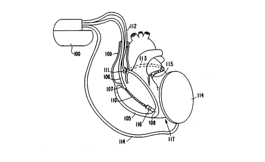
Figure 1 illustrates an implantable pacemaker/
cardioverter/defibrillator 100 and its ~cL-ociated lead system,
as implanted in and adjacent to the heart. As illustrated,
the lead system comprises a co~G.. ary sinus lead 112, a right
~ .icular lead 106, and a -~hc~taneous lead 114. The
coronary sinus lead 112 i8 provided with an elongated
ele ~Lo~e located in the co~G-.ary sinus 111 and great vein
region at 113, extenAin~ around the heart until a~ oximately
the point 115 at which the great vein L~ downward toward
the apex of the heart 117. The right ventricular lead 106
includes an elongated defibrillation ele_~n~e 110, a ring
el~ .o~e 116, and helical el~ v~e 108, which is screwed
into the ~ ç of the right ve..~.icle at the right
25 ~ -icular apex 117. ~D~ 106 and 112 may COL~e_~O.. d to the
leads disclo~ in U.S. Patent No. 5,014,696 by Mehra for an
"Endocardial Defibrillation Elc~t.G~e Systemn, i~ e~ May 14,
1991 and il-~O ~o~ated herein by refere~ce in its entirety. A
r~h~taneous ele~ode lead 114 is alDo illustrated, implanted
in the left chest. Lead 114 ~ay correD~ond to the lead
illustrated in U.S. Patent No. 5,044,374 by Lindemans et al.
for a "Medical Electrical Lead", i~ september 3, 1991 and
i~o.~o~ated herein by reference in its entirety.
W093/2~1 PCT/US93/~
2102~92
- G i;
In eonjunetion with the ~Fçnt invention, the lead
system illustrated provides several eleetrode pairs which may
be employed in the praetiee of the present invention. The
far-field ~en~ing el~_~.G~e pair may eompri~e ring electrode
116 paired with an ele_~lGde loeated on the housing of the
i~plantable r~l~? generator. Ele_L~odes 108 and 116 may be
used for near-field sensing. El~ G~e 116 in eonjunetion
with el~ e 108 or in eonjunction with an el~ o~e loeated
on the hou~ing of the r~ generator will generally be used
for delivery of eardiae paeing r-l~ec. The ele_~.Gdes on
leads 112, 114 and ele_L,Gde llO on lead 106 wil-l be used to
deliver eardioversion and defibrillation r~
Figure 2 illustrates near and far-field eleetrograms, and
the method by whieh the desired synehronization delay
aeeording to the ~,~-ent h-~el-Lion may be derived. The upper
traeing illustrates a simulated far-field ele~LGyLam, for
example as would be taken using ele_LLGde 116 (Fig. 1) and a
re~ote el~ e. R-waves 300 and 302 are illustrated, along
with a synehronized eardioversion pulse 304. The onsets of R-
waves 300 and 302, respeetively, G~UL at 310 and 312. The
~eeond traeing illustrates a simulated bipolar ventrieular
el~_~,Gy,am, for example as would be taken between eleetrodes
108-and 116 (Fig. 1). R-waves 306 and 308 co~e_~ol~ds to R-
waves 300 and 302, respeetively. The third traeing is an
illustration of the output of an R-wave deteetor, as eoupled
to the near-field el~_~,o~e pair used to derive the near-field
el~_t,G~,~m of the reeo~ traeing. ~enr? deteet sig~l~ o~u~
at 314 and 316, e~Y~-ronAing to R-waves 306 and 308,
re~peetively.
As Ai~ A above,~ the interval "t" is derived by
~ubtraeting the point of onset of far-field R-wave 300 from
~ .p~naing near-field s~n~Q deteet 314. The taehyeardia
eyele length VTCL is illustrated as ex~en~ing between
~h-equent R-wave detects 314 and 316. Both methods of
35 - cAlr~lation of the synchronization delay are illustrated,
W093/2~l PCT/US93/00~
2102~92
q
lea~in~ to delivery of the cardioversion pulse 304, a desired
delay ~ after the onset of R-wave 302. In the first instance,
the synchronization delay (SD) may be equal to (~-t) and
initiated in ~~pon~ to ~n~e detect 316. The result is that
the cardioversion pulse 304 is delivered at a point of onset
of R-wave 302, plu8 ~. The alternative method of calculating
the synchronization delay provides a delay calculated from
sense detect 314. In this case, the synchronization delay is
equal to (VTCL+~-t), and is initiated in res~on-? to the ~0n~e
detect 314 ~-~ociated with the R-wave 300, ~ in~ the R-
wave 302 to which the cardioversion p-~ls? is to be
synchronized. In this case the cardioversion pulse 304 is
also delivered at the onset of R-wa~e 302 ~ he second
method of synchronized delay calculation, as ~iscll~red above,
is believed to be more likely used in those circumstAnces in
which the derived delay (~-t) is negative. However, it may
also be used in those inst~nGes in which the derived delay is
positive, if desired.
For ~L~ ~~s of the ~-ent application, the high voltage
pulse delive.Ll is referred to as a "cardioversion" pl~lse.
However, it s~ d be kept in mind that in some ca~es, the
early ~tages of ventricular fibrillation may be difficult to
distinguish from a rapid ventricular tachycardia, and in such
_ -es, the deli~eLed cardioversion p~ e may actually function
as a defibrillation p~ ç, terminating the early stages of
fibrillation. Therefore, for ~u~ s of the invention, the
specific nature of the heart rhythm being monitored
(tachycardia ~1D~S fibrillation) is less important than that
ability to r~l~ahly ~ense and syn~h~o..ize delivery of the high
voltage p~ e. .~c~fore, for ~ s of the ~a-ent
application, the term "cardioversion" should be ~on~ ed
broadly. -
Figure 3 is a functional schematic diagram of an
i~plantable pacemaker/cardioverter/defibrillator in which the
~L2-?nt i-~ -Lion may usefully be practiced. This diagram
WOg3/2 ~ 1 PCT/US93/00~8
2102~92 ~ ~
should be taken as exemplary of the type of device in which
the i..~e..~ion may be embodied, and not as limiting, as it is
believed that the invention may usefully be practiced in a
wide variety of device implementations, including devices
having ~ Lional organization similar to any of the
implantable pacemaker/defibrillator/cardioverters ~ ntly
being implanted for clinical evaluation in the United States.
The i.-~e--~ion is also believed practicable in conjunction with
i~plantable pacemaker/cardioverters/ defibrillators as
disclosed in prior U.S. Patent No. 4,548,209, i~r~e~ to
Wielders,et al on October 22, 1985, U.S. Patent No~ 4,693,253,
issued to Adams et al on September 15, 1987, U.S. Patent No.
4,830,006, ;~ to Haluska et al on May 6, 1989 and U.S.
Patent No. 4,949,730, i~ e~ to Pless et al on August 21,
1990, all of which are incorporated herein by reference in
their entireties.
Thc device is illustrated as being provided with six
ele_~v~es, 500, 502, 504, 506, 508 and 510. Electrodes 500
and 502 may be a pair of ele_~Gdes located in the ventricle,
for example, ~o,.~ o~ ng to electrodes 108 and 116 in Figure
1. Ele_L,G~e 504 may co,,.~pon~ to a remote, indifferent
ele~.o~e located on the housing of the implantable
pacemaker/cardioverter/defibrillator. Electrodes 506, 508
and 510 may ~G~L~-ronA to the large surface area
defibrillation ele_~ odes located on the ventricular, coronary
~inu~ and ~-~cl~taneous leads illustrated in Figure 1.
Ele_~,~des 500 and 502 are shown as hard-wired to the R-
wave detector circuit, comprising b~n~r~s filter circuit 514,
~uto threshold circuit 516 for providing an adiustable sensing
threshold as a function of~the measured R-wave amplitude and
comparator 518. A signal is generated on R-out line 564
whenever the signal ~~n~e~ between electrodes 500 and 502
e~ the ~ ~-ent sensing threshold defined by auto
thre~hold circuit 516. As illuxL,ated, the gain on the band
pass amplifier 514 is also adjustable by means of a signal
WO93/208gl PCT/US93/00~
2102492
q '
from the pacer timing and control circuitry 520 on GAIN ADJ
line 566.
The operation of this R-wave detection circuitry may
~o,-~-ponA to that disclosed in commonly assigned, cop~nAin~
U.S. Patent Application Serial No. 07/612,760, by Keimel, et
al., filed November 15, for an Apparatus for Nonitoring
Electrical Physiologic Si~nAl~, incorporated herein by
reference in its entirety. T-~rcve~, alternative R-wave
detection circuitry such as that illustrated in U.S. Patent
No. 4,819,643, i~--~eA to ~enken on April 11,1989 and U.S.
Patent No. 4,880,004, i~ 6~ to Baker et al on November 14,
1989, both incorporated herein by reference in their
entireties, may also usefully be employed to practice the
~ ?nt invention.
The th.e-~old adjustment circuit 516 sets a threshold
~o..asronA;ng to a predetermined percentage of the amplitude
of a r?r-eA R-wave, which threshold decays to a minimum
thre~hold level over a period of less than three rs-on~C
thereafter, similar to the automatic sensing threshold
circuitry illustrated in the article "Reliable R-Wave
Detection from Ambulatory Subjects", by Tha~or et al,
publ~-heA in Biomedical Science I~ ~mentation, Vol. 4, pp
67-72, 1978, i~ y~ated herein by reference in its entirety.
In the context of the ~.~-?nt invention, it is preferable
that the threshold level not'be adjusted in re pon~Q to paced
R-waves, but instead should continue to approach the minimum
threshold level following pAcQA R-waves to enhance sensing of
low ' level ~pontaneous R-waves ar-oçiatea with
ta~hya~l.y~hmias. The time cG~ ant of the th~ ld circuit
is also preferably sufficiently short so that minimum sensing
threshold may be reached within 1-3 -~conA~ following
ad~ustment of the sensing th~ old equal to 70-80% of the
amplitude of a detected D~o..~aneous R-wave. The invention may
also be'practiced in conjunction with more traditional R-wave
W093/2~l PCT/US93/00288
- 2 1 0 2 4 9 2 /D
sen~ors of the type comprising a band pass amplifier and a
comparator circuit to determine when the hAn~p~ signal
exceeds a predetermined, fixed sensing threshold.
Switch ~atrix 512 is used to select which of the
avAilAhle el~_tLo~es are to be coupled to ~n~? amp s34, for
use in measuring the point of onset of the far-field R-wave.
Selection of which ele_~odes are 80 employed is ~on~ olled by
the microp-G~A-cr 524 via data/ad~ bus 540. Si~ C from
the selected el~ ,odes are pAsr~A through h~n~pAss amplifier
534 and into multiplexer 532, where they are converted to
~ultibit digital si~n-ls by A/D oon~e~er 530, for storage in
random ~c~-:s memory 526 under ~c..~ol of direct memory
aadress circuit 528. Microp~ G~ Qr 524 analyzes the
digitized ECG signal stored in random ~cecs memory to
identify the points of onset and termination of R-waves ~ e~
between the far-field ele_t G~es.
For example, the micro~o~e--or 524 may analyze the ECG
stored in an interval exten~in~ from minus loO milliseconds
previous to the o~ul~el~e of an R-wave detect signal on line
564, until 100 milli~econ~ following the o~c~lence of the R-
wave detect ~ignal. The time window thus extends from a time
prior to R-wave dètection to a point following R-wave
detection, and includes a sufficient time (e.g. 200 ms) to
aD-uLa that the entire R-wave is recorded. After detection of
an R-wave and the expiration of the A~ociated time window,
the device examines the digital value~ stored during the time
window and determines the width of the R-wave, identifying
both the start and end points for the R-wave. The difference
betw~en the ~tart and o,.-l~oints defines the width of the
stored R-wave, which may~be used for ~i~J.o-Lic ~ r or
for classification of a~ Lhmias. In particular, the width
of the R-waves may be used to distinguish sinus tachycardia
from v~..~Licular tachycardia. Cop e nd i ng p a t e nt
application No. for a NMethod and Apparatus
for Discrimination of Ventricular Tachycardia from
W093/2089l PCT/US93/~ ~X
" 2102~.~2
$upraventricular Tachycardia and for Treatment thereof" by
Mader, et al filed on the date of this application discloses
a particularly advantageous method of identifying the points
of onset and termination of digitized R-waves for use in an
implantable device. This application is incorporated herein
by reference in its entirety.
In particular, the point of on~et may be identified at
the point at which a y~e~etermined number of s~cce6cive
digital values eYc~e-7 the preceding stored digital value by
~0 more than a predetermined amount. For example, each digital
value may be compared to the most recently stored previous
digital value but one, the diffe~ence between these two values
détermined, and their diffe~e--~e compared to a predetermined
tl.~hold. If the sign of the difference so calculated
remains constant and the difference so calculated remai~s
above a desired threshold for a predetermined number of beats,
onset is identified.
For purposes of the ~t~-~nt i,.ve..~ion, a ~ampling rate of
256 Hz should be ~ufficient, al~ho~ somewhat lower or
~ubstantially higher sampling rates may be used, depenA;n~ on
the amount of data storage capacity in RAM 526 and on the
processing speed of microploc~Qr 524. However, any method
of identifying the start or onset of a digitized R-wave may
u~efully be employed in conjunction with the present
i,.~ ion.
The identified point of onset To is ~tored and is
co~pared to the time of o~LLence of the near-field R-wave
~detect Td signal (on R 0UT line 564) which tri~e~ed storage
of the digitized waveform. The aifference "t~ between these
two times is calculated by microp~ ror 524 and stored in
memory 526. The desired delay ~ from onset of the far-field
R-wave is ~imilarly stored in the memory 526. The value of ~
may be set by the physician by means of an external ~oyLammer
or may be a function of the rate of the ~en~
tachya~ hmia. Generally, it is believed that the optimal
WO g3/208gl Pcr/uss3/002ss
2 10 2 ~9 2 ,~
value for ~ will be between 80 and 120 ms. The value of t is
~ubtraoted from the value of ~, to ~o~uce a derived delay, as
Ai-c~q~ above. The functioning of the software used to
determine the synchronization delay from the~e values is
S ~ ~A in more detail below in conjunction with the flow
charts of Figures 4, 5 and 6.
The remainder of the circuitry is dedicated to the
provision of cardiac pacing, cardioversion and defibrillation
therapies. The pacer timing/control circuitry 520 includes
~0 ~c~mmable digital counters which control the basic time
intervals A~-oçiated with W I mode cardiac pacing, including
the pacing escape intervals, the refractory periods d'uring
which -~n~e~ R-waves are ineffective to restart timing of the
escape intervals and the r~ e width of the pacing pulses.
1~ The durations of these intervals are determined by
miCrop~G- --or 524, and are communicated to the pacing
circuitry 520 via addle~s/data bus 540. Pacer timing/centrol
circuitry also determines the amplitude of the cardiac p~cing
p~lre~ and the gain of hA~~r~rr amplifier, under control of
microp~ r~~r 524.
During W I mode pacing, the escape interval counter
within pacer timing/control circuitry 520 is reset upon
~ensing of an R-wave as indicated by a signal on line 564, and
on timeout triggers generation of a pacing pulr-~ by pacer
output circuitr~ 522, which is coupled to electrodes 500 and
502. The e~cape interval counter is also reset on generation
of a pacing pulse, and thereby ~o..~.ols the basic timing of
c~rdiac p~~ functions, including antitachycardia pacing.
The ~L~ion of the interval defined by the escape interval
ti~er is determined by mi~o~Lo. _ror 524, via data/address
bus 540. The value of the count present in the escape
interval counter when reset by ~?~-e~ R-waves may be used to
mea~ure the duration of R-R intervals, to detect the presence,
of ta_hy~ardia and to determine whether the minimum rate
WO93~20891 PCT/US93/002
~ /3 ~1 n 2 ~ 2
eriteria are met for activation of the
taehyeardia/defibrillation diserimination funetion.
~ierop~ or 524 operates as an interrupt driven
deviee, and responds to inte~ s from paeer timing/eontrol
eireuitry 520 ~G~L~-po~Aing to the oeeurrenee of 6e~e~ R-
waves and eoL.~-ro~Aing to the generation of eardiac paeing
p~lAe~. These inte~Lu~s are provided via data/add~e_s bus
540. Any nc~ ~ry mathematieal ealeulations to be performed
by mierop~ -ror 524 and any updating of the values or
intervals eG.. L.olled by paeer timing/~G.. -Lol eireuitry 520
take plaee following sueh inte~Lu~Ls.
In the event that a taehya~ h~Lhmia is deteeted, ànd an
antitaehya~.h~hmia paeing regimen is desired, a~p~opLiate
timing intervals for ~G.-LLolling generation of antitaehyeardia
paeing therapies are loAA~A from mi~.u~o~ or 524 into the
paeer timing and ~GlL~ol eireuitry 520, to ~u~.~Lol the
operation of the eseape interval eounter and to define
re~raetory periods during whieh deteetion of an R-wave by the
R-wave deteetion eireuitry is ineffeetive to restart the
eseape interval eounter. Similarly, in the event that
generation of a eardioversion or defibrillation r~ e i8
required, mierop~-c~~~or 524 employs the eounters in timing
and ~O~LO1 eireuitry 520 to co~ ol timing of sueh
eardiove~sion and defibrillation p1l-~s, as well as timing of
-~oiated refraetory periods during whieh ~Qnse~ R-waves are
ineffeetive to reset the timing eireuitry.
- In response to the deteetion of fibrillation or a
ta~h~ardia requiring a eardiover~ion p~lse, mierop~oc~ or
524 aetivates eardio~el~ion/defibrillation co..~ûl eireuitry
,
554, whieh initiates ebarging of the high voltage eapaeitors
556, 558, 560 and 562 via eharging eireuit 55C, under co..L~ol
- of high voltage eharging line S52. The voltage on the high
voltage eapaeitors is monitored via VCAP line 538, whieh is
p---e~ through multiplexer 532, and, in response to reaehing
a predetermined value set by miC~o~o~eCcor 524, results in
W093/2 ~ 1 21 ~ 2 ~ ~ 2 PCT/USg3/00~8
/~ ~
generation of a logic signal on CAP FULL line 542, terminating
charging. Thereafter, delivery of the timing of the
defibrillation or cardioversion rll~e is controlled by pacer
timing/control circuitry 520, using the ~elected
S ~ynchronization delay as di-c~r~~~ above~ One embodiment of
a ~y~tem for delivery and synchronization of cardioversion and
defibrillation rll~e~, and cG..~olling the timing functions
related to them i8 disclosed in more detail in coren~ing~
commonly assigned U.S. Patent Application Serial No.
07/612,761, by Keimel, for an Apparatus for Detecting and
Treating- a Tachya~hy~hmia, filed November 15, 1990 and
in~ol~o~ated herein by refe~e..~a in its entirety. This basic
~ystem, with the addition of the derived synchronization delay
provided by the ~L~~ent invention provides a workable
implementation of the p~-?nt invention. However, many known
cardiv~.~ion or defibrillation pulse generation circuits are
believed usable in conjunction with the ~ nt invention.
For example, it is believed that circuitry ~G.J~ olling the
timing and generation of cardioversion and defibrillation
p~l~e~ as disclosed in U.S. Patent No. 4,384,585, i r~ to
Zipe~ on May 24,1983, in U.S. Patent No. 4949719 ;~ A to
Pless et al, cited above, and in U.S. Patent No. 4,375,817,
~ to Engle et al, all i..~ol~olated herein by reference in
their entireties may also be adapted to practice the present
i--vi.ltion. Similarly, known circuitry for ~ol-~olling the
timing and generation of antitachycardia pacing p~ e~ as
de~cribed in U.S. Patent No. 4,577,633, i~ to Berkovits et
al on March 25, 1986, U.S. Patent No. 4,880,005, i fi~'le~ to
- Ple~ et al on November 14, 1989, U.S. Patent No. 7,726,380,
i~ued to Vollmann et al on February 23, 1988 and U.S. Patent
No. 4,587,970, issued to Holley et al on May 13, 1986, all of
which are i~ yo~atea herein by reference in their entireties
may also be used to in a device for practicing the present
.,Lion.
WO93/2~1 PCT/US93/00288
21~02~92
~ ' .
In the illustrated embodiment of the present invention,
selection of the particular electrode configuration for
delivery of the cardioversion or defibrillation p~ es is
controlled via output circuit 548, under control of
cardioversion/defibrillation control circuitry 554 via control
bus 546. Output circuit 548 determines which of the high
voltage ele_~Gdes 506, 508 and S10 will be employed in
delivering the defibrillation or cardioversion p~ ç regimen,
and may also be used to Qpecify a multielectrode, simultaneous
pulse regimen or a multielectrode sequential r~ re regimen.
Monophasic or biphasic pulses may also be generated. One
exa~ple of circuitry which may be used to perform this
function is set forth in commonly assigned copen~ing Patent
Application Serial No. 07/612,758, filed by Keimel, for an
Apparatu~ for Delivering Single and Multiple Cardioversion and
Defibrillation P~llre~, filed November 14,1990, incorporated
herein by reference in its entirety. However, ou~u~ control
circuitry as disclosed in U.S. Patent No.4,953,551, issued to
Mehra et al on September 4, 1990 or U.S. Patent No. 4,800,883,
~ to Winstrom on January 31, 1989 both incorporated
herein by ref~ e in their entireties, may also be used in
the context of the ~ ent invention. Alternatively single
monophasic pulse regimens employing only a single ele~L~o~e
pair a~o.ding to any of the above cited references which
disclo~e implantable cardioverters or defibrillators may also
be used.
Fi~ 4, 5 and 6 are intenh~ to functionally represent
that portion of the software employed by microp~ or 524
(F~g. 3) which is relevant to or implements the
synchronization delay. ~ This portion of the software
~ -Lt~ed in Figure 4 is illustrated functionally and deals
with the o~r~ all organization of tachyarrhythmia detection
functions during WI mode pacing, as indicated at 600. The
tachya~ yUhmia detection functions are activated in response
to an inte-~u~- indicative of a ~en-~~ or paced beat at 602.
W093/20891 PCT/US93/00288
2 102 492 /6 ,~t~
In ~ponr~ to this interrupt, the value of the prece~ing R-R
interval, ~G~ L ~ -ponA i n~ to the current time on the escape
interval counter in pacer timing/~G.-~ol circuitry 520 may be
stored at 602 and used as a measurement of the R-R interval
for tachyarrhythmia detection functions. In addition, the
time of detection (Td) of the ~en~ed ventricular
depolarization, as indicated by means of a real time clock
within microp~._ ror 524 is also stored at 602.
Stored information reflective of the previous series of
R-R intervals, ~uch as information regarding the rapidity of
on~et of detected short R-R intervals, the s-tability of
detected R-R intervals, the duration of cont~ e~ detection of
short R-R intervals, the average R-R interval duration and
information derived from analysis of stored ECG segments are
used to determine whether tachya~ hmias are p~-rnt and to
di~tinguish between different types of tachyarrhythmias. Such
detection algorithms for ~c~ ing tachycardias are
described in the above cited U.S. Patent No. 4,726,380, is~le~
to Vollmann, U.S. Patent No. 4,880,005, issued to Pless et al
and U.S. Patent No. 4,830,006, issued to Haluska et al,
in~o,yo~ated by refe,en~e in their entireties herein. An
additional ~et of tachycardia ~ ~J~.i tion methodologies is
di~clo~~A in the article "Onset and Stability for Ventricular
Ta~hy~ hmia Detection in an Implantable Pacer-
Cardiov~,Ler-Defibrillator" by Olson et al., publi~he~ in
Com~uters i n Cardiology, October 7-10, 1986, IEEE Computer
Society ~ , pages 167-170, al~o incGLyo~a~ed by reference
in its entirety herein. Ho~l_ve~, other criteria may also be
J~asured and employed in conjunction with the y~ nt
i-.~e.l~ion.
For p~ ec of the y~ nt invention, the particular
details of implementation of the rate and/or R-R interval
ha~A VF and VT detection methodologies are not of primary
importance. Ilo~ er, it is required the VF and VT rate hA~e~
detection methodologies employed by the device allow
W093/2 ~ 1 2 1 0 2 ~ ~ 2 PCT/US93/oo~
/~
identification and d¢tection of ventricular tachycardia
requiring cardioversion. One of the advantages of the ~ nt
~ e~Lion i8 that it is believed practicable in conjunction
with virtually any prior art tachycardia detection algorithm.
The microp~G~ or ch~c~ at 604 to determine whether the
previous ~eries of R-R intervals are indicative of
fibrillation. If 80, an appropriate defibrillation therapy is
~elected at 606, the high voltage capacitor6 are charged at
608, and a defibrillation r--~e i8 delive.el at 610. The
a~e _ited application Serial No. 07/612,761 by Keimel also
di~closes a method and apparatus for delivery of synchronized
defibrillation r~l~e~. This or other prior systems for
delivery of defibrillation p~ e~ may be beneficially be used
in the context of the ~ ent invention. If fibrillation is
not detected at 604, the mi~Lo~. 8 --or chec~ at 612 to
determine whether the ~c~-lin~ series of detected vel.~Licular
depolarizations meet the criteria for detection of
tachycardia. If CO, a therapy is selected at 614.
In modern implantable antitachyarrhythmia devices, the
part;c~lAr therapies are ~o~mmed into the device Ah?~ of
ti~e by the physician, and a menu of therapie is typically
provided. For example, on initial detection of tachycardia,
an antitachycardia pacing therapy may be selected. On
redetection of tachycardia, a more aggressive antitachycardia
pacing thela~y may be ~che~ ed. If repeated attempts at
antita~y~ardia pacing therapies fail, a higher level
cardio~eL~ion r~l-e-therapy may be selected thereafter. Prior
art patents illu~t.a~ing such ~e -et therapy menu~ of
antitachya~ h~hmia therapies include the aLo~e _ited U.S.
Patent No. 4,830,006, i~ ?~ to Haluska, et al, U.S. Patent
No. 4,727,380, issued to Vollmann et al and U.S. Patent No.
4,587,970, i~ueA to Holley èt al. The plP-ent invention is
believed practicable in conjunction with any of the known
antitachycardia pacing and cardioversion therapies, and it is
W093/2 ~ 1 PCT/US93/00288
2102~92
believed most likely that the invention of the present
application will be practiced in conjunction with a device in
which the choice and order of delivered therapies i8
~c~&mmable by the physician, as in current implantable
pacemaker/cardioverter/ defibrillators.
In the context of the pl~s~nt invention, it is
anticipated that the ~ynchronization delay itself may be
varied as function of the ~he~tled se~ence of delivered
therapies. For example, with each sU~ ccive cardioversion
atte~pt, the synchrsn~7~tion delay may be s~lcce-~ively
increa~ or d__~~~ . Alternatively, the synchronization
delay be incr~e~ or d__,e~-e~ as a function of the
amplitudes of the ~c~6~ ed cardioversion p~ r.
If the selected therapy is a cardioversion pulse, at 616,
the high voltage vu~u~ capacitors are charged at 626, and a
cardioversion pulse is delivered at 628, timed from a near-
field R-wave detect as described above. Figure 6, below and
Fi~ 7 and 8 illustrate the synchronization of
cardioversion pulses to detected R-waves, using the ~ ent
inv .. ~ion, in some detail. In the event that a therapy other
than cardioversion, for example antitachya. h~Lhmia pacing, is
~elected, this therapy is similarly delivered at 622.
Following delivery of the antitachycardia for defibrillation
therapies, detection criteria are updated at 632 to reflect
delivery of the.previous therapies and the therapy schedule is
updated at 634, as de~cribed above.
In the ~-ent il.~e..~ion, it is desired to calculate the
~ea~ l interval ~t" in the form of a rl~nning average value
(tAVG), taken over a series of detected R-waves preceding
delivery of the cardio~eI~ion ~ se. As ~i~cvc~ in
conjunction with the above-cited Mader application, the pulse
width is a~e~&~ed over a ~eries of approximately 8 beats prior
to delivery of antitachycardia therapy. This number of values
would appear to be workable in conjunction with -the
c~iC~ tion of an average value of tAVG as well. However, it
WO93/2~1 2 1 ~ 2 4 9 ~CT/US93/00288
J9
~ay be that in some cases a greater or lesser number of R-
waves are desired in order to calculate tAVG.
In the event that the prese~n~ series of R-waves does
not ~eet the criteria for delivery of an antitachycardia
therapy, the mi~G~oc~Qr çhec~ at 616 to determine whether
there has been substantial ~o~ toward detection of a
tachycardia so that onset measurements may be initiated.
Regardles~ of the detection criteria for triggering delivery
of an antitachycardia therapy, it is important that the
"predetection" criteria which activates measurement of R-wave
on~et using the far-field ele_~odes i8 defined~such that it
a~D~e~- that a significant number of R-waves will be available
for measurement, prior to the criteria for delivery of
antitachycardia therapies is met. For example, if the
criteria for delivery of antitachycardia therapies is a
predetermined number of R-waves (NID) having a duration less
than a predetermined tachycardia interval (TDI), the
~eaD~ement of onset of the far-field R-waves can be initiated
when the ongoing count of R-waves less than TDI equals NID-X,
wherein X is the desired number of values to be employed to
calculate tAVG. If the predetection cQ~Aitions are satisfied,
the onset of the stored R-wave is measured and an updated
calculation of the synchron;~tion delay (SD) i8 calculated at
620. The block indicated at 620 ~G~.2-ponAC to the functional
flowchart illu_~a~ed in Figure 5, which deecribes the method
of calr~lAtion of the ~yl.~hronization delay in more detail.
In the event that the predetection criteria are not met
- at 618, the microp~ocE--or c~e~k~ at 624 to determine whether
ter~ination of a previou~ly detected tachycardia or a ~e~n
to normal sinus rhythm during detection of a tachycardia has
v~ ed at 624. As ~;~c~se~ above, this is typically
accomp~ eA by sensing a series of R-waves separated by
intervals less than the minimum intervai for tachycardia
detection (e.g., less than VTDI). In the event that
termination of previously detected- tachycardia or the
W093/2~1 PCT/US93/002~
~~.
210249~ ~D
oc~ ence of a normal sinus rhythm is recog~ized at 624, the
output capacitors are internally Ai ~ch~rged at 630, if they
had been previously charged. In the context of delivery of
synchronized cardioversion, it is possible that the capacitors
may be charged, without the p~ e actually being delivered.
This again is Ai~c~ 6A in more detail in conjunction with the
flowchart of Figure 6. After Ai Ec~rging the ou~u-
capacitors, the detection criteria i8 updated at 632 and the
therapy menu is updated at 634 to reflect the return to normal
sinus rhythm, a~ A i ~ ~~e~ above. At this time, it is also
envisioned that any stored measurements of t will also be
cleared.
Figure 5 is a functional flowchart illustrating the
operation of the functional block 620 in Figure 4, dedicated
to determination of the synchrsni~tion delay. At 700, the
microp~ or ~heo~s to determine whether the preced;n~ R-R
interval qualifies as a tachycardia beat. For example, this
may be accomplished by comparing the R-R interval to a
predetermined interval indicative of a tachycardia, e.g., the
TDI interval Ai ~ e~ above. If R-R is less than TDI,
measurement of onset is undertaken. If not, the device
retuL~- to WI mode pacing.
At 702, the microp~ e-~or measures the time of onset.
As A~r~ A above, this ~ 6 involves waiting the time out
of the mea~ur _ ent window, (e.g. 100 milli~econds after Td),
and examining the digitized R-wave to determine the point of
onset, as Ai~ A in the abGve _ited Mader application. At
704, the time of on~et To i8 subtracted from the R-wave
detection time Td, to yield a value ~tn. At 706, the rl~n~;ng
ave~a~e tAVG of the values of "t" is updated, and at 708 the
~e.ltLicular cycle length VTCL i8 updated. This may be a
r~n~n~ ave~a~e of the most ecc..~ series of R-R intervals
a~~~iated with the tachyaL~hy~hmia. For example, it may be
the ~,~ n~ 8 R-R intervals less than TDI. At 710, the
microprocessor ~h~cks to determine whether the value of ~ is
W093/2089l 2 1 0 2 ~ 9 2 PCT/US93/00288
~/
in~nd~A to be fixed, or to vary as a function of the detected
tachycardia rate. If the value i8 preset, it is simply looked
up at 712. ~f the value is intend~ to be variable, it is
calculated at 714 as a function of VTCL. For example, as VTCL
decrea~c, the value of ~ may decrease from 120 to 80
milliseconds. These values are purely exemplary, and it is
b~l~eved that the physician will wish to optimize the range of
values for ~ and their co L~ pon~nçe to the value of VTCL, on
a patient-by-patient bas~s.
At 716, the micro~ or c~eckc to determine whether ~
- tAVG is greater than zero. If not, the synchronization
del~y is set equal to VTCL + ~ - tAVG. If - tAVG is greater
than or equal to zero, the synchronization delay is set equal
to ~ - tAVG at 718.
Pigure 6 illustrates the functional operation of the
tachycardia synchroni~ation method of the present invention.
In performing this method, the mi~ or employs time~s
within pacer circuitry 520 to define synchronization intervals
as ~~c~ e~ above. Following detection of ventricular
tachycardia and su.~ ful charging of the o~L~ capacitors,
the ~icrop~ or 524 sets the timers in pacer circuitry 520
to define a first synchroni7~tion interval SYNC-1, a first
refractory interval REF-l and a first blAn~in~ interval Rr~K-
1. During time out of BLANK-1 at 800, vel.~Licular sensing is
disabled. In response to an inte~u~ indicating ventricular
sensing dur~ng REF-1 at 802, the microp~ or 524 notes the
o~ ence of the refractory sense at 804, and initiates
ti~ing of a second synchronization interval SYNC-2, a ~con~
refractory interval REF-2 and a ~eccn~ bl~nkin~ interval
RT ~NK-2.
In the event that no inte~ indicating the c~ ence
of ~ icular senging C~L during REF-l, the mic~oyLGces~or
continues to wait for the o~ Lence of an inte~u~
ir~ ting ventricular sensing during SYNC-l. If an inteL~u~
O~ at 806, the microp~oc~-~ror notes it at 808, and
;
W093/2 ~ 1 21 0 2 ~ g 2 PCT/US93/00~
J2
initiates SYNC-2, REF-2 and ~m~NK-2, as di~ above. In
the absence of ~ events G~U~ ~ ing during SYNC-l, the
micro~ C~e,or resets any internal flags set at 810, and
~e~ the function of the device to the ~ ammed
bradycardia pacinq mode at 820.
In the event that a reconA synchronization interval is
initiated, after expiration of BLANK-2 at 812, the
m~croprocescor waits for an inte~u~t during REF-2 indicative
of ~-.L~icular sensing. If such an inte~u~L G'~U~S at 814,
~0 the microp~ or checks at 816 to determine whether an
internal flag (Csl) has been set at 816 indicative of previous
~en~ing during the post-refractory portion of SYNC-l. If this
flag has been set, microplc ~--or ,c~u~ the operation of the
device directly to the ~G~ammed W I brady pacing mode at
820. If the flag has not been set at 816, the microprocessor
increments the count (RS) of ~n-~~ events o~ ing during
refractory periods at 818, and check~ to see whether three
~ucces~ive refractory r?n~e events have o~u.~ed at 822. In
the ~-?nce of three refractory ren~? events (that is, R-wave
inte~ s oc~u~ing during the refractory intervals of three
8U~ ive synchronization intervals) the microproceC~or
d~rectly ~e~u~ the operation of the device to the ~G~ammed
VVI bradycardia pacing mode at 820.
Assuming that the o~u~ence of three ~ccessive R-wave
inte~lu~s during refractory intervals has not o~ e~,
microproce~or 524 initiates timing of a third synchronization
interval, having the same parameters as the second
~ynchronization interval. In the event that no R-waves are
se.n~ed dur~nq REF-2, the micro~ 80r continues to wait at
824, for an inteLLu~ indicating the occu~lence of an R-wave
during the post-refractory portion of SYNC-2. In the event
that no such R-wave is ~?n~ the mi~ o~.,sor resets all
internal flags at 810, and re-initiates W I bradycardia
pacing.
WOg3/2 ~ 1 PCT/~S93/~ ~8
o23 2 ~ 2 ~ ~
In the event that the mi~. G~ oceC~r receives an
inte~ indicating the oc~L~ence of an R-wave during SYNC-2
at 824, the microp~ ror ch~ck~ at 826 to determine whether
a previous R-wave has been ren,-ed in the post-refractory
s portion of a synchronization interval at 826. If an R-wave
previously has been -en-~ in the post-refractory portion of
a previous ~ynchronization interval, at 828 the microp~ rrQr
reads the value of the synchronization delay SD previously
calculated and at 830 initiates delivery of a cardioversion
pulse timed from the most recent R-wave interrupt.
Microp~ or 526 then re~et~ all internal flags at 810, and
then ~ to ~ mmed W I Ll~dy~ardia pacing at 820.
In the event that an R-wave is re~-0~ during the post-
refractory portion of SYNC-2 at 824, but there is no internal
flag set indicatinq the O~UL ~ ence of a previous post-
refractory Qensed R-wave at 826, the mi~c~o~ ~or sets a
flag indicating R-wave sensing in the post refractory portion
at 808 and initiates timing of the third synchron~z~tion
interval, employing the same time parameters as the second
synchroni7ation interval.
The microp~ or 526 continues to define
synch~s..i7--tion intervals having the time parameters of the
second ay..~llroni7Ation interval until either one of the
~ynchronization intervals expires without ventricular ~enDing,
three R-waves o~ within refractory intervals within
~ynchronization intervals, or two R-waves are sensed during
the non-refractory portions of -~ccDc-ive Dynchro~ization
intervalD. As a practical matter, given the method as
illu~trated ~n Figure 6, a maximum of four syncl~ol,;7--tion
intervals may be reguired~ in some c~ cue~er, in most
cA~~s two or three synchronization intervals will be adequate
in order to determine whether a synch~& ~ls cardioversion
pulse is deli-~e~ed.
Figure 7 shows a simulated near-field EGM strip,
evidencing a ventricular tachycardia indicated by closely
WOg3/2 ~ 1 PCT/US93/00~8
2102492 ~y
~paced R-waves 900a, 902a, 904a, 906a and 908a. Figure 7
lllustrates the operation of the invention to synchronize
delivery of cardioversion r~ in the case where the
synchronization delay is initiated in ~ e to the near-
field R-wave detect co~ rQnAi~ to the R-wave to which the
pulse is ~ynchronized. In other words, this figure
illu_~.ates the ca~e in which the synchronization delay SD =
(~ - tAVG).
It ic assumed that microplG~s~or 526 has already
detected the o~L-e~.ce of this tachyarrhythmia, and has
enabled the high voltage charging circuitry *o initiate
rharging of the ou~u~ c~racitors. At point gl6, the voitage
on VCAP line 538 (Fig.l) reaches the ~.c~ ammed voltage,
terminati~g the charging ~oce~. Following the charging
~ , an initial blAn~ing interval BLANK-l, 922a, is
defined. An appropriate duration for this blAnkin~ interval
can be 300 ms. An initial refractory interval REF-l, 924a, is
also defined. An appropriate duration for this interval may
be 400 ms.
An initial synchronization interval SYNC-l, 926a, is also
defined. The duration of this interval is preferably a
function of the rate criterion for tachycardia detection. In
par~irvlar, it is recommended that ~his interval be equal to
the tachycardia detection interval (TDI) plus a predetermined
25 - tiae increment, for example 360 ms.
-R-wave 906a o~.s during the post-refractory portion of
the first ~ynchronization interval 926a, and initiates the
~econd synchronization interval s32a. A ~econ~ bl ~n~i n~
inter~al BLANK-2, 928a, is also initiated. This interval may
be, for example, 120 ms.,~and may ~oL~-ron~ to the blA~in~
interval u~ed following ventricular sensing during brady
pacing or may be a ~eparately defined value. Also initiated
is a ~oconA refractory interval REF-2, 930a, which may be, for
example, 200 ms., and a~in may co-Le~pond either to the
normal refractory interval employed by the device following
W093/2089l PCT/US93/00288
2102~92
8en~ed ventricular co,.~lsctions, or may be separately defined.
The ~econ~ synchronization interval SYNC-2, 932a, is
preferably also a function of the tachycardia detection rate
criterion as ~;~C~A-e~ above, and may be, for example, the
tachycardia detection interval (TDI) plus 60 ms. Because R-
wave 908a i8 the second ~ c~ffive R-wave ~e~ within the
po~t-refractory portion of a ~ynchronization interval, it
initiates timing of the synchronization delay SD, 934a, and
triggers delivery of a cardioversion p~llre at 938a on its
expiration. Following generation of the cardioversion r~ Q
at 938a, the device ~e~ to WI bradycardia pacing.
It should be noted that following delivery of the
cardioversion pulse at 938a, some voltage may remain on the
output capacitors. This voltage will not be di~ch~rged until
detection of VT termination. As di~cnsr~ above,
micropL._ ~r 526 may detect VT termination in ~-ponse the
G~ ence of a predetermined number of sequential R-R
intervals greater than the tachycardia detection interval.
Following detection of termination, the ou~l capacitor~ may
be ~;r~h~~ged internally as ~ r~ above.
Figure 8 illustrates the operation of the invention to
~ynchronize delivery of cardioversion pulses in the case where
the synchronization delay is initiated in res~o -e to the
near-field R-wave detect ~o~ ~pon~ing to the R-wave previous
to the R-wave to which the r~ 1 ~0 is ~ o.... ized. In other
words, this figure illustrates the case in which the
~yn~h~.i7Ation delay equals (VTCL ~ ~ - t). All numbered
ele~ent~ of Figure 8 ~G~ rond to similarly numbered elements
of figure 7, with only one difference. All steps 1eA~;ng to
delivery of the cardioversion rll~e co~ on~ exactly to
those oo~ --ponAingly numbered in Figure 7, but in this case,
the Fynchronization delay SD is calculated to result in
~y~hlGl~ization of the pulse 938b with R-wave 910b, rather
than R-wave 908b.
W093/2 ~ I PCT/US93/00~8
21~2~92 ~,~
It should be rec~nized that although the disclosed
embodiment deals with fibrillation and tachycardia in the
lower chambers or ventricles of the heart, the invention may
be usefully practiced in the context of the upper chambers or
S atria of the heart, which are al~o prone to tachycardia and
fibrillation in some patients. Similarly, it should be
understood that the ~ nt i~.~e..~ion, while particularly
adapted for use in or in conjunction with an implantable
cardio.el~er/defibrillator may also be usefully practiced in
conjunction with an external cardioverter. It is believed
that ~y..~hronization ha~e~ on the point of onset-of the far-
field EGM is of value in this context as well.