Note: Descriptions are shown in the official language in which they were submitted.
~~ Q2~ 5 ~
- 2 -
BACKGROUND 'TO THE INVENTION
THIS invention rE:lates to a system for scanning bodies.
In one application of the invention, it may be used to scan human bodies
to detect the presence thereon or therein of certain articles or
substances.
~1 ~2~ 51
-3-
SUMMARY OF THE INVENTION
According to a first aspect of the invention, there is provided a method
of scanning a body, possibly a human body, the method comprising the
steps of scanning the body with a beam of X-radiation, converting X-
radiation transmitted by the body into an optical image, intensifying the
optical image, converting the optical image into an electronic image and
manipulating the electronc image to produce a scanned image of the
body or a portion thereof.
The intensified optical image is preferably converted to a digital
electronic image using CCD array detectors operated in drift scanning
mode. The scanned image which is produced by manipulation of the
digital electronic image is preferably a visual image of the body or
portion thereof.
In a preferred form of the method, the transmitted X-radiation is
converted to an optical image by scintillator means. It is also preferred
that intensification of the optical image is by means of an optical image
intensifier and possibly also by one or more fibre-optic tapers used to
convey the optic~~l image.
In one embodiment, a series of first fibre-optic tapers conveys an optical
image from a corresponding series of scintillators to an optical image
intensifier which intensifies the optical image, and a series of second
fibre-optic tapers conveys the intensified optical image to a
corresponding series of CCD array detectors, operated in drift scanning
mode, for conversion to an electronic image.
2102851
- 4 -
Preferably, an X-ray source and imaging apparatus are located in
opposition to one another on opposite sides of a scanning station and
are supported by a carrier which is caused to move relative to a body
located at the scanning station, thereby to scan the body. In a case where
the human body is scanned, a person whose body is to be scanned will
stand upright at tile scanning station, and the carrier is caused to move
vertically relative to the: body.
Alternatively, the body may be caused to move, possibly on a conveyor
belt or other trap<,~portation system, relative to a stationary X-ray source
and opposing imaging apparatus.
The invention also extends to apparatus for scanning a body, the
apparatus comprising an X-ray source for scanning the body with a beam
of X-radiation, ;end i:maging apparatus which includes means for
converting X-radiation transmitted by the body into an optical image,
means for intensifying the optical image, means for converting the
intensified optic~~l image into an electronic image and means for
manipulating the electronic image to produce a scanned image of the
body or a portion thereof.
In a case in whi<;h the apparatus is used to scan a human body, the
intensity of the X-radiation is preferably such that the dose of X-
radiation absorbed by the body during the scanning procedure is below
physiologically unacceptable levels. For instance, the beam of X-
radiation may be one which is of sufficiently low intensity for the human
body to absorb an equivalent dose of 10 x 10-s sievert or less.
~102~51
-5-
BRIEF DESCRIPTION OF THE DRAWINGS
The invention will now be described in more detail, by way of example
only, with reference to the accompanying drawings in which:
Figure 1 illustrates a system of the invention, used to scan a
human body, in a side view;
Figure 2 shows a diagrammatic plan view of the irradiation
and detection equipment used in the system of
Figure 1;
Figure 3 diagrammatically illustrates a single detector
channel of the system;
Figure 4 shows a plan view of a preferred collimation
system; and
Figure 5 shows a cross-sectional view of the preferred
collimation system, at the line S-5 in Figure 4.
DESCRIPTION nF AN EMBODIMENT
The system described hereunder is specifically designed to scan a human
body to detect the presence thereon or therein of one or more specific
articles.
X902851
-6-
The system as depicted in Figure 1 includes a scanning station
designated with the numeral 10. Each person 12 who is to be scanned
is brought to this station where, as illustrated, he stands upright during
the scanning proc~:dure. Alongside the scanning station 10 is an upright
post 14 which supports a carrier 16.
A suitable vertical drive unit (not illustrated) is provided on the post 14
or on the carrier 'l6 to move the carrier vertically up or down the post
at a predetermined, constant linear speed.
Referring also to lFigure 2, the carrier 14 incorporates an X-ray tube 18
which produces a generally horizontal, laterally fanned beam 20 of X-
radiation and which directs the beam 20 through the scanning station 10
towards an arcuat~~ detector and imaging unit 22 on the opposite side of
the scanning station. It will be noted from Figure 1 that the person 12
stands in the path of the beam 20. During scanning, the carrier is moved
vertically, either up or down, so that the beam 20 traverses the full
height, or a selected portion only, of the person's body.
The beam of X-radiation produced by the X-ray tube 18 is initially
collimated in the vertical sense by a collimator 24 which limits the
fanning of the beam 20 in a vertical plane. Having traversed the
scanning zone, the beam 20 impinges on a series of laterally adjacent
scintillator screen:~ 26 which define, as illustrated, an arcuate scintillator
screen array 28. A,s photons of X-ray energy impinge on the scintillator
screens 26, the screens generate photons of light energy, in the form of
an optical image, with considerable gain, i.e. many light photons are
generated for each impinging X-ray photon.
~'~ ~p 2 8 51
In the illustrated case, where the body is scanned by an X-ray beam
moving vertically relative to the body, it is important to avoid parallax
effects by collimating the X-ray beam in the vertical sense to limit the
vertical height of the fanned X-ray beam 20.
An X-ray tube anal collimator system which has been found to operate
satisfactorily in practice is illustrated diagrammatically in Figures 4 and
S of the drawings. The preferred X-ray tube 18 is a known line focus X-
ray tube. With an X-ray tube of this kind, X-rays are emitted from a
source having the shape of a straight line, indicated in this case by the
numeral 60. The line source 60 is orientated vertically as will be
apparent from Figures 4 and 5, and has an emitting length corresponding
to the desired height of the fanned X-ray beam 20.
As seen in Figure: 4, the divergent X-ray beam transmitted through the
window 62 of the tube 18 is projected into a collimator 24 consisting of
an outer housing 64 accommodating a stack 66 of closely spaced, thin,
heavy metal plates 68. A collimator of this type is characterised by its
grid ratio, which the ratio of the plate length in the direction of beam
propagation to the clear spacing between the adjacent plates 68 in the
stack. X-rays with a divergence exceeding the grid ratio impinge on the
horizontal plate surfaces and are absorbed.
As will be apparent fram Figure 5, the shape of the collimator is such
that lateral fanning of the beam can take place. Thus the X-ray beam 20
which finally emerge from the downstream end of the collimator 24 is
a laterally fanned beam with a very low vertical divergence, determined
in each case by the particular grid ratio of the collimator plate stack 66.
X902851
-g_
It will be appreciated that the intensity of the optical image which is
produced is proportional to the intensity of the impinging X-radiation.
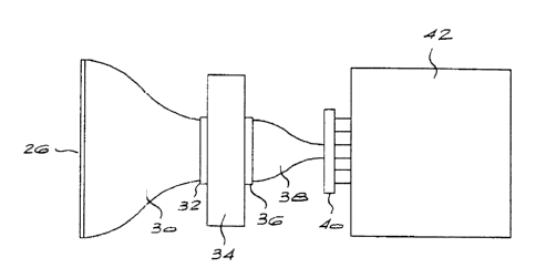
A first fibre-optic taper 30, i.e. a tapering bundle of optical fibres, is
connected to each of the scintillator screens 26. Fibre-optic tapers are
known to achieve good optical collection of light while maintaining good
spatial resolution and low distortion of the light.
The fibre-optic taper 30 directs the optical image via a suitable coupling
32 to the input window of an image intensifier 34. It will be appreciated
that the optical image presented to the input window of the image
intensifier is de-magnified as a result of the shape of the fibre-optic
taper. For each photon of light input to the image intensifier, the image
intensifier output, a nurnt~er of light photons, via a further coupling 36,
to a second fibre-optic taper 38. In other words, the image intensifier 34
intensifies the optical image presented to it by the fibre-optic taper 30.
The second fibre-optic taper 38 directs the intensified optical image,
again with de-magnification, to the front face of a CCD (charge-coupled
device) array detector 40 which is coupled to an electronic interface
module 42.
Figure 3 of the drawings diagrammatically illustrates a single detector
channel as described above.
The CCD array detectors are driven in so-called drift scanning mode.
Drift scanning moves the electronic image generated at the back of the
CCD array detector and continuously enhances, or updates, this image
~C102851
- 9 -
from information gathered from the optical image that "moves"
concurrently across the front face of the CCD detector as the carrier 16
moves.
Since the electronc image is moved along by a constant electronic clock
frequency, each point in the optical image must "move" in a straight line
at a constant velocity i.e. the velocity at which the carrier 16 moves.
Those skilled in the art: will recognise that the gain in image brightness
effected by the image intensifier must be carefully controlled in order to
avoid saturation of the CCD array detectors.
The modules 42 interface, through a databus 46 and line 48, with a
processing unit 50. In the unit S0, each line of the electronic image is
read out and enhanced by suitable digital electronic image processing
enhancement ap~~aratus. The unit 50 generates a video image on a
monitor 52 which is viewed by an operator 54.
It will be appreciated that an important consideration in any personal X-
ray scanning technique is to ensure that X-ray dose absorbed by the
body does not exceed physiologically harmful levels. This is particularly
so in the case of a detection system in which frequent scanning of the
body is necessary. For instance, where the system is used to detect the
presence of goods which may have been stolen from the workplace,
scanning may take place .on a daily basis, each time a person leaves the
workplace.
~102~51
- 10 -
It is anticipated that the system described above, in which X-radiation
is converted to an optical image which is intensified and converted to an
electronic image by a CCD array detector operated in drift scanning
mode, will be able to provide the required accuracy and resolution with
safe levels of initial X-ray intensity.
In practice, it is believed that in non-medical applications equivalent X-
radiation dose levels of 10 x 10-s sievert can be sustained with adequate
levels of signal resolution and accuracy.
In one application of the: invention, the human body scanning system
described above c:an be: used to scan the bodies of persons with a view
to detecting the presence thereon or therein of specific articles prone to
theft. For instance, the bodies of persons may be scanned to detect
concealed diamonds. In such a case, it is known that any diamond
particles in or on the person's body will absorb a greater proportion of
the incident X-radiation than the surrounding body tissue or bone. Thus
the diamonds will cause relative attenuation of the X-ray signal and this
will be visibly dis~~ernible in the final video image which is produced at
the monitor 52. rJot orrsy will the presence of a diamond be detected,
but also its location on the person's body. In such an application, the X-
ray tube voltage may be selected to be in the range 150KV to 160KV.
Also, although specific reference has been made to the person standing
during a vertical scanning procedure, it is equally possible to have the
person in a prone: position with horizontal scanning, or indeed to have
the person moving at a constant speed, for instance on a conveyor belt,
past a stationary scanning apparatus.
~10285'B
- 11 -
The detection of <iiamonds on the human body is but one application of
the invention. In ether applications, the human body could be scanned
for the presence of other foreign objects, for instance pieces of metal, or
for the presence of broken bones or other physiological irregularities, in
a medical examination procedure.
Generally speaking the X-ray tube voltage will be chosen to suit the
particular application. In medical applications such as those mentioned
in the preceding paragraph, the X-ray tube voltage could, for instance,
be as low as 80h:V. On the other hand, for location and analysis of
heavy metals such as iron, tungsten or platinum, voltages as high as
600KV could be employed.
The human body could also be scanned for the presence of drugs or
other prohibited substances or articles such as weapons at airport
security checkpoints.
The body itself i.s not necessarily a human body. For instance, the
method and apparatus described above could be used to examine articles
of luggage at airport security checkpoints.
In such cases, a version of the invention in which the body moves
through the scarring station would normally be preferred to the
illustrated system in which the scanning apparatus moves relative to the
body.
~102~~~
- 12 -
In each applicatia~n of the invention, the contrast presented on the visual
image may be set at an optimum level for accurate detection of a
particular article or substance by appropriate initial setting of the
operating voltage and current of the X-ray tube.
It should be noted that the video or electronic images produced by the
apparatus described above can, if desired, be stored in memory by the
processing unit 50.