Note: Descriptions are shown in the official language in which they were submitted.
WO93t18821 ~ ~ ~7 3 ~ PCT/US93/02109
Multiple frequency impedance measurement for
physioloqical monitoring of the condition of
a patient s body tissue
CROSS-REFERENCED~RELATED APPLICATIONS
Attention is drawn to the commonly assigned co-pending
U.S. Patent Application Serial No. 07/566,636, for a "Field
Density Clamp for sensing Cardiac Depolarizations", filed
August l0, l990 in the name of Terrence R. Hudrlik, U.S.
Patent Application Serial No. 07/626,061, for "Electronic
Capture Detection for a Pacer", filed December 12, l990 and
U.S. Patent Application Serial No. 07/730,160, for a "Medical
Stimulator With Operational Amplifier Output circuit", filed
July 15, 1991 in the name of Terrence R. Hudrlik, all three of
which are incorporated herein by reference in their
entireties. The present application is a continuation-in-part
of all three of these cited applications.
BACKGROUND OF THE INVENTION
This invention relates to diagnostic and tissue
stimulation devices such as implantable pacemakers,
cardioverters and defibrillators, implantable monitoring
devices and implantable drug dispensers, and more particularly
2Q to rate-responsive implantable pacemakers that vary their
pacing rate as a function of the patient's metabolic demand
for oxygenated blood.
Early pacemakers provided a fixed rate stimulation pulse
generator that could be reset on demand by sensed atrial
and/or ventricular depolarizations. Modern pacemakers include
complex stimulation pulse generators, sense amplifiers and
leads which can be configured or programmed to operate in
single or dual chamber modes of operations, delivering pacing
stimuli to the atrium and/or ventricle at fixed rates or rates
that vary between an upper rate limit and a lower rate limit.
W093/t8821 PCT/US93/02109
? ,[~ 3 ~3 ~
More recen~ y, single and dual chamber pacemakers have
been developed that respond to physiologic sensors which, with
greater or lesser degrees of specificity, sense the body's
need to deliver more or less oxygenated blood to the
cardiovascular system. For example, rate responsive pacing
systems have been developed and marketed which rely upon the
patient's rate of respiration. Such pacemakers are described~
for example, in U.S. Patent No. 3,593,718 and 4,596,251 and
have been commercialized by Biotec and Telectronics. These
pacemakers use an impedance pneumograph for acquiring a
respiration signal. More recently, it has been proposed to
employ the variation and the amplitude of the peak-to-peak ECG
signals as a rate control signal on the premise that the
amplitude varies as a function of the patient's activity
and/or respiration as disclosed in U.S. Patent No . 4,757,815.
The impedance pneumograph measurement technique of the
prior art involves the injection of a pulse or pulse burst of
alternating current at subthreshold stimulation energy levels
across a pair of electrodes and measuring voltage or current
levels to derive an impedance measurement. The electrodes may
be located one on either side of the chest, as in U.S. Patent
No . 4,596,251, issued to Plicchi, et al., both located in the
heart as in U.S. Patent No. 4,919,136, issued to Alt, or one
electrode may be located in contact with the heart and one in
the chest. U.S. Patent 4,702,253 describes a system employing
bipolar pacing electrodes. A constant voltage pulse train is
injected into the tissue between one electrode and the
pacemaker can and a measurement of the current taken between
the other electrode in the heart and the pacemaker can. The
impedance varies with exhalation and inhalation.
U.S. Patent No. 4,805,621, issued to Heinze et al.
suggests an approach to minimizing the effects of long term
changes in overall tissue impedance on the accuracy of such
systems. In the Heinze et al. device, the drive signal, as
modulated by the tissue impedance is passed through a first
' ~ .
WO93/18821 ~~ PCT/US93/02109
. . .
low pass filter to strip off the drive signal frequency and
produce a signal indicative of impedance variation over time,
stated to correspond to respiratory activity. This impedance
signal is then passed through a high pass filter to strip off
the extremely low frequencies at which overall changes in
tissue impedance are stated to occur.
SUMMARY OF THE INVENTION
In the study of the nature of the tissue-electrode
interface and, in particular, in developing the theoretical
basis for the operation of the field density clamp as set
forth in U.S. Patent application Serial No. 07/566,636, the
inventor has explored the frequency dependant impedance
characteristics of body tissue. If the wide band impedance is
measured across two electrodes in contact with body tissue and
displayed in a Cole-Cole plot, the impedance plot or impedance
"signature" typically displays several peaks and valleys. The
frequencies associated with the peaks are referred to in the
literature as "turnover frequencies".
The inventor has determined that gross tissue insults,
such as significant reduction of blood supply to the tissue,
causes a dramatic shift in the impedance signature. It is
expected that this type of change will also occur as a result
of gradually developed ischemic conditions. It is also
expected that in some cases, where the tissue is not
permanently damaged, the original signature will return when
blood flow is restored.
The inventor has also noted that different tissues may
have impedance peaks at substantially different frequencies.
A measurement of impedance and frequency across two spaced
electrodes will reflect several impedance peaks depending on
the tissue types within the sensing field of the electrodes.
There will be corresponding impedance minimums between the
peaks. The impedance signatures indicate that the tissues
adjacent and between the electrodes can be modeled as a series
' '
WO93/18821 PCT/US93/02109
2 ~ 9 2 ~
of parallel R-C blocks. Different tissue types, with
different impedance peaks, would corresponding be modeled as
series of R-C blocks having different component values.
The present invention takes advantage of this
physiological phenomenon to provide an impedance sensing
system which may be optimlzed to sense impedance variations
associated with one or more desired tissue types. The present
invention is also believed to provide an increased signal to
noise ratio with regard to sensing of impedance modulations of
each tissue type. The present invention employs multiple,
spectrally selected excitation signals toaccomplish these results.
It is proposed in the present invention that electrodes
be used to apply a plurality of spectrally selected excitation
signals simultaneously or sequentially over a period of time.
The measured impedances at the selected frequencies may be
used either to sense the condition of the tissue to which the
excitation signals are applied or to sense other physiologic
parameters related to the impedance of the tissue. Two or
more excitation frequencies are typically used, one at an
impedance peak, one displaced from the impedance peak,
preferably at a frequency which defines an impedance minimum.
The excitation frequencies should be chosen such that the
event of interest causes a substantial change in the relative
values in the impedances at the excitation frequencies.
For example, multiple frequency excitation signals may be
used to detect cardiac tissue distress, such as ischemia or
the response of the tissue to drug treatment. Multiple
frequency excitation signals may be alternatively be used to
measure respiration characteristics or other parameters
related to the level of the patient's activity. In
conjunction with this aspect of the invention, the invention
may provide a heart pacemaker which varies the pacing rate in
dependence upon the impedance measurements.
The invention is preferably practiced using an
operational amplifier input/output circuit as described in the
WO93/18821 ~ PCT/US93/02109
above cited Application Serial No. 07/730,160. The amplifier
may be used both to provide the drive or excitation signal and
measure the impedance of the body tissue in question, allowing
the use of a tWQ electrode system. The amplifier may also be
used to pace the heart and to sense depolarizations of heart
tissue, in those embodiments taking the form of cardiac
pacemakers.
BRIEF DESCRIPTION OF T~E DRAWINGS
The above and still further objects, features and
advantages of the present invention will become apparent from
the following detailed description of a presently preferred
embodiment, taken in conjunction with the accompanying
drawings.
Figure l is an illustration of a circuit simulating the
various source impedances of several tissue types, within the
field of the electrodes used to apply excitation signals.
Figure 2 illustrates the impedance between the electrodes
used to apply excitation signals to body tissue as a function
of the frequency of the excitation signals.
Figure 3 is a graphical depiction of the effect of
ischemia on the impedance versus frequency curve illustrated
in Figure 2.
Figure 4a is a schematic diagram depicting the
interconnection between a pacer and the heart.
Figures 4b, 4c and 4d are drawings of possible electrode
configurations for use in conjunction with the present
nvention.
Figure 5 is a block diagram of a pacemaker employing the
present invention.
Figure 6 is a block diagram of an alternative embodiment
of the present invention.
W O 93/18821 PC~r/US93/02109
~ ~2~ 6
DETAILED DESCRIPTION OF THE PREFERRED EMBODIMENT
In the following description, reference is made to an
illustrative embodiment for carrying out the invention. It is
understood that other embodiments may be utilized without
departing from the scope of the invention.
In accordance with the present invention, a technique is
employed to provide time varying signals that contain
information regarding the short or long term modulation of the
inter-electrode tissue impedance properties by physical
movement, such as respiration or patient activity, and/or the
changes related to the modulation of the tissue impedance due
to myocardial depolarizations, ischemia, allograft rejection,
drug therapy or other causes. It should also be noted that
the characteristic impedance of the lead body may vary as a
result of fracture of the lead conductors or insulation
degradation. The impedance measurement system disclosed
herein may also be used to identify such conditions.
As stated above, different tissue types have different
frequency sensitive impedance characteristics that may be
modeled by a series of parallel resistance and capacitive
elements denoted Zl, Z2 and Z3 in the illustration of Figure
1. Each of these impedance elements has a different time
constant, and the local impedance peak of each one of the
elements is referred to herein as the "turnover frequency" in
the literature.
It has also been previously noted by the inventor that
the frequency position of these turnover frequency peaks are
very sensitive to tissue condition, ischemia being
specifically studied. The tracking of these impedance peaks
over time provides information regarding changes in tissue
condition. Similarly, the tracking of these peaks on an
hourly basis in a patient on a drug regimen for treating
cardiac disease may provide an indication of the effectiveness
of the drug's therapy.
.:.~ : -
.. . . ~ '. ~,!
';
WO93/18821 h ~ d ~ 3 ~ PCT/US93/02109
Use of two spectrally selected excitation signals to
"focus" in on and specifically tune in at two frequencies
would provide two views of the same tissue from slightly
different aspects. In addition, when these frequencies are
set at local minima and maxima, the change in impedance due to
the occurrence of the event of interest will be significantly
different for the two frequencies. ~he relative difference
between the impedance changes at the selected frequencies can
be used to provide a self-normalizing ratio, based on the
individual in whom the device is implanted, which can be used
to identify the occurrence of the event.
The selected excitation frequencies are preferably
separated by at least a factor of lO. This separation factor
readily allows tuned filter separation of the applied signals
as modulated by the impedance of the tissue. As digital
filtration techniques improve with time, it is expected that
the separation factor may be substantially reduced. The
spectrally selected excitation - signals may be applied
sequentially in time multiplexed fashion or may be applied
simultaneously between electrodes coupled to the tissue to be
measured.
In those embodiments in which the invention takes the
form of a cardiac pacemaker, the excitation signals may be
applied between the probe and can electrodes or between the
probe electrode and a second electrode on or in the heart. In
embodiments in which the impedance measurement system is used
to detect basal conductance changes or to analyze myocardial
depolarizations, the excitation signals are preferably applied
during defined time intervals closely following delivery of
pacing pulses or detection of the occurrence of
depolarizations. In other embodiments, including embodiments
directed toward assessing the impact of drug therapies, other
measurement times may be more optimal.
Figure 2 is a Cole-Cole plot of real versus imaginary
impedance, taken across a band of frequencies. In accordance
WO93/18821 ~ pcT/uss3/o2lns
with the present invention, it is contemplated that the
spectrally selected excitation signal may include a first
drive frequency signal (Fl) tuned to a turnover frequency (a
local maximum) and a second drive frequency signal (F2) tuned
to a frequency corresponding to a local minimum of the
impedance plot set forth in Figure 2. In this fashion, two
views of the same tissue from slightly different aspects may
be obtained. Similarly, if two tissue types are to be
monitored, three or more frequencies may be employed, with
each tissue type having an associated pair of frequencies
particularly adapted to reflect changes in that tissue's
condition.
The increased signal to noise (S/N) ratio provided by the
two views provides isolation of the electrokinetic
lS disturbance, noise caused dy electrode and/or tissue movement
at their interface. In embodiments directed toward respiration
monitoring, the electrodes are mounted such that a significant
portion of lung tissue is between the electrodes. The result
is impedance measurement that modulate with a high degree of
correlation to respiration or activity. In embodiments
directed toward detection o cardiac ischemia, at least one of
the electrodes is located adjacent heart tissue.
An increased S/N ratio compared to single frequency
systems is realized due to the fact that long term changes in
overall tissue/electrode impedance generally result in moving
the impedance plot illustrated in Figure 2 upward or downward,
so the relative impedance difference between the two chosen
frequencies is not greatly affected. By using the relative
amplitudes of the measured impedance at the selected turnover
point as compared to the measured impedance at the selected
second frequency, the effects of changes in overall
tissue/electrode impedance are minimized. Thus, the present
invention provides a new and unobvious method of dealing with
this problem in a fashion unrelated to that suggested in the
above-cited Heinze et al. Patent No.4,805,621.
WO93/18821 ~ PCT/US93/02109
Figure 3 illustrates a simulated plot of frequency versus
impedance taken across normal and ischemic intestinal tissue,
using sinusoidal excitation siqnals. As illustrated, normal
tissue displays a local impedance peak at frequency F-l and a
S local impedance minimum at a frequency F-2. Ischemic tissue,
on the other hand, displays a substantially different
frequency plot. In general, changes in tissue condition, for
example due to ischemia or drug therapy may be reflected by a
change in frequency dependent impedance characteristics. For
example, the frequencies at which local, minima and maxima of
impedance occur may be substantially shifted or rearranged, as
illustrated in Figure 3. Therefore, by monitoring the
impedance characteristics across a plurality of frequencies,
such changes can be discerned.
The frequency dependant impedance characteristics as
illustrated in Figures 2 and 3 are believed to occur generally
in body tissues, although the frequencies at which local
maximums and minimums in the impedance plot will vary. Local
impedance maximums and minimums may be determined empirically
for the body tissue coupled to the electrodes. Given that
such measurements may be readily accomplished with available
apparatus, it is believed within the capabilities of one of
skill in the art to measure and derive impedance plots as
illustrated in Figures 2 and 3 for any particular electrode -
tissue system.
In the context of Figure 3, ischemia could be detected by
placing electrodes on or in the tissue to be monitored or
placing the tissue between the electrodes, and generating
drive or excitation signals at frequencies at F-l and F-2. By
monitoring the relative impedance levels at frequencies F-l
and F-2, changes in the frequency dependent impedance
characteristics of the tissue can readily be discerned. By
selecting excitation signals associated with a local impedance
maximum and a local impedance minimum, a shift in the
frequencies of the minimum and the maximum may be readily
.
. . . :
WO93/18X21 PCT/US93/~2109
~ 3~ 0
detected due to their substantial effect on the relative
impedance values at the two chosen frequencies.
For purposes of monitoring tissue impedance to ascertain
changes in tissue condition, the measured impedance amplitudes
can be averaged over extended periods of time, for example in
the range of days to weeks, so that short term modulation of
impedance characteristics of the tissue being monitored due to
physical movement, respiration, peristaltic motion, etc. can
be disregarded. Alternatively, short term modulation of the
relationship between the impedances measured at the two
frequencies due to the normal functioning of the tissue (e.g.
modulation due to respiration or heartbeats) may be measured
and recorded. Changes in the modulation amplitude and rate
associated with such tissue-related activities may be measured
and similarly be used to detect short or long term changes in
tissue condition or overall metabolic functioning.
For example, short term changes in the measured
modulation of the relationship of the impedances at the two
frequencies due to heartbeats or respiration may be used to
control the pacing rate of a cardiac pacemaker.
Alternatively, longer term changes in the average modulation
characteristics associated with heart contractions may be
indicative of cardiac ischemia or other factor related to the
condition of the heart tissue.
Figure 4a is a drawing depicting the interconnection
between a cardiac pacer and the heart. For purposes of
illustration, a composite unipolar/bipolar ventricular
inhibited pacer is shown with a lead bearing two electrodes
situated in the ventricle. Typically, the pacemaker 14 is
implanted beneath the skin, outside the rib-cage. A pacing
lead 12 is passed transvenously into the right ventricle of
the heart 10. The pacing lead 12 is used for supplying pacing
pulses to the heart and for conducting electrical signals
resulting from the depolarization of the heart to the
pacemaker 14. Traditionally, there are two basic electrical
WO93/18821 2 ~ a ~ PCT/US93/02109
1 1
configurations for pacing leads. A unipolar configuration
would include tip electrode 22 and a can or case electrode 24.
In a bipolar configuration, ring electrode 21 is used with tip
electrode 22. Electrode 22, in direct contact with cardiac
tissue is referred to herein as the "probe" electrode.
In unipolar configurations the implanted pacer is
implanted with the can electrode surface 24 disposed toward
the ribs 18 and generally toward the heart 10. This electrode
configuration places at least the tip electrode 22 within the
heart, and the case electrode 24 proximate the outside of the
heart, with the syncytium of the heart and a significant
amount of lung tissue located between the electrode poles.
Typically, the distance between the distal tip electrode 22
and the pacer can electrode 24 is between 10 and 30 cm.
lS In bipolar configurations, the case electrode 24 is not
used and the tip and proximal ring electrodes 22 and 21 are
connected to the pacemaker pulse generator output circuit and
sense amplifiers. Typically, the tip and ring electrodes 22
and 21 are spaced apart between 0.5 and 3.0 cm. In dual
chamber pacemakers, unipolar and/or bipolar electrodes are
similarly situated in or on the atrium or coronary sinus.
More recent pacemaker models often have the flexibility
to employ combinations of unipolar and bipolar electrode
configurations, under control of external programming
commands. For example, unipolar pacing might be selected in
conjunction with bipolar sensing. The present invention, if
embodied as a pacemaker may employ any of the various unipolar
and bipolar electrode configurations and combinations thereof.
Similarly, any two of the electrodes may be employed to
deliver the excitation signals and to measure the tissue
impedance.
Figures 4b, 4c and 4d illustrate alternative electrode
configurations for use in conjunction with the present
invention. The location of the electrodes will of course
depend on the tissue being monitored, but in the context of
WO93/18821 PCT/US93/02109
~ 12
measurement of heart tissue impedance, it is believed to
locate both electrodes so that they directly contact heart
tissue. The illustrated electrode configurations are intended
for use in applying the excitation signals to the heart tissue
and for measuring the impedance of the heart tissue, but may
be valuable in measuring other tissue as well.
All three embodiments employ a pair of closely spaced
electrodes mounted to the distal end of the lead body (12a,
12b, 12c). Electrodes 21a and 22a (Fig. 4b) take the form of
a split ring. Electrodes 21b and 22b (Fig. 4c) are a pair of
closely spaced rectangular electrodes. Electrodes 21c and 22c
(Fig.4d) include a ring electrode surrounding a small. central
electrode. The electrodes of Figures 4b, 4c and 4d may also
be used for R-wave sensing and cardiac pacing functions,
either paired with one another or paired with other electrodes
located on the lead body or on the pacer housing.
If the invention is embodied in the form of device which
monitors other tissue-types, other electrode configurations
may be required, such electrodes mounted on separate leads,
individually mounted to or inserted in the tissue to be
monitored. For example, existing intramuscular electrodes,
nerve stimulation electrodes and so forth or modified versions
thereof may be employed.
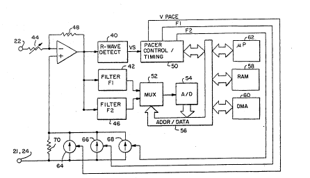
Figure 5 is a schematic diagram of an illustrative
embodiment of a cardiac pacemaker practicing the present
invention. An operational amplifier top amp) 38 is configured
to operate as a field density clamp amplifier as discussed
extensively in the above-cited Hudrlik applications.
Amplifier 38 has its positive input connected to the reference
or case electrode 21 or 24. The negative input to the
amplifier 38 is connected to the tip or probe electrode 22.
The tip electrode 22 is coupled through a variable resistor 44
which is used to set a virtual load impedance for the system.
A feedback path is provided for the amplifier 38 by a
resistance 48 which converts current through the virtual load
. .
.... .. ., .: . . . . .
'~
WO93/18821 PCT/US93/02109
resistor to a proportional voltage. Operational amplifier
38 maintains its inputs at the same voltage. Therefore, in
response to a disturbance in the electrical equilibrium
condition at the tissue-electrode interfaces, the amplifier 38
applies current to electrode 22 through virtual load resistor
44 in an amount sufficient to maintain its inputs at equal
potentials. As described in the above-cited Hudrlik
applications, the current applied to electrode 22 both
reflects the power of the passing depolarization wave-front
and reestablishes the equilibrium condition at the electrode
tissue interface. In operation, the op amp 38 provides a
voltage signal at its output which reflects the amount of
current applied through virtual load 44 in response to the
passage of a cardiac depolarization wave front.
At least in medical applications, it is conventional that
input amplifiers have very high input impedances. Ordinary
biomedical engineering design practices dictate that a sense
amplifier's input impedance must be at least an order of
magnitude higher than the source impedance. See for example,
"Bioelectric Amplifiers," in Introduction to Biomedical
Equipment Technolooy by Carr and Brown, John Wiley & Sons,
1981, pages 41-44 at 42. In accordance with the present
invention, however, the input impedance of operational
amplifier 38 may be varied by adjustment of virtual load
resistor 44 to be even less than the source impedance which,
in the case of heart tissue, typically is in the range of 500
- lOOO ohms, resulting in sharply enhanced peaks in the ECG
signal. Similarly, a low virtual load impedance is also
believed beneficial in the present case in which the amplifier
is used both to apply the excitation signals and to provide a
signal output indicative of tissue impedance.
While the illustrated embodiment employs an operational
amplifier 38 to monitor R-waves and to measure the tissue's
impedance, other embodiments of the invention may employ other
gain cells. While the inventor has employed the particular
. ,.
' ' , :'~
W093/t8~21 PCT/US93/02109
f.l I ~ id ~ 4
approach illustrated, it is believed to be within the scope of
the invention to employ other circuitry for measurement of the
tissue impedance. Similarly, it is anticipated that in some
embodiments of the invention, separate amplifiers may be
employed for R-wave sensing and impedance measurement.
The invention may be practiced with conventional pacing
leads and electrodes. However, the operational amplifier
circuit illustrated may advantageously be used with electrodes
of smaller than conventional surface area, for example as
illustrated in Figures 4b 4c and 4d. The electrode surface
areas of the electrode or electrodes in or on the heart are
preferably in the range of l.0 mm2 squared to lO.0 mm2, for
human use. The optimum electrode size will also vary as a
function of electrode material, with optimum electrode size
generally increasing as the conductivity of the electrode
material decreases. For example, the active surface of a
vitreous carbon electrode would optimally be somewhat larger
than a corresponding platinum electrode. The specific
electrode sizes to be employed will vary with application, and
should be empirically determined in conjunction with the
specific application of the invention. As described in
the above-cited application Serial No. 07/730,160 by Hudrlik,
the operational amplifier 38 may also be used to generate
stimulation pulses. In the present case, this is accomplished
by means of a controlled signal generator 64 which generates
a defined voltage across resistor 70. This voltage, applied
to the positive input of operational amplifier 38 results in
amplifier 38 delivering current through virtual load resistor
44 in an amount sufficient to maintain the positive and
negative inputs of operational amplifier 38 at equal
potentials. The current delivered through virtual load
resistor 44 serves as a stimulation pulse corresponding to the
signal applied to the positive input of amplifier 38. As
discussed in the above-cited Hudrlik application, this allows
a amplifier 38 to function both as a sense amplifier and an
,~ . .
-, ~ ,
~. ' .
WO93/18821 ~ PCT/US93/02109
output amplifier, with the amplifier 38 rapidly restoring the
electrode/tissue interface to its original equilibrium state
following delivery of the stimulation pulse. Signal generator
64 is preferably configured to deliver rectangular pulses of
adjustable duration, however, other wave-forms may be employed
such as triangular, sinusoidal, or trapezoidal, if desired.
one advantage of the use of amplifier 38 to generate the
stimulation signal is that it a~lows generation of stimulation
pulses of arbitrary wave-form, constrained only by the output
capabilities of the amplifier 38.
Generation of the excitation signals is accomplished in
a similar fashion. Signal generators 66 and 68 are adjuste~
to provide sinusoidal excitation signals at frequencies F-l
and P-2, respectively. Preferably, the excitation signals
take the form of sinusoidal signals applied across resistor
70, and to the positive input of amplifier 38. Amplifier 38
will correspondingly provide- sinusoidal signals to probe
electrode 22 via virtual load resistor 44 in order to maintain
its inputs at equal potentials. Preferably the amplitude of
the excitation signals applied to the tissue by amplifier 38
is substantially below the threshold for electrical
stimulation of the associated tissue. Excitation signals in
the range of less than l mv to about lO0 mv are believed
appropriate.
Signal sources 66 and 68 may be activated simultaneously
or sequentially. The impedance of the tissue as measured
between electrodes 22 and 21/24 will be reflected in the
current applied through virtual load resistor 44, thus
modulating the amplitude of the output signal from amplifier
38. Thus, the amplitude of the signal output of operational
amplifier 38 during application of the drive signals provides
a measurement of the impedance of the tissue between the
electrodes. It should be understood that in this embodiment,
while multiple independent current sources 64,66,68 are
illustrated, a single voltage to current converter driven by
.. .. ~ .
,,
' ~:; ~ : . :
WO93/18821 PCT/US93/02109
~ 6 ~
as summed signals of the selected excitation frequencies would
also be workable.
While the inventor has employed the operational amplifier
circuit as illustrated to generate pacing and excitation
signals, it is anticipated that other embodiments of the
invention may employ other voltage or current sources to
perform these functions. It is also anticipated that other
embodiments of the invention may employ separate current or
voltage sources to pace and to generate excitation signals.
Similarly, it is anticipated that other embodiments of the
invention may employ separate circuits for delivering pacing
pulses and for sensing R-waves, as well as separate circuits
for delivering excitation signals and for measuring impedance.
The output of operational amplifier 38 is provided to an
R-wave detection block 40, a filter 42 tuned to frequency F-l
and a second filter 46 tuned to frequency F-2. The output of
R-wave detection circuit 40 is coupled to pacer control/timing
circuitry 50, which performs all timing and control functions
necessary for both cardiac pacing and for activation and
control of the various signal generators 64, 66 and 68. The
outputs of filters 42 and 46 are provided to a multiplexor 52,
and thereafter to an analog to digital converter 54 so that
the resulting signals may be digitized for storage and
analysis. Overall control of the operation of the pacemaker
is provided by microprocessor 62, under control of stored
programming in random access memory 58. Entry of programming
information into random access memory 58 and readout of memory
stored in random access memory 58 is accomplished by direct
memory addressing 60, which allows data storage even while
microprocessor 62 is in a quiescent state.
Operation of the pacemaker is best understood by
beginning with the sensing of a depolarization of the
ventricle in which electrode 22 is located. The resulting
disturbance in the electrode-tissue equilibrium condition at
electrode 22 results in delivery of current by amplifier 38
;. ., ~ ~
'~, ' ,
, .:
W O 93/18821 ~ ~ 13 r~ PC~r/US93/02109
!~: 1 7
through virtual load resistor 44 in order to maintain the
inputs of the amplifier at equal potentials. The output of
amplifier 38 is coupled to R-wave detector 40, which compares
its output amplitude to a predetermined sensing threshold. R-
wave detection circuitry 40 may correspond to any known R-wave
detection circuitry, and is a conventional portion of most
cardiac pacemakers. The threshold to which the output of
amplifier 38 is compared may be fixed or may vary depending
upon the detected amplitude of previous R-waves. In any case,
a digital signal (VS) is provided by R-wave detector 40 to
pacer control/timing logic 50.
Pacer control/timing logic 50 includes the basic timers
which control operation of the pacemaker. In particular, it
includes at least one programmable timer into which
microprocessor 62 may load predetermined time intervals and
also includes the decoding logic associated with the timer for
decoding the expiration of various predetermined time
intervals following resetting of the timer. Alternatively,
pacer control/timing logic 50 may include separate timers,
each individually controllable, for defining the various time
intervals necessary for implementation of cardiac pacing
functions. At the very least, pacer control/timing circuitry
50 should be capable of providing an escape interval,
indicative of the interval between adjacent pacing pulses and
the interval separating a sensed depolarization from the
subsequent pacing pulse, a blanking period associated with the
delivery of a stimulation pulse, during which the output of R-
wave detector 40 is not considered, and one or more time
intervals controlling activation of signal sources F-l and F-2
to delivery excitation signals via amplifier 38.
In response to the detection of a depolarization by R-
wave detector 40, or delivery of a pacing pulse, pacer
control/timing circuitry 50 generates an interrupt on an
address/data/bus 56, which awakens microprocessor 62, which in
turn calculates appropriate values for the next subsequent
:
. .
. ~ , . ~ .
. '
.. .
W093/18821 PCT/US93/02109
3 l d ~ ~ 18
pacing interval, and loads it into pacer control/timing
circuitry via address/data bus 56.
To obtain impedance information, following detection of
a depolarization or the delivery of a pacing pulse, pacer
control/timing circuitry also generates signals on lines 74
and 76 for activation of signal sources 66 and 68. Signal
sources 66 and 68 may be activated sequentially or
simultaneously and preferably generate signals at frequencies
F-l and F-2, as discussed above. During application of the
excitation signals, the output of amplifier 38 is applied to
the inputs of filters 42 and 46. Filters 42 and 46 are band
pass filters having center frequencies tuned to F-l and F-2
respectively. Their outputs are applied to multiplexor 52,
under control of microprocessor 62, and are digitized by
analog/digital converter 54. The digitized signals may be
stored in random access memory 58 under control of direct
memory addressing circuitry 60.
If the device is intended to measure the impedance of
heart tissue and/or blood during depolarization or
repolarization, measurement will be initiated immediately or
soon after delivery of a pacing pulse or sensing of a
ventricular depolarization. Similarly, if respiration is to
be measured, impedance measurements may be taken at this time.
In the event that a more complete record of the impedance
changes associated with a depolarization is desired, impedance
measurements may be taken continuously, except for a time
interval associated with the delivery of a pacing pulse. The
measured impedance values may be stored in a looping memory of
- the type in which the most recent data is written over the
oldest data, preferably with the capacity to store at least
200 ms. of measurements. Upon detection of the occurrence of
a spontaneous depolarization, either by analysis of the
impedance measurements or by means of a conventional R-wave
detector, a time delay of lO0 ms or greater may be specified,
with the memory frozen after this time interval, analogous to
,,'-' ~, ''.: '
WO93/18821 2 ~ PCT/US93/02109
;~ 19
the system disclosed in U.S. Patent No. 4,223,678, issued to
Langer et al. and incorporated by reference in its entirety.
The data available for analysis would include impedance
measurements before, during and after the depolarization.
If the device is intended to measure the condition of the
tissue beginning prior to depolarization, the expected time of
the next depolarization may be calculated and the signal
sources 66 and 68 may be activated just prior to the expected
depolarization to yield information on passive tissue
conductance properties. The device may alternately activate
the signal sources 66,68 during the period between
depolarizations,to provide baseline data which could be
compared to the impedance changes associated with
depolarizations and used to quantify or identify ischemia.
As illustrated, the outputs of filters 42 and 46 are
applied directly to multiplexor 52. However, in some
embodiments it may be desirable to pass the outputs of filters
42 and 46 through low-pass filters to strip off the carrier
frequencies F-l and F-2, performing an envelope demodulation
of the band pass filters' outputs for presentation to
multiplexor 52. In either case, the filter outputs are stored
in random access memory 58 and analyzed under control of
microprocessor 62 to measure the relative impedance at the two
selected frequencies.
As discussed above, the measured impedance values may be
employed to detect changes in tissue condition, such as those
induced by ischemia and by drug therapies, allograft rejection
and lead fractures or insulation degradation. For example, in
the context of cardiac pacemaker, microprocessor 62 may
average the stored impedance valves at the two frequencies
over a period of hours, days, weeks, or even months, compare
the measured impedances at the two frequencies, and in
response to detection of a predefined change in the
relationship of the relative average impedance values over
these extended time periods, may provide an increase in
'
.
: .
WO93/18821 PCT/US93/02109
2 ~
minimum or base pacing rate in an attempt to counteract the
detected ischemia.
Alternatively, in the event that the reflected change in
relative measured impedances of the two frequencies reflects
S a variation in tissue condition as a function of an applied
drug therapy, the microprocessor 62 may set an internal flag
for external telemetry to notify the physician at a later
date. Additionally, although not illustrated herein, it is
contemplated that the pacemaker may provide an informational
signal to an associated implanted drug dispenser, allowing for
modulation of the drug therapy in response to detected changes
in tissue impedance.
In the event that the impedance measurement system is
employed to measure respiration for control of pacing rate, it
lS is envisioned that ele~trode 22, located within the right
ventricle and the can electrode 24 of the pacemaker will
preferentially be used, to allow for a substantial volume of
lung tissue to be located within the sensing field of the
electrodes. In such case, microprocessor 62 will similarly
control processing of the outputs of filters 42 and 46,
however, the time scale employed will be significantly
shorter. For example, the measured differential between the
output of filter 42, centered at the selected local impedance
maximum and filter 46, selected for a local impedance minimum
will both be processed to determine their amplitudes over much
shorter time periods. For example, the amplitude of the
output of filter F-2 may be subtracted from the amplitude of
the output of filter F-l to derive a measurement of
instantaneous, frequency dependant impedance characteristics.
The modulation of the instantaneous impedance characteristics
may be measured, and the amplitude and rate of modulation of
the frequency dependant impedance characteristics may used to
calculate respiration rate and/or minute volume, in a manner
analogous to that described in the above-cited Plicchi,
Nappholtz and Alt patents.
. ,
- - ,
.
.
.
W093t1~21 PCT/US93/02tO9
For example, the signal indicative of the instantaneous
impedance difference at the two excitation frequencies may be
applied to a delta modulator, and the measurement to
measurement change in measured instantaneous impedance
difference may be summed over a short period of time, for
example 30 seconds, to derive a measurement of minute
ventilation. As noted above, this approach should provide a
self referencing measurement system for the particular
individual in whom the device is implanted. Alternatively,
any of the signal processing techniques employed by the above-
cited Alt, Plicchi and Nappholtz patents may be employed in
order to derive a measurement of respiration and/or minute
volume from individual measurements of instantaneous impedance
difference at the two excitation frequencies.
Figure 6 illustrates an alternative embodiment of the
present invention, employing a somewhat different impedance
measurement system. System components which are identical to
those illustrated in Figure 5 are labelled with the same
numbers as used in Figure 5. Only the additional or
alternative structures are discussed in detail below.
In Figure 6, a pink noise generator 80 is substituted for
signal sources 66 and 68 in Figure 5. Similarly, tunable
band pass filters 82 and 84 are substituted for fixed band
pass filters 42 and 46 in Figure 5. Control of the band pass
characteristics of filters 82 and 84 is provided by
microprocessor 162 via address/data buss 56. In a system
such as illustrated, it is contemplated that the selection of
the frequencies of tunable band pass filters 82 and 84 will be
accomplished in response to an initial scan through the
frequency range provided by pink noise generator 80. In
response to such a scan, as discussed above in conjunction
with Figure 3, frequencies indicative of local impedance
maxima and minima may be identified, and used to specifically
select the center frequencies of filters 82 and 84.
Thereafter operation of the device may correspond to that of
'. . :' ., ' : ' .
WO93/18821 PCT/US93/02109
~ j3 ~ ;~ 22 ``
the device illustrated in Figure 4, with the exception that
rather than sequentially or simultaneously activating two
separate signal sources to excite the tissue to be measured,
pink noise generator 80 would instead be activated.
In a first embodiment, the outputs of tunable filters 82
and 84 may be stored in random access memory 58 and analyzed
by microprocessor 62 to determine whether a shift in local
minima and maxima has occurred, as discussed above. The
occurrence of a shift in frequency dependant impedance
characteristics may trigger a change the base pacing, or other
corrective action may be taken as described in conjunction
with Figure 5. In this case, the corrective (increased pacing
rate or alteration of drug regimen) would be directed toward
restoring the original frequency dependent impedance
characteristics of the tissue, as indicative of a return to
normal tissue condition. Upon detection of a return to normal
condition, variation in the pacing or drug therapy would be
discontinued pending detection of a subsequent change in
frequency dependant tissue impedance.
In a second embodiment, it is envisioned that in response
to detection of a change in the frequency dependant impedance
of the tissue, a new scan through the frequencies associated
with pink noise generator 80 may be undertaken under control
of microprocessor 62, by varying the center frequencies of
tunable band pass filters 82 and 84. In such case, a
measurement of the frequency shift of local impedance minima
and maxima may provide useful additional diagnostic
information.
The invention as disclosed in Figures 5 and 6 will be
understood to be implemented in a multi-programmable, multi-
mode, processor based system of the type described in
Medtronic U.S. Patent No. 4,754,753 for example, and the
various timing intervals and signal processing to be described
may be effected by the processor under software algorithm
control. Although these exemplary embodiments of the present
- . ..................... ;: , .
.. . . .
WO93/18821 2 ~ PCT/US93/02109
23
invention have been shown and described, it will be apparent
to those having ordinary skill in the art that a number of
changes, modifications, or alterations to the invention as
described herein may be made, none of which depart from the
spirit of the present invention. For example, substitution of
other circuit architectures, such as dedicated function
digital or analog circuitry for the microprocessor based
system illustrated is believed within the capabilities of
those of skill in the art. It is also envisioned that the
diagnostic value of the impedance related information
generated by the present invention will have broad
applicability. Therefore use of the impedance measurements to
affect other types of therapies than those disclosed is
believed likely as the benefit of the invention becomes widely
understood. All such changes, modifications and alterations
should therefore be seen within the scope of the present
invention.
.. . . . .
.. .: . ~
:.:. . . .
,.. , ~ -
.. ~. .