Note: Descriptions are shown in the official language in which they were submitted.
~ 21 ~34 ~4
.~
The invention relates to the area of :
blood filtration units or devices.
More precisely, within this area, the
invention relates to devices which can be
implanted in a patient's body and comprises a
blood filter which can be positioned in a vessel,
at the end of a catheter or a filament extending
along the access route followed by the filter to
the area where it is to be implanted in the
vessel.
.
This type of device is frequently known
as "a temporary filtern since it is more
15 particulary used within the context of vascular -.
treatments which only require the filter to be -~
present for a period restricted a priori in time,
frequently of the order of a few days to a few
monthY, for example within the context of the . ~- ;;-~
treatment of a thrombus, it being noted, however,
that the retention of the device over a longer
term can be envisaged, for example within the
context of an anti-coagulant treatment.
:,., ' . ,:
Whatever the case, one of the advantages
of thi~ type of filtration unit i8 that, as the
filter normally does not have any means for . ~
definitively attaching it to the wall of the . ~ :
`; 21~34~
vessel where it is to be installed, the -
practitioner can remove it when desired, using its
transporter catheter for this purpose. ~ :
., ' ~. '
Examples of the production and use of `
filtration unit of this type are described in
particular in patents FR-A-2 580 504 and FR-A-2
643 250.
lo The devices described in these two
documents are particularly advantageous in that
they propose using as a means of connecting the
filter "to the exterior~ a catheter which is open
over its entire length and at its two opposite
ends if necessary, utilising its inner duct for
injecting a treatment product towards the filter `~ -`
implantation area, the filter being designed to be
secured to the catheter such that it does not
block Qaid inner duct thereof.
However, a_ far aQ the applicants are
aware, account hai not hitherto been taken of a
:
problem which can prove to be important in
particular with thi~ type of device, namely the
25 migration of the filter. -~
- .: .
If, fo~ a circulation of a liquid, or at least for holding the
filter, a catheter is used and this catheter is
' ~ , ,' '''' .':'
--` 21~3~
made of a flexible material wh~ch helps it to
slide along the access route to the vessel,
looping effects can appear over its length, whicX
risks moving the unit progressively towards the
heart, with the obvious inherent risks. If, on
the other hand, the catheter used is more rigid
and therefore normally no longer tends to roll up
in loops, in contrast, there is a risk of its
attenuating or even assisting the thrust which the
filter tends to exert thereon, the catheter then
tending to pull on the tissues, which is
unfavourable for the patient and can lead to
additional complications.
It is.in particular to overcome these
migration problems that the invention proposes a -
device which can be implanted in a patient's body
as far as a blood vessel in order to retain any ~ :
clots there, this device comprising:
_ a catheter (or any equivalent elongated blood filter holding
0san) having an axis and a lengtn in the direction of the axis, ~-
with a proximal end and an opposite distal end;
- a blood filter secured to the distal
end of the catheter;
the devicQ being characterised in that the
catheter used is stiffened in a variable manner
over its length so as to be stiffer at its distal
end than at its proximal end. :
~` 4 2 1 ~
Furthermore, in order, preferably, to
enable a liquid or a treatment product possibly to
be injected into the filter implantation area (or
even a possible blood tap), this variable rigidity
can be brought about by progressive longitudinal
stiffening of the catheter wall, externally of its
internal duct which is thus kept free for the
passage of said liquid, in one direction or the
other. --~
,, ~ ,,
In addition to simple and reliable
distribution of a treatment product, a catheter of ~-
this type with variable rigidity offers a further
advantage if it is effectively open.
-
Given that it represents an advantageous ~-~
solution to the problem of the possible migration
of the filter, it will allow an implantable means
for distributing said product to be positioned
subcutaneously.
:" ' ': '"~''~,,'.' .'..''` '
Thus, the risks of infection from the
surrounding environment during the implantation
period are to a large extent restricted, the
entire filtration unit being protected at least by
the cutaneous ~urface of the patient, the
treatment product being in~ected at the suitable
moment through the skin and, via said unit, to the
: .
~.'',';,.',',, .
- 2103~0~
area in which the filter is implanted in the
vessel.
Preferably, a capsule, which can in
particular comprise an inner chamber for
accommodating the treatment product and closed by
a self-closing wall, which can be perforated by
the needle of a syringe for in~ecting said product
through the patient's skin, can be used as the
10 implantable distribution means. ~
., .
To return briefly to the variable ~ :
stiffening of the catheter, it will be noted that ~ :
.- . . .~; . .
this stiffening can be achieved in particular in
two ways: by internal stiffening of the very wall
of this catheter without added auxiliary means -
(for example, by varying the thickness of this
wall or by using telescopic portions), a further
solution being to use auxiliary stiffening means ~ :
which can in particular be embedded in the
~; ~ :-,...
catheter wall in question.
Advantageously, and in order to obtain
the best effect with respect to the retention of ~ ; .
the filter, the rigidity of the catheter wall
preferably decreases progressively to a greater or ;
lesser extent form its distal end to its proximal :.
end.
:.
~ .
.
21 ~34~
For whatever purpose it may serve, it
will be noted that the "distal" end of the device ~ `
designates the end to which the blood filter is
secured and which is thus, with this filter,
implanted most deeply in the vessel to be :
filtered, the "proximal" end evidently being the ~ :
opposite end located closest the surface of the
skin, in the vicinity of the outlet of the access ~.
route leading to the vessel in question.
Further characteristics and advantages of : :
the invention will appear from the following
description given with reference to the attached
drawings, in which: -
Figure l is a schematic overall view of a
detachable blood filtration unit equipped with an .
implantable chamber;
Figure 2 is a median view in section of ..
the detail marked II in Figure l;
'' . ,~ '
Figure 3 shows an internal view of the
implantable chamber and the manner in which it can
25 be u~ed;
- ..
Figure 4 i-~ a partial view in
longitudinal section of the catheter with variable
~ q~
- 2103~
rigidity equipped with the filter according to a
first embodiment;
Figure 5 shows a schematic perspective
view of an alternative embodiment of the catheter
equipped with the filter; -
Figure 6 shows, in a view similar to .
Figure 5, a further alternative embodiment of the ..
catheter with its filter;
:-... ~: . .
Figure 7 shows the stiffening means used ~.
in the embodiment in Figure 6;
Figures 8, 9 and lO are three views in
section along the lines VIII-VIII, IX-IX and X-X .---;~
respectively in Figure 6;
Figure ll shows in longitudinal section a ~: .
fourth alternative embodiment of the catheter;
Figure 12 is an enlarged view of the
detail marked as XII in Figure ll;
.
Figures 13 and 14 ~how two further
possibilities of the variable stiffening of the ;~ :
catheter, here using a helical spring having "?",
', . ~
~ :
-` 210349'1 :
either a thickness which decreases (Figure 13) or
a widening pitch); and
.: . ' ,. .:
Figures 15, 16 and 17 show the principal
5 means used for positioning, or withdrawing, the - ~ -
filtration unit according to the invention.
Since one of the advantages of the use,
according to the invention, of a catheter with
lo variable rigidity in conjunction with a blood
filter, is thus to permit the use of a chamber
which can be implanted under the skin of a patient
who is, for example, to undergo thrombolysis, the
invention will only be described hereinafter
within the context of an application of this type
even if it is clear that, if necessary, the
proximal end of the catheter may not be connected -~
to a chamber of this type.
.
Figure 1 firstly shows, in its entirety,
a blood filtration unit 1 which can be positioned
percutaneously via the jugular vein.
The unit 1 essentially comprises a
qectile catheter 3 with variable rigidity which is
connected at its proximal end 3a to an implantable
chamber 5, the catheter further being securely
- 21 03~0~
connected at its distal end 3b to a blood filter
- 7. ~:
: ' . ,
Since, in accordance with the invention, -
5 the internal longitudinal duct 9 of the catheter 3 ;
has to be left free for the passage of the
treatment product, such as a medicinal liquid
substance used for the lysis of thrombi, it will
be seen from Figure 2 that the filter 7 is secured
lo to the distal end 3b in such a way that the
catheter is not blocked (this feature should not, :.
however, be necessarily considered as essential or - -:
limiting). ;:
To this end, in the example illustrated,
the filter comprises six feet or arms ll which can ~ -
expand radially and automatically, so as to .-
constitute a sort of umbrella, such that, in their
expanded state (illustrated, for example, in
Figure 1), the arms adopt a substantially conical
shape such that via their free ends, which in this :
case are curved, they can match the corresponding
wall of the ves-~el where the filter is to be
implanted, without becoming attached thereto
definitively (the feet not having any fa~tening
means at all in the example in question). ..
', '` '`''~' '',.'' '.'
lo 2~3~
Opposite their free ends, the feet of the ~ :
filter are connected together at an apex or head
14 through the centre of which there passes a -:
passage 15 having a cross-section comparable to
5 that of the duct 9. . . `~
:. ~ '.;~':''`'.'.',.
Since the filter 7 is normally adapted ;
such that it can be made of metal, for example of
steel or cobalt, it can be secured to the catheter
by the crimping and/or bonding of its head 14 such
that the aperture 15 is in the extension of the
conduit 9. ~ .
Figure 3 shows a possible embodiment of
15 the implantable chamber 5. --
In this Figure, it can be seen that the
chamber in question is in this case formed in the -~
manner of a sealed capsule containing an inner
20 chamber 17 which is closed at the top by a self- ~ ~
closing wall l9 (for example made of silicone .~.
plastics material) which can be perforated by the
needle 21 of any suitable systQm, such as syringe,
for in~ecting through the patient' 9 ~kin, ~hown
schematically at 23.
The base 25 of the capsule can in
particular be made of metal, it being possible for
:~. :
. .
~: . .. .
21~34~
11 .
the remainder, particularly its lateral wall 27, ~ -
to be made of electrically insulating material,
such as a bio-compatible plastics material.
In order to distribute the product ;
received from the syringe, the space l7
communicates here with the catheter 3 via a
conventional union 29.
If, in this construction, the unit 1 is
implanted without particular precautions with
respect to the rigidity of the catheter 3 being
taken, it thus risks coiling up in Ioops at the
location of the heart or, if it is too rigid,
exerting a thrusting effect on the chamber,
risking transforming the latter into a
subcutaneous "tunneller". ~- :
To avoid the above occurring, the
catheter wall is stiffened such that it is stiffer
at the distal end 3b than at its proximal end 3a
~where the catheter is thus more flexible), in
order to attenuate the thrust effect on the -::~
chamber 5. ~It will be appreciated that a further
25 solution might consist in stiffening the catheter :
by introducing a metal cable of variable
~tiffness, which, for example, might have a
' '
.::
-` 21034~
12 :
diameter which is larger at its distal end than at
its proximal end, into its duct 9).
Figures 4 to 13 show (within the context ~:
5 of stiffening the wall outside the inner duct) - . :
various ways of stiffening the catheter, namely in
this case either by structural stiffening of the
wall itself, without added auxiliary means ;~ .
(Figures 4 and 5), or by using added stiffening ~ ` .
lo means which are connected to this wall and can, in ~: :
particular, be embedded therein (solutions shown
in Figures 6 to 14).
In Figure 4 firstly, the solution used :
15 consists in producing the catheter 3 in a -
plurality of portions, in this case three 31, 33,
35 connected end-to-end coaxially, for example by -
bonding or welding, each tube portion being ~ `
produced from a different material having its own
degree of rigidity.
. ,~ ~, .
The difference in rigidity between the
portions 31, 33, 35 can be obtained as a result of
polyolefins (such as polyethylene) of high,
25 average and/or low density, or even seguenced :~
polymers (such as A-B) of which one seguence is
more rigid than the other and is variable (for
--`` 21~3~
13
example polyether block amide: PEBA), being
proportioned or cut to varying proportiQnS.
A further solution, illustrated in Figure :
S, consists in providing a catheter of which the
5 wall thickness decreases from the distal end 3d to ~ :
the proximal end 3a.
In the embodiment illustrated, this
variation in thickness has been produced by the
lo use of three tubes 37, 39, 41 of different lengths
and different cross-sections (both internal and .
external), such that a telescopic assembly is
obtained, the tube with the largest cross-section ~ ~
and the shortest length 41 being secured to the ~ :~-
filter 7 and accommodating tightly the second tube
39 with a smaller cross-section and a longer
length inside which is fitted the third tube 37 ;
with an even smaller cross-section and, a greater
length.
.; . ~ .
`
If necessary, the last tube 37 can extend ~ :
to the distal end of the shortest section 41, such .
that the cross-section of the inner duct 9 of the
catheter is constant over its entire length, it
25 being possible for the thicknes~ of each of the :::
tubes 37, 39, 41 to be identical. - ~
~ : .
21~34~ `
14
In Figure 6, the variable stiffening of -
the catheter 3 is brought about by means of added
auxiliary means, the catheter itself consisting of .
a single piece of flexible bio-compatible plastics
material having a wal.l thickness which is constant
from one end to the other. .. ..
Stiffening is brought about by the use of ~ : .
fine filaments or metal rods shown at 43 in Figure
10 7, the~e filaments having to be positioned in .. .
passages of suitable cross-section 45, provided in . ;
the wall 13 of the catheter surrounding its inner
duct 9, these passages, and thus the rods,
extending substantially from the distal end 3b in
the direction of the proximal end 3a.
In order to vary this rigidity, the use
of rods 43 of different lengths can be provided. .
In the version illustrated, there are
thus provided at the distal end 3b eight channels
45 each enclosing tightly a rod 43 over
approximately one third of the length of the -:
catheter (Figure 8), the -~econd, intermediate ~ :~
25 third only comprising four channels and four rods ::.
~Figure 9), and the final third extending to the
proximal end 3a only enclosing two channels 45
each with a rod 43 (Figure 10). - ~ :-
~'
2 1 0 3 ~
Although a priori it appears more simple
to provide for the channels to extend ~ ~:
substantially parallel to the general longitudinal ;~
axis 3c of the catheter and of its internal duct,
it is evidently also possible to imagine a helical
arrangement.
:~: - . :" :
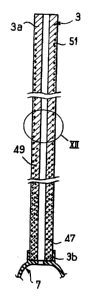
In the version illustrated in Figure 11, ~ :
a plurali~y of helical springs embedded inside the :-~ :
lo catheter wall, here consi~ting of single part of -~ ~ -
conventional bio-compatible plastics material
having a constant thickness, is used instead of
the metal rods.
In the version illustrated, there are - .
three portions of helical springs 47, 49, 51 of . -
different lengths disposed coaxially about one
another and thus embedded in the wall 13 such
that, for example, the outermost spring 51 extends .~
20 sub~tantially to the vicinity of the two ends of ~ ~-
the catheter, the intermediate spring 49 only
extending over two thirds of the length from the
distal end, the third, innermost spring 47 only
extending over the lower third, from this same
distal end.
For greater clarity, Figure 12 shows, in .-;
an enlarged view, a detail of the catheter showing -.
. ~
16 2 1 0 3 ~
the relative arrangement of the longest helical
spring 51 and the medium length helical spring 49. ;-
'"
As Figures 13 and 14 show respectively,
5 it would also be possible to use only a single ~ ; ;
helical spring, still embedded in the thickness of
the catheter wall but which may comprise a pitch
between turns and/or have a variable thickness.
~ .1."d
Thus, Figure 13 shows a helical spring 53
consisting of three metal portions welded end-to- -
end, namely a first portion 55 with a relatively
large thickness (a few tenths of a millimetre), - -~
. ~ .
then a second portion 57 with a reduced thickness,
itself connected to a third portion 59 with an
even smaller section. ~
With respect to Figure 14, the helical ~- -
spring 61 illustrated there has a pitch "p"
between turns which increases in one direction.
It will be appreciated that it is either
the thickest portion of the spring or the portion
having the closest pitch which is disposed at the
distal end of the catheter.
With reference to Figures 15, 16 and 17,
one possible method of implanting the filtration
. .. . , , . I
- 21~3~
17
unit 1 will now be described, this implantation
being performed under, at least local,
anaesthetic. .
Firstly, the.operator can commence by .`
making in the neck a percutaneous access route to
the ~ugular vein, the filter in this case being
implanted in the inferior vena cava (it will be
noted that access could also be provided by
denuding).
Once this has been performed, the
operator firstly introduces a metal guide wire
marked 63 in Figure 15 and having a curved end 65 .
via the access route provided (jugular vein, then
superior vena cava and finally interior vena
cava), the cable 63 being inserted until the end
65 is slightly downstream of the filter :-
implantation area. :
When the aperture in the access route :
through the skin has been slightly enlarged, the ~ .
operator then fits onto the proximal end of the
wire 63 (which then emerges from the ~ugular :
25 vein), an assembly consisting of a relatively :~
rigid mandrel 67 and an outer sheathing 69, made
of bio-compatible material, the respective
.
''' '~
~ . .
21~34~
18
proximal ends 67a, 69a of the mandrel and of the
outer sheathing then abutting one another.
- - ~ '.
The operator then gently lowers this
S assembly along the cable 63 until the radio-opaque
mark 71 provided on the mandrel reaches the area
provided for the implantation of the filter.
: .
The operator can then withdraw the wire
63, which is nevertheless flexible, and the
mandrel 67 surrounding the latter from the
sheathing 6g.
Once the outer sheathing 69 is thus in - ~ -
position, it is used as a guide for positioning
the filter, which is then conventionally pre-
arranged in the state in which its feet are
radially folded in a sort of packaging syringe 73
open at its two opposite ends so as to enable the
catheter 3, which was previously secured to the
filter, to be passed through from one side.
:::
Pre-arranged in this way, the body of the
syringe 73 is then screwed on at its threaded end
75 to a complementary thread provided on the
proximal end piece 69a of the sheathing 69, this
operation, evidently, being performed externally
of the patient's body.
19 21034~
The operator then lowers into the ~ ~
... ,.. ~ ~ .
sheathing the filter followed by its catheter
until said filter (still in its retracted state)
reaches the distal end 69b of the sheathing where
it then naturally expands, taking account of its
structure which is compact here, its feet
naturally unfolding owing to their flexibility
whilst bearing against the inner wall of the vena
cava at the desired location.
, .
It will be noted that, in view of the
relative rigidity of its wall, the catheter should
be able to slide inside the sheathing 69 without
necessarily requiring for this purpose the --
addition of an auxiliary stiffening device, such
as a very fine mandrel or detachable metal
filament which is frequently slid into highly ~-
flexible catheters to facilitate their ~ -
positioning. If a mandrel of this type is to be
provided, it is selected a priori such that it is
relatively short and the device is arranged such
that, at the end of its travel, it abuts the
proximal end of the catheter.
At all events, once the filter/catheter
assembly is in place, the operator can then remove
the sheathing 69 from the vein.
. . .' "~.
20 2~ a34~4
In now remains for the operator to
- connect correctly the proximal end of the catheter
to the injection means used.
: .
If these means consist of a chamber 5,
the operator firstly provides a subcutaneous
housing. Once this has been performed, the
catheter 3 i8 cut to a suitable length. The
operator fits the capsule 5 to the free cut end,
via the union 29, then conceals the assembly in
the housing, subsequently closing off the access
route in such a way that the capsule is trapped ~ :
under the skin after suturing, as illustrated in `
Figure 3.
By way of indication it will be noted
: : :: ', . . .
that the mandrel 67 and the sheathing 69 can have ~ -
a length of the order of 50 to 60 cm, the length
of the catheter may be 60 to 80 cm, the cable 63
preferably being slightly longer with a diameter
which may be of the order of 0.5 mm, the diameters ;
of the mandrel 67 and of the sheathing being,
respectively, of the order of 3 and 4 mm, that of
the catheter being slightly les~, for example of
the order of 2 mm, and its length after cutting
po~ibly being 40 to 50 cm. Especially, if the catheter 3 is not used
as a "support" for allowing a fluid circulation therethrough, any equivalent
blood filter ho~ding:mea~.coRld be-used,- suc~ as a flexible tube or rod, made
~o~.example-o~.`.silicone.
.