Note: Descriptions are shown in the official language in which they were submitted.
210~618
~LAR~ ~LIMINATION DEVICE
F~ld of Invention
This invention relates to methods and sterile devices
for use in video assisted minimally invasive surgery, for
example, endoscopy, laparoscopy or other optically
assisted viewing procedures. More specifically, the
invention relates to a sterile device which reduces the
lQ glare present when utilizing an endoscope in an endoscopic
procedure. The term "endoscopic" as used herein is meant
to refer to any surgical procedure using either natural
body openings and/or small artificial openings made by
puncture or incision; the term "endoscope" as used herein
1~ is meant to refer to the viewing devices for an endoscopic
procedure.
~Qkg~ound of th- Invention
Endoscopic surgery has been gaining wide acceptance
as an improved and cost effective technique for conducting
certain surgical procedures. In endoscopic surgery, a
trocar, which is a pointed piercing device, is inserted
into the body with a cannula placed around the trocar.
After the trocar pierces the abdominal wall, it is removed
and the cannula remains in the body. Through this
cannula, endoscopic procedures are possible. Often
multiple openings are produced in the body with a trocar
BO that an endoscopic instrument may be placed in one
cannula, appropriate viewing and illuminating means placed
in another cannula and so forth. As more is learned
about endoscopic surgical procedures and more instruments
developed, the type of procedures that may be performed
will increase. Presently, some procedures include gall
END-17
. .
.
,' ' ' '
2104618
bladder, diagnostic procedures, bowel resection, joint
repair, tissue repair, and various sterilization
procedures.
An endoscope includes a relatively long tubular
member which is placed through a cannula or natural body
opening. The tubular member carries fiber optics from an
appropriate light source to the end of the tube to provide
illumination of the surgical site. The tube also carries
~Q imaging means for viewing the surgical site. Many scopes
will have the outer annular portion at the end of the tube
used for illumination and the central portion of the tube
used for imaging. There are also scopes which have one
circular area of the tube used for illumination with
appropriate fiber optics and an adjacent circular area of
the tube used for imaging. Still other scopes may include
an open channel in the tube through which one or more
small instruments may also be introduced into the surgical
site.
In endoscopic procedures, glare often presents a
problem and reduces the information in the image being
provided to the surgeon during the procedure. Glare may
obscure not only the area of interest but also surrounding
areas. All of the internal organs reflect light and
produce glare due to their wet surfaces. Also,
instruments being utilized within the surgical environment
will cause glare because of their polished and/or smooth
eurfaces.
One type of reflection, termed "diffuse" is a
scattered reflection from the surface or sub-surface of
the illuminated object and contains desirable information.
A second type of reflection is what is known as mirrored
~ND-17
.
- ~
. ~ , . -
- . ~ - , . :
- ~ . : . .: ::
.
2104618 ;
reflection or "specular" reflection and does not change
the nature of the incident or illuminating light. This
reflection obscures the image.
S Another problem with endoscopes is the difficulty of
sterilizing such devices. As these devices are expensive
they are used over and over again. They are not
sterilized but at the present time and are only
disinfected between uses.
It is an object of the present invention to reduce
glare present in an endoscopic image. It is another
object of the present invention to improve or enhance the
image produced during an endoscopic procedure. It is a
further ob~ect of the present invention to produce a low
cost disposable device for use with an endoscope. It is
yet a further object of the present invention to provide
a device that will allow or provide that the endoscope
will be sterile during use.
8u~ary of th- Pr-~-nt rnventlon
The present invention provides a method for reducing
glare and enhancing visualization during endoscopic
procedures. In certain embodiments of the present
invention, the diagnostic capabilities of certain
endoscopic diagnostic procedures may also be enhanced.
In the method of the present invention, the illumination
present in an endoscopic procedure is polarized with a
first polarizing means or filter and through a second
polarizing means or filter the reflected illumination is
analyzed. By "polarize" or "polarizing means" it is meant
an optical device whose input is natural light and whose
output is some form of polarized light. Natural light is
END-17
-
.,..,
- . ., . . ~.
.. . . . .
.
21~46~8
electromagnetic radiation with wavelengths between 300 and
1200 nanometers and which contains two or more
polarization states. The terms light, illumination and
the such are used herein to include, not only visible
light, but the entire range of electromagnetic radiation
as described above. In the device of the present
invention, a first polarizer polarizes light in a first
direction. A second polarizer analyzes the reflected
illuminations. In certain embodiments of the present
invention, the first polarizer linearly polarizes light in
a first direction. The second polarizer linearly
polarizes light in a direction at 90 to the first
direction. Some devices of the present invention
comprises a hollow tube with one end of the tube open and
1~ the tube sized to fit over the illuminating end of an
endoscope. The opposite end of the tube is at least
partially closed by polarizers. In a preferred embodiment
of the sterile medical device of the present invention,
the end of the tube is totally closed with polarizers.
~Q The outer annular portion of the closed end of the tube is
a polarizer which linearly polarizes light in a first
direction while the central portion of the closed end is
a polarizer which linearly polarizes light in a direction
at 90to the first direction. The second polarizer which
is polarizing the reflected light is often termed an
analyzer.
Briof Desori~tion Or the Drawinas
Figure 1 i8 a cross-sectional view of a ~terile
device of the present invention positioned on an
endoscope;
END-17
. . : . .
: . . , , . ~ . - . .
. .
,, . . . ~
. . , . ~ - : .
.
. . .
, ~ . . .. :
2~04~18
Figure 2 is an enlarged end view of the device
depicted in Figure l;
Figure 3 is a perspective view of an embodiment of a
sterile medical device according to the present invention;
Figure 4 is a perspective view of another embodiment
of a medical device of the present invention;
~0
Figure 5 is a perspective view of another embodiment
of a sterile medical device of the present invention;
Figure 6 is a cross-sectional view of the device
depicted in Figure 5;
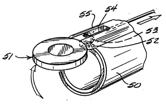
Figure 7 is a perspective view of the device depicted
in Figure S with the polarizer assembly in an open
position; and
Figure 8 is a cross-sectional view of the device
depicted in Figure 7.
Dat~iled De~cription of the Drawings
Referring to the drawings, in Fig. l there is shown
an endoscope lO. The endoscope comprises an elongated
portion ll which may be inserted in a cannula, or natural
body opening, with the distal end 12, placed in the
surgical environment. Through the center of the endoscope
is an imaging mechanism 13 which, at the distal end, has
an appropriate lens 14 and at the proximal end has a
suitablQ eyepiece lS. In many e~bodiments, the viewing
mechani~m is attached to a camera and imaging monitor.
E~D-17
: . ' . -, ' . '
: .. . - . - :
' : . ~: '" ~ -
21 ~4618
-- C --
The endoscope also comprises illuminating means which, in
this embodiment, is fiber optics 16 positioned at the
outer periphery of the endoscope around the imaging means.
The fiber optics are connected to a suitable light source
17 as is well known in the art.
The sterile medical device of the present invention
is an elongated hollow tube 22. The tube fits snugly
about the endoscope. The open end of the tube has an
~Q outwardly tapered portion 20. A ring 21 is mounted on the
tube as shown and may be slid over the tapered portion to
insure a snug fit of the tube on the endoscope. The tube
terminates at the distal end of the endoscope. The distal
end of the sterile medical device is the polarizer
assembly 24. As more clearly shown in Figure 2 the
polarizer assembly comprises an outer annular portion 25
about the periphery of the assembly which linearly
polarizes light in a first direction (in the horizontal
direction in this embodiment). The polarizer assembly
~Q includes a central analyzer 26 whose polarizing axis is 90
to the first direction (in the vertical direction in this
drawing.) If desired, the analyzer, instead of being
located at the distal end of the tube, could be located
ad~acent the eyepiece; or anywhere within the imaging
means.
Referring to Fig. 3, there is shown another
embodiment of a sterile medical device of the present
invention. In some endoscopes, the illumination is
provided in one channel of the endoscope while the lens or
imaging mechanism of the endoscope is provided in an
adjacent channel. When utilizing such a endoscope, the
sterile medical device of the present invention could be
constructed as shown in Fig. 3. The distal portion 30 of
D -17
. -, ... . . ~ .. ...
.
. . .
- . ~ . . .
- .. .. .
21~ 1618
the medical device includes a polarizer assembly 31 having
a first circular portion 32 which linearly polarizes light
in a first direction. An adjacent portion 33 analyzes
light in a direction 90 to the first direction. The
device would include along its outer periphery a suitable
indent 34 or alignment means in order to locate or
position the device on the endoscope so that the
polarizers align themselves with the light source and the
imaging means of the endoscope. Also in some endoscopes,
there may be included an open channel. As shown in Fig.
4, a medical device 40 according to the present invention
could include an open channel 41. The open channel is
used to accept small instruments used in an endoscopic
procedure. Adjacent the first channel is a first circular
~5 area 42 which linearly polarizes light in the first
direction. A second circular area 43 which analyzes light
in a direction 90 to the first direction is disposed
ad~acent said open channel. Again, the medical device
would include an indent 44 to appropriately align the
device on the endoscope.
The medical device of the present invention may be a
tube which covers substantially the entire length of the
endoscope and which is attached to the endoscope by
suitable coupling means such as shown in Fig. l. In some
embodimentæ of the present invention, it may be desirable
to use a very shortened tube that only fits on the distal
end of the endoscope but is sized to fit snugly on that
end to avoid inadvertent removal. It is preferred that
the tube be a relatively rigid tube for ease of use. A
rigid tube is often easier to place on an endoscope and
more easily inserted through a cannula or natural body
opening.
-17
, .
.
. : , ' ' -
.
. ' ~ . :: ' ~ :: '
21~4~1 8
- 8 -
The material used to make the tubular and coupler
portions of the medical devices of the present invention
may be any of the standard polymeric or metallic materials
used in medical instruments. It is preferred that a
S material be used that can be easily sterilized; for
example, by cobalt irradiation. One such suitable
material would be polyethylene. The tubular portion of the
medical device will have a length of about 11 inches with
an outside diameter of 7/16 of an inch and an inside
diameter of 13/32 of an inch so that it will fit a
standard 10 mm operative endoscope. The polarizer may be
made from polyvinylalcohol laminated in an acrylic resin
or other appropriate substrate. The outer annular
polarizer for a 10 mm laparoscope would have an outside
lS diameter of about 7/16 of an inch and an inside diameter
of 1/4 of an inch with a thickness of about .01 inch. The
inner central polarizer would have a diameter of 1/4 of an
inch with a thickness of .01 inch.
~Q Referring to Figures 5 through 8, there is shown
another sterile medical device of the present invention.
It may be desirable during certain surgical procedures
that the illumination not be polarized throughout the
entire procedure. The device depicted in Figures 5-8
allows the surgeon to polarize or not polarize the
illumination as desired. The device shown comprises a
hollow tube 50 which is sized to fit snugly over the
distal end of an endoscope. The polarizer assembly 51
which closes the distal end of the tube is pivotally
mounted on the tube by a hinge 52. Extending from the
periphery of the polarizer assembly is a pin 53. An
activation rod or wire 54 is attached to the pin and
extends through an opening 55 in the tube to the proximal
end of the endoscope. As seen in Figures 7 and 8, if the
~ND-17
. ~ ~ - . . . . . ~ -, -
. , . ~ . . .
2~0~6~ -
surgeon does not desire polarized illumination at some
point in the surgical procedure, it is a simple matter to
pull the activation rod and pivot the polarizer assembly
out of the path of illumination.
While the polarizing assemblies disclosed above
provide for linear polarization in different directions,
the present invention is not meant to be limited to linear
polarization. Polarization may occur in various states;
such as linear, circular, elliptical etc. The polarizing
assemblies of the present invention polarize the
illumination in one state and analyze the reflected
illumination in a different polarization state.
While the present invention may have its primary use
in reducing glare and enhancing visual representation
during endoscopic surgical procedures, it also has use in
endoscopic diagnostic procedures. For example, in certain
diagnostic procedures that use either infra-red or ultra-
~0 violet light, it may be de6irable to polarize such light
in a given state and then analyze the reflected light with
an analyzer that polarizes such light in a different
polarization 6tate.
Although this invention has been shown and described
with respect to certain embodiments thereof, it should be
under6tood by those skilled in the art that various
changes in form and detail may be made without departing
from the spirit and scope of the claimed invention.
END-17
- . :
. .
.
,
.
.
, .
- .
.
. -
