Note: Descriptions are shown in the official language in which they were submitted.
2104'767
RAN 4095/94
This invention relates to the process of estimating the quantity and quality
of human
nuclear and mitochoncirial DNA contained in a sample. More specifically, it
relates to the
use of biotinylated probes that hybridize to a human alpha satellite locus,
such as D17Z1, or
to a conserved sequence in the ;mitochondrial control region and a
chemiluminescent
detection assay to analyze immobilized DNA. Additionally, it relates to the
use of gel
electrophoresis followed by Southern Blotting to assess the degradation state
of DNA in a
sample.
An increasingly detailed analysis of human genetic diversity has been made
possible
by techniques of molecular biology such as amplification of DNA by the
polymerase chain
reaction (PCR) (Saiki et al., 1988, Science M:487-491, U.S. Patent Nos.
4,683,195;
4,683,202; and 4,965,188) and DNA typing based on restriction fragment length
polymorphisms (RFLP) (Jeffereys et al., 1985, Nature 314:67-73). The
specificity and
sensitivity of PCR amplification have resulted in widespread use of that
method in the fields
of forensic science (Bl;ake et al., 1991, Journal of Forensic Sciences
37(3):700-726), studies
of ancient DNA samples (Paabo et al., 1988, Nucleic Acids Research 1~:9775-
9787),
analysis of genetic diseases (Gibbs et al., 1989, in Erlich (ed.), PCR
Technology:
Principles md ApplicaLtions fig DNA Amplification. Stockton Press, New York:
153-169),
and in studies of population genetics (Helmuth et al., 1990, American Journal
of Human
Genetics 47:515-523).
The field of forensic science, in particular, has been revolutionized by the
ability to
extract and type DNA from forensic evidence samples (Reynolds et al., 1991,
Analytical
Chemistry 0:1-15). In forensics, where many biological evidence samples
contain either
extremely small quantides of DNA or DNA that has been degraded, the quantity
and quality
of DNA in a sample can be important factors in selecting suitable analytical
methods. For
example, RFLP-based typing methods require relatively large quantities of
undegraded
DNA, typically greater than 50 nanograms. Analysis of samples containing only
a few
nanograms of possibly degradeci DNA may require the additional sensitivity and
specificity
of PCR-based methods. In general, the efficiency of a PCR amplification is
influenced by
the quantity, quality, and purity of the sample DNA. Because the success of
most DNA
analysis methods is dependent c-n the quantity and quality of the DNA sample,
it is important
to be able to quantitate the DNA and assess the quality of DNA in samples
prior to analysis.
Current methocls for quantitation of DNA include UV spectroscopy, fluorometry,
Mey/21.7.93
CA 02104767 2003-12-17
2 -
and semi-quantitation by agarose gel electrophoresis followed by staining with
ethidium
bromide. However, these methods require multi-nanogram quantities of non-
denatured
DNA for analysis and are not specific for human DNA. In addition, they do not
distinguish
between nuclear and mitochondrial DNA. Quantitation methods specific for human
DNA are
important for the analysis of samples which sometimes contain bacterial or
fungal DNA,
such as forensic evidence samples, ancient DNA samples, and clinical samples.
Recently,
Waye et al., 1989, BioTechniques 7.(8):852-855, reported a method that is
relatively specific
for human DNA. However, this method requires a radioactive label and takes
several hours
to obtain results. There is a need for a rapid, sensitive, and human-specific
method for
quantitating the DNA in a sample which does not require the use of
radioactivity.
The quality of a DNA sample is conventionally evaluated by agarose gel size
fractionation of the DNA followed by ethidium bromide staining. High quality,
i.e.
undegraded, genomic DNA consists pritnarily of high molecular weight DNA.
Degradation
of the DNA produces fragments of random lengths which yield a smear of lower
molecular
weight DNA on the gel. Because ethidium bromide does not readily stain
denatured (single-
stranded) DNA, conventional methods are unsatisfactory for samples that are
extracted using
methods that require heating or boiling steps or alkaline treatment. For
example, the Chelex*
method of DNA extraction for amplification using the polymerase chain reaction
requires
boiling for cell lysis (Walsh et a1.,1991, BioTechniques 1Q(4):506-513). Some
DNA
extraction methods require heating to 95 C for inactivation of proteinase K
(see Higuchi,
1979, in Erlich (ed.) PCR Technology: Principles and Applications for DNA
Amplification,
Stockton Press, New York: 31-38). There is a need for a sensitive method of
assessing the
quality of both single- and double-stranded DNA samples. The present invention
meets
these needs.
The present invention provides sensitive methods for the quantitation of human
nuclear and mitochondrial DNA contained in a sample that are both simple and
rapid to carry
out. The DNA is immobilized on a membrane, hybridized with a biotinylated
probe that
hybridizes to a repeated human genomic sequence or to a mitochondrial sequence
and
detected using a chemiluminescent assay. In one embodiment, the repeated human
sequence
is the human alpha satellite locus, D17Z1. In another embodiment, the sequence
is part of
the control region of the mitochondrial genome. The quantity of DNA contained
in the
sample is estimated from the amount of hybrldized probe detected. The entire
procedure can
be completed in 1.5 hours and can detect less than 75 pg of human DNA.
In addition, the present invention provides methods of evaluating the quality
of DNA
contained in a sample. The DNA is size fractionated by gel electrophoresis,
immobilized on
*Trade-mark
2104767
- 3 -
a membrane, hybridizc:d with a biotinylated probe that hybridizes to a
repeated human
genomic sequence, and detected using a chemiluminescent assay. In one
embodiment, the
repeated human sequence is the human alpha satellite locus, D17Z1. The quality
of the DNA
is estimated from the fragment size pattern observed.
The present invention also provides biotinylated oligonucleotide probes that
hybridize to sequences within the human alpha satellite locus, D17Z1, and to
sequences
within the mitochondrial control region for use in the methods provided.
The present invention also provides ldts containing reagents used in the
methods of
the present invention.
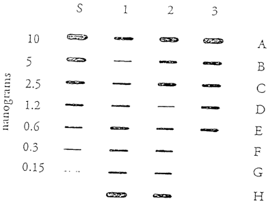
Figure 1 illustiates the results of a DNA quantity assay as described in
Example 2.
Sample DNA was immobilized on a nylon membrane and hybridized with the probe,
SW49
(SEQ ID No. 1). Bancis were visualized using chemiluminescent detection with a
15 minute
exposure to film Coliimn "S" is a human genomic DNA titration series ranging
from 10 to
0.15 nanograms of DNA. Columns 1-3 are samples in which the DNA quantity was
unknown. The sources of the extracted DNA samples in columns 1-3 were as
follows: 1A-
1E were bloodstains, :lF-2C were whole blood, 2D-3B were single hairs, 3C-3E
were
buccal samples, 3F was 1 g of cow DNA, 3G was 1 g mouse DNA; no sample was
added to 3H.
Figure 2 illustrates the results of a DNA quality assay as described in
Example 3.
Figure 2A illustrates a photograph of a 1% agarose geL Human genomic DNA (14
nanograms) was boiled in either 5% Cheletor water for 0,1, 3, or 8 minutes and
then
subjected to electrophoresis. DNA size markers were run concurrently on the
gel and are
indicated by "M". The DNA was subsequently transfesred to a nylon membrane,
hybridized
with the probe, SW49 (SEQ ID No. 1), and visualized by chemiluminescent
detection with a
15 minute exposure to film. Figure 2B illustrates the resulting photograph.
Photograph
labels are as in Figure 2A.
The term "sample" as used herein refers to any substance containing or
presumed to
contain nucleic acid including, but not limited to, tissue or fluid isolated
from one or more
individuals, in vitro cell culture constituents, as well as evidential,
clinical, archival, and
ancient samples.
The terms "oligonucleotide" and "nucleic acid" as used herein refer to
molecules
comprising two or mare deoxyribonucleotides or ribonucleotides. The exact size
will
* Trade Mark
2104767
- 4 -
depend upon many factors, which in turn depend on the ultimate function or use
of the
oligonucleotide. The terms refer to both single- and double-stranded DNA and
RNA.
Oligonucleotides may be derived by any suitable technique including, but not
limited to,
isolation of an existing or natural sequence, chemical synthesis, DNA
replication or
amplification, reverse ti-anscription, or a combination thereof. Chemical
synthesis methods
may include, for example, the phosphotriester method described by Narang et
al., 1979,
Methods in Enzymology ik$:90,the phophodiester method described by Brown et
al., 1979,
Methods in Enzymology ff 109, the diethylphosphoramidite method described by
Beaucage
et al., 1981, Tetrahedron Letters 22:1859, and the solid support method
disclosed in U.S.
Patent No. 4,458,066. Oligonucleotide synthesis is described in Levenson and
Chang,
1990, in PCR Protocols, Innis et al. (eds.), Academic Press, New York:99-112.
The term "subsequence" as used herein refers to a nucleotide sequence which is
wholly contained within anotlier nucleotide sequence. As defined, a sequence
is also a
subsequence of itself. As used herein, a subsequence suitable as a
hybridization probe is
about 10-140 nucleotidi-Is in length and preferably 40-130 nucleotides in
length.
The term "quality" as used herein refers to the degree of degradation of a DNA
sample. A high quality sample contains DNA which has undergone little or no
degradation.
The quality of a DNA sample can be assessed by measuring the molecular weight
of the
sample DNA by gel electrophoretic size fractionation. For example, using a 1
7o agarose gel,
a high quality sample o1' human genomic DNA migrates with a 20 kb DNA marker
and
forms a relatively tight lband when visualized either by ethidium bromide
staining or by the
methods of the present invention, whereas in a low quality human DNA sample,
degradation
of the DNA yields fraginents of varying length which appear as a smear of
lower molecular
weight DNA, i.e., less than 20kb.
The terms "probe" and "oligonucleotide probe" as used herein refer to labeled
oligonucleotides which are sufficiently complementary to a specific target
sequence
contained in a DNA sarnple to form a stable hybridization duplex with the
target sequence.
The hybridization is uniier stringent conditions. Stringent hybridization
conditions are well
known in the art and air described, for example, in Sambrook et al., 1989,
Molecular
Cloning: A Laboratory Manual, Second Edition, Cold Spring Harbor Laboratory,
New
York. The term "hybridizing region" refers to that region of an
oligonucleotide probe which
is complementary to, and therefore hybridizes to, the target sequence.
Although the
hybridizing region typically refers to the entire oligonucleotide, the probe
may include
additional nucleotide sequences which function, for example, as the label
binding site to
provide means for fixing the probe sequence to a solid support. In the
preferred mode, the
2104767
-5-
hybridizing region of the oligonucleotide probe is completely complementary to
the target
sequence. However, in general, complete complementarity is not necessary;
stable duplexes
may contain mismatched bases or unmatched bases. Modification of the stringent
conditions
may be necessary to pei-mit a stable hybridization duplex with one or more
base pair
mismatches or unmatched bases. Sambrook et al., 1989, supra, provides guidance
for
suitable modification. Stability of the target/probe duplex depends on a
number of variables
including length of the oligonucleotide, base composition and sequence of the
oligonucleotide, tempeiature, and ionic conditions.
The oligonuclec-tide probes of the present invention are labeled to permit
detection of
probe-target hybridization duplexes. In general, a label can be any atom or
molecule which
can be attached to the oligonucleotide probe and used to provide a detectable,
quantifyable
signal. Labels may be attached to an oligonucleotide directly or indirectly by
a variety of
techniques. Depending on the type of label used, the label can be attached to
a terminal (5'
or 3' end of the probe) or a non-terminal nucleotide, and can be attached
indirectly through
spacer arms of various sizes and compositions. Using commercially available
phosphoramidite reage-its, one can produce oligomers containing functional
groups (e.g.,
thiols or primary amines) at either the 5' or 3' terminus via an appropriately
protected
phosphoramidite, and can label such oligonucleotides using protocols described
in, for
example, PCR Protocols: A(3uide to Methods and Applications, Innis et al,
eds., Academic
Press, Inc., 1990. In a preferred embodiment, the label consists of a biotin
molecule
covalently bound to the oligonucleotide at the 5' end. The term "biotinylated
probe" as used
herein refers to a probe with one or more biotin molecules bound either
directly to the
oligonucleotide or indirectly through intervening "spacer" molecules.
Detection of the probe is preferably by a chemiluminescent assay using a
luminol-
based reagent as described in Whitehead, et al., 1983, Nature ,305:158-159,
and available
commercially (ECL, Amersham, Arlington Heights, IL, USA). Following
hybridization of
the probe with the target DNA, the biotin molecule attached to the probe
oligonucleotide is
conjugated to streptavictin-horseradish peroxidase (SA-HRP). Alternatively,
the
oligonucleotide probe can be labeled with horseradish peroxidase directly,
thereby
eliminating the separate, conjugation step. In either case, subsequent
oxidation of luminol by
the horseradish peroxidase enzyme results in the emission of photons, which is
then detected
on standard autoradiography film. The intensity of the signal on the film is a
function of
DNA quantity. A series of DNA standards containing known amounts of DNA are
assayed
along with one or more unknown samples, blotted on the same membrane. The
signal
intensities of the known. DNA standards allows an empirical determination of
the functional
relationship between signal intensity and DNA quantity, which enables the
quantitation of
2104767
- 6 -
the unknown samples.
Alternatively, :probe/target hybridization duplexes may be detected using a
color
development reaction as described in Sheldon et al., 1986, Proc. Natl. Acad.
Sci. USA
5U:9085-9089, which utilizes 3,3',5,5'-tetramethylbenzidine (TMB) and hydrogen
peroxide.
The probes of the invention are complementary to a highly-repetitive human
genomic
sequence. High sensilivity is obtained from the use of a repetitive sequence
because the high
copy number per genome of a repeat sequence provides a large number of target
sequences
for hybridization. Numerous repetitive sequences are known to occur in the
human genome,
such as satellite DNA and the Alu repeat sequences (Brilten et a1.,1988, Proc.
Natl. Acad.
Sci. USA $J:4770-47 74). Alpha satellite DNA is a complex family of tandemly
repeated
DNA located primarily at the centromeres of primate chromosomes (Waye and
Willard,
1986, Molecular and Cellular Biology f2(9):3156-3165, Willard, 1985, American
Journal of
Human Genetics 31:524-532). In a preferred mode, the probes are complementary
to
D 17Z1, a primate specific alpha satellite DNA sequence located on chromosome
17. The
sequence is estimated to be present in 500 to 1,000 copies per chromosome 17
(Waye and
Willard,1986, supra). The primary D17Z1 repeat sequence is 2.7 kb in length
and is
arranged as 16 contigiious monomers; a less abundant (approximately 100 copies
per
chromosome 17) repeat consisting of 15 tandem monomers is also found.
In another embodiment of the invention, the probes are complementary to
sequences
specific to the mitochondrial genome. The mitochondrial control region has
been extensively
charactmized at the sequence level, and it contains two hypervariable regions
surrounded by
relatively conserved r,-,gions (Vigilant et a1.,1989, Proc. Natl. Acad. Sci.
USA $¾:9350-
9354). Two pairs of primers complementary to the conserved regions have been
used to
amplify the hypervariable regions using the PCR. These regions can be analyzed
subsequently by dirw: DNA sequencing or by hybridization to a collection of
sequence-
specific oligonucleotide probes (Vigilant et al., supra., and Stoneking et
al., 1991, Am J.
Hum. Genet. 4$:370-:582). In a preferred mode, the quantitation probes contain
sequences
overlapping the control region primer sites or sequences within the conserved
areas of the
control region.
In a preferned DNA quantitation method, a DNA sample is immobilized on a nylon
membrane before hybridizing with the labeled probes as described in Example 2.
A
commercially availablle apparatus (e.g., the Convertible, GIBCO BRL,
Gaithersburg, MD,
USA) may be used to immobilize the DNA sample on the membrane in a specified
location.
* Trade Mark
CA 02104767 2003-12-17
-7 -
Thus, a large number of samples can be immobilizod on the same membrane in a
defined
array. Simultaneous hybridization of numerous samples can be effected by
immersion of the
membrane in a hybridization buffer containing the quantitation probe. The
methods of the
present invention are particularly suited to applications in which a large
number of samples
need to be analyzed on a routine basis, such as in a commercial environment.
In a prefenred DNA quality assay method, after size fractionation of the DNA
sample
by agarose gel electrophoresis, the fiactionated DNA is tranaferred to a nylon
membrane
before hybridizing with the probes of the present invention. The DNA may be
transfered
using a commercially available apparatus (e.g., the Posiblot transfer system,
Stratagene, La
Jolla, CA, USA). After the DNA is transfenred to the nylon membrane, the DNA
may be
fixed to the membrane by baking at 80 C, as described in Example 3, or by
crosslinking of
the thymidine residues to the membrane by UV uradiation (Church and
Gilbert,1984, Proc.
NatL Acad. Sci. USA $1:1991-1995).
Reagents employed in the methods of the present invention can be packaged into
kits. Kits include the labeled oligonucleotide probe or, if unlabeled,
specific labeling
reagents may be included. The lats may also include suitably packaged reagents
and
matesials needed for DNA immobilization and detection, such membranes,
buffers,
enzymes, DNA standards, and a hybridization tray, as well as instructions for
conducting
the assay.
The following examples are offened by way of illustration and are not intended
to
limit the invention in any manner.
~ixg~e 1
P-robes
The probes of the present invention are complementary to a region contained
within
the 2.7 kb D17Z11ocus. Each oligonucleotide is bound to a biotin molecule at
the 5' end
either directiy or through a phosphoramidite "spacer" molecule. Probes SW49
(SEQ ID No.
1), SW1000 (SEQ ID No. 1), SW1001 (SEQ ID No. 1), SW1002 (SEQ ID No. 1),
SW1003 (SEQ ID No. 1), SW1004 (SEQ ID No. 1), and SW1005 (SEQ ID No. 1) were
constructed with the identical oligonucleotide sequence, differing in the
details of the label.
The nucleotide sequence common to each of the probes is provided below and in
the
Sequence Listing.
*Trade-mark
-8- 2104767
SEO ID No. SNUence
1 5'TAGAAGCATTCTCAGAAACfACITTGTGATGATTiGCA17C
Oligonucleotid-. synthesis was performed on an automated DNA synthesizer
(either
Milligen/Biosearch 8750, Applied Biosystems 394; or Eppendorf Biotronik D) on
a
micromole scale using 500 angstrom controlled-pore glass supports and 0-
cyanoethyl N,N
diisopropyl phosphoramidites as described in Beaucage and Caruthers, 1981,
Tetrahedron
Letters, 22:1859-1862 and Sinha et a1.,1984, Nucleic Acids Research, 12:4539-
4557.
Supports and phospho,amidite derivatives of dA, dC, dG, and T were obtained
commercially from Millipore/Waters, Bedford, MA, USA; or Cruachem, Sterling,
VA,
USA. Biotin and "Spacer" phosphoramidites were obtained from Glen Research,
Sterling,
VA. Two biotin reagents were used: Biotin Phosphoramidite and BioTEG
Phosphorawidite. These two biotin reagents differ in that the BioTEG product
includes a
longer spacer separating the biotin from the oligonucleotide. Oligonucleotides
were
synthesized with the terminal dimethoxytrityl group left intact and were
purified by lipophilic
selection using solid-phase extraction cartridges (PREP-NENSORB, Dupont).
Biotinylation
and oligonucleotide purification are described in Misiura et al., 1990,
Nucleic Acids
Research 1$:4345-4354; Alves et a1.,1989, Tetrahedron Letters, 2Q:3089-3092;
and Pon,
1991, Tetrahedron Letters 12:1715-1718.
Quantitation probes SW49 (SEQ ID No. 1), SW1000 (SEQ ID No. 1), SW1001
(SEQ ID No. 1), SW1002 (SEQ ID No. 1), SW1003 (SEQ ID No. 1), SW1004 (SEQ ID
No. 1), and SW1005 (SEQ ID No. 1) are shown schematically in Table 1, below.
In Table
1, B 1 refers to Biotin Phosphoramidite, B2 refers to BioTEG Phosphoramidite,
Spacer
refers to a phosphoranudite spacer. Oligo refers to the oligonucleotide (SEQ
ID No. 1)
common to the probes., mtOiigo refers to mitochondrial DNA oligonucleotides
(sequences
provided below). Probes SW1000 (SEQ ID No. 1) and SW1003 (SEQ ID No. 1) are
identical in sequence and label to SW49 (SEQ ID No. 1).
* Trade Mark
9 2104767
Table 1
SEQID No. Probe Construct
1 SW49 B 1-Oligo
1 SW 1000 B 1-Oligo
1 SW 1001 B 1-Spacer-Oligo
1 aW 1002 B 1-Spacer-B 1-Spacer-Oligo
1 SW1003 B 1-Oligo
1 ,3W 1004 B2-Oligo
1 SW 1005 B2-Spacer-Oligo
12-20 RR64-RR72 B2-mtOligo
Additional oligonucleotide probe sequences useful in the methods of the
present
invention are provided below.
Probe SEQ ID No. Sgquence
S W31 2 5'CACTATITGTAGAATGTGCAAGTGGATATTTAGGCCTCTC
SW32 3 5'CAGAAGCATTCTCAGAACCTTCTTCGTGATGTTTGCATTC
SW33 4 5'TAGAAGCATTCI'CAGAAACTACTI'PGTGATGATTGCATTCAAGTCACA
G;AGTTGAACATTCCCITPGACAGAGCAGTTTGGAAAC='CTMTGTAG
A,ATCTGCAAGTGGAGATATGGACCGCTITAGG
SW34 5 5'CAGTAGCATTCACAGAAAACTCTTGGTGACGACTGAGTTTAACTCACA
G~AGCTGAACATTCGTITGGATGGAGCAGTTI'CGAAACACACTATTTGTAG
AATGTGCAAGTGGATATTTAGGCCTC=AGG
SW35 6 5'CAGAAGCATTCTCAGAACCTTCTTCGTGATGTITGCATTCAACTC
A,CAGTGTTGAACCTTTCTI'PGATAGTTCAGGTITGAAACGGTCTTPCTGT
A GAAACTGCAAGTAGATATTTGGACCGCTCTGAGG
SW56 7 5'GAAACTCTY.TITGTGTAGAATCTGCAAGTGGAGATATGGA
SW59 8 5'AAGTCACAGAGTTGAACATTCCCTI'TGACAGAGCAGTITG
SW52 9 5'TAGAAGCATTCTCAGAAACTACI'I'IY'TGATGATTGCATTCAAGTC
ACAGAGTTGAACATT
-10 - 2104767
SW57 10 5'TAGAAGCATTCTCAGAAACTAC:TITGTGATGATTGCATTCA
AGTCACAGAGTTGAACATTCCCI'ITGACAGAGCAGTTTG
S W 5 8 11 5' TAG AAGCATTCTCAGAAACTACITTGTGATGATTGCATTCAAGTCAC
AGAGTTGAACATTCCCTTTGACAGAGCAGTTTGGAAACTCTCI'I'I'GTGTA
GAA
The oligonucleotide probe sequences are subsequences of the D17Z1 constituent
monomers. SW33 (SEQ ID No. 4) is a subsequence of monomer 11, SW34 (SEQ ID
No. 5) is a subsequence of monomer 12, and SW35 (SEQ ID No. 6) is a
subsequence of
monomer 13. The other oligonucleotide sequences are subsequences of one of the
above 3
oligonucleotides. SEQ ID No. 1, SW56 (SEQ ID No. 7), and SW59 (SEQ ID No. 8)
are
non-overlapping subso4uences of SW33 (SEQ ID No. 4). SEQ ID No. 1 is a
subsequence
of SW52 (SEQ ID No. 9), which is a subsequence of SW57 (SEQ ID No. 10), which
is a
subsequence of SW58 (SEQ ID No. 11), which are all subsequences of SW33 (SEQ
ID No.
4). SW31 (SEQ ID No. 2) is a subsequence of SW34 (SEQ ID No. 5), and SW32 (SEQ
ID
No. 3) is a subsequence of SW35 (SEQ ID No. 6).
Additional protes of the present invention are complementary to various
conserved
sequences within the mitochondrial control region.
Probe SEO ID No. Sequence
RR64 12 5'GGC:GGTATGCACITITAACAGTCACCCCCCAACTAACAC
RR65 13 5'GTC:TITAACTCCACCATTAGCACCCAAAGCTAAGATTCTA
RR66 14 5'CGTGAAATCAATATCCCGCACAAGAGTGCTACfCTCCTCG
RR67 15 5'GAACTGTATCCGACATCTGGTTCCTACTTCAGGGTCATAAAGC
RR68 16 5'GAC:ATCACGATGGATCACAGGTCTATCACCCTATTAACCAC
RR69 17 5'CAT'CCTCCGTGAAATCAATATCCCGCACAAGAGTGCTAC
RR70 18 5'GTC'TTTAACTCCACCATTAGCACCCAAAGC
RR71 19 5'CTCCACCATTAGCACCCAAAGCTAAGATTC
RR72 20 5'GTATCCGACATCTGGTTCCTACTTCAGGGTC
RR70 (SEQ ID No. 18) and RR721 (SEQ ID No. 19) are overlapping subsequences of
RR65 (SEQ ID No. 13). RR72 (SEQ ID No. 20) is a subsequence of RR67 (SEQ ID
No.
15). RR66 (SEQ ID No. 14) and RR69 (SEQ ID No. 17) overlap.
-11 2104767
xamFle 2
DNA antiEstimation
To estimate the quantity of DNA in a sample, extracted sample DNA was
immobilized on a nylori membrane along with a titration series of a human
genomic DNA
standard and hybridized to a'biotinylated probe, SW49 (SEQ ID No. 1). The
synthesis of
the probe was as described in. Example 1, above. Hybridization was visualized
using a
chemiluminescence decection protocoL The quantity of DNA present in the sample
was
estimated by comparison of the hybridization signal obtained from the sample
DNA to those
obtained from the DNA standards. Details of the experimental protocol are as
follows.
The quantities of human DNA in extracts from 5 human bloodstain samples, 6
human whole blood samples, 7 human hair samples, and 3 human buccal samples
were
estimated In additionõ samples consisting of 1 g cow DNA and 1 g mouse DNA
were
also used as a test of pnobe specificity. DNA was extracted from samples
either by the
Chelex method described in Walsh et al., 1991, supra., or by a salting out
method as
described in Miller et al., 1988, Nucl. Acids Res. _6(3):1215. In the Chelex
method, a 3
mm2 bloodstain, a buccal scraping, or a 1 cm hair root section were incubated
in 200 l 5%
Chelex at 56 C, follovied by boiling for 8 minutes.
Five l of each extracted DNA sample was added to 100 l spotting buffer (0.4
N
NaOH, 25 mM EDTA). DNA standards were prepared by adding the following
quantities
of human DNA to 101) l spotting buffer. 10, 5, 2.5,1.2, 0.6, 0.3, 0.15 ng. A
blank was
also prepared which c;ontained no DNA added to 100 l spotting buffer. A piece
of Biodyne *
B Membrane (Pall Biosupport, Glen Cove, NY, USA) was prewet in distilled water
and
placed in a slot blot apparatus (The Convertible, 0.75 x 0,75 mm, GIBCO BRL,
Gaithersburg, MD, USA). The sample and standard preparations were added to the
welis
(entire volume) then die vacuum was applied. With the membrane still in the
apparatus, 200
l of 15% hydrogen peroxide was added to each well, and the vacuum was applied
again.
The membrane was removed from the apparatus and immediately placed in 200 ml
of
prehybridization soluiion consisting of 5x SSPE (20x SSPE is 3.6 M NaCI, 200
mM
NaH2PO4-H20, 20mM EDTA, pH 7.4) and 0.5% sodium dodecyl sulfate (SDS)
prewarmed to 50 C and incubated in a shaking water bath for 15 minutes at 50
C. The
membrane was transfirrred to 30 ml hybridization buffer (5x SSPE, 0.5% SDS)
containing
15 pmoles of the probe, SW49 (SEQ ID No. 1), incubated in a shaking water bath
for 15
minutes at 50 C to allow hybridization to occur, and then rinsed briefly in
1.5x SSPE, 0.5%
* Trade Mark
- 12 - 2104767
SDS. The stringent wash and conjugation (biotin to SA-HRP) steps were carried
out
simultaneously. The niembrane was placed in 30 ml of 1.5x SSPE, 0.5% SDS
containing
90 l of SA-HRP (Perldn Elmer, Norwalk, CT, USA) and incubated in a shaking
water
bath for 10 minutes at ;50 C. The membrane was rinsed briefly in 1.5x SSPE,
0.5% SDS
and then washed in 200 ml of 1.5x SSPE, 0.5% SDS on an orbital shaker for 15
minutes at
room temperature. Thi-, membrane was then rinsed in 0.1 M Sodium Citrate, pH
5.
*
Detection of the hybridized probe was carried out using ECL (Amersham,
Arlington
Heights, IL, USA), which is a luminol-based reagent used for enhanced
chemiluminesce,nt
detection. The membrane was placed in a mixture of 10 ml of ECL Reagent 1 and
10 ml of
ECL Reagent 2 and sh3ken for 1 minute at room temperature. The membrane was
placed on
a sheet of Benchkote ('Whatman, Maidstone, England), covered with Saran Wrap,
and
wiped free of excess moisture. To visualize the DNA, the membrane was exposed
to
Hyperfilm~(Amersham, Arlington Heights, IL, USA) or Kodak XAR5 film (Kodak,
Rochester, NY, USA) for 15 minutes at room temperature. Results are shown in
Figure 1.
DNA quantitation was determined both by visual comparison of sample DNA slot
blot intensities to those of the DNA standards and by computer image analysis
of the slot
blot results on film. For the computer analysis of the slot blot results, the
film was scanned
using an 8-bit gray-sc2ae flatbed scanner (available from Abaton Corporation,
Fremont, CA,
USA) and the resulting picture analyzed using the computer program Image 1.41
(written by
Wayne Rasband and available from NIH, Bethesda, MD, USA) running on a
Macintoslt
computer. The mean signal density (actually scanned pixel values ranging from
0 to 255) of
each slot blot signal was measured. The mean signal density was defined over a
rectangle of
constant size that wholly contained the slot blot signal. A comparison of mean
signal
densities defined and measured in this manner is equivalent to a comparison of
the total
signal from each slot blot. The background density, measured next to each
slot, was
subtracted from each inean density. Background signal was not observed on the
membrane
directly, and was most likely an artifact of the scanning of the film. This
data was then
exported to another computer program (Kaleidagraph,Abelback Software) fit into
an
equation describing the relationship between slot blot mean signal density and
the DNA
quantity to the data from the DNA standards. The data closely fit the
exponential equation
Y=C=e(r=x) where Y is the DNA quantity, in nanograms, C = 0.1787 nanograms, r
=
0.0308, and X is the raean signal density. Signal density, defined here as
scanned pixel
values, is a dimensionless number between 0 and 255. Once determined, this
exponential
equation was used to determine the quantity of DNA in the unknown samples from
the mean
signal density measurements.
* Trade Mark
-13 - 2104767
The quantity of DNA in the each of the samples containing human DNA was
estimated from the obsesrved slot blot results using the above equation. The
DNA quantity
estimates are shown below. Sample positions refer to the column and row
positions
indicated in Figure 2. No signal was observed from cow or mouse DNA samples,
indicating that the probe SW49 (SEQ ID No. 1) hybridized specifically to human
DNA only.
.Sample Source Quanti (ngl
1A Bloodstain 2.24
1B " 0.26
1C " 1.07
1D " 0.88
1E " 1.74
1F Whole Blood 0.83
1G 1.09
1H 7.17
2A 6.32
2B 1.32
2C 2.33
2D Hair 0.28
2E " 0.94
2F " 0.52
2G " 0.68
2H " 2.49
3A " 6.34
3B " 1.64
3C Buccal 1.46
3D " 1.49
3E " 1.00
3F Cow DNA 0
3G Mouse DNA 0
To estimate the: quantity of mitochondrial DNA in a sample, the protocol
described
above for quantitation of nuclear DNA is used with the following exceptions:
1. Prehybridization is performed at 46'C instead of 50'C.
2. Hybridization is performed at 46'C with 20 pmoles of the probe RR70 (SEQ ID
No. 18).
14 - 2104767
-
A commercially, available preparation of placental DNA (SIGMA) is diluted and
used
for the DNA standards. Since the extracted placental DNA contains a mixture of
nuclear and
mitochondrial DNA, the standards can be used for both types of quantity
estimates.
Mitochondrial DNA is the minor component of the total DNA preparation and is
present in
an unknown quantity. 'rherefore, to use placental DNA (or other total DNA)
preparations as
a standard for the mitoc:hondrrial DNA quantitation assay, the amount of
mitochondrial DNA
present must be determined. 'This value can be obtained using a purified
mitochondrial DNA
preparation that has beE:n quantitated spectrophotometrically. Dilutions of
the purified DNA
can then be hybridized with the mitochondrial DNA-specific probe as described
above at the
same time as the total DNA dilutions, and the signal intensities can be
compared to determine
the quantity of mitochondrial DNA in the total DNA preparation. Obviously, a
purified
mitochondrial DNA sample would be the ideal quantitation standard, but the
procedure for
isolating mitochondrial'. DNA is extremely time consuming, expensive, and
provides a very
low-yield.
Ex=p?3~
DNA Qualitv Estimation
Purified human genomic DNA was diluted to 2 ng/ l in both 59'o Chelex and
glass
distilled water, and thc:n boiled for 0, 1, 3, or 8 minutes in a boiling water
bath. Seven l
(14 ng) of each sample was subjected to electrophoresis on a 1% agarose gel
containing 0.5
g/ml ethidium bromide in lx TBE for 30 minutes at 100 volts. The gel was
photographed,
soaked in 0.25 M HCl for 15 minutes to depurinate the DNA, and then soaked in
0.5 N
NaOH,1.5 M NaCl fcr 10 minutes to denature the DNA. The DNA was transfeired to
a
Biodyne B membrane using the Posiblot*transfer system (Stratagene, La Jolla,
CA, USA).
Transfer was perform:d at 75 mm Hg for 1 hour using lOx SSPE as the transfer
buffer.
The membrane was baked in a vacuum oven for 15 minutes at 80 C to fix the DNA.
The
membrane was wetted with 2x SSPE and then soaked in 15% hydrogen peroxide for
2
minutes. Hybridization and detection of bound SW49 probe (SEQ ID No. 1) was
performed essentially as described in Example 1, above, except that the blot
was exposed to
film for 30 minutes.
The results, sh own in Figures 2A and 2B, indicate that the methods of the
present
invention for the evaluation of DNA quality provide improved detection
sensitivity,
particularly for the analysis of denatured DNA. In this analysis (1% agarose
gel),
undegraded genomic ]DNA runs as a relatively tight band at about 20 kb
relative to an
appropriate molecular marker, degraded DNA appears as a smear of DNA below 20
kb in
~ Trade Mark
2104767
- 15 -
molecular weight. The photograph of the ethidium bromide-stained agarose gel,
shown in
Figure 2A, shows weak or absent band intensities for the boiled samples using
ethidium
bromide detection, particularly for the samples boiled in water. This is
because ethidium
bromide does not readily stain denatured (single-stranded) DNA. As shown in
Figure 2B,
DNA boiled in Chelex remains relatively intact. The double band for the sample
boiled in
Chelex for 1 minute presumably corresponds to both denatured and non-denatured
DNA.
After 3 to 8 minutes of boiling in Chelex, all DNA is denatured. The presence
of a single
band of high molecular weight indicates that little degradation occurred from
boiling in
Chelex. In contrast, DNA boiled in water shows slight degradation after 3
minutes and
significant degradation after 8 minutes, as indicated by the smear of lower
molecular weight
DNA apparent. The increased detection sensitivity realized by blotting and
probing using the
methods of the present invention allow for a greatly improved ability to
evaluate the effects
of boiling on DNA quality.
ExaMle 4
Probe LabelinQ
A comparison of the effect of different probe label moieties on the
sensitivity of the
DNA quantity assay was done using probes SW49 (SEQ ID No. 1), SW1000, SW1001
(SEQ ID No. 1), SW1002 (SEQ ID No. 1), SW1003 (SEQ ID No. 1), SW1004 (SEQ ID
No. 1), and SW1005 (SEQ ID No. 1). As described in Example 1, above, the
oligonucleotide sequence of each of these probes is identical SW49 (SEQ ID No.
1); these
probes differ in the number and spacing of the biotin labels bound to the
oligonucleotide.
For comparison, subsets of the probes listed above were used in the quantity
assay
essentially as described in Example 2, above.
One comparisoii was done using probes SW1000 (SEQ ID No. 1), SW1001 (SEQ
ID No. 1), and SW1002 (SEQ ID No. 1). Probe SW1001 (SEQ ID No. 1) showed a
slight
increase in sensitivity compared to probe SW1000 (SEQ ID No. 1). Because these
probes
differ only in the preseiice of a spacer in SW1001 (SEQ ID No. 1), the
improvement in
assay sensitivity most likely resulted from an increase in the space between
the
oligonucleotide and the: biotin label. Probe SW1002 (SEQ ID No. 1), which
contains two
biotin molecules per probe, stiowed increased sensitivity (increased signal
for 150
3 5 picograms with a 15 m:inute exposure) over both of the single-biotin
probes.
Another comparison was done using probes SW1001 (SEQ ID No. 1), SW1004
(SEQ ID No. 1), and SW1005 (SEQ ID No. 1). From the comparison of SW1001 (SEQ
ID
21()4'767
- 16 -
No. 1) and SW1004 (SEQ ID No. 1), it was observed that biotinylation using
BioTEG
phosphoramidite (SW1.004 - SEQ ID No. 1), which, in effect, contains a spacer,
is
equivalent to, or even superior to, biotinylation using biotin phosphoramidite
and a separate
phosphoramidite spacer (SW1001 - SEQ ID No. 1). Probe SW1005 (SEQ ID No. 1),
which incorporates both Bio7EG phosphoramidite and a separate phosphoramidite
spacer,
provided the greatest sensitivity.
Fxmple 5
Assay Sensitivity
In each of the I)NA quantity assays described in Examples 2 and 4, above,
visualization of the slot blot results was accomplished using a 15 minute film
exposure.
With a 15 minute exposure time, assay sensitivity in the range of 75 picograms
was seen for
nuclear DNA quantitation. Increasing the exposure time can increase the
sensitivity of the
assay. This was demonstrated by a DNA quantity assay essentially as described
in Example
2 using probe SW 1MG (SEQ ID No. 1) and with both 15 minute and 3 hour
exposures.
Whereas sensitivity down to 75 picograms could be detected with a 15 minute
exposure, the
3 hour exposure showi:d sensitivity down to between 9 and 18 picograms.
Example 6
Preferred Method for Determining the uantity of DNA in a Sample
(Protocol)
Slot Blot
1. Add 1 to 5 l of each DNA sample to 150 1 of spotting buffer (0.4 N NaOH,
25 mM
EDTA, 0.0015 % l3romophenol Blue). Also add the following quantities of DNA
standard (in 5 l) to 150 l of spotting buffer: 10, 5, 2.5, 1.2, 0.6, 0.3,
0.15 ng.
2. Pre-wet Biodyne Ft membrane in 50 mL of 0.4 N NaOH, 25 mM EDTA (5-30
minutes).
3. Place the membrane in the slot blotter, and pipette the entire volume for
each sample into
the wells. Apply spotting buffer containing no DNA to some of the empty wells
as a
negative control. 'Curn on the vacuum only after all samples have been
applied.
4. Begin the pre-hybiidization step immediately (see below).
-17- 2104767
Hvbridization and Detelaion
1. Pre-Hybridization: Place the membrane in 150 mL of pre-warmed 5X SSPE, 0.5%
SDS. Then add 5 r,nL of 30% H202. Shake in a water bath (70 rpm) for 15
minutes at
50 C.
2. Hybridization: Incubate in 30 mL of 5X SSPE, 0.5% SDS containing 20 pmoles
SW1004, for 20 mi.nutes at 50 C in a shaking water bath (70 rpm).
Rinse: Briefly rinse in 1.5X SSPE, 0.5% SDS.
3. Stringen Wash/Ccn' i n: Incubate in 30 mL of 1.5X SSPE, 0.5% SDS containing
90 l SA-HRP, for 10 minutes at 50 C in a shaking water bath (70 rpm).
Rinse: Briefly rinse in 1.5X SSPE, 0.5% SDS.
4. Wash: Incubate in 150 mL of 1.5X SSPE, 0.5% SDS at room temperature for 15
minutes on an orbital shaker (100 -125 rpm).
Rinse: Briefly rin;ce in approximately 150 mL of 0.1 M NaCitrate, pH 5.
5. E'L: Add 10 mL ECL reagent A to 10 mL ECL reagent B. Shake the membrane in
the
ECQ, reagents for exactly 1 minute at room temperature.
6. Exose i~: Place the membrane on the plastic side of benchkote and place
Saran Wrap
over the membrane. Use a paper towel to smooth out any wrinkles in the Saran
Wrap.
Expose to Hyperfilm or Kodak XAR5 film for 15 minutes.
2104767
- 18 -
SEQUENCE LISTING
(1) GENERAL INFOR14ATION:
(i) APPLICAN'C:
NAME: F. HOFFMANN-LA ROCHE AG
STREET: Grenzacherstrasse 124
CITY: Basle
COUNTRY: Switzerland
POSTAL CODE: CH-4002
TELEPHONE: 061 - 688 25 11
FAX: 061 - 688 13 95
TELEX: 962292/965542 hlr c
(ii) TITLE OF INVENTION: A Chemiluminescent Method for the
Quantitation of Human DNA
(iii) NUMBER OF SEQUENCES: 20
(iv) COMPUTER READABLE FORM:
(A) MEDIUM TYPE: Floppy disk
(B) COMPUTER.: IBM PC compatible
(C) OPERATING SYSTEM: PC-DOS/MS-DOS
(D) SOFTWARE: PatentIn Release #1.0, Version #1.25
(v) CURRENT APPLICATION DATA:
(A) APPLICATION NUMBER:
(B) FILING DATE:
(C) CLASSIFICATION:
- 19 - 2104767
(2) INFORMATION FOR SEQ ID NO:l:
(i) SEQUENCE CHARACTERISTICS:
(A) LENGTH: 40 base pairs
(B) TYPE: nucleic acid
(C) STRANDEDNESS: single
(D) TOPCLOGY: linear
(ii) MOLECULE TYPE: DNA (genomic)
(xi) SEQUENCE DESCRIPTION: SEQ ID NO:1:
TAGAAGCATT CTCAGAAACT ACTTTGTGAT GATTGCATTC 40
(2) INFORMATION FOR SEQ ID NO:2:
(i) SEQUENCE CHARACTERISTICS:
(A) LENGTH: 40 base pairs
(B) TYPE: nucleic acid
(C) STRANDEDNESS: single
(D) TOPOLOGY: linear
(ii) MOLECULE TYPE: DNA (genomic)
(xi) SEQUENCE DESCRIPTION: SEQ ID NO:2:
CACTATTTGT AGAATGTGCA AGTGGATATT TAGGCCTCTC 40
-20- 2104767
(2) INFORMATION FOR SEQ ID NO:3:
(i) SEQUENCE CHARACTERISTICS:
(A) LENGTH: 40 base pairs
(B) TYPE: nucleic acid
(C) STRAbIDEDNESS: single
(D) TOPOLOGY: linear
(ii) MOLECULE TYPE: DNA (genomic)
(xi) SEQUENCE DESCRIPTION: SEQ ID NO:3:
CAGAAGCATT CTCAGAACCT TCTTCGTGAT GTTTGCATTC 40
(2) INFORMATION FOR SEQ ID NO:4:
(i) SEQUENCE CHARACTERISTICS:
(A) LENGTH: 130 base pairs
(B) TYPE: nucleic acid
(C) STRANDEDNESS: single
(D) TOPOLOGY: linear
(ii) MOLECULE TYPE: DNA (genomic)
(xi) SEQUENCE DESCRIPTION: SEQ ID NO:4:
TAGAAGCATT CTCAGAAACT ACTTTGTGAT GATTGCATTC AAGTCACAGA GTTGAACATT 60
CCCTTTGACA GAGCAGTTTG (3AAACTCTCT TTGTGTAGAA TCTGCAAGTG GAGATATGGA 120
CCGCTTTAGG 130
-2i - 2104767
(2) INFORMATION FOR SEQ ID NO:5:
(i) SEQUENCE CHARACTERISTICS:
(A) LENGTH: 1:31 base pairs
(B) TYPE: nucleic acid
(C) STRANDEDNESS: single
(D) TOPOLOGY: linear
(ii) MOLECULE 'TYPE: DNA (genomic)
(xi) SEQUENCE DESCRIPTION: SEQ ID NO:5:
CAGTAGCATT CACAGAAAAC TCTTGGTGAC GACTGAGTTT AACTCACAGA GCTGAACATT 60
CCTTTGGATG GAGCAGTTTC GAAACACACT ATTTGTAGAA TGTGCAAGTG GATATTTAGG 120
CCTCTCTGAG G 131
(2) INFORMATION FOR SEQ ID NO:6:
(i) SEQUENCE CHARACTERISTICS:
(A) LENGTH: 130 base pairs
(B) TYPE: nucleic acid
(C) STRANDEDNESS: single
(D) TOPOLOGY: linear
(ii) MOLECULE TYPE: DNA (genomic)
(xi) SEQUENCE DESCRIPTION: SEQ ID NO:6:
CAGAAGCATT CTCAGAACCT TCTTCGTGAT GTTTGCATTC AACTCACAGT GTTGAACCTT 60
TCTTTGATAG TTCAGG7.'TTG AAACGGTCTT TCTGTAGAAA CTGCAAGTAG ATATTTGGAC 120
CGCTCTGAGG 130
~1Q4'767
- 22 -
(2) INFORMATION FOR SEQ ID NO:7:
(i) SEQUENCE CHARACTERISTICS:
(A) LENGTH: 40 base pairs
(B) TYPE: nucleic acid
(C) STRA]VDEDNESS: single
(D) TOPO:LOGY: linear
(ii) MOLECULE 'PYPE: DNA (genomic)
(xi) SEQUENCE DESCRIPTION: SEQ ID NO:7:
GAAACTCTCT TTGTGTAGAA TCTGCAAGTG GAGATATGGA 40
(2) INFORMATION FOR SEQ ID NO:8:
(i) SEQUENCE CHARACTERISTICS:
(A) LENGTH: 40 base pairs
(8) TYPE: nucleic acid
(C) STRANDEDNESS: single
(D) TOPOLOGY: linear
(ii) MOLECULE TYPE: DNA (genomic)
(xi) SEQUENCE DESCRIPTION: SEQ ID NO:8:
AAGTCACAGA GTTGAACATT CCCTTTGACA GAGCAGTTTG 40
- 23 - 2104767
(2) INFORMATION FOR SEQ ID NO:9:
(i) SEQUENCE (:HARACTERISTICS:
(A) LENG'CH: 61) base pairs
(B) TYPE: nucleic acid
(C) STRAI.JDEDNESS: single
(D) TOPOLOGY: linear
(ii) MOLECULE 'PYPE: DNA (genomic)
(xi) SEQUENCE DESCRIPTION: SEQ ID NO:9:
TAGAAGCATT CTCAGAAACT ACTTTGTGAT GATTGCATTC AAGTCACAGA GTTGAACATT 60
(2) INFORMATION FOR SEQ ID NO:10:
(i) SEQUENCE CHARACTERISTICS:
(A) LENGTH: 80 base pairs
(B) TYPE: nucleic acid
(C) STRANDEDNESS: single
(D) TOPOLOGY: linear
(ii) MOLECULE TYPE: DNA (genomic)
(xi) SEQUENCE DESCRIPTION: SEQ ID NO:10:
TAGAAGCATT CTCAGAAACT ACTTTGTGAT GATTGCATTC AAGTCACAGA GTTGAACATT 60
CCCTTTGACA GAGCAG7'TTG 80
- 24 - 210 047V7
(2) INFORMATION FOFt SEQ ID NO:11:
(i) SEQUENCE CHARACTERISTICS:
(A) LENG'.CH: 100 base pairs
(B) TYPE: nucleic acid
(C) STRAWDEDNESS: single
(D) TOPO:LOGY: linear
(ii) MOLECULE TYPE: DNA (genomic)
(xi) SEQUENCE DESCRIPTION: SEQ ID NO:11:
TAGAAGCATT CTCAGAAACT ACTTTGTGAT GATTGCATTC AAGTCACAGA GTTGAACATT 60
CCCTTTGACA GAGCAGTTTG GAAACTCTCT TTGTGTAGAA 100
(2) INFORMATION FOR SEQ ID NO:12:
(i) SEQUENCE CHARACTERISTICS:
(A) LENGTH: 39 base pairs
(B) TYPE: nucleic acid
(C) STRP.NDEDNESS: single
(D) TOPOLOGY: linear
(ii) MOLECULE TYPE: DNA (genomic)
(xi) SEQUENCE DESCRIPTION: SEQ ID NO:12:
GGCGGTATGC ACTTTTAACA GTCACCCCCC AACTAACAC 39
- 25 - 210 47 67
(2) INFORMATION FOR SEQ ID NO:13:
(i) SEQUENCE CHARACTERISTICS:
(A) LENG'CH: 40 base pairs
(B) TYPE: nucleic acid
(C) STRANDEDNESS: single
(D) TOPO:LOGY: linear
(ii) MOLECULE TYPE: DNA (genomic)
(xi) SEQUENCE DESCRIPTION: SEQ ID NO:13:
GTCTTTAACT CCACCATTAG CACCCAAAGC TAAGATTCTA 40
(2) INFORMATION FOR SEQ ID NO:14:
(i) SEQUENCE CHARACTERISTICS:
(A) LENGTH: 40 base pairs
(B) TYPE: nucleic acid
(C) STRANDEDNESS: single
(D) TOPOLOGY: linear
(ii) MOLECULE TYPE: DNA (genomic)
(xi) SEQUENCE DESCRIPTION: SEQ ID NO:14:
CGTGAAATCA ATATCCCGCA CAAGAGTGCT ACTCTCCTCG 40
2104767
- 26 -
(2) INFORMATION FOFt SEQ ID NO:15:
(i) SEQUENCE CHARACTERISTICS:
(A) LENG'.CH: 4:3 base pairs
(B) TYPE: nucleic acid
(C) STRANDEDNESS: single
(D) TOPO:LOGY: linear
(ii) MOLECULE 'rYPE: DNA (genomic)
(xi) SEQUENCE DESCRIPTION: SEQ ID NO:15:
GAACTGTATC CGACATCTGG TTCCTACTTC AGGGTCATAA AGC 43
(2) INFORMATION FOR SEQ ID NO:16:
(i) SEQUENCE CHARACTERISTICS:
(A) LENGTH: 41 base pairs
(B) TYPE: nucleic acid
(C) STRANDEDNESS: single
(D) TOPCLOGY: linear
(ii) MOLECULE TYPE: DNA (genomic)
(xi) SEQUENCE DESCRIPTION: SEQ ID NO:16:
GACATCACGA TGGATCACAG GTCTATCACC CTATTAACCA C 41
2104767
- 27 -
(2) INFORMATION FOR SEQ ID NO:17:
(i) SEQUENCE CHARACTERISTICS:
(A) LENGTH: 39 base pairs
(B) TYPE: nucleic acid
(C) STRA,vDEDNESS: single
(D) TOPOLOGY: linear
(ii) MOLECULE TYPE: DNA (genomic)
(xi) SEQUENCE DESCRIPTION: SEQ ID NO:17:
CATCCTCCGT GAAATCAATA TCCCGCACAA GAGTGCTAC 39
(2) INFORMATION FOR SEQ ID NO:18:
(i) SEQUENCE CHARACTERISTICS:
(A) LENGTH: ;30 base pairs
(B) TYPE: nucleic acid
(C) STRANDEDNESS: single
(D) TOPOLOGY:: linear
(ii) MOLECULE TYPE: DNA (genomic)
(xi) SEQUENCE DESCRIPTION: SEQ ID NO:18:
GTCTTTAACT CCACCATTAG CACCCAAAGC 30
2104767
- 28 -
(2) INFORMATION FOR SEQ ID NO:19:
(i) SEQUENCE C:HARACTERISTICS:
(A) LENGTH: 30 base pairs
(B) TYPE: nucleic acid
(C) STRAttDEDNESS: single
(D) TOPOLOGY: linear
(ii) MOLECULE TYPE: DNA (genomic)
(xi) SEQUENCE DESCRIPTION: SEQ ID N0:19:
CTCCACCATT AGCACCCikAA GCTAAGATTC 30
(2) INFORMATION FO;R SEQ ID NO:20:
(i) SEQUENCE CHARACTERISTICS:
(A) LENG'TH: 31 base pairs
(B) TYPE: nucleic acid
(C) STRANDEDNESS: single
(D) TOPOLOGY: linear
(ii) MOLECULE TYPE: DNA (genomic)
(xi) SEQUENCE DESCRIPTION: SEQ ID NO:20:
GTATCCGACA TCTGGTTCCT' ACTTCAGGGT C 31