Note: Descriptions are shown in the official language in which they were submitted.
CA 02104960 2002-10-03
- 1 -
SYSTEMS AND METHODS OF MOLECULAR SPECTROSCOPY
TO PROVIDE FOR THE DIAGNOSIS OF TISSUE
Background of the Invention
In the United States heart attacks, almost entirely
attributable to coronary atherosclerosis, account for 20-25% of all
deaths. Several medical and surgical therapies are available for
treatment of atherosclerosis; however, at present no in situ methods
exist to provide information in advance as to which lesions will
progress despite a particular medical therapy.
Objective clinical assessments of atherosclerotic vessels are at
present furnished almost exclusively by angiography, which provides
WO 92/15008 PCTlUS92/0042:~ : .
w:~~;l 4
-2-
anatomical information regarding plaque size and
shape as well the degree of vessel stenosis. The
decision of whether an interventional procedure is
necessary and the choice of appropriate treatment
modality is usually based on this information.
However, the histological and biochemical
composition of atherosclerotic plaques vary
considerably, depending on the stage of the plaque
and perhaps also reflecting the presence of multiple
to etiologies. This variation may influence both the
prognosis of a aiven lesion as well as the success
of a given treatment. Such data, if available,
might significantly assist in the proper clinical
management of atherosclerotic plaques, as well as in
the development of a basic understanding of the
pathogenesis of atherosclerosis.
At present biochemical and histological data
regarding plaque composition can only be obtained
either after treatment, by analyzing removed
material, or at autopsy. Plaque biopsy is
contraindicated due to the attendant risks involved
in removing sufficient arterial tissue for
laboratory analysis. Recognizing this limitation, a
number of researchers have investigated optical
spectroscopic methods as a means of assessing plaque
deposits. Such "optical biopsies" are non-
destructive, as they do not require removal of
tissue, and can be performed rapidly with optical
fibers and arterial catheters. With these methods,
the clinician can obtain, with little additional
risk to the patient, information that is necessary
to predict which lesions may progress and to select
the best treatment for a given lesion.
"_~< 92/15008 PGT/US92/00420
.y . ~s
~~.Q~9G~
-3-
Among optical methods, most attention has
centered on ultraviolet and/or visible fluorescence.
Fluorescence spectroscopy has been utilized to
diagnose disease in a number of human tissues,
including arterial wall. In arterial wall,
fluorescence of the tissue has provided for the
characterization of normal and atherosclerotic
artery. However the information provided is limited
by the broad line width of fluorescence emission
signals. Furthermore, for the most part,
fluorescence based methods provide information about
the electronic structure of the constituent
molecules of the sample. There is a need for non-
destructive real time biopsy methods which provide
more complete and accurate biochemical and molecular
diagnostic information. This is true for
atherosclerosis as well as other diseases which
affect the other organs of the body.
Summary of the Invention
The present invention relates to vibrational
spectroscopic methods using Fourier transform
infrared (FT-IR) attenuated total reflectance (ATR)
and near-infrared (IR) FT-Raman spectroscopy. These
methods provide extensive molecular level
information about the pathogenesis of disease. Both
of these vibrational techniques are readily carried
out remotely using fiber optic.probes. In
particular, a preferred embodiment utilizes FT-Raman
spectra of human artery for distinguishing normal
and atherosclerotic tissue. Near IR FT-Raman
spectroscopy can provide information about the
tissue state which is unavailable from fluorescence
WO 92/15008 PGT/~JS92/004 ~f ~,~~s
,r~, ~
-4-
methods. In situ vibrational spectroscopic
techniques allow probing of the molecular level
changes taking place during disease progression.
The information provided is used to guide the choice
of the correct treatment modality.
These methods include the steps of irradiating
the tissue to be diagnosed with radiation in the
infrared range of the electromagnetic spectrum,
detecting light emitted by the tissue at the same
frequency, or alternatively, within a range of
frequencies on one or both sides of the irradiating
light, and analyzing the detected light to diagnose
its condition. Both the Raman and ATR methods are
based on the acquisition of information about
molecular vibrations which occur in the range of
wavelengths between 3 and 300 microns. Note that
with respect to the use of Raman shifted light,
excitation wavelengths in the ultraviolet, visible
and infrared ranges can all produce diagnostically
useful information. Near IR FT-Raman spectroscopy
is ideally suited to the study of human tissue.
Raman spectroscopy is an important method in
the study of biological samples, in general because
of the ability of this method to obtain vibrational
spectroscopic information from any sample state
(gas, liquid or solid) and.the weak interference
from the water Raman signal in the "fingerprint"
spectral region. The FT-spectrometer furnishes high
throughput and wavelength accuracy which might be
needed to obtain signals from tissue and measure
small frequency shifts that are taking place.
Finally, standard quartz optical fibers can be used
to excite and collect signals remotely.
r'10 92/1500$ PCT/US92/00420
2~~~~~~
-5-
Near IR FT-Raman spectroscopy provides the
capability to probe biological substituents many
hundred microns below the tissue surface. In
particular, for atherosclerotic tissue, calcified
deposits below the tissue surface are easily
discerned. Thus, it becomes possible to detect
pathologic conditions which would not be apparent
using angioscopic methods, as well as to study the
detailed molecular basis of the pathology.
In contrast with electronic techniques, the
bands in a vibrational spectrum are relatively
narrow and easy to resolve. Vibrational bands are
readily assigned to individual molecular groups.
The ATR technique offers several features
especially suited to sampling of human tissue
in vivo. Being a surface technique, the ATR method
can non-destructively probe internal human tissue
either by direct contact in a hollow organ (e. g.
artery), or by insertion of a needle probe. In the
mid-IR region, strong water absorption dominates the
spectra of highly hydrated samples such as arterial
tissue, obscuring the absorption from other tissue
components (see Figure 8). Accurate subtraction of
the strong water absorption from FT-IR ATR spectra
is relatively easy and very reliable, with the high
dynamic range, linearity, stability, and wavelength
precision of available FT spectrometers.
Furthermore, high quality mid-IR spectra of aqueous
protein solutions can be collected with fiber optic
ATR probes. Such probes are easily adaptable to
existing catheters for remote, non°destructive
measurements in vivo. The mid-IR ATR technique
allows clinicians to gather precise histological and
WO 92/15008 PGT/US92/004 <r=.
-6-
biochemical data from a variety of tissues during
standard catheterization procedures with minimal
additional risk.
The present methods relate to infrared methods
of spectroscopy of various types of tissue and
disease including cancerous and pre-cancerous
tissue, non-malignant tumors or lesions and
atherosclerotic human artery. Examples of
measurements on human artery generally illustrate
the utility of these spectroscopic techniques for
clinical pathology. Results obtained demonstrate
that high quality, reproducible FT-IR ATR spectra of
human artery can be obtained with relative ease and
speed. In addition, molecular level details can be
reliably deduced from the spectra, and this
information can be used to determine the biochemical
composition of various tissues including the
concentration of molecular constituents that have
been precisely correlated with disease states to
provide accurate diagnosis.
Another preferred embodiment of the present
invention uses two or more diagnostic procedures
either simultaneously or sequentially collected to
provide for a more complete diagnosis. These
methods can include the use of fluorescence of
endogenous tissue, Raman shifted measurements
and/or ATR measurements.
Yet another preferred embodiment of the present
invention features a single stage spectrograph and
charge-coupled device (CCD) detector.to collect NIR
Raman spectra of the human artery. One particular
embodiment employs laser light in the 810 nm range
to illuminate the tissue and thereby provide Raman
.;<'.-',10 92/15008 PCT/US92/00420
,,. .,
a.,.
~.,.n
..
spectra having frequency components in a range
suitable for detection by the CCD, other
wavelengths can be employed to optimize the
diagnostic information depending upon the particular
type of tissue and the type and stage of disease or
abnormality. Raman spectra can be collected by the
CCD at two slightly different illumination
frequencies and are subtracted from one another to
remove broadband fluorescence light components and
thereby produce a high quality Raman spectrum. The
high sensitivity of the CCD detector combined with
the spectra subtraction technique allow high quality
Raman spectra to be produced in less than 1 second
with laser illumination intensity similar to that
for the FT-Raman system also described herein.
Brief Description of the Drawings
Figures lA-1C are schematic illustrations of
preferred systems for providing the spectroscopic
measurements of the invention.
Figure 2 graphically illustrates FT-Raman
spectra of human aorta: a) normal artery;
b) atheromatous plaque; c) FT-Raman spectrum solid
cholesterol (Sigma).
Figure 3 graphically illustrates FT-Raman
spectra of normal human aorta: a) irradiated from
intimal side (spectrum multiplied by 3); and b)
irradiated from adventitial side (primary adipose
tissue). c) NIR FT-Raman spectrum of triglyceride,
triolein.
Figure 4 graphically illustrates FT-Raman
spectra from human aorta: a) fibrous plaque; and
WO 92/15008 PCT/US92/0043~~",.:',
_8_
b) atheromatous plaque, c) FT-Raman spectrum of
cholesterol monohydrate powder.
Figure 5 graphically illustrates FT-Raman
spectra of calcified human aorta: a) calcified with
fibrous cap; b) excised calcification from a
different plaque; c) spectra of the same tissue as
in a) taken from adventitial side.
Figure 6 graphically illustrates FT-Raman
spectra of calcified human aorta: a) calcified
plaque with a fibrous cap (spectrum multiplied by
8); and b) exposed calcification.
Figure 7 graphically illustrates the measured
NIR Raman intensity of the 960 cm' band (A(960 cm')
indicates the area of this band) in a calcified
deposit as a function of depth below the irradiated
surface. The dashed curve corresponds to the fit of
an exponential function to the data with an exponent
of 2.94 mm'. '
Figure 8 graphically illustrates FT-IR ATR
spectra (4000 - 700 cm'') of (a) normal aorta,
intimal surface; and (b) buffered saline (0.14M
NaCl,pH 7.4).
Figure 9 graphically illustrates FT-IR ATR
spectra (1800 - 800 cm'') after water subtraction:
(a) Normal aorta, intimal surface; (b) SUb-
adventitial fat; (c) Saline rinsed from the intimal
surface of normal aorta.
Figure 10 graphically illustrates FT-IR ATR
spectra (1800 - 800 cm''): (a) Two consecutive
water-subtracted spectra of normal aorta, intimal
surface, collected immediately after placement on
ATR element (solid line) and 10 minutes later
pup 92/15008 PGT/US92100420
~~~~\1~
_g_
(dashed line); (b) Same two spectra as in (a) after
lipid subtraction, scaled to have equal maxima.
Figure 11 graphically illustrates FT-IR ATR
spectra (1800 - 800 cm''), water-and lipid-
s subtracted: (a) Normal aorta, media layer; (b)
Atherosclerotic plaque, intimal surface; (c)
Atheromatous plaque with intact fibrous cap, intimal
surface.
Figure 12 graphically illustrates FT-IR ATR
to spectra (1800 - 80o cm''): (a) Necrotic core of
atheromatous plaque, water-and lipid-subtracted;
(b) Dry film of cholesterol.
Figure 13 graphically illustrates scatter plot
for all samples of the area, A(1050), of the 1050
15 cm'' cholesterol band (integrated from 1075 to 1000
cm~) ratioed to the area, A(1550) of the
1548 cm' protein amide II band (integrated from 1593
to 1485 cm''). The intensities were calculated from
the water-and lipid-subtracted spectra. NORMAL
20 denotes normal aorta specimens, intimal side,
FIBROUS includes atherosclerotic and atheromatous
plaques with intact fibrous caps, and NECROTIC
includes exposed necrotic atheroma cores and
necrotic material isolated from atheromatous
25 plaques.
Figure 14 graphically illustrates FT-IR ATR
spectra (1800 - 800 cm''): (a) Second derivative
spectrum of normal aorta intima (Figure 8a); (b)
Water-subtracted spectrum of same normal aorta
30 intima specimen (same as Figure 9a).
Figure 15 graphically illustrates a scatter
diagram for all the specimens of the area, A(1050)
CA 02104960 2002-10-03
-10-
of the 1050 cm-1 cholesterol band plotted versus the area, A(1382), of
the 1382 cm-1 cholesterol band. Both cholesterol bands have been
normalized to the area, A(1550), of the protein amide II band. All
band intensities were calculated from the water- and
lipid-subtracted spectra. Tissue categories are the same as in
Figure 13. The solid line represents a linear least squares fit to the
data.
Figures 16A and 16B are additional preferred embodiments of
ATR probes adapted to make the diagnostic measurements of the
present invention.
Figure 17 is a schematic diagram of the system of Figure lA
modified to use the spectrograph/CCD Raman detector of the present
invention.
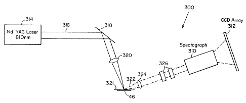
Figure 18 is a schematic diagram of a preferred system for
implementing the spectrograph/CCD Raman detector of the present
invention.
Figure 19 graphically illustrates spectrograph/CCD-Raman
spectra of normal human aorta: A) Raman plus fluorescence
spectrum produced by illuminating the tissue sample with 810 nm
laser light; B) Raman difference spectrum produced by subtracting
spectra produced by illuminating the tissue sample with 810 and
812 nm laser light; C) resulting Raman spectrum produced by
integrating the Raman difference spectrum of B).
Figure 20 graphically illustrates spectrograph/CCD-Raman
spectra of an atherosclerotic plaque with a calcified deposit exposed
at the surface: A) Raman plus fluorescence spectrum produced by
illuminating the tissue sample with 810 nm laser light; B) Raman
difference spectrum
CA 02104960 2002-10-03
- 11 -
produced by subtracting spectra produced by illuminating the tissue
sample with 810 and 812 nm laser light; C) resulting Raman
spectrum produced by integrating the Raman difference spectrum of
B).
Figure 21 graphically illustrates a spectrograph/CCD-Raman
spectrum of adventitial adipose tissue.
Detailed Description
The spectroscopic methods of the present invention can be
performed on a system such as that for laser treatment of
atherosclerosis which is illustrated in Figure lA. Figure 1A includes
separate block diagrams for the system of the invention which
utilizes laser light for spectroscopic diagnosis as well as for
treatment and/or removal of tissue. The ablation laser 225, pulse
stretcher 229 and the pulse filler/multiplexer 231, 233 produce an
output laser ablation pulse of sufficient energy and intensity to
remove tissue and sufficient pulse duration to propagate through a
fiber optic laser catheter delivery system without damaging the
fibers. These systems and methods are more fully described in
U.S. Patent No. 5,312,396 published on May 17, 1994.
To remove plaque, a device 219 is used to contact the tissue
such as multiple-fiber laser catheter 10 of Figure 1B with an optical
shield. The catheter 10 is inserted into the artery and the distal end
of the catheter is brought into contact with the lesion. Next, a
determination is made as to the type of tissue at which each optical
fiber
CA 02104960 2002-10-03
-12-
20a-c' is aimed. Only fibers aimed at diseased tissue are activated.
Thus, selective tissue removal is obtained. Furthermore, this
technique is also applicable for guiding surgical procedures in other
organs and tissue types such as the colon and bladder.
The present invention relates to systems and methods of
performing spectral diagnostics to diagnose the tissue in front of
each fiber. A preferred embodiment is a laser light source 207 that
is coupled to the fibers. The diagnostic light is sent to the fiber of
choice by the optical fiber selector 217.
The diagnostic light exits the selected optical fiber and falls on
the tissue. The tissue absorbs the light and a fraction of the
absorbed light is re-emitted, by Rayleigh fluorescence, Raman or
other elastic or inelastic light scattering processes. This light is
returned to the optical fibers and exits from selector 217, and is
detected by a photodiode, photomultiplier or other detector 203.
Returning light could use different optical fibers than those
employed for illumination. Diagnostic subsystem produces the
entire spectral signal which is coupled to computer 215.
The computer stores the information in a memory as a
spectrum, which is a graph of light intensity vs. wavelength. This
can be displayed immediately on the video display 82 or compared to
an existing spectrum stored in the computer and the difference
displayed on the spectra display 86. Temporal display 88 can
display corrections made for the wavelength dependent sensitivities
of the source.
CA 02104960 2002-10-03
-13-
Information from either the temporal or spectral display can be
stored in the computer 215. The comparative data is shown on
numerical display 84 to provide a quantitative measure of the health
of the tissue observed.
With a multichannel detector and a computer, or with
appropriate multiple filters and detectors, it is possible to gather
this information in a fraction of a second. Thus, a spectra or
numerical display is provided which indicates the type of tissue at
which the fiber of interest is aimed. If the tissue is plaque, and is to
be removed, then fiber selector 217 will align this fiber with the
output beam of the high power laser 225. Then, the high power
laser 225 is turned on and an appropriate power level is selected for
a predetermined amount of time to remove a certain amount of
diseased tissue. The beam of laser 225 is transmitted to pulse
stretcher 229 and pulse filler/multiplexer 231, 233 to properly adjust
the beam fluence.
The procedure is repeated for different fibers. Where diseased
tissue is detected, it is quickly removed. The laser catheter 10
nibbles away at the plaque, leaving the healthy artery wall intact.
If the artery 30 makes a bend 31 as shown by Figure 1B, the
laser catheter 10 will tend to make contact with artery wall 32 at the
outside wall of the bend. To prevent the catheter from contacting
the artery wall, the optical fiber 20c is not fired. The lesion is
removed asymmetrically. This allows the laser catheter 10 to follow
the lumen 39, 39a around the bend. Thus, the artery wall 32 is not
irradiated and is not perforated. Additional
CA 02104960 2002-10-03
-14-
details of this fiber optic catheter 10 are disclosed in U.S. Patent
No. 4,913,142.
In both Raman and ATR methods, information is contained in
the spectral lines which are observed in their intensities, and also
their linewidths and center frequencies (and how they change in
different environment). Further, Raman and infrared ATR have
different "selection rules". Some vibrations seen in infrared won't
show up in Raman, and vice versa. In other cases the same
vibration can be detected by both techniques, but with different
relative intensities (e.g. a strong Raman line will be weak in ATR).
So in this sense the two techniques provide complementary
information and combining the two techniques (or using either or
both with laser induced fluorescence) is valuable in diagnosing
pathology.
The methods can utilize Fourier transform detection to observe
the radiation thereby providing improved signal/noise ratios. Other
techniques (e.g. diode array detection and CCD detection) can also be
used.
As described in more details below contributions from major
tissue constituents can be "subtracted out" to reveal information
about molecules which are present in small concentrations. For
example, in ATR water contributions are removed before the "dry"
tissue constituents could be studied. Also, derivative spectroscopy is
used to eliminate background signals and low frequency filters.
Note that these techniques deconvolute the observed spectra into its
individual constituents, an
~:;'~ 92/15008 . PCf/US92/00420
..
-15-
essential step for optimal extraction of diagnostic
information.
While Kaman can sample deeply into tissue, ATR
samples only a very thin layer (a few microns).
Thus, ATR is "naturally" suited to probe surface
disease, such as the superficial cancers of the
bladder and GI tract, whereas Kaman is well suited
to providing information about conditions deep
inside tissue (such as breast cancer or stones).
This is important for 3D imaging. Furthermore, the
ATR tissue sampling depth can be controlled by
properly matching the probe surface material to the
tissue type.
Generally, the ATR signal is very sensitive to
the surface of the waveguide or probe. For example,
if the probe surface has an affinity for lipids in
the tissue, lipids can migrate to the probe surface,
creating a thin lipid layer and producing a.large
signal. This can be a problem (it can give
misleading information if not properly recognized
and guarded against). Conversely, it can be used to
advantage: Probes with special surfaces can be
developed to prevent this effect or to promote it,
in order to search for particular substances in the
tissue.
In a preferred method one can adjust depth
probed by ATR by varying refractive index of ATR
probe. Alternatively as discussed below one can use
a "waveguide needle" to get subsurface information.
Kaman diagnostic methods permit adjustment of
Kaman depth by varying the wavelength. Penetration
depth is wavelength dependent, and can be varied by
choosing different excitation wavelengths between
WO 92/15008 A l ~ ~ ~ PLT/US92/004 Aa~~
~s~~
-16-
about ~=700nm and 2~Cm. Another potentially
important way of adjusting Raman depth is to vary
the collection angle. In the near IR, incident
(exciting) light is strongly scattered out of the
forward direction into larger angles, so Raman
signals sampled at smaller angles come from tissue
closer to the surface. Therefore, the Raman
sampling depth can be controlled to a large extent
by probe design.
Depth information is important if one desires
to provide imaging by creating 3D images of small
tumors in the brain or breast. Differential
techniques based on the ideas of the preceding
paragraph might allow accurate localization of such
tumors in three dimensions. Near-IR Raman can be
combined with a sound wave technique (time of flight
or standing waves set up in the tissue)--the sound
wave would modulate the Raman signal emanating from
a point in the tissue when it passes that point, and
the modulated signal could be used to establish the
depth of the tissue producing the signal.
A system employed for the collection of Raman
spectral data from excised tissue samples is
illustrated in Figure 1C. FT-Raman spectra were
measured from 0 - 4000cm'1 below the laser excitation
frequency with a FT-IR interferometer 40 equipped
with a FT-Raman accessory. The accessory employed
at 180 back scattering geometry and a cooled (77K)
InGaAs detector 42.
A 1064 nm CW Nd:YAG laser 44 can be used for
irradiating a sample 46: utilizing 500 mW to 1 W
laser power in a 1.0 to 2.5 mm spot 48 at the sample
CA 02104960 2002-10-03
-17-
46 to collect Raman data. Alternatively, a pulsed laser source can
also be employed. Laser 44 generated a beam 45 that is directed
through plasma filter 48, mirrors 50, 52, focussing lens 54 and
mirror or prism 56 before irradiating the sample 46. The radiation
received by sample 46 undergoes various mechanisms of absorption,
reflection and scattering including Raman scattering. Some of the
light emitted by the tissue is directed toward lens 60 and then
through one or more Rayleigh filters 62. The collecting lens 60
collects this backscattered light 64 and collimates it. The Rayleigh
filters 62 removes the elastically scattered light and transmits the
inelastically scattered, frequency shifted (Raman) light. The Raman
scattered light enters the interferometer 40. No visible sample
degradation was observed under these conditions.
Excised human aorta was chosen of atherosclerotic artery
tissue. Samples were obtained at the time of post mortem
examination, rinsed with isotonic saline solution (buffered at
pH 7.4), snap-frozen in liquid nitrogen, and stored at -85°C until use.
Prior to spectroscopic study, samples were passively warmed to room
temperature and were kept moist with the isotonic saline. Normal
and atherosclerotic areas of tissue were identified by gross
inspection, separated, and sliced into roughly 8 mm x8 mm pieces.
The tissue samples were placed in a suprasil quartz cuvette
with a small amount of isotonic saline to keep the tissue moist, with
one surface in contact with the sample irradiated by the laser 44.
The spectra shown in Figures 2 through 6 were collected
WO 92/15008 PCT/US92/004 ,,~~~
CPU
-18-
with 512 scans at 8 cue' resolution (approximately 35
minutes total collection time).
Human aorta is composed of three distinct
layers: intima, media, and adventitia. The intima,
normally less than 300 ~m thick, is the innermost
layer and provides a non-thrombogenic surface for
blood flow. It is mainly composed of collagen
fibers and ground substance. The medial layer,
typically about 500 ~m thick, is quite elastic and
l0 serves to smooth the pulsatile blood flow from the
heart. The structural protein elastin is the major
component of aortic media, with some smooth muscle
cells present as well. The outermost adventitial
layer serves as a connective tissue network which
loosely anchors the vessel in place, and is mainly
made up of lipids, lipoproteins and collagen.
During the atherosclerotic process, the intima
thickens due to collagen proliferation, fatty
necrotic deposits accumulate under and within the
collagenous intima, and eventually, calcium builds
up, leading to calcium hydroxyapatite deposits in
the artery wall.
Figure 2a shows the FT-Raman spectrum of a full
thickness section of aorta grossly identified as
normal. Laser irradiation was on the intimal side.
The dominant bands appear at 1669 cm's and 1452 coal
and can be assigned to an amide I backbone and C-H
in-plane bending vibration from proteins,
respectively. Weaker bands at 1331 and 1255 cnii are
assigned to C-H wagging and amide III vibrations
from proteins, respectively. The frequencies of
t'!O 92/15008 ~ ~ ~ ~ ~ ~' ~ P(_'T/LJS92/00420
~ . ~.~ 7 f
v
-19-
amide I and III are consistent with those observed
for disordered proteins.
Another example of a typical NIR FT Raman
spectrum from normal aorta is shown in Figure 3.
When irradiated from the intimal side, Figure 3a,
the major vibrational bands observed in normal aorta
are all attributable to protein vibrations: the
band at 1658 cm'' is assigned to the amide I
vibration of the polypeptide chain, the 1453 cm''
band to a C-H bending mode of proteins, and the 1252
cm's band to the amide III vibration. The spectrum
of normal aorta is at least 25% weaker than any of
the pathologic samples. The peak frequency of the
C-H bending band, which averaged for all the normal
specimens is 1451~1 cm'', is specific to the protein
C-H bending mode (See below). The weak band near
1335 cm'', which appears as a shoulder in many of the
normal specimens, appears to be specific to elastin,
and the weak band at 1004 cm~ is likely due to
phenylalanine residues. In general, the relative
intensities of the bands in the region between 1250
and 1340 cm'' appears very much like that observed in
the FT Raman spectrum of elastin. This observation
is consistent with the thin collagenous intima in
normal aorta, the elastic nature of the media of
aorta, and the deep penetration depth of 1064 nm
radiation. Band assignments for all tissue types
presented here are listed in Table 2.
Figure 3b displays the NIR FT Raman spectrum of
the adventitial side of normal aorta. In this case,
the irradiated adventitial surface consisted of
several millimeters of visible adipose.tissue. In
WO 92/15008 PCT'/US92/004 ..
-2 0-
contrast with the spectrum collected from the
intimal side, the bands observed in this adipose
material appear to be mainly due to lipid, and in
particular triglyceride, with almost no contribution
from protein. This is not unexpected, as the
triglyceride content of adipose tissue is on the
order of 60%. The sharp band at 1655 cm' is due to
stretching of C=C groups in unsaturated fatty acid
chains. This band is distinguished from amide I by
its peak frequency and its width, which in this case
is 17 cm'' FWI~i. Amide I, in contrast, is roughly 60
cm'' wide. The dominant C-H bending band is shifted
to 1440 cm'', characteristic of lipids. This band is
about 3 times more intense in adipose tissue than in
normal intima, probably a result of the greater
number of C-H groups per unit volume in
triglycerides. The bands as 1301/1267 cm'' and 1080
cm'' are assigned to C-H bending and C-C stretching
vibrations of fatty acids, respectively.
The 1746 cm'' band, assigned to the C=O
stretching mode of the triglyceride ester linkages,
indicates that most of the lipid observed in the
adventitial adipose tissue is of the triglyceride
form. Specifically, the integrated .intensity of
this band relative to the C-H bending band at 1440
cm' is equal to 0.103, while this same ratio for
triolein is 0.136, which indicates that roughly 75%
of the C-H band is due to triglyceride. The NIR FT
Raman spectrum of triolein (a triglyceride
containing fatty acid chains of 18 carbons and a
single double bond) is shown for comparison in
Figure 3c. Additional molecular information
O 92/15008 PCT/US92/00420
~-~~~~~J~
-21-
regarding the state of the fatty acid chains is
readily deduced from the adventitial adipose
spectrum. For example, the relative intensity of
the C=C band at 1655 cm' indicates that there are on
average roughly 0.7 unsaturated double bonds per
fatty acid chain, assuming 16-18 carbon fatty acids.
In addition, the frequencies and structures of the
C-H bending and C-C stretching bands suggest that
most of the fatty acid chains are in the gauche
conformation. The sharp 1129 cm'' band,
characteristic of all-trans chains, is not observed
in the spectrum.
The FT-Raman spectrum obtained from a diseased
artery, an atheromatous plaque, with a thick fibrous
connective tissue cap and an underlying necrotic
core is shown in Figure 2b. The necrotic core of an
atheromatous plaque contains cellular debris as well
as large accumulations of oxidized lipids and
cholesterol. The band in the amide I region,
peaking at 1665 cm'', is distinctly narrower in this
spectrum compared to normal aorta. In addition, the
in-plane C-H bend, at 1444 cm', is relatively more
intense and has a distinct shoulder at higher
frequency. The two weaker bands at 1307 and
1267 cm' are shifted in frequency from those found
in the spectrum of normal aorta. The band
frequencies and shapes in the FT-Raman spectrum of
cholesterol, shown in Figure 2c, coincide with some
of those observed in the atheromatous plaque,
consistent with the expected composition of the
necrotic core.
WO 92/15008 PCT/US92/00f .~,.
,. a
-22-
The NIR FT Raman spectra of other fibrous
plaque specimens exhibit a range of features as
shown in Figures 4 and 5. Figure 4a shows a
representative spectrum from one of the types of
fibrous plaques. These fibrous plaque spectra are
quite similar in both relative and absolute band
intensities to the spectra of normal aorta. The
most significant differences are that the C-H
bending band, peaking near 1448 cm'' on average, is
shifted by 2 cm' to a slightly lower frequency.
This may be the result of a small increase in the
lipid content of these plaques relative to normal
aorta. In addition, the band near 1340 cm',
attributed to elastin in normal aorta, is decreased
relative to amide III at 1265 cm''. The putative
explanation is that the collagenous intima is
thickened in these specimens, so that the spectral
contribution from the elastic media is reduced
relative to that of normal aorta.
The NIR FT Raman spectra of other fibrous
plaque specimens appeared similar to atheromatous
plaques' spectra (Figure 2b). These are
substantially different than either normal aorta, or
adipose tissue. In these cases, the intense C-H
bending band occurs at 1440 cm'', characteristic of
lipid material. This band is roughly twice as
intense as the C-H bending band in normal aorta.
The complete absence of a band at 1746 cm'' indicates
that this lipid is not triglyceride. In fact, this
lipid appears to be predominantly cholesterols, as
identified by the sharp, characteristic band at 700
cm'' and comparison to the cholesterol spectrum shown
.'~O 92/ 15008 PGT/US92/00420
-23-
in Figure 4c. Again, this is not surprising, since
cholesterols accumulate in high concentrations in
atherosclerotic lesions. Several of the bands
between 1000 and 500 cm'' are assignable to
vibrational modes of the sterol rings. These
include the bands at 959, 882, 844, 805, 700, 605,
and 546 cm''. In addition, the 1666 c~1 band is
attributed in part to the C=C stretching vibration
of the steroid nucleus.
l0 The presence of fatty acid chains in the
atheromatous plaque spectra is evidenced by bands at
1300/1262 cm'' and 1130/1088 cm'', due to C-H bending
and C-C stretching vibrations, respectively. These
bands may contain contributions from cholesterol as
well. The relative intensities of the fatty acid
band at 1300 cm'' and the sterol ring bands suggest a
mixture of free cholesterol and cholesterol-fatty
acid esters. Moreover, the relative intensities of
the 1130 am' C-C stretching and the 700 cm'' sterol
bands indicate that most of the fatty acid chains
are in the gauche conformation, consistent with the
predominance of unsaturated fatty acid chains in the
cholesterol esters in these plaques. It is
particularly noteworthy in the atheromatous plaques
that the cholesterol deposits are detected from
material below a thick fibrous cap indicating the
ability of the NIR FT Raman method to probe
materials several hundred microns below the tissue
surf ace .
In addition to the cholesterol and cholesterol
ester bands, the NIR FT Raman spectra of some of the
fibrous plaques contained two unique bands, at 1519
cm'' and comparison to the ch
wo 92nsoog Pcrius9aioo4 c j
-24-
and 1157 cm''. The intensities of these bands are
highly correlated, which suggests that they are due
to a single component. These bands, which have been
previously observed in visibly-excited Raman spectra
of atherosclerotic plaques, are assigned to
carotenoids. The amount of carotenoid in these
plaques is probably much smaller than the amounts of
cholesterols or proteins, but may be strongly pre-
resonance enhanced (14). The carotenoid bands are
observed only in this subset of fibrous plaques.
In an advanced plaque, calcium may begin to
accumulate leading to the formation of calcium
hydroxyapatite crystals in the tissue as shown by
the spectra of Figures 5 and 6. The FT-Raman
spectrum of a calcified plaque with a thick (several
hundred microns) fibrous connective tissue cap
overlying a calcified deposit is shown in Figure 5a.
The spectrum clearly indicates bands due to the
protein in the fibrous cap, amide I and III at 1665
and 1255 cm', respectively. However, additional
bands are observed between 1250 and 1350 coal and
around 1100 cm'', as well as a strikingly sharp band
at 961 cm'. The latter is readily assigned to the
symmetric phosphate stretching vibration associated
with phosphate groups in the calcium hydroxyapatite
deposits, while the band around 1100 cm' is an
asymmetric phosphate stretch. These assignments are
confirmed by excising the solid "rock" from a
different calcified plaque, and obtaining its
spectrum as shown in Figure 5b. A strong Raman
signal from the phosphate stretching vibration in
solid calcifications in advanced atherosclerotic
'~~.0 92/15008 PGT/US92/00420
."~,~~.
y',~ ~.~,
-25-
plaques can also be observed utilizing standard
visible Raman instrumentation. The ability to
detect the calcifications several hundred microns
below the tissue surface when using near IR FT-Raman
spectroscopy, however, is a diagnostic measurement
which can be utilized to guide treatment.
A measurement was attempted to determine if the
calcification might be detected when the tissue was
irradiated from the adventitial side. The resulting
FT-Raman spectrum is shown in Figure 5c. No
evidence of the strong phosphate vibration is
apparent. In contrast, sharp vibrational bands at
1745, 1656, 1444, 1303, 1267 and 1082 ciri' are
observed which are mainly associated with the lipid
material that constitutes the majority of the
adventitia.
The NIR FT Raman spectrum of calcified plaque,
containing a subsurface calcified deposit and an
overlying soft fibrous cap, exhibits an intense,
sharp, new band at 960 cm'' (Figure 6a). This band,
specific to calcified tissue, is assigned to the
symmetric stretching vibration of phosphate groups
(15), which are present in high concentrations in
the solid calcium salts. The weaker phosphate
antisymmetric stretch is also present at 1072 cm''.
A symmetric stretching vibration of carbonate groups
may also contribute to this latter band. The
phosphate vibrations are easily observed from
subsurface deposits in the calcified plaques: the
960 cm'' band can be observed from deposits up to 1.5
mm beneath a soft tissue cap with the current
signal-to-noise level (See below). The calcified
WO 92/15008 ~ PCT/US92/004
-26-
plaque also displays protein vibrations from the
fibrous tissue cap. These include amide I at 1664
cm' and amide III near 1257 cm''. The C-H bending
band at 1447 cro' suggests a mixture of protein and
lipid, and the weak band at 699 cm'' is likely due to
cholesterol that is either in the fibrous cap, the
calcified deposit, or both.
The NIR FT Raman spectra of exposed
calcifications (Figure 6b) display a range of
features. In all cases, the major bands are due to
the calcium salts. These include the 960 cm'
phosphate and 1072 cm'' phosphate/carbonate bands as
well as the band at 587 cm'', which is assigned to
another phosphate vibrational mode. On the other
hand, several differences are apparent in the weaker
bands, which are presumably due to soft tissue
components which are embedded in the calcification.
In some cases (not shown), the C-H bending band
occurs at 1450 cm's, and the band at 1663 cm'' is
similar in shape to amide I for some of the
calcifications, indicative of protein vibrational
modes. In other calcified plaques, such as that in
Figure 5b, the C-H bending band occurs at 1440 cm',
and the 1667 cm'' band, which is much sharper, is
more like due to C=C stretching vibrations. In this
plaque, the bands are due to lipid, in particular
cholesterols, as evidenced by the 700 cm'' and 1300
cm'' bands.
In our histological examinations of aorta, two
distinct types of exposed calcifications have been
noted. In one type, the fibrous tissue cap is
calcified. In the other, the necrotic core of an
'~Q 92/150(?8 '.~F :# ;T , :;a ,. Pi'fI~JS92/00420
-27_
atheromatous plaque is calcified, and the calcified
deposit is exposed by ulceration of the soft tissue
fibrous cap. A positive explanation for the two
spectral types of exposed calcifications is that the
specimens which exhibit protein bands are of the
former histologic type, while the specimens which
exhibit lipid bands are of the latter type.
The present methods provide an IR FT-Raman
technique for differentiating various stages of
atherosclerosis in human aorta. They demonstrate
that molecular level information is available using
these methods. This information is useful for
following the pathogenesis of the disease and in
guiding the treatment of different lesions. The
near IR FT-Raman method, with its relatively deep
penetration depth, is able to obtain spectroscopic
signals from below the tissue surface, yielding
details about the atheromatous necrotic tissue and
sub-surface calcifications. These signals can be
utilized with an optical fiber based imaging system
to determine the content and composition of
different atherosclerotic plaques in vivo.
With the observation that several of the
biochemical species important in the atherosclerotic
process, including cholesterol and calcium
hydroxyapatite, can be easily detected below the
tissue surface, we wished to determine the depth
limit of detection using the NIR FT Raman technique.
In order to address this question, ten 200 ~Cm
sections of aortic media were cut and placed one at
a time over a large calcified deposit (6x6x3 mm),
and the FT Raman spectra of the 960 cml band
CA 02104960 2002-10-03
-28-
monitored as a function of depth below the surface. As indicated by
the plot of FT Raman intensity versus depth shown in Figure 7, the
signal from the calcified deposit was detectable until the deposit was
greater than 1.6 mm below the irradiated surface. Even slightly
deeper depths could be probed if the focus of the collection optics was
moved into the tissue.
The two dimensional resolution of the NIR FT Raman signal
for material below the tissue surface was then tested by placing
1 mm of aortic media above another calcified deposit, and moving
the tissue transversely in two dimensions through the laser beam
and collection lens. The FT Raman signal was observed to drop-off
rapidly as the beam and collection optics moved from the calcified
deposit. The detected FT Raman signal closely followed the
geometry of the calcified deposit below the surface, despite the
significant scattering of the overlying layer of tissue. This result
suggests that the Raman scattered light may be utilized for imaging
objects below the tissue surface with minimal image blurring due to
elastic scattering in the tissue.
A second spectroscopic method is also used to obtain molecular
vibration information, attenuated total reflective (ATR) of infrared
light.
Human aorta was chosen as an example to illustrate the
diagnosis of atherosclerotic artery tissue. As in the samples
obtained for the Raman spectral measurements human aorta
samples were obtained for ATR measurements at the time of post
mortem examination. Sample storage and preparation procedures
are identical to those set forth for the
CA 02104960 2002-10-03
-29-
Raman spectral measurements. These reflectance measurements
can be used by themselves to provide diagnostic data in conjunction
with either the Raman spectroscopic measurements described above
or with fluorescence measurements, or with both types of
measurements to enhance diagnosis for specific applications.
The medial layers of a normal artery and the necrotic cores of
atheromatous plaques were exposed by blunt dissection and
spectroscopically examined. ATR spectra were also collected from
several purified tissue components including collagen, elastin, and
cholesterol to assist in analysis of the spectra.
Mid-infrared ATR spectra were measured from 4000 to
700 cm-1 with a commercially available FT-IR spectrometer and a
horizontal ATR accessory. The sampling area was purged with dry
nitrogen gas to control background absorption from atmospheric
water vapour and C02. Spectra were collected at 4 cm-1 resolution
with 50 scans. The artery specimens, kept physiologically moist
with isotonic saline (buffered at pH 7.4), were placed in contact with
the ATR element (ZnSe crystal 45 ends). A 5 gram weight placed on
the tissue sample ensured uniform sample contact with the ATR
element. An ATR spectrum of the saline solution with absorbance
similar to that of the artery samples was also obtained and used for
subtraction of spectral components due to water. Collagen
(CALBIOCHEM; trade-mark: type I, bovine achilles tendon) and
elastin (SIGMA; trade-mark: bovine neck ligament) were prepared
as saline
CA 02104960 2002-10-03
-30-
slurries. Cholesterol (Sigma) was prepared as a dry film on the ATR
element by evaporation of a benzene solution. These elements can
be clearly identified in the resulting spectra.
The ATR sampling crystal is a rod of high refractive index
material which acts as a waveguide for the infrared sampling beam.
This waveguide can be in the form of a needle that is adapted for
penetration into the tissue to be diagnosed. Alternatively, the probe
will have a geometry suitable for contacting the surface of exposed
tissue sites or for contacting internal locations with a catheter.
The devices shown in Figures 16A and 16B illustrate preferred
embodiments of the invention adapted for ATR diagnostic
measurements within the human body. In Figure 16A a
single-ended probe 100 is shown where one or more optical fibers
102 coupled both the incident light to, and the transmitted
(reflected) light from the ATR element 104. A 100% infrared
reflector 106 such as gold is placed at the distal surface 108 of the
ATR element 104 functions to return the transmitted light back
through the same fiber as well as to provide double pass sampling.
The ATR element 104 can be a separate component optically
fastened to the optical fibers 102, or alternatively, it can be
constructed from the end of the optical fiber by removing the
cladding material. Sampling is provided by placing the ATR element
in contact with the tissue 110 of interest. Radiation is transmitted
112 and collected 114 in a radial direction from element 104. The
probe can either be inserted through a
'~'~O 92/15008 PCT/US92/00420
~a
Tl_y,'
I
.u
-31-
standard endoscope or catheter to sample a hollow
organ, or, if made with sufficiently thin optical
fiber, it can be directly inserted directly into a
solid organ as in the case of needle biopsy. In
this particular embodiment the distal tip 108 is in
the form of a needle. The cone or needle
configuration on the end of the catheter can be long
or shallow.
A double-ended probe is illustrated in Figure
16B. Incident IR beam from FT-IR is transmitted
through IR optical fiber 122 to ATR element 128
positioned at the distal end of catheter body 120.
The ATR element is placed in contact with tissue 126
surface to be sampled. Transmitted light is
conducted through a second IR optical fiber 124 back
to an IR detector. The ATR element may be a
separate component optically fastened to the two
optical fibers 122, 124, or it may be simply a
region of a single optical fiber in which the fiber
cladding material has been removed. The entire
apparatus can be inserted through a standard
endoscope or outer catheter.
For methods of measuring excised samples, the
specimen to be sampled is placed in optical contact
with the surface of the waveguide or ATR element.
The evanescent wave which extends outside of the
waveguide surface is absorbed by the sample in
proportion to its absorption coefficient. The
penetration depth of the evanescent wave into the
sample depends on the wavelength of the infrared
radiation and the refractive indices of the
waveguide and the sample; for a ZnSe-water
interface, this depth is roughly 1 ~cm from 1800 to
WO 92/15008 PCT/YJS92/004
i~ ~~~)~
'~
-32-
700 cm''. The 1/e penetration depth of the
evanescent wave into the sample is given by
~/2n (nZ2sinz6-nW2)''~, where ~ is wavelength, 9 is angle
of incidence and nZ and nW are the refractive
indices of ZnSe and water respectively.
Consequently, only tissue that is in good optical
contact with the ATR element will be sampled. In
addition, individual components in the sample can
exhibit different affinities for the waveguide
material (ZnSe in this case), which can influence
the relative concentrations of the components at the
waveguide surface. Despite relatively high
concentrations in the bulk tissue, components with
poor optical contact can be difficult to measure in
the ATR spectrum.
Figure 8 shows FT-IR ATR spectra of (a) normal
aorta (intimal side) and (b) buffered saline. A
comparison of these spectra shows that a majority of
the IR absorption of normal intima can be attributed
to water, which comprises roughly 80% of the tissue
by weight. The large, broad bands peaking at 3300
cm'' and 1636 cm' are due to the O-H stretching and
H-O-H bending vibrations, respectively, of water,
and the weak band at 2120 cm'' is due to a water
combination vibration. The 3300 cm'' and 1636 cm''
bands also include contributions from the N-H
stretching and amide I vibrations. The relatively
flat absorption between 1500 and 900 cm'1 and the
rising absorption below 900 cm'' is also due
primarily to water; however, in the intima, a number
of very weak bands due to other tissue components
are also present in this region.
CA 02104960 2002-10-03
-33-
Most biomolecules give rise to IR absorption bands between
1800 and 700 cm-1, which is known as the "fingerprint region" or
primary absorption region. The dominant absorption of tissue water
in this region obscures the absorption brands from other tissue
components. To observe the IR bands from these components, one
must eliminate the water interference. With the ATR method,
spectral deconvolution or subtraction of the water component is
particularly easy. By using the 2120 cm-1 band, which is due solely
to water, as an internal intensity standard the spectrum of buffered
saline (Figure 8b) can be accurately and reliably subtracted from the
spectrum of aorta intima (Figure 8a), yielding a water-subtracted
spectrum of intima (Figure 9a).
In the water-subtracted spectrum, the previously weak bands
are easily observed. Band assignments, based on the spectra of the
major tissue components are listed in Table I. Most of the
vibrational bands observed in the spectrum of normal intima
(Figure 9a) can be divided into two broad categories: lipid-associated
bands and protein-associated bands. All of the strong bands in
normal intima are associated with one of these moieties (see
Table I). This can be seen as a simple consequence of the large
concentrations of these two materials. Aside from water, a large
fraction of tissue can be divided into one of these two groups.
Moreover, both protein and lipid components contain repeating
molecular units which are common to all members of the group. For
~'~~~ ~'~~~~) ~ PGT/US92/004?~;
WO 92/15008
-3 3 . 1-
Table I. Preliminary assignments of IR absorption peaks in
the ATR spectra of normal aorta intima.
V Preliminary Vibrational Associated Tissue
ji-1cm-'~Assignment Components
2923(x) C-H stretch Lipid, Protein, Others
2853(x) C-H stretch Lipid, Protein, Others
1744(x) C=O (ester) stretch Lipid
165I('s)Amide I Protein
1635(sh)Amide I, H-O-H bend Protein, Water
1548(x) Amide II Protein
1465(x) CH2 bend Lipid
1457 CH2 bend, CH3 anti- Lipid
(s)
symmetric deformation.
1454(x) CH bend, CH3 anti- Protein, others
symmetric deformation
1417(w) CHz bend adjacent to C=O Lipid
1401(m) COO' symmetric stretch, Protein, others
amide C-N stretch
1378(w) CH3 symmetric Lipid
deformation
1244(m) Amide III, POz anti- Protein, others
symmetric stretch
1239(m) CHZ wag, P02 anti- Lipid
symmetric stretch
1159(s) CHz wag, C-O-C Lipid
antisymmetric stretch
1117(w) C-C stretch, O-C-o Lipid
stretch
1096(w) Lipid
1083(w) POa symmetric stretch Protein, others
1030(w) Lipid
965(w) C=CH deformation (trans) Lipid
722(m) CHz rock Lipid
SUBSTITUTE SHEET
~.'u0 92/15008 '~ ;~ '~ PCT/US92/00420
,....:V,~
-33.2-
r~
O
ro
-~
N -. ~o ,~ x
~ v -~
~ ~ o
x '~ p
p y ~ U _ a U .-. U H _ p
U ~ ~ ~ T3
N f.1 H y, ~ ~ ~ p ~
1~ -1 ,i N ' N tl
~
~ CS ~ H .
K3 ~ ~
~ ~ ~ H N
U ri
.-I ~
~' ~ ~'a ~ N p ~ v w p a
~' ~ v d ~~ ~r
~ c;~ a
~a
. , ~ , ~o ~, v
, ,r
.~ ..1
U t~ O U ~rV ~ + a b
N .~
v ti a w v x~ a ~ ~ v
o a ~
~w a
a
x
H
b 'p
H m 3 3
o 0
v
f0 ~ V' 1"1 N
V
x~
r' w
~a
U
O
,~
H
p ~ ~ N 3 3
c~ c~ o .~ co
p to vD tf1 1D N
V
H
N k
G w
U
,G
N 3 3 3
3
' o o ~ '
~
~ ~ o
tr o
b ~' f"~ N r1
O
H v-i ri r-1
ri
r-1
DI
N
N
O ~ 3 N 3 3 3 3
~ 3
p ~
p ' ~ ~ o W i ~
~ ~ m
~ " 1 l
O
(n .L1 y 0 ll1 C f r r
1 N
H H ~i ~i H H H
H
U
' 'ro
v ~ 3 ~ ~ N ~ 3
~ o o ~ 0
"., .~ ~ ~ v o
0
at c'1 N O
ro
,o
'
a
H ro ~ ~
.-
H O r
is y p et N
H O H ri r-1
z
H
US.S'i'1TUTE SHE:E'~'
WO 92/15008 ~~ ~ PCT/US92/004,~
c~; ~ ~ ~ -33 . 3- ,.
m
II
N U N d N N ~ N
-r~1 G ~ r.l
+~
N
c c ro c ;'~x ~ ~ ~ .
~~ ro .
o . ro , ~,... b N..,N
~o d ~~,~
v N N...
~ ~~ ~ '' ~ ~m~ n,~~ o
~''' oc ~~'
'
o, ~ N
_ .
~ ~ V
N N U ~ N N O .4" N
N ~.. -r1 N U G ,..
W +~ ~
H ~ W
b , . U
U U
U W N
H
N N 3 3 3 l~ ~ N
3
N W H ~ CO A sr 01 r G
O r co tv
~
O -.i r ~ r w O rn lfl
V O to t0 t0
tD ll1
X r- H w
I
wro
U
+I
Tt
G.
H O
rt
N
r O
d
O r O N .~
il.~ O tr1 >
p
W r-' Ov ~
~
U U
~
~
r
1
N
b
3.1
~
N ~ '~' 1 O 1G ~0
l .x
Ql
. y r ~
~D N r ~
nco~oooa N
rtf o~ co N co N
~ r vo m 3
O
i 'O
_
U
N p 3 O.
G
N
~
r N N l'1 H
O ~D ~D
~
~
c ro~
o o~v
~c
ro
~
.~
>;
'
a
w~
0
.N N
G
U
O
U
N
~
O
H fIf cT ~
W
~
.i
G
' H ~
~
,(?, .O
~ .
~,
N
a
~t 1BSTITU'TE SHE~~
~ S ~d n ''? ,;~
:~''RO 92/15008 '~' -~ ~~ '~,w :,~3 (9
PGT/US92/00420
-34-
protein, the polypeptide backbone of repeating amide
groups is the dominant element. In lipids, the
repeating hydrocarbon chain is the defining quality.
The end result is that these molecular units are
present in very large concentrations, and their
vibrational bands tend to dominate the spectrum.
Note that this does not imply that no further level
of detail is derivable from the IR spectrum of
tissue. For example, the frequencies of the amide
l0 group vibrations are sensitive to protein
configuration and conformation. Therefore, shifts
in protein makeup might be expected to produce
observable changes in the amide bands.
The water-subtracted spectrum of sub-
adventitial fat shown in Figure 9b, more clearly
illustrates the division of bands into lipid and
non-lipid categories. This fat can be considered as
the model of the lipid component. Protein '
contributions, as judged from the intensities of the
amide I and II bands, are negligible for the
purposes of this model. All of the bands observed
in the fat spectrum can be attributed to the lipid
component. These include the strong bands at 1744
cm~ (C=O stretch), 1465 cm~ (C-H bend), 1161 cm'' (CHZ
wag, C-O-C stretch), as well as the weaker bands at
1378 cni~, 1239 cm'', 1118 cm'', 1099 cm's, 966 cm'', and
722 cm'1.
The bands observed in the water-subtracted
spectrum of intima constitute less than 30% of the
total absorption, the rest being due to water. Any
conclusions regarding these relatively weak bands
depends critically upon the accuracy of the water
WO 92/15008 PGT/US92l004 ;a:;_1,
~~ i~~J~
-35-
substraction. The accuracy of this subtraction can
be judged from the reproducibility of spectra
obtained sequentially from the same sample. Two
consecutive water-subtracted spectra collected 10
minutes apart from a sample of normal aorta (intimal
side) are shown in Figure l0a (solid and dashed
curves). Most of the IR bands exhibit a substantial
increase in absorbance with time. This trend
continues for consecutive spectra collected more
than an hour after the placement of the sample on
the ATR element. However, not all of the bands
change by the same fraction, so that the relative
intensities differ between consecutive spectra. For
instance, in Figure 10a, the C=O band at 1744 cm' is
relatively constant, while the amide bands at
1650 cm' and 1547 cm'' increase by 50% in the later
spECtrum. Although these changes might seem to
indicate that the water subtraction is inaccurate,
the changes with time are systematic and
predictable, which suggests that the optical contact
between the sample and the ATR element is changing
regularly with time.
In fact, the constancy of the 1744 cue' C=O
band, which is due solely to lipid, and the
increases in the amide bands, which are due to
protein, indicate that the lipid contributions to
the IR absorption remain unchanged while the non-
lipid contributions increase between consecutive
scans. This is confirmed by subtracting the
spectrum of lipid (Figure 9b) from the water-
subtracted spectra of aorta intima (Figure l0a),
using the 1744 cm' band for normalization. The
~'~,K~O 92/15008 '~ 1 ~ ~ . ~ PCf/L1S92/00420
Y~~F
-36-
resulting lipid-subtracted spectra of aorta intima
are shown, normalized to peak absorbance, in Figure
lOb. As can be seen, the relative peak absorbencies
and spectral bandshapes in the lipid- subtracted
spectra reproduce quite well, reflecting the
accuracy of both the water and the lipid-
subtraction procedures. ,
The constancy of the lipid bands and the
variation of the non-lipid bands between successive
scans may seem somewhat puzzling. An explanation of
this apparent anomaly can be inferred from a water-
subtracted spectrum of saline solution which is
rinsed off the surface of the tissue (Figure 9c).
This spectrum, aside from the weak amide I and II
bands, matches quite closely with that of
adventitial fat. The lipid component observed in
the tissue appears to be due to free lipid particles
that have equilibrated with the tissue surface
water, forming a thin water-lipid film on the tissue
surface which is in full optical contact with the
ATR element immediately after the tissue specimen is
placed upon the crystal. The tissue components
beneath this film presumably achieve better optical
contact with the ATR crystal as the sample settles.
As a result, the content of lipid in a spectrum of
aorta intima or media may be influenced by the
presence of sub-adventitial fat in the specimen, and
the relative lipid-protein absorbencies are accurate
to 50~ at best with the present experimental design.
For the reason, all of the remaining spectra shown
are both water and lipid subtracted.
With the lipid bands removed, assessment of the
non-lipid bands in the spectrum of normal intima
,.
WO 92l150i18 PCT/US92/004
~.ri
-37-
(Figure 10b) is greatly simplified. The major bands
in the spectrum may be assigned to protein backbone
vibrations. These include the bands at 1648 cm'
(amide I), 1549 cm' (amide II), 1455 c~i' (C-H bend),
1401 cm' (amide C-N stretch), and 1244 cai' (amide
III). The frequency of the amide I peak (1648 cm'),
which is sensitive to protein secondary structure,
may indicate contributions from a-helix, disordered,
and collagen helix conformations. This band also
exhibits a shoulder near 1634 cm'', which may be due
to the (3-sheet regions of proteins or water. The
protein C-H bending band at 1455 cm' is distinct
from the corresponding vibration in lipid, which
occurs as a double-peaked band at 1465/1457 cni'.
Note that all of these bands may include
contributions from other moieties. For instance,
the symmetric stretch of carboxylate groups and the
antisymmetric stretch of phosphate groups may also
contribute, respectively, to the 1401 cm'' and 1244
cm'' bands. This correlation of components is
summarized in Table I above.
A typical spectrum of the medial layer of
normal aorta is shown in Figure lla. A comparison
of this spectrum to that of normal intima (Figure
lOb) fails to reveal any significant differences.
This result is somewhat surprising, considering that
normal aorta intima and media have significantly
different compositions. Typical spectra of an
atherosclerotic plaque and a non-ulcerated
atheromatous plaque are shown in Figures llb and
llc, respectively. For these plaques, only the
intact fibrous cap at the intimal surface is probed
CA 02104960 2002-10-03
-38-
due to the short penetration depth (1 Vim) of the beam. Any necrotic,
atheromatous material beneath this fibrous cap is not sampled.
Even so, the fibrous caps of these plaques are known to be
compositionally different than normal intima and one might expect
these differences to be reflected in the IR ATR spectrum. However,
as in the case of media, no consistent differences are observed in the
spectra of these plaques (Figures 11B and 11C) and normal intima
(Figure lOb). This issue will expand upon in the discussion below.
On the other hand, substantial differences are obvious in the
spectrum of the necrotic, atheromatous core of an atheromatous
plaque (Figure 12a) as compared with the corresponding spectra of
normal intima (Figure lOb) as well as those of intact atherosclerotic
(Figure l lb) and atheromatous (Figure llc) plaques. In this case,
the necrotic core was presumably exposed in vivo as disease
progressed by ulceration of the overlying intimal fibrous tissue cap.
(The spectrum of necrotic core exposed by dissecting away the
fibrous cap of a non-ulcerated atheromatous plaque is similar).) A
new band appears at 1050 cmu, with a secondary peak at 1023 cm-1.
In addition, the necrotic core spectrum exhibits an increase and
frequency shift in the 1466 cm-1 band as compared with the
1455 cm-1 protein band in normal intima as well as a set of unique
bands near 1382 cm-1. These characteristic bands are found in the
spectra of all the exposed necrotic core samples and in none of the
other samples (see below).
WO 92/15008 PCT/US92100d_
-39-
The source of these unique bands in the
necrotic core spectra may be cholesterol, which is
known to accumulate in large amounts in atheromatous
cores. An ATR spectrum of cholesterol (dry film) is
shown in Figure 12b. The three major bands unique
to the necrotic core, near 1463 cm'', 1382 cm'', and
1050 cm'', match closely in position and relative
intensities with the three main cholesterol bands at
1466 cm'', 1377 cm'', and 1056 c~i'. Each of the main
l0 cholesterol bands has a secondary peak, which also
appear to be present in the necrotic core bands.
These secondary peaks occur at 1445/1436 cm'', and
1023 cm'' in the cholesterol spectrum and at 1441 c~
1367 cm'', and 1023 cm~ in the necrotic core
spectrum. In addition, several of the weak bands in
the necrotic core spectrum, including the peaks at
1334 cm'', 1109 cm'', 954 cm'', and 797 cm'', are
associated with the weaker cholesterol bands near
these frequencies. Other components in the necrotic
core may also contribute to some of these distinct
bands.
The consistency of the spectral differences
which are attributed to cholesterol between the
necrotic core specimens and the normal,
atherosclerotic, and non-ulcerated atheromatous
specimens are illustrated in the scatter plot in
Figure 13. This plot depicts the integrated
intensities (areas) of the 1050 cm' cholesterol band
ratioed to the total protein content, as measured by
the area of the amide II band at 1548 cm''. The 1050
cai' band was integrated from 1075 to 1000 cai' and
baseline subtracted using these endpoints, and the
'.'O 92/15008 PGT/bJS92/00420
21~~~~~~
-40-
amide II band was integrated from 1593 to 1485 cm'
with a similar baseline subtraction. This ratio is
a measure as the relative cholesterol contribution
to the spectrum, and is proportional to the relative
cholesterol concentration of the sample with the
assumption that the area of the 1050 cm'' band is due
solely to cholesterol. As can be seen in Figure 13,
this ratio is higher for all the exposed necrotic
core specimens than for all the other specimens.
l0 The consistent results of this sample analysis,
which is possible because of the separation and
molecular identification of the bands, indicates the
potential of TR spectroscopy for tissue
characterization.
Investigations of human arteries and
atherosclerosis by mid-IR spectroscopy have been
limited to date. It has been reported that ATR
spectra have been recorded from partially dried
human artery, among other tissues. In comparing a
normal aorta from an infant to an atherosclerotic
plaque in an adult, they observed increases in
several bands in the atherosclerotic aorta. Most of
these bands were associated with lipids and
lipoproteins. IR spectroscopy has been employed to
determine the chemical composition of calcified
atherosclerotic deposits. A more detailed IR study
of atherosclerotic aorta involves recorded IR
transmission spectra from thin layers sectioned at
different depths into the arterial wall. Results
showed increased absorption near 1739 cm' in the
fatty (atheromatous) regions of plaque, which was
attributed to absorption by cholesterol esters in
WO 92/15008 PCT/US92/00420
-41-
the plaque. IR spectra from the fibrous tissue cap
at the surface of the plaques were similar to normal
intima.
One of the main difficulties in measuring mid-
:infrared spectra of intact human tissue is the
intense water absorption, which dominates and
obscures the absorption of other tissue components
of interest. In most of the studies cited above,
the water absorption was not eliminated, limiting
the quality and amount of information available from
the spectra. With the ATR sampling method, this
water interference is easily removed (see Figure 9).
The ATR method is also naturally amenable to
sampling with fiber optic probes in vivo. Water
interference in fiber optic probe ATR spectra of
aqueous protein solutions has been accurately
eliminated with a water subtraction procedure
similar to the one employed in the present study.
While the ATR method is well suited to in vivo
sampling and to accurate subtraction of the water
signal, spectra collected with the ATR method are
not equivalent to TR absorption spectra, but depend
on properties of the ATR material and the sample in
addition to the sample absorption coefficient. For
instance, the penetration depth of the evanescent
sampling wave depends on the refractive indices of
the ATR material and the sample. However, the
refractive indices of both ZnSe and human tissue are
expected to vary slowly with frequency between 1800
and 700 cm'' and such variations will at most affect
the relative intensities of bands at different
frequencies. All of the structure observed in the
~~O 92/15008 PCT/US92/00420
,~ yY
-42-
tissue spectra is attributed to absorption bands in
the tissue.
The component absorptions observed in an ATR
spectrum also depends upon the optical contact of
the sample and ATR element. The small penetration
depth of the evanescent wave into the tissue sample
implies that only a 5 ~,m thick layer, and preferably
about 1 micron, of material at the surface is
observed. This is referred to as the near surface
region of the tissue for the purposes of this
application. The tissue deeper than 5 microns from
the surface is defined as the sub-surface region.
This thin, sampled near-surface layer may differ in
composition with the bulk sample. For example, a
film of free water may be present on the surface of
wet tissue, with different levels of some molecular
species of the tissue relative to their
concentrations in the bulk tissue. In addition, the
varied affinities for the ATR material of different
2o moieties in the tissue may play an important role in
the intensities of the observed bands.
These considerations may explain the lack of
substantial differences among the ATR spectra of
normal intima, plaque fibrous cap, and media. For
instance, normal aorta intima is composed of roughly
25% collagen (dry weight) and 20% elastin, while
aorta media has 20% collagen and 50% elastin. The
ATR spectra of purified collagen and purified
elastin (not shown) differ substantially. In
particular, amide I/II occur at 1657/1556 cm'' in
hydrated collagen (type I) and 1653/1543 cue' in
hydrated elastin (spectra not shown).
WO 92/15008 PCTlU592/004
-43-
One might expect these differences to be
reflected in the intima and media ATR spectra. A
possible explanation of why this is not the case is
that the thin layer in optical contact wit the ATR
element is compositionally different from the bulk
tissue, and collagen and elastin make only a minor
contribution to the IR ATR bands of this layer.
Such an effect may also explain the lack of
significant differences among the plaque fibrous cap
intima and normal intima ATR spectra. In ATR
elements made of other substances with different
biochemical affinities, the spectral differences
among these tissues can be substantially enhanced
depending on the tissue type.
The results of the present investigation
demonstrate that high quality water-subtracted
spectra can be readily obtained from human tissue
with the ATR technique. Similar results have been
obtained in other mammalian tissues. Accurate
removal of the water interference is crucial to
isolating the relatively weak tissue absorption
bands of lipid, protein, as well as other tissue
components. It is worth noting that the observation
of these relatively weak bands via spectral
subtraction depends entirely upon quality of the
tissue and saline spectra. For instance, the
absorbance of the normal intima specimen (Figure 8a)
between 1500 and 900 cm'' is approximately 0.06. In
the water-subtracted spectrum (Figure 9a), the peak
absorbencies range from 0.018 (30%) for the
strongest bands to 0.003 (5%) for the weakest ones.
The detection of a 0.003 absorbance peak in a
~~4 92/15008 IPCTiUS9210042U
-44-
subtracted spectrum with a 0.06 absorbance
background requires a signal-to-noise ratio of 700
or better in the 100% baseline. Such a signal-to-
noise is easily achieved with an FT-spectrometer.
The high linearity, baseline stability, and
wavelength precision of the FT-spectrometer are also
obviously critical for accurate background
subtraction.
While water subtraction is relatively easy and
accurate with ATR, it may be substantially more
difficult with other clinically applicable sampling
techniques such as diffuse reflectance or
photoacoustic sampling. These alternative sampling
techniques are clinically useful, however, because
of their longer tissue penetration depths
(approximately l0um). As an alternative to water
subtraction, one can exploit the properties of the
spectral lineshape of water to eliminate the water
signal by other computational methods. .
Specifically, the spectral lineshape of water varies
rather slowly with frequency over much of the region
of interest, especially between 1500 and 700 ciri'.
Therefore, any method which filters this slower
variation and spares the sharper features of the
non-water bands can separate the water and non-water
components.
One such method is second derivative
spectroscopy. Differentiation of a spectrum is
typically used to narrow absorption bands and
3o resolve overlapping peaks. Differentiation also
tends to deemphasize broad bands relative to sharper
ones. In IR spectra of artery, the broad,
WO 92/15008 PCT/IJS921~104
..
., .,
-45-
.featureless absorption of water can be nearly
eliminated in favor of the sharper non-water bands
by computing the second derivative of the spectra.
This is clearly demonstrated in Figure 14, which
shows the second derivative of a spectrum of normal
aorta intima (Figure 14a), along with the water-
subtracted spectrum of the same specimen (Figure
14b). Essentially only the 1633 cm'' water band is
left, partially obscuring the amide I band.
Elsewhere in this spectrum, the water contribution
is minimal. All of the bands identified in the
water-subtracted spectrum are easily observed in the
second derivative spectrum.
In addition to elimination of water
interference, several of the unresolved double peaks
and shoulders in the water-subtracted spectrum
appear as distinct peaks in the second derivative
spectrum. For example, the amide II band in normal
intima (Figure 14b) has a very weak shoulder near
1518 cm'', and the C-H bending region near 1468 cm''
appears to include two overlapping peaks. In the
second derivative spectrum (Figure 14a), the 1518
cm'' band is clearly visible, and the C-H region
exhibits two separate peaks at 1469 and 1456 cm''.
Moreover, by sharpening the bands, the second
derivative spectrum allows a more precise
determination of peak frequencies, so that
relatively small frequency shifts are observed.
Such frequency shifts can be of importance in
detesting and characterizing subtle molecular
alterations involved in certain tissue conditions.
CA 02104960 2002-10-03
-46-
The observation of individual, resolved bands in the artery IR
ATR spectra is of considerable interest, since separation of bands is
the first step determining the composition of a sample from its
spectrum. Once a band has been isolated, its integrated intensity is
S proportional to the concentration of the moiety responsible for that
band. In particular, since the amide I and II bands are due entirely
to protein, these bands can be used to isolate the overall protein
content in the spectrum. The sharp, well resolved 1744 cm-1 C=O
ester band appears to be due solely to lipid, and the integrated
intensity of this band should be proportional to the relative lipid
content. Finally, it should be remembered that the relative water
content of the tissue sample is automatically computed from the
2120 cm-1 band in the water subtraction algorithm. However, as
noted earlier, the composition of tissue as determined from an ATR
spectrum may not be precisely identical to the composition of the
bulk tissue.
The tissue composition can also be determined from
overlapping bands by first deconvolving the bands of interest into
their individual components. This is especially easy if one
component has an additional, isolated band elsewhere in the
spectrum. An example is the 1465 cm~l C-H bending region, which is
due to different tissue components with distinct spectral features in
this region. In the normal intima spectrum (Figure 9a), this band is
attributed to a combination of lipid and protein components.
CA 02104960 2002-10-03
-47-
Since the lipid component also exhibits the isolated 1744 cm-1 band,
this band can be used to subtract the lipid C-H bending component
and isolate the protein C-H bending component at 1455 cm-1
(Figure lOb), effectively deconvolving this band. Note that this
deconvolution depends on having a reliable spectrum of one of the
individual components, which in this example, is the lipid spectrum
in Figure 9b.
The detection of distinct bands attributed to cholesterol in
necrotic core may provide a useful means of determining cholesterol
concentrations in vivo. Both the 1050 cm-1 and 1382 cm-1 cholesterol
bands appear to be fairly isolated in the necrotic core spectrum after
lipid-subtraction (Figure 12). If these two bands are due to a single
component, namely cholesterol, the ratio of their integrated
intensities should be a constant for all the samples. The
baseline-subtracted area of the 1050 cm-1 band, A(1050), is plotted
versus that of the 1382 cm-1 band, (A(1382), for all the samples,
normalized to the protein content in Figure 15. As can be seen in
the plot, there is a roughly linear relationship between A(1050) and
A(1382). A linear least squares fit to this data yields the line shown
in the plot, with a high regression coefficient of r=0.967. The slope of
this line is 2.8, while the ratio A(1050)/A(1382) for the pure
cholesterol ATR spectrum is 2.3. The reasonable agreement between
these two numbers provides additional evidence for the assignment
of these bands to cholesterol. Moreover, it indicates that the relative
spectral content of cholesterol is reasonably approximated by
CA 02104960 2002-10-03
-48-
the integrated intensities of either of these bands. Figure 15 also
shows that the ATR spectra of all the specimens other than exposed
necrotic core exhibit almost no intensity in both the 1050 and
1382 cm-1 bands, in contrast to the necrotic specimens, all of which
have significant bands at both frequencies.
The present systems and methods demonstrate that infrared
spectra of moist, bulk tissues can be reliably obtained with the ATR
technique. Although water is the dominant absorber throughout
much of the mid-infrared region, the high quality spectra acquired
with the FT-IR ATR technique allow for accurate subtraction of the
water signal. Elimination of the water interference is critical for
identifying and assigning the absorption bands of other tissue
species. The isolation and designation of these relatively sharp
bands provides a means of analyzing spectroscopically the
composition of arterial tissue non-destructively. These methods are
also applicable to the study and diagnosis of other tissues and tissue
conditions, such as neoplasia.
The observation of both lipids and cholesterol in the spectra of
necrotic atheromatous core samples is particularly exciting, because
lipids and cholesterol are thought to play major roles in the
pathogenesis of atherosclerosis. The spectral observation of these
components, cholesterol in particular, provides a reliable means of
detecting and characterizing advanced atheromatous plaques in
which ulceration of the fibrous cap has occurred (as demonstrated in
Figures 13 and 15). Intimal
wo 92/isoos Pcr/u~92/ooa
-49-
accumulations of lipid and cholesterol occur early
in the atherogenic process. Therefore, the mid-IR
ATR technique can also be useful in detecting and
studying the early fatty streak lesion.
Suectroaraph/CCD Svstem for NIR Raman Spectra
NIR Raman spectroscopy using a single stage
spectrograph and a charge coupled device (CCD)
detector offers superior sensitivity over the Nd:YAG
excited FT-Raman system of Figures lA and 1C. By
shifting the wavelength of the laser excitation from
1064 nm to the 800-900 nm region, a CCD can be used
to detect the Raman scattered signals while still
avoiding fluorescence excitation in most molecules.
The system can operate usefully in the range of 750
nm to 1050 nm. Although the fluorescence emission
from tissue is significantly higher with 810 nm than
with 1064 nm excitation, the Raman signals are
readily observed. This is because the dominant
noise source in the spectrograph/CCD system is shot
noise associated with the fluorescence emission,
which is 2-3 orders of magnitude smaller than the
dark current noise of the InGaAs detector, which is
the dominant noise source in the FT-Raman system.
Figure 17 shows the laser diagnosis and
treatment system of Figure lA modified to use the
spectrograph/CCD system of this invention. The
diagnostic subsystem 201' includes a single stage
spectrograph 310 and charge-coupled device (CCD)
detector 312 for collecting near-infrared (NIR)
Raman spectra from intact human arterial tissue.
With 810 nm laser light excitation, preferably
pulsed, the fluorescence emission from human artery
~'"~ 92/15008 PCTlUS92/00420
-50-
tissue is sufficiently weak to observe Raman bands
more rapidly with the spectrograph/CCD system than
with the 1064 nm excited FT-Raman system of Figures
lA and 1C. A method for removing the broadband
emission from the spectra by computing the
difference of two emission spectra collected at
slightly different excitation frequencies was used
to enhance observation of the Raman bands. This
method relies on the stability, linearity, and low
noise characteristics of the CCD detector. The
results indicate that high quality NIR Raman spectra
can be collected in under 1 second with the
spectrograph/CCD system and an optical fiber probe,
as compared to 30 minutes with the FT-Raman system
at similar laser power levels, further improving the
use of the technique for in vivo clinical
applications.
A preferred embodiment of a spectrograph/CCD
system 300 employed for the collection of near
infrared (NIR) Raman spectral data from excised
tissue samples using a spectrograph and a charge
coupled device (CCD) array is illustrated in Figure
18. NIR Raman spectra were measured from 100 - 2000
c~~ below the laser excitation frequency with a
single stage imaging spectrograph 310 (Acton Model
ARC275, 0.25 m, F/3.8) and a CCD array 312
(Princeton Instruments EEV Model 88130).
System 300 can use a NIR 810 nm Nd:YAG pumped
pulsed dye laser 314 operating at l0 Hz for
irradiating a sample 46. Alternatively, a CW or
diode laser source can also be employed. Laser 314
generated a laser beam 316 which is directed by
CA 02104960 2002-10-03
-51-
mirror 318 through focusing optics 320 to impinge on sample 46
mounted behind a transparent window 321. The laser beam was
focused on the sample at a 70° angle of incidence, yielding a spot size
of 0.7 mm x 2 mm on the tissue surface. The average incident power
at the sample was maintained at 20 mW to avoid excessive peak
intensities during an individual pulse. The spectral signals were
observed to be linear over a range of average incident powers from 2
to 20 mW.
A portion of the scattered light 322 emitted by sample 46 was
collected by collecting optics 324 at a 90° angle relative to the
incident laser beam. Collecting optics 324 collimates and F/matches
the collected light for the spectrograph 310. Prior to entering the
entrance slit of the spectrograph 310, the collected light was passed
through a series of SCHOTT; trade-mark, glass filters 326 which
1 S attenuated the elastically scattered component of the collected light.
The combined effect of the Schott glass filters provided an optical
density of 7 at 810 nm, a transmission of 20% at 850 nm (580 cm-1
from 810 nm), and a transmission of 85°/ above 900 nm (1200 cm-1).
The spectrograph 310 utilized a 200 ~m slit width and a 600
groove/mm grating blazed at 1 ~.m and could be scanned to provide
spectral coverage over different wavelength regions. The 200 ~m slit
width provided a resolution of roughly 15 cm-1.
The CCD array 3I2 consisted of 298 (column) by 1152 (row)
pixel elements having a total active area of 6.7 mm x 26 mm, with
the short axis p arallel to
CA 02104960 2002-10-03
-52-
the slit. The CCD array was cooled to -110°C to eliminate dark
current. Each row of pixels was binned to reduce readout noise.
Commercially available CCD detectors offer extremely low detector
noise and usable quantum efficiencies out to 1050 nm and provide
substantial advantages over InGaAs and other NIR detectors. These
advantages outweigh the lower throughput of the grating
spectrograph, provided that broadband fluorescence interference is
not too great with the shorter excitation wavelengths.
Excised human aorta samples 46 obtained at the time of
post-mortem examination were rinsed with isotonic saline solution
(buffered at pH 7.4), snap-frozen in liquid nitrogen, and stored at
-85°C. Prior to spectroscopic examination, samples were passively
warmed to room temperature and were kept moist with the saline
solution. Normal and atherosclerotic areas of tissue were identified
by gross inspection, separated, and sliced into roughly 8 mm x 8 mm
pieces.
The tissue samples 46 were placed in a suprasil quartz cuvette
with a small amount of isotonic saline to keep the specimens moist,
and with one surface in contact with the transparent window 321
and irradiated by the laser 314.
Raman spectra were typically measured between 100 cm-1 and
2000 cm-1 below the laser excitation frequency. Each spectrum was
background subtracted to remove the DC offset of the A/D converter
of the CCD controller. In addition, hot pixels due to high energy
2S radiation events were removed from the
WO 92/15008 PCT/LJS92/004~
~ ~.~; ~ ;~
-53-
recorded spectrum by applying a median filter having
a 7 pixel wide window as to each spectrum. Raman
frequencies were calibrated with the spectra of
benzene and barium sulfate powder and are accurate
to ~5 cm'. The spectra were not corrected for the
wavelength dependent response of the filters,
spectrograph, and CCD. For each spectrum shown in
the following Figures, Raman signals were
accumulated for 5 minutes. Substantially shorter
collection times can also be used as described
herein.
Figure 19A shows the Raman spectra of a normal
aorta sample excited with 810 nm laser light and
collected with the spectrograph/CCD system 300. In
this case, the broadband background emission, which
is presumably due to tissue fluorescence, is roughly
five times more intense than the strongest Raman
bands at 1650, 1451, 1330, and 1253 cm~. In
contrast, the 1064 nm FT-Raman study of normal human
aorta shown in Figure 2a exhibited Raman signals
with the peak intensities of the strongest bands,
amide I at 1650 cm's and C-H bend at 1451 cm'1, being
roughly three times larger than the broadband
background emission. However, this background
emission in the spectrograph/CCD system is
relatively weak with respect to the Raman signals
(i.e., on the order of the Raman signals) and
therefore the shot noise associated with detecting
this background emission is substantially smaller
than the Raman signals, allowing the Raman bands to
be made distinct after the background emission
signals are removed through filtering or
4~p 92/15008 .~ PCT/US92/00420
-54-
subtraction. The shot noise is typically random
noise exhibiting a Poisson distribution and is
associated with the detector and/or the background
emission itself.
In contrast, with visible excitations, the
fluorescence background emission from the arterial
pathology tissue types described is 3 to 4 orders of
magnitude larger than the Raman signals, and the
shot noise associated with this stronger background
emission completely obscures the Raman bands even
after the background emissions are removed.
However, certain other types of tissue, e.g., colon
and bladder, do not exhibit such high level
fluorescence reactions at visible excitation
frequencies, and therefore can prolbably operate with
visible excitations.
The signal-to-noise ratio of the spectrum of
normal aorta collected with the spectrograph/CCD
system 300 with 20 mW incident power and 5 minutes
collection time (Figure 19A) is similar to that
obtained with the FT-Raman system of Figure 1C with
500 mW incident power and 35 minute collection time.
Since the observed spectral signal-to-noise ratios
are similar, we estimate that the noise level
observed with the CCD detector 312 of Figure 18 is
roughly 3400 times less than that observed with the
InGaAs detector 42 of Figure 1C. For the InGaAs
detector, the major noise source is the shot noise
of the dark current, while with the CCD detector the
dominant noise source is the shot noise of the
broadband tissue emission, as the dark current and
readout electrons of the CCD are much smaller than
this emission.
WO 92/15008 PCT/US92/004
~~~~:,~'~'~
-55-
This simple analysis has several important
implications. First, since the major noise source
encountered with the spectrograph/CCD system is shot
noise from broadband emission by the tissue sample,
the spectral signal-to-noise ratio is proportional
to the square root of the product of incident
intensity and the collection time.
The FT-Raman and spectrograph/CCD systems can
be compared as follows. For the FT-Raman system,
the incident intensity is 640 mW/mm2. The quantum
efficiency of the InGaAs detector at 1200 nm is 0.7,
and the FT-spectrometer throughput is 1.1 mm2sr, and
the transmission efficiency of the FT-spectrometer
and filters is roughly 0.062. For the
spectrograph/CCD system, the incident intensity is
14 mW/mmz. The CCD quantum efficiency is 0.15 at
900 nm, the spectrograph throughput is 0.043 mmZSr,
and the transmission efficiency of the spectrograph
and filters is 0.24. Combining these factors and
taking into account the v4 dependence of the Raman
cross-sections, the signal level measured by the FT-
Raman spectrum is estimated to be 3400 times greater
than that of the spectrograph/CGD spectrum.
Therefore, if the laser intensity is increased
to the level employed in the. FT-Raman experiments,
the collection time could be reduced by a factor of
40, to 8 seconds, with no change in the spectral
signal-to-noise ratio. Second, the noise level can
be further reduced by using longer excitation
wavelengths which minimize the tissue fluorescence
emission. However, such reductions in fluorescence
emission must be balanced against the decreasing
FH'~'~:O 92/15008 PCTl~JS92100420
h:
e[~r
~rt.vl~
~1~!~~~~
-56-
quantum efficiency of the CCD at longer wavelengths,
and the optimum excitation wavelength also depends
on the fluorescence excitation profile of the
tissue. For tissue types that exhibit little
fluorescence emission at visible wavelengths, such
as colon and bladder tissue, the GCD can be operated
at visible or near visible wavelengths to take
advantage of increased quantum efficiency of the CCD
at these wavelengths. Finally, the throughput of a
500 ~Cm core, 0.2 numerical aperture fused silica
optical fiber is 0.03 mm2sr, which is roughly the
same as that of the spectrograph/CCD system. This
means that the present lens collection system can be
replaced with an optical fiber probe, as is required
for in vivo operation, with no additional loss in
signal.
Figure 19A shows that although the shot noise
due to the broadband tissue emission is relatively
small, the sloping broadband fluorescence emission
still obscures the sharper Raman signals and
complicates determination of peak frequencies and
identification of weak bands. Furthermore, given
the complexity of human tissue, it is likely that
this broadband emission will be significant
throughout the useful range of the CCD. Any
quantitative analysis of the Raman bands in Figure
19A requires that this broadband emission be first
removed from the spectrum. The standard methods of
removing fluorescence emission from Raman spectra
utilize mathematical filters, which rely upon the
fluorescence emission being relatively featureless.
In an alternative method the excitation frequency is
WU 92/ 15008 ;~ PCT/ 1JS92/044 ~l
t~h ~'_~~~~'J
'J~
varied over a narrow range (10 - 30 cm'). The Raman
band positions vary directly with the excitation
frequency, while the fluorescence emission remains
fairly constant with such small changes in
excitation frequency, allowing it to be efficiently
subtracted out. In contrast with mathematical
filters, this operation requires no assumptions
about the emission lineshape.
To implement this method, the Raman spectrum of
the normal aorta specimen is recorded with
excitation wavelengths of 810 nm (Figure 19A) and
812 nm. The Raman bands shift with the exci~cation
frequency by 30 cm'', while the fluorescence emission
remains fairly constant. By subtracting these two
spectra, the broadband emission is greatly reduced,
and the Raman bands are more readily observed
(Figure 19B). This operation is mathematically
analogous to taking the derivative of the Raman
spectrum, so that the original Raman spectrum can be
recovered by integrating the difference spectrum, as
shown in Figure 19C. The fluorescence background is
greatly reduced in Figure 19C as compared with
Figure 19A, allowing easier identification of the
Raman bands and their peak frequencies. The
integration also smooths the Raman spectrum over a
bandwidth similar to the excitation frequency shift
and causes some linewidth broadening, as is evident
from Figure 19C. Note that the accuracy of this
method depends upon the high linearity and stability
of the CCD array.
The NIR Raman spectrum of an atherosclerotic
plaque with a calcified deposit exposed at the
'..~:0 92/15008 PCT/US92/00420
~?~'
-58-
surface collected with the spectrograph/CCD system
is shown in Figure 20A. In this case, the broadband
emission is nearly 10 times greater than that
observed in normal aorta ('Figure 19A), resulting in
increased noise. However, the intense phosphate
stretching vibration at 960 cm's, due to the
calcified salts, is readily identified. This band
is sufficiently intense to be observed in real time
and was used in aligning the collection optics.
Some weaker bands may also be identified, such as
the phosphate/carbonate band at 1070 cm'', although
these are obscured by the large fluorescence
background. By subtracting out this fluorescence
(Figure 20B), as above, these bands are much more
easily distinguished. The Raman spectrum obtained
by integrating the difference spectrum is shown in
Figure 20C. The broadband emission is reduced by a
factor of 50 relative to Raman bands, and several
weaker bands are readily observed. This spectrum is
remarkably similar to that of Figure 5a which was
observed with the FT-Raman system and 1064 nm
excitation.
As another example of the sensitivity of the
spectrograph/CCD system 300, the Raman spectrum of
adventitial adipose tissue is shown in Figure 21,
which can be compared to the FT-Raman spectrum shown
in Figure 5c. The broadband emission is similar to
that of normal aorta, while the Raman bands, due
mainly to triglycerides in the tissue, are very
strong, resulting in an excellent spectral signal-
to-noise ratio.
WO 92/15008 PCT/US92/00420
dl
-59-
Thus, the spectrograph/CCD system with 810 nm
excitation offers a faster alternative to FT-Kaman
with 1064 nm excitation and which has greater
sensitivity. Even in complex mixtures such as human
tissue, the level of background emission observed
With 810 nm excitation is low enough to observe the
Kaman signals. This fluorescence emission does not
excessively degrade the signal-to-noise ratio. By
subtracting two spectra collected at slightly
different excitation wavelengths, and then
integrating the difference spectrum, this broadband
emission is rejected, yielding high quality Kaman
spectra. Deconvolution techniques can also be used
to selectively remove, or reduce, Kaman,
fluorescence, or noise light components.
Improvements such as using a CW laser to increase
the incident intensity and a back-thinned CCD having
better red response allows Kaman spectra to be
collected from intact human tissue in under 1
second. Longer excitation wavelengths may reduce
the background emission further. Implementation of
the spectrograph/CCD system with a high power diode
laser and an optical fiber probe will provide a
compact, mobile system for rapidly acquiring NIR
Kaman spectra remotely from human tissues and will
provide a powerful tool for in vi.vo clinical
applications.
f~~~0 92/15008 PGT/US92/00420
h.) ~
2 ~ ~;~ i~~
-60-
E~guivalents
Those skilled in the art will recognize, or be
able to ascertain using routine experimentation,
many equivalents to the specific embodiments of the
invention described herein. These and all other
equivalents are intended to be encompassed by the
following claims.
°e : . .
.,. n ..
..