Note: Descriptions are shown in the official language in which they were submitted.
2l~5l-~39
P-2349
CHROMATOGRAPHIC ANTIGEN SANDWICH TEST
FOR DETECTION OF SPECIFIC ANTIBODY AND :DEVICE THEREFOR
BACKGROUND OF INVENTION
Immunoassays are now well known tools of the
diagnostic industry. Such assays take advantage of the
speci~icity of the antigen antibody reaction to detect the
presence of the analyte in the test sample. This provides
a simple method for measuring the analyte.
When the target analyte is an antibody, the assay
ordinarily uses an anti-species antibody coated on the
detector, such as anti-IgG or anti-IgM. This type of
detector is difficult to incorporate into a single step
assay, as it would tend to bind both specific and
non-specific antibody, if the non-specific antibody is in
excess in the sample. Such is generally the case with
serum.
Thus, there exists a need for alternate technologies
to utilize such a single step assay.
SUMMARY OF INVENTION
This invention presents a simple and rapid single step
assay for the detection of specific antibodies in a
sample. Specifically, the test utilizes a two zone
chromatogra~hic device wherein the two zones are a
detector zone and a capture zone.
The detector zone is comprised of detector
particulates coated with an antigen or ~antigen analog and
.
, .. ... .. . , ... . . . . . .................. . .. , .. ~.. ..
.
21~3 9
the capture zone is antigen, or antigen analog, bound to
the chromatographic matrix. In the assay, the sample
containing the specific antibody is applied to the device
upstream of the detector zone, which, in turn, is upstream
of the capture zone. The specific antibodies will first
bind to the detector zone particulates, and these
complexes will become bound to the capture zone, where the
~ detector signal can be read.
BRIEF DESCRIPTION OF THE FIGURES
FIGURE 1 depicts a chromatographic device o~ this
invention showing the chromatographic matrix (1), detector
zone (2), and capture zone.
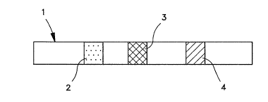
FIGURE 2 depicts the complex formed in the capture
zone showing the detector (1), antigen on the detector
surface (2), antigen bound to the chromatographic matrix
(3) and specific antibody (4).
0
DETAILED DESCRIPTION OF INVENTION
The detector zone comprises particles of detector
coated with antigen. For a true one step assay, the
detector should provide a visible readout, although other
detectors (e.g. 1uorescent, W ) can also be used, the
only criterion being the ability to attach antigens to its
surface.
The detector is a particulate material. One useful
particulate is a sac which includes a dye or other colored
substance as a readout. The sac which is used may be any
one of a wide variety of sacs, including but not limited
to intact erythrocytes, erythrocyte ghosts, liposomes
(single walled, sometimes called vesicles or
' ' ' ' ' ' ' '" ' ,
21 ~ i39
multilamellar), polymer microcapsules (for example, thcse
made by coascervation, or interfacial polymerization), etc.
Also useful are colored particles (e.g., latex or other
polymeric particles) and precipitated or insoluble metals,
nonmetals and alloys.
The primary eriterion for selection of the deteetor
are the ability to bind the antigen and the visibility of
the readout under test eonditions.
The signal-generating substance can be any substance
which exhibits a deteetable signal under test eonditions,
but is preferably a visibly eolored dye or pigment. Other
dyes, such as W or fluorescent, ean also be used, but
will complicate detection by necessitating the ~se of
specialized equipment. Most preferably the detector is a
colored latex particle.
The antigen is attached to the surface of the
detector, by means whieh are well known in the art, in a
geometrie orientation whieh renders it aeeessible to
reaction with specifie antibody in the sample to form a
eomplex. This eomplex then mi~rates through the
ehromatographie matrix to the eapture zone whieh eontains
antigen immobilized on the matrix by means whieh are well
known in the art (in the ease of low moleeular weight
antigen, it may be necessary to eouple the antigen to a
earrier macromolecule before immobilization on the
ehromatographie matrix). The multivalent speeifie
antibody also binds to the bound antigen in this zone to
form a labeled antigen/antibody/bound antigen "sandwieh,"
whieh ean then be deteeted.
- - .................. ..... ......... .. ... . ... .
' . . . ' ' ~' ' . ' ' : .
21~ 39
The assay is highly selective for specific antibody,
since it relies on the formation of an antigen/antibody
complex which is similarly highly specific.
The chromatographic matrix can be any of the various
matrices known in the art including silica, polyacrylam:;de
alumina, and, preferably, nitrocellulose. Further, while
the assay is preferably performed in a thin layer
chromatograph device (TLC) format, it can also be
. performed in any other compatible chromatographic formats
such as column chromatography.
In the preferred embodiment, the TLC device is
; arranged as shown in FIG. 1~ Briefly, the sample is
introduced to the chromatographic medium (1) upstream of ~l
the detector zone (2). After migrating through the
detector zone, the sample continues on to the capture zone
(3) where any antibody/antigen complexes are bound and
detected.
EXAMPLES
The following examples illustrate certain preferred
embodiments of this invention, but are not intended to be
illustrative of all embodiments.
EXAMPLE 1 - Anti-biotin system
In this example, a TLC device containing 8 um
nitrocellulose as the chromatographic matrix, was arranged
as in FIG. 1. The detector zone contained 3 ul of
biotinylated red latex, and the capture zone contained
biotinylated BSA sprayed onto the device as a straight
line with a CAMAG TLC applicator. The density of
.. , ..... ... ... . .. .. -- .,
.
.. . . ~ .. . . . . . . . . .
21~39
biotinylated BSA on the membrane was lug/cm.
The sample was placed on the surface of the device
. upstream of the detector zone, and the system was visually
observed for the development of a red color in the capture
zone. Two samples were tested, normal rabbit serum, and
rabbit serum containing anti-biotin antibody.
Only the serum containing the specific anti-biotin
antibody gave a positive result.
A third sample containing normal rabbit serum seeded
with 20 ug/ml of affinity purified anti-biotin antibody j,
was similarly tested, and yielded a positive result
indicating the sensitivity is at least 20 ug/ml. Triton il
X-100 was added (final conc. 1% (w/V) to all samples in
this example to ensure rapid migration of detector latex.
EXAMPLE 2 - Anti-rabbit IqG system
In this example, a device similar to that used in
Example 1 was utilized, except that the detector was blue
colored 0.48 um latex particles (O.48 um) coated with
rabbit IgG and the capture zone was rabbit IgG spotted by
micropipettor on the nitrocellulose (0.4 mgJml IgG, 3
ul/spot); the blue latex detector was mixed 1:1 (V/V) with
a solution consisting of PBS, 10% BSA, 5% sucrose, 0.2%
Triton X-100, pH 7.7, prior to application to the
nitrocellulose.
Three sera were tested, normal goat serum (diluted
1/20), goat serum containing anti-rabbit IgG (diluted
1/20), and goat serum containing anti-rabbit IgG (diluted
1/20) with normal rabbit serum added to 10% (V/V).
, . , . :
... . , , : .
~`~
21~ll39
Only the second sample produced a positive result,
indicating the specificity of the anti-rabbit antibody,
since normal rabbit serum mired in the third sample (which
contains rabbit IgG) will bind to the anti-rabbit IgG and
block the reaction.
It is apparent that many modifications and variations
of this invention as hereinabove set forth may be made
without departing from the spirit and scope hereof. The
specific embodiments described are given by way of example
only and the invention is limited only by the terms of the
appended claims.
: - . ; ~ . . -
' ~ ' . -