Note: Descriptions are shown in the official language in which they were submitted.
- 1 - 2119~i962
DEVICE AND METHOD FOR AFFINITY SEPARATION
Background
Separation processes are used widely in
biological science to isolate one component from
complex mixtures using a single or combination of
unique characteristics of the desired entity. These
characteristics can be size, shape, charge,
hydrophobicity, solubility, density etc. all coming
into the categorisation of chromatography. Usually
these characteristics are not totally unique and so a
series of different sequential separations must usually
be taken to refine the purification. Often the choice
of which steps to use is empirically determined and
devising a new purification route is very laborious,
each choice having many variants within it. The labile
nature of many biological substances also makes this a
difficult procedure, the desired substance sometimes
decaying as fast as it can be purified.
A variant of these techniques is affinity
chromatography. In this a more specific characteristic
of the desired item is utilised in the separation
strategy. Usually this involves a specific binding
capability which is instrumental in effecting the
separation. Biological systems employ specific binding
very regularly as part of their natural functioning,
such as in antibody/antigen interactions in disease
resistance, or receptor/ligand interactions for cell
signalling. This can be harnessed in a separation
technique to obtain 100% separation in one step and is
ther~fore a particularly powerful method. With such a
powerful separation method it is important when
performing these activities that all contaminating
~ . : -, , . , ,.... - . . . . ... .. ..
2 2 ~ 2
unbound materials are removed with high efficiency.
Preferably there should be 100% effici~ncy both of
contaminant removal and of capture of desired substance
in the first pass. This requires maximal interaction
5 between the desired substance or entity with its
binding partner as well as very efficient washing.
A method is therefore needed where sufficient
capture molecules to capture all the target molecules
are held on a solid phase. This is done in such a way
that the target molecule or entity has easy access to
the binding site but also so that non-binding moieties
can be washed off vigorously without trapping or
spuriously interacting.
There are many formats for performing
affinity chromatography procedures. All of them share
the general feature of having one side of the binding
pair immobilised to a solid phase. This most commonly
consists of a bead material sometimes packed into
columns. The liquid containing the desired entity can
then flow over, around and in some cases inside the
beads coming into contact with the capture entity and
contaminants can then be washed by allowing washing
solutions to flow over the bead surfaces. The
stringency of the washing procedure can be influenced
by the nature of the washing solution such as by
temperature, ionic strength, pH, solvent mix etc.
Beads are in many cases a preferred option as
this format maximises the surface on which the capture
entity is immobilised.
In general the major requirement is for an
insoluble material to which the capture entity can be
attached such that a fluid containing the target be
passed over the solid phase to allow maximum contact
between the compartments. Also the solid phase
normally requires a maximal surface area to allow
sufficient capture entity to be available. In most
...... -, , :
.
~l~a~62
cas~s the purpose of the procedure is for the removal
of the target from a complex mixture in pure form and
then elution of the target from the capture entity
without the capture entity also being removed. For
this to be possible conditions have to be found where
the binding forces can be overcome without damage to
either binding partners.
In recent times, especially in molecular
biology, techniques have arisen which are manyfold more
sensitive than before and so do not need such large
amounts of sample as starting material. Also more and
more of subsequent treatments are able to be done while
still attached to the solid phase. This is the basis
of the burgeoning fields of downstream processing and
biotransformations where chemical modifications are
made often enzymically by materials held on a solid
phase.
There have appeared in the last few years
several new formats for affinity chromatography based
on flltratlon membranes especially for antibody
purification. Most commonly these involve the use of a
filtration cartridge format, familiar to those working
in biological fields. This consists of either a
disposable or reusable cassette within which is mounted
a disc of filtration membrane.
The membrane is supported on both sides by
plastic meshes within the cassette and leading out from
the upper and lower surfaces of the cassette is a
nozzle designed to be attached to a syringe and an
outlet designed to be directed into the collection
vessel. These cassettes sometimes include in the
design, channels of liquid flow to maximise the
interaction of the fluid across the membrane
(US 4,690,757).
Later developments have lead to new versions
with either capture moieties already permanently
21~9~
attached or in a chemically activated form for custom
derivatisation. The discs are usually about 5 cm in
diameter and are claimed to have as high a binding
capacity as a column. This is consistent with their
use with a syringe for the application of large samples
of between 1 and 50 mls of solution at a time.
As mentioned earlier the trend now is towards
smaller samples, more sensitive detection and
amplification techniques especially in molecular
biology. In biology large samples are difficult to
obtain and involve significant derangement to the
biological entity which is the source of the sample.
This is especially true if repetitive samples need to
be taken to follow a trend or reaction. Large samples
are also slower to process and involve exposing fragile
biological entities to inhospitable environments during
the process.
As mentioned earlier existing affinity
processes involve removal of the target from the
capture entity as the final step. This requires
empirical discovery of conditions which will perturb
the binding without damaging the desired purified
moiety. This can be extremely difficult for example
with antibodies where strong binding is often
particularly desirable. Obviously the stronger the
binding the more denaturing the eluting solution.
Sometimes numerous combinations of elutant have to be
tried to find a good set of conditions. Sometimes
however no combination achieves the right effect.
As well as molecular purifications cells are
also used in affinity processes. This can take a
variety of forms based on different characteristics of
the cells. Usually but not always the cells must be
recovered intact from the process for further analysis.
Frequently the separation is based on presence of cell
surface molecules for which antibodies can be obtained.
.: , .. .. .
.,
. . .. ... ..
This can also be combined with size and density
measurements. Methods for affinity purification of
cells include "Fluorescence Activated Flow Cytometry"
which can combine size with the existence of one or
more cell surface markers. The problems of cell
purification are severe due to their fragility and if
antibody selection has been used the selected cells
have to be used with the label still attached.
Many areas of science use natural affinities
for binding. In genetics complementarity of nucleic
acids is commonly utilised as the basis of a method of
analysis. For example mRNA is isolated by virtue of
the fact that it always has a tail of adenine
nucleotides at the end which can be bound to a row of
thymidine nucleotides.
Specific gene nucleotide sequences can be
captured by the complementary nucleotide sequence.
These hybrlds can usually be removed very easily by
reducing the ionlc strength of the elutant allowing the
natural charge-driven repulsion between DNA strands to
take effect.
As mentioned earlier, sometimes elution
conditions cannot be found to remove without damage.
This can be turned into an advantage however if the
capture is done in the situation where it can act as
the linker immobilising the desired activity in place
so that subsequent steps can be performed in situ. For
example this could be an enzyme reaction in the new
science` of biotransformations which uses immobilised
enzymes for chemical synthesis.
If it is essential to remove the target
material then a final resort is to use a membrane
material which is itself soluble in a solvent not
damaging to the target. This case however does release
the capturing material also.
Frequently the coupling of the material to
:' ' ~- ' -';- ' ,
.,: ... . ~ , ..... .
-
~' ` ' , .
- `21~`962
-- 6
its solid phase would be by covalent linker to avoid
any problems of the leaching out of the capture moiety
leading to contamination of the process.
If removal of the target moiety is difficult
or unnecessary, analytical work can be done in situ.
Many biological analyses can be performed on membranes.
For example one item is immobilised on the membrane and
used to capture the other. Using further labelled
binding moieties the presence of an entity can be
revealed using fluorescent markers or colourimetric
enzyme markers or radiolabels. These can be visualised
by eye, by machine or by microscopic analysis or
counted by appropriate instrumentation. The
requirement for being on a membrane is to allow
efficient washing as well as to provide for information
of localisation.
Other processes are now carried out on
membranes such as enzyme reactions, gene amplification
reactions, chemical syntheses etc.
Often the results of a separation especially
if it involves cells must be verified by direct
observation. This is used to tell whether the cells
are still in good condition, whether the separation
looks "clean" etc. Sometimes further specific tests
must be done on the separated cells to verify their
identity by a different route such as specific staining
or enzyme activity. If a cell sorter has been used
then the produced cells can be examined. If a column
has been used they must be eluted to be observed. If
however a membrane has been used they-can be visualised
directly as most membranes are semi-transparent or
translucent.
Most capture-based techniques still utilise
the column format. This requires the sample to be
slowly trickled over the matrix and then washing to be
done by trickling over the matrix a succession of
- .
:
.. . ,: ~- . ~ , . . ..
_ 7 _ 2103~62
washing steps. To improve the washing and the elu~ion,
gradients of solvents are often used with varying pH,
ionic strength and hydrophobicity.
Also several changes of small volumes of wash
solution are far more efficient than one large one
which is difficult to implement in a column situation
without extending the time further.
These processes can take a long time
especially if the binding affinity is weak and
sometimes requires the circulation of the sample
solution over and over the capture surface to maximise
contact. During this long time many constituents will
deteriorate and possible denature and consequently many
of these processes are now carried out in cooled rooms.
These are very unpleasant environments to work in and
only partially solve the problem.
One solution to this has been the development
of HPLC techniques which among their other
characteristics are faster, as the liquids are
transferred under pressure. These systems are
expensive however and subject the substances to high
pressures as well as temperature.
Membrane capture processes are usually faster
and therefore better for labile materials but they
suffer from problems of dead space which means that the
smallest samples cannot easily be used and that the
material eluted is lower in concentration.
~ s existing cartridges are contained, it is
not easy to see when they are full of liquid and this
3~ can result in air being drawn through.and partial
drying out of the membrane in an attempt to reduce the
minimum volume. Also, because the membranes are
contained and supported it is not easy to remove the
membrane for visualisation either by light electron
microscopy. Similarly they cannot easily be used for
subsequent reactions. Some of the cartridges can be
.. ~.. , ,...... , , ,, : , , , - - -.- .; .. :, ., - . . ... ,, - - -
- 8 ~10~9~`2
disassembled and hence the membrane removed. The true
purpose of this is re-use of the cartridge however and
usually results in some damage to the membrane.
In some samples the desired constituent is
present in minute amounts or numbers. This results in
large volumes being drawn over the capture moieties.
In addition to the time involved this has the
additional disadvantage that the process of liquid
flowing in some cases is sufficient to cause the
removal of hitherto bound components. The severity of
this depends on the nature and strength of the
binding, but as biologically significant affinities are
often subtle there are many cases where this method of
purification cannot be achieved for these reasons.
Another route to solving these problems is to
increase the amount of available capture partner by
increasing the amount of solid phase. This results in
slower flow rates, longer reaction time and greater
dilution of the desired material.
Samples for these types of purifications are
often clinical and potentially infective and the
washing and especially eluent chemicals are also
frequently of a hazardous nature. They may be organic
solvents, acids, ion-pairing molecules, chelators,
detergents etc. Traditional processes using columns
offer the potential of injury to the operators as they
are exposed to the whole system which often involves
the use of significant amounts of the hazardous
material. Membrane cartridge devices are better but
still rely on squirting out liquids with possibilities
for spillages and aerosols.
Many targets once purified will be used in
further analysis such as electrophoresis or reactivity
for example. For most of these subsequent reactions
the target, which is usually in limiting amounts, is
preferably at a high concentration. For column and
-,, .. .... ~ - , . - - ~ . . . . .
, , . ~ , , ,- . . . :. ,: .,
.
9 21~9~
membrane affinity systems, elution will result in the
sample being collected at less than maximal
concentration. This frequently means that
concentration has to be performed before further work
can be done. The concentration of a highly purified
substance at low starting concentration is very
inefficient often leading to losses in excess of 50~.
These small amounts of dilute material also
suffer from the disadvantage of being easily denatured
on storage and usually have to be mixed in with other
molecules such as bovine serum albumin to increase
their stability. This defeats some of the object of
purifying them in the first place. They are also very
liable to adsorb to the surfaces of their storage
vessels which sometimes necessitates pre-treatment with
toxic silicon compounds (silanes) as a prevention.
As mentioned earlier most chromatographic
procedures are reached empirically and frequently
involve multiple stages. Strategles are designed by
the individual testing of single steps under a variety
of conditions on a small scale to optimise both the
type and order of steps sometimes for subsequent large
scale operations.
This usually means that large numbers of a
variety of small columns have to be made, equilibrated,
and run both singly and in combination and the elution
analysed and monitored. Constituents often need to be
radio-labelled to be able to detect them in such
processes and this results in a considerable amount of
radio-active waste. There are automated systems to
perform these reactions but they are very expensive,
complex to run and the produced materials still need to
be analysed.
The concept of having a separation on the end
of a tip has been utilised before in patent
specification No. WO8809201. In this case however the
2 ~ 6 2
tip contains column material between two frits and is
therefore a miniature column. Usage of the column is
by solution flowing by gravity as commonly employed for
column processes.
The Inventio~
This invention provides a device for
capturing a component present in a fluid, comprising a
pipette tip having an open rearward end adapted to be
fitted on a pipette for drawing fluid into the pipette
tip, an open forward end, and at least one membrane
extending across the pipette tip at or adjacent its
forward end.
The membrane is preferably porous, since it
is necessary that the fluid be able to flow through,
over or round the membrane and into the pipette tip.
Preferably the membrane is a woven or non-woven mesh of
flbres, which term is used to include threads and
filaments which may be discrete or continuous. The
membrane may be a mesh weave, a spun bonded mesh, a
nuclear track etched membrane, or an electrolytic mesh.
Membranes of various pore sizes are possible; it will
be understood that the membrane is not normally used
simply as a filter to physically separate from a fluid
particles that are too large to pass through the pores.
Membrane pore size is chosen rather to ensure intimate
contact between the fluid and the membrane. The larger
is the pore size, the easier is passage of fluid
through the membrane but the lower is the capture
efficiency of the membrane for the desired component.
Preferably the membrane is adapted to bind
and thereby capture a component present in the
fluid. For example, the membrane may incorporate a
specific binding partner of the component to be bound.
The membrane may incorporate capture entities which are
ion exchange molecules, affinity proteins such as anti-
- - . . . . . .
~: . . . . - . , . . . . "
. . ;. .. ~ . . . . :
. . . . . . . . ..
- 11 2~ 2
bodies or biotin-binding molecules, enzymes, nucleic
acids, nucleotide oligomers, cell attachment
molecules,receptors, chelators etc. V
In one embodiment, the membrane is of a
material which is capable of binding DNA, for example
by chemical interaction or hydrophobic bonding or
physical absorption or by a charge interaction. DNA
binding to plastics materials is complex and involves
various combinations of these phenomena. For example a
highly charged polymer surface may favour charge
interaction, while an uncharged polymer surface may
favour hydrophobic bonding.
Many materials are known which have nuclear
capture properties, including polyester, polyamide,
polycarbonate, cellulose, nitrocellulose,
polyvinylidine difluoride, and glass. Alternatively,
the membrane can be made of any material which can be
activated, chemically or physically in such a way that
lt binds the component to be captured. Immobilization
of the capture entity, e.g. antibody or other specific
binding species, on the membrane is readily effected by
a variety of chemical and physical means which are well
described in the literature.
The membrane may be chosen with a view to the
specific requirements of the separation involved. For
example, the membrane may be chosen to be non-
inhibitory to subsequent enzyme reactions or culture
requirements. The membranes can also be selected to be
non-fluorescent, transparent, heat or chemical-
resistant.
Capture membranes which have been usedsuccessfully are 1, 5, 6 and 11~m polyester woven
membranes and 1~m nylon woven membrane. Either 1~m
membrane is preferred. Less preferred but effective to
a lesser extent are 50, 100,um random mesh polycarbonate
membranes and 5, 10 ,um track-etched polyester membranes.
; ., ;:: .. . . .
. .
21~ 2
- 12 -
Also 0.45~m nitrocellulose has been used successfully,
as has 0.45~m nylon, although the flow rate and hence
washing efficiency were reduced (see Example 1).
It is an advantage of the invention that the
desired component is captured on or at or in the
forward-facing surface of the membrane. The membrane
is mounted at or adjacent the forward end of the
pipette tip, that is to say, close enough to the
forward end to be easily visible or accessible for
subsequent treatment, reaction or analysis. The
membrane is preferably bonded to the forward end of the
pipette tip, either at right angles to the axis of the
pipette tip or set obliquely (i.e. not perpendicular to
the longitudinal axis of the pipette tip). An oblique
mounting increases the surface area of the membrane,
for a given tip diameter and may help to avoid
contamination when the device is inserted into a dirty
solution. Or the membrane may be mounted on the
forward end of a short tubular section, the back end of
2~ which is a friction fit on the forward end of the
pipette tip of the invention. Several tubular sections
comprising several membranes may be mounted on the
pipette tip in this way.
The first ~or only) membrane is preferably
bonded to the pipette tip at or adjacent its forward
end. The membrane may be made peelable from the
pipette tip for subsequent processing. But in this
embodiment it would not be practicable to replace the
same or another membrane on the pipette tip. The
device of the invention is thus designed to be
disposable rather than re-usable.
Alternatively, the membrane may be secured to
the forward end of the pipette tip by means of a
securing collar.
The pipette tip is preferably a one-piece
moulded structure preferably formed of a plastics
' '
.
~ ' ~
210~2
- 13
material, which should withstand autoclaving (a 120-C
for 20 minutes) as well as repeated heating and cooling
between 95 and ambient. No mould release or plastizer
should be used in the manufacture of the pipette tip,
and the plastics material should not inhibit enzyme
reactions such as PCR either by removal of crucial
components by adsorption or by chemical inhibition.
Preferred plastic membranes include polycarbonate,
polypropylene, nylon, polyester, PTFE. Polycarbonate
and polyester can have the advantage that the tube is
transparent. The pipette tip should preferably not
fracture on freeze-thawing.
The pipette will usually be an adjustable
volume or non-adjustable volume disposable tip
micropipette, as produced by Companies such as Gilson
and Eppendorf, and well known to those working in
biological laboratories. Preferably the rearward end
of the pipette tip is internally tapered so as to be a
friction fit on a micro pipette. The rearward end of
the pipette tip may carry external axial reinforcing
ribs.
Preferably the pipette tip is of a brittle
plastics material and has between its ends a
circumferential line of weakening, e.g. provided by an
external groove, along which the tip can be broken
manually. The length of the forward end of the tip may
be chosen to enable it, complete with the membrane, to
be inserted in an eppendorf tube for further processing
in such a way that the lid can be shut. The pipette
tip is preferably conical, with the rearward end sized
to fit a micro pipette, with the tip decreasing in both
internal and external diameter towards its forward end.
The small diameter of the end of the tip
allows the membrane to be immersed in very small
samples. The tip is adapted to contain the fluid that
is drawn into it by the pipette, thus avoiding any
21û~9~2
- 14 -
contamination of the pipette itself. This also allows
liquid to be taken up into the tip past the membrane
with very little required force. This also allows the
liquid to be forced out again past the membrane and
hence double the interaction of the sample with the
capture partner. This can be repeated any number of
times pipetting up and down so the sample has multiple
chances to interact with the capture entity. The speed
of pipetting can be varied easily or for very slow
kinetics the tip could be left incubating in the
solution. Once the binding step has been allowed the
tip is easily transferred to the required number of
washing solutions in turn pipetting up and down each
time as required. Several consecutive small volume
washes are much more effective in washing terms than
the same volume once. This method therefore minimises
the required amounts of wash solutions whilst
maximising the washing effect. The pipetting for
washing can be as vigorous and numerous as desired.
A particular advantage is that the presence
of the membrane at the end of the tip results in any
entities attaching by whatsoever means to the membrane
do so predominantly on the external surface. This is
important for any visualisation purposes, for efficient
elution or for subsequent reactions or for example
cellular entities they can continue to be cultured Ln
on the membrane.
The nature of the fluid, containing a
component to be captured, is not material to the
invention. The fluid may, for example, be a biological
fluid such as a body fluid.
In some cases, the fluid may be so "dirty" by
virtue of containing so much insoluble matter, that the
step of sucking it through the membrane would be slow
or difficult. In this case, it may be sufficient to
bring the fluid into contact with the outer surface of
.... , . . .. .. , .. ,, . .- - . . - - . -
.. . ~. - i . ' -
- ~ ~ . . .
.. . .
,, . ~ , ~ ~ ' !
~105~2
, 5
the membra~e without applying any positive or negative
pressure through the pipette tip. As the fluid is
stirred in contact with the pipette tip, the membrane
captures the desired component. The pipette tip can
then be removed from the "dirty" fluid, and immersed in
a washing fluid which is sucked into the pipette tip
and then ejected through the membrane, usually several
times.
For applications where many conditions need
to be tried for optimal purification perhaps for a
subsequent large scale scheme, trial of many conditions
can be performed in rapid succession. For multiple
preparations the tips can be utilised with a multi-
channel version of the micropipette such as are common
among those experienced in the area. These commonly
allow 8 or 12 purifications to be done simultaneously.
The membrane-ended tips can be manufactured attached in
a row for easy attachment to the multi-pipettes.
For particularly difficult separations or
ones wh0re different components need to be removed from
the same sample multiple layers can be used separated
by a small spacer. Each of these layers can be
derivatised differently so the sample contacts all of
them and they could be assembled in a head-to-tail
manner. This is also useful where samples are
undesirably particulate or otherwise contaminated and
the first layer can be chosen to remove some of this
unwanted material.
These layers can be used either to remove
30 multiple specific contaminants to improve the --
separation by capturing the target on two or more
layers of the same materials to capture different
materials simultaneously or to remove one material
while capturing another. The layers can then be
separated after treatment by "unslotting" them.
This can be extended to include membranes
21~9~
- 16 -
which have attached to then. molecules or entities which
are preservatives or enzyme inhibitors to help the
desired material to survive. They can also have
detergents, surfactants or anti-bacterial agents
available on the solid phase membrane which allows
their activity to be utilised without contaminating the
sample.
An additional variant of this is to have an
enzyme activity attached to a section of membrane so
that the enzyme can perform its reaction and can then
be removed from the sample to avoid sample
contamination. In some cases these sections can be
stored for re-use.
Reference is directed to our European patent
application 92 308 537.7, filed on 18 September 1992
and entitled "Capture Method and Device". That
invention describes a method of separating components
of cells, which method comprises
a) treating a fluid contalnlng whole cells so as
to selectlvely lyse the cytoplasmic membrane together
wlth a small proportion of the nuclear membranes but
leaving a large proportion of the cell nuclel intact,
b) applying the treated fluid to a surface
whereby a mesh of DNA from the lysed nuclei is formed
on the surface and captures intact cell nuclei,
c) washing the DNA mesh on the surface to
separate the captured cell nuclei from other components
of the cells.
The said patent application also describes a
device for use in the method. The device of the
present invention is very suitable for that purpose;
the forward-facing surface of the membrane herein
described can constitute the surface to which the
treated fluid is applied in step b) above. In that
case however the membrane would usually not be
derivatised.
- 17 2 1 0 5 9 6 2
Reference is dlrected to the accompanying
drawings, in which:
Figures 1 and 2 show sectional and end
elevations respectively of a preferred form of the
device.
Figures 3, 4 and 5 are perspective sectional
side views of three different devices according to the
invention.
Pigure 6 is a perspective sectional side view
of such a device in position in an eppendorf tube.
Referring to Figures 1 and 2, the device
comprises a pipette tip 10 the bore 11 of which
decreases in diameter along its length from its
rearward end towards its forward end 12. The forward
end face of the pipette tip has an annular pip 13 for
the attachment of a permeable membrane 17 extending
across the bore and selected according to the nature of
the component to be captured. The pip has in this
construction a triangular cross-section. A line of
2~ weakening in the form of a peripheral groove 14 is
formed in the wall of the pipette tip at a selected
distance from the forward end. In its rearward end
portion 15 the external surface of the pipette tip is
cylindrical and has a series of axial stiffening ribs
16 to enable that end of the pipette tip to be secured
in a friction fit on the end of a micro-pipette. The
pipette tip is made from a transparent and brittle
thermoplastic plastics material. Polycarbonate is
particularly suitable for this purpose.
In use of the device, a biological fluid is
drawn into the pipette tip by a micro-pipette through
the membrane which captures a component of the fluid.
The captured component is then washed by drawing a wash
solution through the membrane. The pipette tip is then
broken at the line of weakening and the whole forward
end part of the pipette tip with the membrane and
21~59~
- 18 -
captured component is placed in a standard eppendorf
tube for further treatment. To enable the lid of the
eppendorf tube to be closed it is particularly
advantageous for the groove 14 to be formed at a
distance of 18mm from the forward end of the pipette
tip.
Figures 3, 4 and 5 show generally similar
embodiments. In Figure 3, a capture membrane 17 is
shown extending across the forward end 12 of the
pipette tip. An aerosol filter 18 is mounted in the
pipette tip between the fracture point 14 and the
rearward end 15; the purpose of the filter being to
preserve the tip of the micro-pipette from
contamination with biological fluid being sucked
through the capture membrane.
In Figure 4 the pipette tip does not carry
any membrane bonded thereon. A tubular portion 19
carries a membrane 20 at its forward end; its rearward
end is a friction fit over the forward end of the
pipette tip.
In Figure 5, the device is as shown in Figure
3, with two tubular sections 19 and 21 pushed over and
a friction fit on the forward end. The device thus has
three membranes, 17, 20 and 22, and the biological
fluid is sucked through these in sequence.
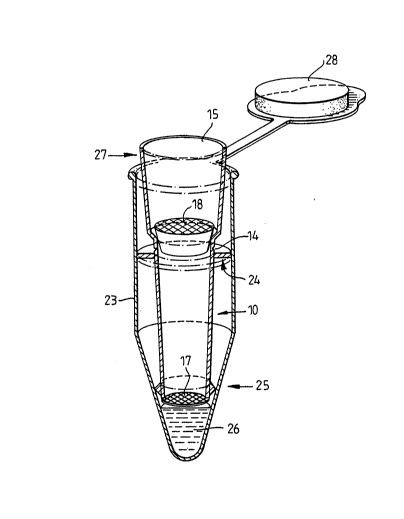
Figure 6 shows a device similar to that
illustrated in Figure 3, inserted into an eppendorf
tube 23. The device includes an external peripheral
disc 24, just forward of the fracture point 14, which
serves as a fracture fulcrum. The membrane 17 is
secured to the forward end of the pipette tip by a
securing collar 25 which also acts as an anti-reflux
seal. The eppendorf tube contains 50 ul of reaction
fluid 26 for further treatment of the capture membrane.
In use, sideways pressure is applied to the
rearward end 15 of the pipette tip, e.g. in the
~, ... . .. . - . . ... . . ~
- - 2 1 ~
- 19 -
direction marked by the arrow 27, to break the pipette
tip at the fracture point. The rearward end of the
pipette tip is then removed, the lid 28 of the
eppendorf tube closed, and the capture membrane 17
further treated as required.
EXA~P~
Use of Mem~rane ~ips for Affinity Purification of a
Cytoc~Qm~ p450 variant from Two Protei~.Mixtures
a) A mixture of proteins containing the protein
cytochrome P450 was obtained as a series of molecular
weight markers. These were obtained as an aqueous .-
solution at a concentration of around 1mg/ml for each
protein and consist of P450 (55kd), ovalbumin (46kd),
carbonic anhydrase (30kd), trypsin lnhibitor (21.5kd),
lysozyme (14.3kd), aprotinin ~6.5kd), insulin chain A
(3.4kd), & insulin chain B ~2.3kd) in Tris buffer
pH 8Ø
b) Phenobarbitone treated rats (Guengerich, F.P.
and Martin M.V. Arch. Biochem. Biophys., 205, 365-379,
1980) were sacrificed and the livers removed. Extracts
were prepared ~Guengerich, F.P.J. Biol. Chem. 252,
3970-3979, 1977) of the microsomes known to contain
overexpressed P450 protein. This method involves
successive homogenisation and centrifugation steps in a
saline solution. The extracts once prepared are stored
at -70 C till required.
Tips were prepared as follows: tips were
constructed as described with nylon reinforced
nitrocellulose membrane attached across the forward
end. Antibody was attached to the surface of the
membrane by immersing in 100,ul of a solution of a
partly purified polyclonal antibody to P450 (Ryan,
D.E. and Levin, W. Pharmac. Ther. 45, 15-239, 1990)
, ,,........ . .. , . ,. ~ . . , . .. ~ .. . ~ . .. . - .. .. - .
2~
- 20 -
1Omg/ml in 100mM carbonate buffer pH 9.0 containing
1Omg/ml bovine serum albumin. This was held in the
solution for 15 minutes and excess was then washed off
in phosphate buffered saline pH 7Ø and stored in
saline at 4'C to avoid drying.
lO,ul of the protein mixtures in a) and b)
were diluted to 500,ul in phosphate buffered saline
(PBS).
Separate tips with antibody-coated membrane
attached were then used to aspirate up and down the
extracts.
The aspiration was repeated five times. The
tips were then transferred to wash solution of 3xSml
PBS and one aspiration up and down was done in each.
The membrane was then peeled off the end of the tip and
added to sample loading buffer for PAGE
electrophoresis.
The samples were boiled in 20,ul of loading
buffer containlng SDS, DTT, glycerol and bromophenol
blue for 2 minutes. Control samples of extract
aspirated through an untreated tip as well as depleted
extract after "tipping" were prepared by boiling equal
amounts of sample and loading buffer together for
2 minutes.
10,ul of each sample was run on a denaturing
12~ PAGE gel at 100V for three hours and subsequently
stained in Coomassie blue fixative to reveal the
protein bands.
The first three tracks showed mixture A: 1)
Mixture after "tipping" :2) Mixture after "tipping"
using a "blank" tip without antibody and 3) Material
extracted by antibody-treated tip.
The following three tracks showed 5) Liver
extract after "tipping" 6) Liver extract after
"tipping" using a "blank" tip without antibody 7) Tip
extracted material (the additional material in this
.. . . . . ..
... ~ . . . . .. . .. ...
.. . . . . . . ... .. . .. .
2~5962
- 21 -
track may represent antibody contaminants eluting from
the tip membrane. Normal practice would be to perform
these reactions using radiolabelled extracts.
Contamination from the bound antibody would not then be
a problem as it would not be radiolabelled.)
The results demonstrate that ~he antibody
held on the tip has efficiently captured a specific
substance from a complex mixture during the rapid
aspiration steps.
The captured substances can then be removed
from the tip membrane for further study.
The tips themselves are not removing
substances non-specifically by absorption to the
membrane.
E~AMPLE ~
Use o~ Membrane De~ivatised TipS ~or
Immunop~cipi~tio~ of p53 Protein from HeLa Cell
Extract
a) Tips were constructed as described, with a
nylon membrane attached across the forward end.
Antibody was attached to the surface of the membrane by
immersing the tips in either 500 ,ul of a solution of
purified monoclonal antibody to p53, pAb248 (1), or a
solution of purified antibody to Adenovirus ElA, M73,
(2) in 100 mM carbonate buffer pH 9Ø This was held
at 4 C overnight. The excess solution was removed and
the tips washed in phosphate buffered.saline (PBS),
pH 7Ø The membranes were then blocked by immersion
in 500 yl 3% BSA/PBS for 30 minutes at room
temperature. After removing excess blocking reagent
and washing in PBS, the tips were immersed in 1 ml HeLa
cell lysate.
b) Approximately 107 HeLa cells were lysed in
2~0~9~2
- 22 -
2 ml of 150 mM NaCl, 1~ NP40, 50mM Tris pH 8.0 for 30
minutes on ice. The liquid was removed for incubation
with the derivatised tips.
c) The tips and lysate were incubated on ice for
30 minutes, followed by removal of excess lysate. The
tips were then washed with 1 ml PBS, by sucking the PBS
into the pipette tip and ejecting it from the pipette
tip through the membrane three times. Each tip was
then added to a tube containing 40 ,ul of Laemmli sample
buffer (2% SDS, 10% glycerol, 100 mM DTT, 60 mM Tris pH
6.8, 0.001% bromophenol blue). The samples were then
boiled for 2 minutes and loaded onto 3 separate tracks
of a 10 - 15~ polyacrylamide/SDS gel.
The first two tracks show immunoprecipitation
of the p53 protein with tips derivatised with anti-pS3
antibody, pAb248. In track 3 (negative control), a tip
derivatised with an anti-E1A antibody shows no
detectable protein immunoprecipitated as would be
expected, since the HeLa cells do not express the
Adenovirus proteins.
1. Yewdell, J. W., Gannon, J. V. and Lane, D.
P., J. Virol., (1986), 59, 444-452.
2. Harlow, E., Franza, B. R. and Schley, C., J.
Virol., (1985), 55, 553.