Note: Descriptions are shown in the official language in which they were submitted.
CA 02106296 2001-06-27
'' 92/18850 PCT/US92~, .._923
1
10 CONFOCAL IMAGING SYSTEM FOR VISIBLE AND ULTRAVIOLET LIGHT
BACKGROUND OF THE INVENTION
This invention relates generally to confocal
microscopy and more specifically to a confocal scanning
microscope usable with 'visible and ultraviolet (UV) light.
Fluorescent light microscopy is extensively used in
biological research and medical diagnosis. It provides the
selectivity necessary to enable specific components of a cell
or tissue to be visualized and the spatial organization of such
components to be determined. Confocal microscopy operates so
that illumination and detection are confined to a single point
in the sample. This is typically achieved by using spatial
filters (usual.lf~ pinholes) in the optical paths of the
objective and cui~denser,, and a complete image is built up by
sequentially scanning a:L:1 points in the field of view.
A particular confocal microscope is shown in U.S.
Patent No. 5,032,720.
The microscope produces a small (preferably
diffraction limited) spot on a sample, scans the spot over the
3o sample in a raster pattearn, and generates an electrical signal
proportional to the ints:nsity of light emanating and collected
from the region of the spot. The electrical signal is
communicated to a computer which can produce a visual display
on the monitor.
The optical train between the source (or detector)
and the sample comprises focusing optics to form the spot and
scanning elements to scan the beam in two orthogonal directions
to form the raster pattern on the sample. The light emanating
from the sample pas~ec along a return path to a detector, which
generates the eleci:ricai signal. An aperture in the form of an
iris diaphragm is disposed in front of the detector and blocks
light that emanated from, points spatially displaced from the
beam spot.
CA 02106296 2001-06-27
2
A confocal microscope accessory for use in combination
with a conventional microscope has been marketed under the
trade designatior:.s MRC-500 and MRC--600 by Bio-Rad
Laboratories, Inc., Hercules, California. The resulting
confocal microscopE-~ provides a visible excitation beam and
senses fluorescence i.n the visible range.
SUMMARY OF THE INVENTION
The present invention provides a scanning confocal
microscope capable of providing ultraviolet (UV)
excitation. The optical train substantially corrects
scanning and focusing errors over the wavelength range that
includes UV and visible wavelengths.
Accordingly, t:he present invention provides a scanning
confocal microscope comprising:
UV source mea~ms Eor providing a UV beam;
a visible light detector;
means for limiting the effective size of said
detector;
means for direct=ing said UV beam along a forward path
from said UV sourcE:e means to a sample region, said forward
path including first <~nd second segments;
means for dire=pct=iTlg visible light along a return path
from said sample region to said visible light detector,
said return path including said second segment and a third
segment;
focusing means in said second segment for focusing
said UV beam to a ;7pot in said sample region; and
scanning mean; in said second segment for varying the
angle at which saic:~ U~7 beam encounters said focusing means
so as to vary the location of said spot in said sample
region;
said focusing means being corrected for chromatic
scanning errors so that where UV light leaves said scanning
means at a given angle and is focused at a given location
in said sample region, visible light emanating from the
given location encounters said scanning means at the same
given angle.
CA 02106296 2001-06-27
2a
In a further aspect, the present invention provides a
scanning confocal microscope comprising:
a UV illumina.t_icn subsystem for directing a UV beam
along a forward pa~~h to a samp:Le region;
a visible light ;subsystem f_or detecting visible light
travelling along a return path from said sample region;
said UV illum=inat~ion subsystem and said visible light
subsystem having shared elements including a beam scanner
and focusing optic;:;
said focusing optics operating to focus said UV beam
to a spot in said. sample region and to collect visible
light emanating from said spot;
said beam scanner operating to vary the location of
said spot in said ::ample region;
said focusing cpt:ics being corrected for chromatic
scanning errors so that. where UV light leaves said beam
scanner at. a giver angle and is focused at a given location
in said sample rec:~ion, visible light emanating from the
given location encounters said beam scanner at
substantially the same given angle.
In brief, the confocal scanning microscope according
to the present invention includes a UV excitation source
and UV-transmission :canning and imaging optics. The UV
light is directed G.long a forward path and focused to small
spot in a sample plane. Light emanating from the region of
the spot is directed along a return path and detected.
Light emanating from points spatia7_ly displaced from the
spot is rejected by t;he detector aperture. The beam spot
is scanned over thc:~ sample in a raster pattern. Where the
returning light i;~ visible (<~s for example when it is
desired to detect visible fluorescence), the optical train
must be made confecal for both the visible and UV light .
This also makes it possible to provide simultaneous W and
visible excitation.
In a particular embodiment, the lenses in the common
path portion of the optical train are corrected for
chromatically induced scanning errors while extra lenses
are provided in the forward UV path to correct for focusing
CA 02106296 2001-06-27
2b
errors due to ='_ongitudinal chromatic (visible/UV)
aberration. Separating out the correction of scanning
errors and focusing errors makes it easy to accommodate
different objectlVE'_:3 that vary in their degree of
longitudinal chromati~~ aberration.
A further undE:~rst:anding of the nature and advantage of
the present invent.ic>n can be realized by reference to the
remaining portions of the specificai:ion and the drawings.
WO 92/18850 ~ ~ ~ ~ ~ ~ PCT/US92/02923
3
BRIEF DESCRIPTION OF THE DRAWINGS
Figs. lA and 1B are simplified optical schematics of
a prior art confocal microscope;
Fig. 1C is an optical sketch illustrating proper
confocal operation;
Fig. 2 is a simplified optical schematic of an
inverted microscope embodiment of the present invention;
Figs. 3 and 4 are optical schematics of a first
generation eyepiece and adapter lens for the inverted
microscope embodiment;
Fig. 5 is an optical schematic of a first-generation
eyepiece for an upright microscope embodiment of the invention;
Fig. 6 is an optical schematic of a second-generation
6.25x eyepiece for the inverted microscope embodiment;
Figs. 7A and 7B are optical schematics of a second-
generation 8x eyepiece and adapter lens for the inverted
microscope embodiment:
Fig. 8 is an optical schematic of a second-generation
8x eyepiece for the upright microscope embodiment;
Figs. 9A-E are optical sketches illustrating the
effect and correction of focusing errors;
Figs. l0A-C are optical sketches illustrating the
effect and correction of scanning errors;
Fig. 11 shows plots illustrating the effect and
correction of field curvature:
Figs. 12A-C are plots illustrating the effect and
correction of field curvature and magnification errors; and
Fig. 13A and 13B show plots of intensity across the
field.
DESCRIPTION OF SPECIFIC EMBODIMENTS
Prior Art Visible Confocal Microscope
Fig. 1A is a simplified optical schematic of a prior
art scanning confocal microscope lo. The partwcul;~r prior art
confocal microscope discussed here and the confocal microscope
as modified according to the present invention is a Bio-Rad
MRC-600 accessory used in combination with a conventional
WO 92/18850 ' ~ ~~ PCT/US92/02923
w
4
microscope. The term "microscope" will typically be used to
mean the scanning confocal microscope resulting from the
combination.
The microscope operates to focus a beam of visible
light to a point in a sample plane 15, and to detect light
(reflected light and fluorescence) emanating from the point in
the sample plane. To this end, the microscope includes a
visible light source 20 such as an argon ion laser whose beam
is directed along a forward path through an optical train
comprising a beam splitter 22, scanning optics 25, an eyepiece
27, an adapter lens 30, an infinity-correcting lens 32
(referred to as telon lens 32), and an objective 35. Beam
splitter 22 is formed of a dichroic material that reflects the
visible excitation beam but transmits light in the range of
wavelengths characteristic of the fluorescence from the sample.
The objective and eyepiece focus the beam to form a small spot
nominally in the sample plane.
bight emanating from the region of the spot travels
along a return path until it reaches beam splitter 22, from
which point it passes through and is directed to a detector 37a
such as a photomultiplier tube (PMT). An aperture 40a,
preferably an iris diaphragm (variable diameter 0.7-7.0 mm), is
disposed in front of the detector. A dichroic beam splitter 42
may be disposed in the return-only path to direct light in one
wavelength range to detector 37a and light in a different
wavelength range to a second detector 37b and associated
aperture 40b.
The optical path from the scanning optics assembly to
the detector is folded by means of a number of plane steering
mirrors in order to provide a relatively long path length. The
scanning optics comprises a pair of galvanometer-driven plane
mirrors with relay optics, preferably a pair of facing concave
mirrors, therebetween. The first scanning mirror scans the
bea:~ in a p7 arse pernen.3icular to that of the figure while the
second scanning mirror scans the beam in the plane of the
figure. Only the second scanning mirror is shown in the
schematic.
WO 92/18850 ~ ~ ~ ~ ~ ~ ~ PCT/US92/02923
The last scanning mirror is placed near the front
focal plane of the eyepiece so that the beams travel from the
eyepiece to the adapter lens generally parallel to the optic
axis, regardless of scan angle. The collimated laser beam is
5 focused to the back focal plane of the eyepiece; the microscope
is located relative to the confocal accessory so that the front
focal plane of the adapter lens coincides with the eyepiece
back focal plane.
The particular embodiment illustrated is for an
to inverted microscope where the optical path between the eyepiece
and the objective is longer than the distance for that which
eyepieces and objectives are normally designed. An extra
detail view is shown in Fig. 18. Most objectives are corrected
to receive incoming light as if it were coming from a point
source 160 mm away. Telon lens 32 cooperates with the
objective so that the combination of the two is corrected for
incoming parallel light (i.e., is infinity corrected). In a
complementary manner, the adapter lens (which is basically a
160 mm achromat) cooperates with the eyepiece to provide
parallel light. Thus adapter lens 30 and telon lens 32 operate
as a type of a relay optics. In an upright microscope
embodiment (not illustrated), the adapter lens and telon lens
are absent.
It is sometimes convenient to consider the upstream
elements (eyepiece and possible adapter lens) as a unit and the
downstream elements (possible telon lens and objective) as a
unit. The use of an eyepiece, a long return path, and a
collimated return beam allows confocal operation to be achieved
without pinhole spatial filters.
For purposes of discussion, it is convenient to
divide the optical paths into a number of segments. The term
"forward-only" will refer to the path segment from the light
source to beam splitter 22 over which only excitation light
travels _ The term "co~n:no:~ Gath Se went" will refer to the
optical path between the beam splitter and the sample plane
over which light travels in both directions. The term "return-
only" will refer to the path segment between the beam splitter
WO 92/18850 ~ ~ PCT/US92/02923
6
and the detector, over which only light returning from the
sample travels. References to the front surface of a lens will
be taken to mean the surface that is first encountered by the
light on the forward path while references to the back surface
will be taken to mean the surface that is first encountered by
the light on the return path.
~~onfocal Operation
Fig. 1C shows how light emerging from the region of
l0 the beam spot in sample plane 15 passes back through the
optical system along the return path and passes to the
detector. The figure also shows how light diverging from
points in a pair of planes 15u and 15d displaced upstream and
downstream, respectively, from the sample plane is rejected by
the aperture. Sample plane 15 is drawn as a solid line, and
the light emanating from a point in plane 15 is drawn as solid
lines. Planes 15u and 15d are drawn in two styles of broken
lines, and the light emerging from those planes is drawn
correspondingly. As can be seen, the light emerging from
downstream plane 15d is refocused by the lenses and diverges
while the light emerging from upstream plane 15u is diverging
when it leaves the lenses. In both cases, the beams have
diverged greatly by the time they reach the aperture plane and
are largely rejected by the aperture.
The above description is subject to a slight
qualification. To the extent that light from sample plane is
collimated by the optics, light emanating from a plane very
slightly downstream (not as far downstream as plane 15d) is
actually focused into the aperture. What this means is that
the light reaching the detector includes a small component that
is not confocal with the excitation light. The effect can be
avoided by placing a long lens (say 1000 mm) in the return-only
path so that it focuses the collimated (confocal) beam into the
aperture and causes the light from slightly dow:.strea:n to be
diverging when it reaches the aperture. This will be discussed
in greater detail below.
WO 92/18850 ~ ~ ~ ~ ~ ~ ~ PCT/US92/02923
7
W Confocal Microscope Embodiments
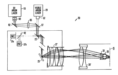
Fig. 2 is a simplified optical schematic of a
confocal microscope 50, modified according to the present
invention so that both visible and W light can be used for
excitation. Elements that are the same as those in Fig. lA
will have the same reference numerals, corresponding elements
that are modified will have primed reference numerals, and
elements that have no counterpart in Fig. lA will have
different reference numerals. As above, an inverted microscope
embodiment is illustrated.
Confocal operation as described in connection with
the prior art visible microscope does not naturally extend into
the realm of UV excitation, much less so when it is desired to
provide visible excitation as well. The first obvious problem
is that eyepiece 27 does not transmit UV light and adapter lens
30 transmits it poorly. However, even if the lenses transmit
UV, a number of aberrations must be corrected for proper
operation to occur. The design details for both inverted and
upright microscope embodiments are described (and illustrated
where appropriate) below.
A W laser 55 provides a W beam that is combined
with the visible beam from visible laser 20 by a beam combiner
57 (which is a visible/UV dichroic reflector). A focusing W
(5x) beam expander 60, steering mirrors 62, and a UV correction
lens 65 are interposed between W laser 55 and beam combiner
57. To the extent that beam splitter 22 does not reflect UV
light, it must be modified so that it reflects both the visible
and W laser beams but transmits the visible fluorescence. The
microscope is further modified by being provided a specially
designed eyepiece 27' and adapter lens 30', to be described
below. The characteristics of beam splitter 42 may be selected
in view of the visible wavelengths to be separated.
Focusing W beam expander 60 provides magnification
t« ~:Llow filling the back aperture of the objective
sufficiently to obtain maximum resolution. The use of a
confocal microscope reduces the smallest detectable distance
between spots to 0.707 times the smallest distance detectable
WO 92/18850 f~ ~ PCT/US92/02923
8
by a conventional microscope. The use of W excitation
improves the resolution relative to that obtainable with
visible excitation due to the shorter wavelength.
A visible beam expander 67 is preferably disposed
between visible laser 20 and beam combiner 57, and provides
1.5x-2x magnification to allow the visible excitation beam to
fill the back aperture of the objective. This is necessary
since UV eyepiece 27~ has a lower magnification than visible
eyepiece 27 (about 6.4x versus 8x). In view of the reduced
l0 magnification, consideration is being given to reducing the
minimum aperture diameter from 0.7 mm to 0.5 mm.
W correction lens 65 is preferably a 500mm lens that
focuses the W excitation light to a point in front of the
eyepiece, from which point it diverges. As noted above, the
visible excitation light is generally collimated until it
reaches the eyepiece. The W light is caused to diverge so as
to correct for longitudinal chromatic aberration effects in the
downstream lens elements. As will be discussed in more detail
below, there are chromatic scanning effects that must be
corrected in the eyepiece and adapter lens, and the correction
of those tends to make it difficult to correct for longitudinal
chromatic aberration as well.
The position of the W correction lens along the
optical axis can be adjusted, either alone or in conjunction
with refocusing of the beam expander in order to adjust for
different amounts of longitudinal chromatic aberration in
different objectives while maintaining appropriate filling of
the back aperture of the objective. This is a relatively
necessary option since objective chromatic aberrations vary
significantly in the UV, and back aperture sizes vary
significantly with different objectives.
First-Generation Lens Designs
Figs ? and 4 are optical schematics of a first-
generation design for eyepiece 27' and UV adapter lens 30' for
the inverted microscope embodiment. The geometrical and
optical parameters for these lenses are set forth in Tables 1
CA 02106296 2001-06-27
92/18850 PCT/US92~. _923
9
and 2. Surfaces are numbered along the direction of the laser
beams and dimensions are in mm unless otherwise stated.
Chromatic correction wa.s in terms of the following design
wavelengths: 330nm, .464nm, 560nm, and 656nm. The lens design
was performed using the. OPTEC optical systems design analysis
software (PC version), available from SCIOPT Enterprises, San
Jose, California.
Fig. 5 is an optical schematic of a first-generation
design for the eyepiece, designated 27", for the upright
l0 microscope embodiment. The geometrical and optical parameters
for this lens are set forth in Table 3.
The design criteria for the eyepiece (alone for the
upright embodiment or in combination with the adapter lens for
the inverted embodiment) can be summarized as follows. The
eyepiece (or combination) is well corrected for the standard
monochromatic aberrations (spherical aberration, coma,
astigmatism, distortion) for each design wavelength for light
traveling in both directions. As noted above, W focus is
corrected by focusing lens 65 and focusing beam expander 60,
although the eyepiece (or combination) is corrected for
longitudinal chromatic: aberration for visible wavelengths. The
eyepiece (or combination) is, however, chromatically corrected
to minimize scanning errors as will be discussed below. Field
curvature is minimized and matched for different colors for a
visible object at infinity and a finite UV object located 195
mm from the front surface of the eyepiece. The front focal
length is sufficiently long to prevent the eyepiece from
touching the scanning mirror motor during adjustment.
The particular lens design was also subject to a
number of more mundane constraints. For example, the glasses
had to be affordable and relatively easy to manufacture into
high quality lenses. Further the lens had to withstand normal
laboratory temperature .arid humidity conditions and exposure to
W and visible laser radiation. Fused silica and calc?um
fluoride were chosen as the lens materials because they pass W
light, are fairly hardy under normal laboratory conditions, do
not cloud with prolonged W light exposure (under 200 mw of
Trade-mark*
WO 92/18850 PCT/US92/02923
to
power), and have sufficiently different dispersion
characteristics to allow chromatic corrections with reasonably
attainable lens surface curvatures.
In the first-generation design, the calcium fluoride
lens elements are all the same shape, being available as stock
items from Janos Technology, Inc., Townshend, Vermont, at the
desired quality (1% tolerances on curvature and thickness). It
was believed, however, that improvements in the monochromatic
aberrations could be achieved (at a cost) by relaxing the
1o symmetry constraint on the calcium fluoride lens elements and
having them custom ground. This was done in a series of
second-generation designs.
Second-Generation Lens Desiqns
A number of second-generation designs were developed,
characterized by the following differences from the first
generation design. First, they are not subject to the
constraint that the calcium fluoride elements be any particular
shape. Second, the order of the last two elements of the
eyepiece is reversed so that none of the calcium fluoride
elements (which are somewhat sensitive to humidity) is exposed
to the atmosphere. Along this line, the adapter lens is
provided with an extra silica element to reduce chromatic
aberrations and enclose the calcium fluoride element. These
second-generation designs include 6.25x and 8x eyepieces and
adapters (same adapter for both) for the inverted microscope
embodiment, and an 8x eyepiece for the upright microscope.
Fig. 6 is an optical schematic of the 6.25x eyepiece
for the inverted microscope embodiment. The geometrical and
optical parameters for this lens are set forth in Table 4.
Surface 1 is the location of the scanning mirror and the front
focal plane of the eyepiece. Surface 14 is the plane where the
eyepiece focuses the beam, which corresponds to the front focal
plane cf the adapter lens (not included in Table 4 - same
parameters as adapter lens for 8x eyepiece). Surfaces such as
1 and 14 are planes in air and are denoted as having a radius
of 100000 mm for convenience.
WO 92/18850 ~ , s. -
PCT/US92/02923
11
Figs. 7A and 7B are optical schematics of the 8x
eyepiece and adapter lens for the inverted microscope
embodiment. The geometrical and microscope parameters for
these lenses are set forth in Table 5. Surface 1 is the
location of the scanning mirror and the front focal plane of
the eyepiece. Surface 14 is the plane where the eyepiece
focuses the beam, which corresponds to the front focal plane of
the adapter lens. Surface 21 is located near the objective
back aperture.
Fig. 8 is an optical schematic of the 8x eyepiece for
the upright microscope embodiment. The geometrical and
microscope parameters for this lens are set forth in Table 6.
Surface 1 is the location of the scanning mirror and the front
focal plane of the eyepiece. Surface 14 is the plane where the
eyepiece focuses the beam. Surface 15 is located near the
objective back aperture.
Focusing Errors.
Figs. 9A-E are optical sketches showing the focusing
errors caused by longitudinal chromatic aberration, as well as
the way that this problem is handled.
Fig. 9A shows how longitudinal chromatic aberration
in the lenses causes focusing errors. This is illustrated in
the context of the inverted microscope embodiment, but the
problem and the solution are the same for the upright
microscope embodiment. More particularly, an incoming
collimated W excitation beam is shown in dotted lines as
coming to a focus in a plane 15u, displaced upstream from the
plane 15 that an incoming beam of visible light would be
focused. The visible light diverging from points in plane 15
would be collimated by the lenses and would reach the detector.
However, the visible light emanating from plane 15u (where the
W beam is focused) is not collimated by the lenses on its
return, and diverges before reaching the detector. As
mentioned above, it is convenient to correct for longitudinal
chromatic aberration through the use of focusing beam expander
60 and W correction lens 65.
P~'~"'~'~ ~ ~ , ~ ~ ~~ ~ 9 ~ 3
~~.~~9;~
IP~~/CJ ~ 16 J ~'~, ~~
12
Fig. 9B shows how W correction lens 65 causes the
W excitation beam (drawn in broken lines) to focus in the
same plane 15 from which emerging visible light (drawn in
solid lines) is collimated by the lenses and detected. This
is accomplished by bringing the W beam to a focus before
eyepiece 27', whereupon the W beam is diverging when it
encounters the lenses. The lenses are not corrected for
longitudinal chromatic aberration out to the W, and thus the
W light is focused in plane 15. It should be noted that the
illustrated visible light could equally well correspond to the
visible excitation beam.
Figs. 9C-E show how focusing beam expander 60 and W
correction lens 65 can be adjusted to vary the position of the
W focus and increase the beam magnification to increase the
filling of the back aperture of the objective. Fig. 9C shows
the situation where beam expander 60 provides an expanded
collimated beam. Fig. 9D shows how by adjusting the beam
expander to provide a divergent beam, it is possible to change
the location of the W focal point. Fig. 9E shows how a
stronger lens can be used to bring the location of the W
focus into a usable location.
Scanning Errors
Figs. l0A-C are optical sketches showing the
i5 scanning errors caused by chromatic effects, as well as the
way that this problem is handled.
Fig. l0A shows how chromatic effects in the lenses
affect confocal scanning. This problem is a separate one from
longitudinal (focusing) errors, which are assumed to have been
taken care of as described above. This is illustrated in the
context of the inverted microscope embodiment, but the problem
is the same for the upright microscope embodiment as well.
For confocal operation, the returning visible light (drawn as
a solid line) resulting from W excitation (drawn as a broken
line) must travel along the same path when it encounters the
scanning optics as the excitation light traveled when it left
the scanning optics. As shown in the figure, this condition is
.~ i i ~.~.~,
~i,, ., ~, ~, . . L ~ ... '
WO 92/18850 ~ ~ ~ ~ ~ ~ ~ PCT/US92/02923
13
met, but at large scan angles, due to chromatic effects in the
lenses, the visible light is collected from a point that is
laterally displaced from the point of maximal W excitation.
This type of scanning error results in a marked drop off in
intensity across the image field of view.
Fig. 10B is an optical sketch showing the way that
scanning errors are corrected for the inverted microscope.
This is accomplished by requiring that UV and visible light
from an object point 80 are focused by eyepiece 27' and adapter
lens 30' at an achromatic image point 82, where the object
point corresponds to the location of the last scanning mirror
and the image point corresponds to a point near the center of
the telon-objective combination. In the first-generation
design, the object point is approximately 18 mm from the front
surface of the eyepiece and the achromatic image point is 167.3
mm from the rear surface of the adapter lens. In the second
generation 6.25x design, the corresponding distances are
approximately 13.5 mm and 148.2 mm. In the second-generation
8x design, the corresponding distances are approximately 21.2
mm and 151.5 mm.
It is noted that this chromatic correction is not
entirely compatible with a correction for longitudinal
chromatic aberration, which as discussed above is instead
handled by the UV correction lens and the focusing beam
expander. The particular way that this optimization is
implemented is as follows. The eyepiece is corrected so that
(a) visible colors and UV parallel light in the backward
direction focus to the same point, and (b) all colors of
visible parallel light in the forward direction focus to the
same plane for large and small scan angles. The adapter lens
is optimized so that (a) parallel W and visible light in the
forward direction are achromatically focused at image point 82
and (b) visible parallel light traveling backward into the
acia?~tr.r lens is focused to a common front focal playa ( iocatsd
between the adapter lens and the eyepiece) for large and small
scan angles. Further the system is substantially achromatic
for all colors of visible light.
WO 92/18850 ~ PCT/US92/02923
14
Fig. lOC is an optical sketch showing the way that
scanning errors are corrected.~for the upright microscope. This
is accomplished by requiring that W and visible light from an
object point 85 are focused by the eyepiece, designated 27", at
an image point 87, where the object point corresponds to the
location of the last scanning mirror and the image point
corresponds to a point near the center of the objective. In
the first-generation design, the object point is 32 mm from the
front surface of the eyepiece and the achromatic image point is
178.25 mm from the rear surface of the eyepiece. In the
second-generation 8x design, the corresponding distances are
approximately 27.3 mm and 160 mm.
In any given design, the different wavelengths
emanating from point 80 (or 85) will not come to a focus
precisely at the same point 82 (or 87), since this constraint
is one of many that must be addressed in the optimization
process. Thus, relative to a reference wavelength, each given
wavelength will be characterized by a chromatic scan focus
error, of, that represents the longitudinal separation between
the given wavelength's focus and that for the reference
wavelength.
Table 7 shows the chromatic scan focus errors (ef)
for 330nm and 656nm relative to the position for 494nm (0 by
definition) for four eyepieces. The first (eyepiece #1), used
for comparison purposes is a four-element fused silica eyepiece
optimized for monochromatic aberrations but not for scanning
chromatic aberration. The other three are the 6.5x first-
generation eyepiece and the 6.25x and 8x second-generation
eyepieces, all for the inverted microscope.
Each eyepiece design was combined with a 160mm lens
design in the OPTEC program, and rays of different wavelengths
were traced along scan lines through the optics. The chromatic
scan focus error (of) was determined using the 494nm reference
waveleng'ch. ~i was more than one inch at UV wavelengths with
the uncorrected fused silica eyepiece, and was reduced to a few
millimeters with the eyepieces corrected according to the
WO 92/18850 ~ ~ ~ ~ y ~ ~ PCT/US92/02923
invention. Table 8 shows of for additional wavelengths with
the 6.25x eyepiece.
Thin lens equations predict that, for maximum
resolution, an 8x eyepiece will require 25% less chromatic
5 correction than that required by the 6x eyepiece. However,
chromatic error was more than halved by designing a longer
focal length 6.5x eyepiece (compare eyepieces #2 and #3 in
Table 8). The 8x design required stronger surface curvatures
to achieve the same chromatic correction as the 6x, increasing
l0 monochromatic aberrations to unacceptably large values.
Therefore, the chromatic correction was necessarily reduced for
the 8x eyepiece. Eyepiece #3 (6.5x) had the least amount of
chromatic aberration but was flawed by significant distortion
and was redesigned to yield eyepiece #4 (6.25x). For eyepiece
15 #4, the chromatic correction was compromised to optimize the
monochromatic corrections.
The correction for chromatic scan focus errors
appears to be accomplished primarily in the eyepiece, even in
the inverted microscope embodiment. Indeed, some tests
indicated that an off-the-shelf 160mm visible achromat that
also transmitted W worked somewhat better than the first-
generation adapter lens, and generally as well as the second-
generation adapter lens.
Field Curvature and Magnification Errors
Field curvature is typically one of the hardest
aberrations to correct, and a certain residual amount is often
acceptable. For example, assuming that all wavelengths are
subject to the same degree of field curvature, the result would
be that the sample plane would not be a plane, but rather a
slightly curved surface. Fundamentally this would not be a
problem since slightly curved sections would generally provide
the same useful information as perfectly flat ones.
However, the real problem wises when all wavelengths
are not subject to the same degree of field curvature. In such
a case, for points removed from the center of the field, the W
excitation would not be confocal with the visible fluorescence.
WO 92/18850 PCT/US92/02923
16
Rather, the visible light reaching the detector would be that
emanating from a point away from the focused beam spot and
would thus be of much lower intensity. This is the same
problem that arises if longitudinal focusing errors are not
corrected except that the result is not uniform across the
field, but increases away from the center of the field.
Accordingly, the problem is handled by accepting some
degree of field curvature and placing a priority on achieving a
field that is similarly shaped for the W and visible
l0 wavelengths.
Fig. 11 shows calculated plots of field curvature for
eyepiece #1 (top three lines) and eyepiece #3 (bottom three
lines), each combined with a theoretical 160mm adapter lens and
a theoretical objective. Also shown are plots for the
objective alone (three almost coincident lines). Field
curvature at the specimen plane was calculated by tracing rays
of light at incremented scan angles through theoretical
thick-lens models (using the OPTEC program). As the scan angle
was incremented, the root-mean-square position of the objective
focus was recorded in the axial and radial directions, and
plotted. To model the full optical train, a theoretical
infinity-corrected, lOx objective (achromatic in visible but
with some residual chromatic aberration in UV) was designed
from calcium fluoride and fused silica. A 160mm lens with
similar chromatic properties was also designed. Rays were
traced through the optical system at three wavelengths: red
(656 nm), blue (488 nm), and uv (330 nm). Chromatic
corrections in the calcium fluoride/fused silica eyepiece
dramatically reduced lateral magnification errors, ey, as well
as field curvature errors, ez, compared to errors introduced by
the uncorrected fused silica eyepiece.
Figs. 12A-C are plots showing calculated field
curvature and magnification errors near the edge of the field
of view for the microscope with a 100x objectize, a 40x
objective, and a lOx objective. The vertical axis represents
the location of the spot along the beam direction. The
horizontal axis represents the transverse position of the spot.
WO 92/18850
PCT/US92/02923
17
The three rows of plus signs represent measurements taken with
red (top), blue, and UV (bottom) for the four-element fused
silica objective. Measurements for each of the three
wavelengths at a particular scan angle are grouped. The
vertical separation between points represents a field curvature
error between wavelengths at that angle: the horizontal
separation represents a magnification error.
The four rows of diamonds represent measurements
taken with the corrected six-element 6.5x eyepiece for UV (top)
and red, blue, and green (grouped below). As can be seen, the
field curvature and magnification errors are much smaller for
the corrected eyepiece. The magnification errors dominate the
field curvature errors for the TOOx objective, while the
reverse is true for the lOx objective. The errors are
comparable for the 40x objective.
Overall Intensity Profile
Confocal sections were collected through the center
of a thick slab of fluorescein dye, excited by blue or UV
light, using a 40X Olympus objective. Curves were fit to a
profile of the fluorescent image, drawn through the center of
the field. The curves were shifted slightly to match their
peaks. (Peak-shifting was caused by small mirror alignment
differences). A full field-of-view was 768 pixels. Fig. 13A
shows the fluorescent field brightness with UV or visible
excitation for chromatically-corrected eyepiece #3 (curves a.v
and a.uv), and chromatically-uncorrected eyepiece #1 (curves
b.v and b.uv,). As can be seen, image intensity across the
field was dramatically improved with the chromatically
corrected eyepiece. Fig. 13B shows data collected using
eyepiece #1 and 100x, 40x, and lOx Olympus objectives. As can
be seen, field intensity improved with increasing objective
power.
Focusing Lens in Detector Path
As alluded to above, collection efficiency may be
improved by placing a lens in the detector path. Collimated
WO 92/18850 ~~ ~~ PCT/US92/02923
18
excitation laser light passes through the eyepiece and
objective, and focuses to an excitation point. Fluorophores
are excited in the region around the excitation point according
to the intensity distribution of a diffraction-limited, three-
s dimensional point spread function (psf). Light is collected by
the objective along this same intensity profile, resulting in a
somewhat narrower confocal psf that is approximately
symmetrical around the excitation point. In a simple pinhole
confocal microscope the center of the psf image is focused to
the pinhole, and light from planes above and below the
objective focus is rejected nearly symmetrically. In the
particular microscope described above, light from the center of
the psf image is focused to infinity, and consequently, light
from planes above and below the objective focus is rejected
asymmetrically.
The radius of the beam at the confocal aperture was
calculated for light emitted from various points along the
optical axis near the excitation point using thin lens
equations. The proportion of the intensity collected from each
point was calculated for the aperture set to the width of the
collimated beam according to I=(rp/re)2, where rp, the radius
of the pinhole, was set to the radius of the collimated beam
from the excitation point, and re was the radius of the
diverging or converging light at the pinhole from out-of-focus
points. Values greater than 1 were truncated to 1 (indicating
all of the light was collected at the PMT). The center of the
resulting pinhole collection function (pcf) was found to be
shifted past the center of the psf along the optical axis. As
the pinhole was opened or closed, the pcf widened or narrowed
around its center. The beam of light from the center of the
pcf was focused on the pinhole. For a 40x objective, the
optical section, when using a 6x eyepiece, was found to be more
than twice as wide as the section from a lOx eyepiece when all
the light from the maximally excited point at the center of the
psf is collected.
The pinhole can also be thought of as a spatial
sampling device. To obtain theoretical resolution, the pcf
WO 92/18850 ~ ~~ ~ ~ ~ ~ PCT/US92/02923
19
must be at least half the width of the theoretical resolution
to satisfy the Nyquist sampling criterion. The pcf generally
has a different shape than the psf, with the result that a
smaller pinhole must be used to attain maximum resolution in
the radial direction than in the axial direction. If the
pinhole size is reduced to attain theoretical axial resolution,
the pcf will narrow around its center, and a large portion of
the excited light from the psf will be rejected.
Thin lens equations were used to calculate the
l0 magnification of the psf in the radial direction at the
pinhole. The theoretical lateral resolution was found to be
attainable with a lOx eyepiece but not with a 6x eyepiece in
this configuration. Similar calculations were made for 100x,
40x, 20x, and lOx objectives with 6x, 8x, and lOx eyepieces.
The model lOx eyepiece could attain theoretical axial and
radial resolution (although not with optimal power) for all but
the lOx objective. The 8x eyepiece resolving power was
marginal for the 40x objective, and was insufficient for the
lOx objective. The 6x eyepiece only attained theoretical
resolution with the 100x objective.
To solve this resolution problem, a 1000mm lens was
placed between the dichroic mirror and the pinhole to focus
collimated light from the center of the excitation spot to the
pinhole. This approach shifted the center of the axial pcf to
the center of the axial psf, allowing the pinhole to be reduced
to any size and yet still collect light from the maximally
excited point in the specimen.
At small apertures, the use of the long lens boosts
the light intensity collected by about 40%. Collecting the
out-of-focus light is not as much of a problem as rejecting
some of the in-focus light because the aperture is smaller than
the width of the returning collimated beam. The final effect
is to increase the light intensity at small apertures while
maintaining or boosting resolution. Due to tha basic
microscope design, which includes the eyepiece and long return
path, the benefit of using an iris diaphragm instead of a
WO 92/18850 ~ ~~ PCT/US92/02923
pinhole is retained even when the long lens is inserted into
the return-only path.
However, the single lens reduced the lateral
magnification of the psf FWHM at the pinhole below 0.5 mm (a
5 practical limit for the variable aperture) with some
objective/eyepiece combinations. The single lens was
subsequently replaced with a two lens optic designed to focus
the psf image to the aperture, and magnify the lateral FWHM of
the image to a size greater than 0.5 mm for all objectives.
10 This design is expected to allow the chromatically corrected 6x
eyepiece design to be used to attain theoretical confocal
resolution.
Conclusion
15 In conclusion it can be seen that the present
invention provides an economical and effective technique for
extending the advantages of confocal scanning microscopy into
the UV.
While the above is a full description of the
20 preferred embodiments, various modifications, alternative
constructions, and equivalents may be used. For example, while
the return-only path is shown as being provided with a single
beam splitter and two detectors, a second beam splitter and a
third detector could be added to exploit more fully the UV
capability of the microscope. Furthermore, while the objective
in the inverted microscope embodiment was shown in combination
with a telon lens, an infinity-corrected objective without a
telon lens could be used.
Therefore, the above description and illustrations
should not be taken as limiting the scope of the present
invention which is defined by the appended claims.
WO 92/18850 ,~ ~ ~~ ~a ~ ~ ~ PCT/US92/02923
l:. rm :!
21
TABLE 1 - FIRST-GENERATION
EYEPIECE FOR INVERTED MICROSCOPE
Center Maximum Edge
Surface Radius Thickness Material Aperture Thickness
1 -66.331
7.00 silica 25.4 5.467
2 -30.597
0.00 air 25.4 6.532
3 23.266
9.54 CaF2 25.4 1.996
4 -23.266
0.00 air 25.4 0.958
5 -30.070
2.00 silica 25.4 7.509
6 15.388
1.00 air 17.4 2.077
7 23.266
9.54 CaF2 25.4 1.996
8 -23.266
1.50 air 25.4 1.186
9 -16.052
1.50 silica 21.4 8.942
10 25.710
0.00 air 25.4 0.416
11 23.266
9.54 CaF2 25.4 1.996
12 -23.266
EFL = 38.59 mm FFL = 22.4 mm
Distance from scanni~~c~ ~t~irror to surface #1 = 18 mm
Magnification = 250/38.59 = 6.5x
Minimum aperture = 12 mm
Maximum tube diameter = 30 mm
WO 92/18850 PCT/US92/02923
TABLE 2 - FIRST-GENERATION
ADAPTER LENS FOR INVERTED MICROSCOPE
Center Maximum Edge
Surface Radius Thickness Material Aperture Thickness
1 37.211
6.40 CaF2 25.4 1.931
2 -37.211
0.134 air 25.4 0.00
3 -37.259
2.50 silica 25.4 5.548
4 120.112
EFL = 134.4 mm FFL = 137.3 mm BFL = 123.5 mm
Distance from eyepiece to surface #1 = 155.30 mm
Distance from surface #4 to telon lens = 167.30 mm
WO 92/18850 . ~ ~,~ ~ ~ PCT/US92/02923
23
TABLE 3 - FIRST-GENERATION
EYEPIECE FOR UPRIGHT MICROSCOPE
Center Maximum Edge
Surface Radius Thickness Material Aperture Thickness
1 -90.609
7.00 silica 25.4 5.467
2 -41.788
0.00 air 25.4 6.532
3 23.266
9.54 CaF2 25.4 1.996
4 -23.266
0.00 air 25.4 0.958
5 -28.449
2.00 silica 25.4 7.509
6 19.287
1.00 air 17.4 2.077
7 23.266
9.54 CaF2 25.4 1.996
8 -23.266
1.50 air 25.4 1.186
9 -16.113
1.50 silica 21.4 8.942
10 22.175
0.00 air 25.4 0.416
11 23.266
9.54 CaF2 25.4 1.996
12 -23.266
EFL = 38.82 mm FFL = 22.75 mm BFL = 18.24 mm
Distance from scanning mirror to surface #1 = 32 mm
Distance from surface #12 to objective = 178.25 mm
Magnification = 250/38.82 = 6.4
Minimum aperture = 12 mm
Maximum tube diameter = 30 mm
WO 92/18850 ~~ , ~ PCT/US92/02923
24
TABLE 4 - SECOND-GENERATION
6.25X EYEPIECE FOR INVERTED MICROSCOPE
Center Maximum Edge
Surface Radius Thickness Material Aperture Thickness
1 100000.000
23.805 air 25.4 23.652
2 -527.977
4.000 silica 25.4 6.475
3 35.895
0.000 air 25.4 0.686
4 23.490
10.500 CaF2 23.0 3.020
5 -17.021
0.000 air 23.0 0.466
6 -18.510
2.000 silica 23.0 9.262
7 26.400
0.000 air 25.4 0.682
8 22.449
9.540 CaF2 25.4 3.991
9 -50.869
0.000 air 25.4 3.386
10 46.320
9.540 CaF2 25.4 4.596
11 -27.035
1.500 air 25.4 0.965
12 -17.279
3.000 silica 21.4 4.584
13 -39.099
~ 25.615 air 25.4 27.735
14 -_
100000.000
WO 92/18850 ~ ~ ~ ~ ~ ~ ~ PCT/US92/02923
TABLE 5 - SECOND-GENERATION
8X EYEPIECE AND ADAPTER FOR INVERTED MICROSCOPE
5 Center Maximum Edge
Surface Radius Thickness Material Aperture Thickness
'~
1 100000.000
21.201 air 25.4 19.943
2 -64.724
10 7.000 silica 25.4 6.800
3 -56.024
0.000 air 25.4 6.458
4 18.630
10.000 CaF2 25.4 1.127
15 5 -22.758
0.440 air 25.4 0.109
6 -20.660
2.000 silica 25.4 8.352
7 20.517
20 1.000 air 17.6 1.096
8 39.746
9.540 CaF2 25.4 4.981
9 -33.820
0.000 air 25.4 5.754
25 10 26.229
9.540 CaF2 25.4 5.138
11 -72.437
1.700 air 25.4 0.409
12 -22.471
2.000 silica 20.3 3.628
13 -103.178
12.021 air 25.4 12.806
14 100000.000
148.896 air 25.4 149.199
15 405.219
WO 92/18850 ~ PCT/US92/02923
'~~ ~ ~ 26
TABLE 5 - SECOND-GENERATION
8X EYEPIECE AND ADAPTER FOR INVERTED MICROSCOPE
Center Maximum Edge
Surface Radius Thickness Material Aperture Thickness
3.000 silica 25.4 4.336
16 53.314
0.063 air 25.4 0.000
17 55.550
7.000 CaF2 25.4 4.545
18 -82.494
0.000 air 25.4 1.032
19 1672.767
4.000 silica 25.4 4.052
20 -805.477
151.452 air 25.4 151.352
21 100000.000
WO 92/18850 ~' ~ ;,~ ~~''~ ~~ PCT/US92/02923
27
TABLE 6 - SECOND-GENERATION
8X EYEPIECE FOR UPRIGHT MICROSCOPE
Center Maximum Edge I
Surface Radius Thickness Material Aperture Thickness
1 100000.000
27.290 air 25.4 26.213
2 -69.162
7.000 silica 25.4 6.908
3 -64.215
0.000 air 25.4 6.268
4 18.630
10.000 CaF2 25.4 1.127
5 -22.758
0.600 air 25.4 0.371
6 -19.983
2.000 silica 25.4 8.212
7 21.556
1.000 air 18.6 0.974
8 39.746
9.540 CaF2 25.4 4.981
9 -33.820
0.000 air 25.4 5.755
10 26.229
9.540 CaF2 25.4 5.138
11 -72.437
1.700 air 25.4 0.447
12 -21.655
2.000 silica 19.7 3.341
13 -78.532
' ~~ 12.180 air 25.4 13.215
14 _ -
100000.000
160.000 air 25.4 0
15 100000.000
WO 92/18850 ~ ~~~ ~ ~ PCT/US92/02923
28
TABLE 7 - SCA.NNING CHROMATIC ERROR
(ef) FOR FOUR EYEPIECES
Silica 8z 6.Sz 6.25z
Wavelength Eyepiece #1 Eyepiece Eyepiece Eyepiece #4
(nm) ef (mm) #2 #3 ef (mm)
ef (mm) ef (mm)
330 -29.77 -9.09 -3.28 -4.22
494 0 0 0 0
656 9.53 1.38 -0.76 -0.21
WO 92/18850 , PCT/US92/02923
29
TABLE 8 - SCANNING CHROMATIC ERROR
FOR 6.25x EYEPIECE
Wavelength (nm) ef (mm)
656 -0.21
540 0.13
494 0
488 -0.05
450 -1.01
351 -3.19
330 -4.22