Note: Descriptions are shown in the official language in which they were submitted.
Wo92/17~6s PCT/US92/02499
21û7~
IMMORTALIZATION OF
ENDOTHELIAL CF~TS
BAC~GROUND OF THE INVENTION
Field of the Invention
The present invention relates, in general,
to endothelial cells. In particular, the present
invention rPlates to immortalized microvascular
endoth~lial cells obtained from human skin.
Backaround Information
In the last 10 years, the accumulation of
information about the endothelium has led to the
realization that this is a tissue which is not just
a target for injury, but by undergoing alterations
in functions, metabolism and structure, it directly
influences the evolution and outcome of vascular
injury, inflammation and immune reactions like graft
rejection and tumor metastasis (Cotran, R. (1987)
Am. J. Pathol. 129, 3: 407-413). Factor VIII-
related antigen is an endothelial cell product
involved in the aggregation of platelets;
megakaryocytes are the only other cell type known to
express this antigen (Cotran, R. (1987) Am. J.
Pathol. 129, 3: 407-413). Upon activation by
molecules like Interleukin-1 and tumor necrosis
factor, these cells up-regulate their expression of
leukocyte specific adhesion molecules like ICAM-l
and ELAM-1 (Pober, J.S. (1988) Am. J. Pathol. 133,
3: 426-433; Butcher, E.C. et al. (1986) J. Cell.
Biochem. 30, 2: 121-131); interferon-gamma is
associated with the expression of Class II major
histocompatibility antigens (Dvorak, H.F. et al.
~U~;ST~TUTE SHEE7
,- . . . - - . . - . . .. . .
~ .......... : . ., .. - . . . : :. ..
. `- - - - ~ ,.
. - . . . . . .
- . . . . . ~: , ... . : . ..
7~- , . , . ` : . ~ -- .: . :
WO92/17~69 PCT/US92/0~ ~
2~07~9
(1986) Hum. Pathol. 17, 2: 122-127).
Morphologically, endothelial cells can express
facultative traits of tissues from which they were
derived, including Weibel-Palade bodies and
cobblestone growth pattern (Cotran, R. (1987) Am. J.
Pathol. 129, 3: 407-413; Xarasek, M.A. (1989) J.
Invest. Dermatol. 93: 335-385).
Primary human microvascular endothelial
cells have a limltod llfe span of about ~-lC
passages and have specific growth requirements.
Early methods of tissue culture required tho US2 sf
high concentrations of serum, Sarcoma 180
conditioned medium and multiple growth factors for
optimal growth. Human serum requirements can be
decreased or substituted with fetal bovine serum by
incorporating 2% pre-partum maternal serum in the
medium (Karasek, M.A. (1989) J. Invest. Dermatol.
93: 335-385). These cells can also be stimulated by
the addition of agents such as cholera toxin,
dibutyryl cAMP and isobutyl methyl xanthine which
activate adenyl cyclase prolonging the growth rate
and morphology (Iuder, R.M. et al. ~1990) J. Cell
Physiol. 142: 272-283). In the absence of cAMP,
cultured vascular endothelial cells undergo
pronounced changes in their morphology and
functioDal properties; cells turn from epithelial to
spindle shape and lose some of their ability to
express HLA-DR antigens in response to interferon-
gamma (Tuder, R.M. et al. (1990) J. Cell Physiol.
142: 272-283).
Various normal and neoplastic, as well as
differentiated embryonic cells of human origin, have
been transformed and immortalized by intact SV40
. . , ~ . . . ..
,,;
SUBSTlTUTc SHEET
~ . ' ' . ,' '
WO92/17569 PCT/US92/02499
21U7 j~9
virus including human umbilical cord endothelial
cells (SacX, G.H., Jr. (1981) In Vitro 17, 1: 1-19;
Gimbrone, ~.A. ~ Jr. et al. (1976) Cell 9: 685-693).
Papova viruses constitute one of the simplest group
of DNA tumor viruses and have been the most studied
(Aaronson, S.A. (1970) J. Virol. 6, 4: 470-475).
The SV40 transfected human endothelial cells did not
exhibi~ ~actor VIII related antigen expression nor
show char~st~rlstic Weibel-~alade bodies (Gi~brone,
îO i~.~., ~.. ~t al. (1976) Cell 9: 685-693). Human
~m~i lic21 vel n endothelial cells have also been
immo' talized by exposure to murine sarcoma viruses
containing the l'v-ras~ or "v-mos" oncogenes (Faller,
D.V. et al. (1988) J. Cell Physiol. 134: 47-56).
These cells expressed Factor VIII related antigen
and contained Weibel-Palade bodies. An endothelial
cell line derived from mouse lymph node stroma which
; retains most functional characteristics of normal
mouse endothelial cells has been described
(O~Connell, X.A., and Edidin, M. (l990) J. Immunol.
144, 2: 521-525); transient infection was performed
~ using whole virus SV40 strain 4A to immortalize
; these cells.
The cells according to this invention
exhibit a number of utilities. For example, the
cells can be used to study the immediate adherence
of HDMEC to graft vascular surfaces, for example:
angioplasty and endarterectomy. The cells can also
be used in pre-coating vascular grafts (with
endothelial cells).
i The cells can be used in metabolic studies
of lipid and lipoprotein metabolism, arachidonic
acid metabolism, hemostasis factors, and endothelial
!
SU8STITUTE SHEET
~'' ' ' ' ' - . '.' : ' ~ , `
,. - .
- . - .- . -. .
W092/1~569 PCT/US92/0~ ~
21~7~
derived vasoactive substances such as endothelin
(ET). The cells can also be used in studies of
angiogenesis, wound healing, leukocyte adherence and
adhesion molecule expression (intracellular
expression as well). Further, the cells can also be
used in genetic studies aimed at the isolation o~
endothelial cell specific gene regulatory products
and creation of cDNA libraries for endothelial cell
specific g2nes.
One sXilled in the art will apprecia t2
that the cells of th2 invention can be used in
pharmacologic s-udies as substrates for the
screening of various agents as inhibitors of
inflammation or modulators of cell adhesion molecule
expression or in the cosmetic industry for toxicity
testing. The cells, if tumorigenic, can be used in
studies of endothelial cell tumor formation and
potentially useful in specific problems, such as
Kaposi sarcoma (or the transforming effects of
chemicals or other agents on diploid human cells).
The cells can also be used for viral or parasitic
growth or detection. The cells can be used to
produce products (for example: cytokines or
lymphokines) which may be secreted into the medium
or isolated from the cell surface.
SUMMARY OF THE INVENTION
It is a general object of this invention
to provide a microvascular endothelial cell.
It is a specific object of this invention
to provide a primate immortalized microvascular
endothelial cell.
SJ~ 5TUTE SHEE ~ :
. . . . . . . . . ., .; .......... .. . ..... .. - ....... .
.
W092/~7S69 PCT/US92/02499
2~7~ ~j9
It is a further object of this invention
to provide a primate immortalized microvascular
endothelial cell line.
I~ is another object of this invention to
S provide a method of establishing a cell line of
primat~ o~t~ li z~d microvascular endothelial
cells.
~ u~th~r objects and advantages of the
~r~s_~ n~ -n ~ e clQar .rom the d~scription
that -~-ollo-~s.
~ -. one Pm~odiment, the present invention
relatQs to 2 microvascular endothelial cell (or cell
line~ ~her2in the c~11 (or cell line) is obtained
from a primate skin and is immortalized.
In another embodiment, the present
invention relates to a method of establishing a cell
line of immortalized microvascular endothelial cells
derived from primate skin comprising:
(1) introducing DNA which encodes SV40 large T
antigen into the cells and
(2) c~lturing the cell line.
ERIEF DESCRIPTIO~ OF THE DRAWINGS
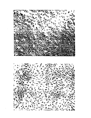
Figure 1. (A) Phase-contrast microscopy of
a pure culture of human dermal microvascular
endothelial cells (HDMEC) showing typical
"cobblestone" morphology. (B) Phase-contrast
microscopy of a culture of SV 4OT-transformed human
dermal microvascular endothelial cells. They also
have a "cob~lestone" appearance and are
indistinguishable from non-transformed human dermal
microvascular endothelial cells.
SUBSTITUT~ S.
' ,, ., . ' , . " .: " . ." ~ ' . ' ' . . ~ ' , . . ' ' ' '~. ' . "
WO92/1756s PCT/US92/0~99
2 !~ '3 7 .~ ~ 9 6
Figure 2. (A) Direct immunofluorescence - -
microscopy of human dermal microvascular endothelial
cells using acetylated LDL. The cells show granular
cytoplasmic staining indicating uptake of the
acetylated LDL, typical of endothelial cells. (8)
Direct immunofluorescence microscopy of CDC/EU.XMEC-
1. The staining is typical of endothelial cells and
indistinguishable from that shown in Figure 2A.
~igure 3. (~) Phase-contrast microscopy
of human dermal microvascular endothelial cells
gro~n on matrigel. Long, tu~ular, c~llular
~xtensions are seen at eight hours of cultur~. This
is typical of the morphologic differentiation that
endothelial cells undergo when cultured on this
matrix. (B) Phase-contrast microscopy of
CDC/EU.HMEC-l cells cultured on matrigel.
; Morphologic differentiation is noted that is very
similar to that seen in Figure 3A. --
Figure 4. CDC/EU.HMEC-1 (Transformed ~ -
HDMEC) cells were grown in HDMEC medium supplemented
with different concentrations of human serum. They ~-
were seeded at 1.7 x 10 cells/flask, cultured for
eight days at 37C, 5% C0" har~ested, and counted. -~
Cells grew at all concentrations of human serum that
were tested but did so in a concentration-dependent
fashion. (Untransformed human dermal microvascular
endothelial cells will not grow below 20% normal
human serum.)
Figure 5. CDC/EU.HMEC-1 or untransformed
human dermal microvascular endothelial cells were
seeded at 5 x 10' cells/25 cm' flask and grown for 10
days at 37C 5% CO, with various concentrations of
human serum. Cells were harvested every 24 hours
SUBSTITUTE SHEET ~ .
. .. .
j' : ' ''. ' ' .' ' ,, , . .. : . . ' . ', '. ' . , ! : .
WO 92/17569 PCT/US92/0~99
21~7rj59
for 10 days and counted. (Open boxes = CDC/EU.HMEC-
1, 30Q0 52~ ; solid boxes = C~C/EU.HMEC-l, 1% serum;
open diamonds = CDC/EU.HMEC-1, 0% serum; solid
diamonds = .~M~C, 307 serum.) CDC/EU.HMEC-1 cells
grow best with 30~ human serum supplementation, but
will surv v~ at 0% human serum.
Figure 6. HLA-DR Expression on CDC-HMEC-
1. Cell-i ~2r_ untr2ated or treat~Ad with Interferon-
gamm2 -^?- 5~tan days. E~prassion of HLA-DR W2S
rerA^~ e~ ACSc-^~n analysis using PE-conjugated
anti-h~an ~LA-DR a~tisar~m.
3~T~ ~ T~n DESCRIPTION OP THE INVENTION
The present invention relates to human
microvascular endothelial cells.
In one embodiment, the present invention
relates to microvascular endothelial cells obtained
from primate skin (preferably, human skin or
foreskin) and immortalized. In a prefeEred
embodiment, immortalization is effected by
introducing DNA encoding SV-40 large T antigen into
the skin-derived endothelial cells. A preferred
cell line comprising such immortalized cell is
designated CDC/EU.HMEC-l. This cell line has been
dQposited in accordance with the Budapest Troaty
with the American Type Culture Collection, 12301
ParXlawn Drive, Rockville, MD 20852, USA as ATCC
D-signation CRL 10636. The deposit was received
January 8, 1991 and was accepted by this
International Depository Author ty.
The deposited human microvascular endothelial
cell line from human foreskin was immortalized by
.
~U''S7-lTlJTE SHEET
- . - , . . . - . . , . , . . , . . `
, . - ... . .
-- - - ,- - - .. - - . , . . ~.
, ~ : : ,. .
WO92/17569 PCT/US92/0~9g
~ ~ ~7559
transfecting freshly isolated cells with an
eukar~otic voctor containing the early region of the
SV40 virus large T antigen. These cells
(CDC/~U.'IM~C~ orm microtubules on matrigel, they
take up acetylated low density lipoprotein, express
Factor VIII-related antigen and express HLA-DR
antigen upon exposure to gamma-interferon; all of
which a ~ characteristics of normal microvascular
endothelial ce1ls. ~dditionally, these cells shaL2
0 'ho s ~ 1~= epi~hPlioid co~blestone growth pattern
of normal microvascular ,ondothelial cells.
Di.f2ren. - om normàl HD~EC, this new cell line
reauires low or no concentration(s) of human or
fetal bovine serum for growth, has a shorter
lS doubling time than HDMEC, do not require epidermal
growth factor or hydrocortisone for growth and do
not require gelatin or fibronectin-coated surfaces
$or attachment. Control cells which were not
transfected and did not contain SV-40T DNA died off
after two additional passages. Immortalized cells
are thus defined as cells which remain alive after
ten passages following introduction of DNA.
Ideally, they replicate or divide indefinitely,
maintain morphologic and physiologic characteristics
of the tissue of origin, and grow at lower serum
reguirements and increased cell density. In
particular, the DNA encodes the SV-40 large T
antigen and can be introduced via transfection.
In a further embodiment, the present
invention relates to a method of establishing a cell
line of immortalized microvascular endothelial cells
derived from primate skin (preferably human skin or
foreskin). The method comprises introducing DNA
SUBSTITUTE SHEET
:: ' ~ , ,,, ', . . :' '
W092~t7569 PCT/US92/02499
21~7~
g
encoding the S~40 large T antigen into the cells
under condit~ons such that the microvascular
endothelial cells ~ecome immortalized and culturing
the c2 11
Th~ pres~nt invention is described in
further detail in the following non-limiting
examol~s.
~ ~T~T ~;
~h_ -.olio~ing orotocols and experimental
details are re-rerenced in the examples that follow.
~ .
endothelial cells (HDMEC!. HDMEC have been isolated
from human foreskins by the following technigue.
Foreskins are cut into 3 mm squares and placed in
phosphate-buffered saline (PBS) containing 0.3%
trypsin (Sigma Chemical Co., St. Louis, M0) and 1%
ethylenediamine tetracetic acid (Sigma) at 37C for
10 minutes. The skin segments are washed with HBSS
several times and placed in a petri dish in HBSS
with the keratinized surfaced down. They are then
individually compressed with the side of a scalpel
blade to express microvascular fragments from the
cut surfaces of the skin. The microvascular
segments in 1 ml of HBSS are layered onto a 35%
solution of Percoll (Pharmacia AB; Uppsala, Sweden)
in H9SS that has been previously spun at 30,000 g
for 10 minutes at 4C to create a gradient. The
gradient is then spun at 400 g for 15 minutes at
room temperature. The fraction with a density less
than 1.048 g per ml, which is rich in microvascular
fragments, is removed. Those portions of the
SUBSTITUTE SHEET
. . . - - . -- .
. . .
.. . ., - -: ... : . . - ,.. . ~ .
, - . ~ ,' ' ~ ' '
WO92/17569 PCT/VSg2/0~99
21~75'a9
gradient containing the microvascular segments are
applied to a human fibronectin (Advanced
Biot~chnologies; Silver Spring, MD) precoated area
10 mm in diame.Pr in the center of a 60-mm tissue-
culture dish. The dishes are then incubated at 37Cin a moist incubator in 5% C02 overnight. Unattached
cells are removed by washing with HBSS. The
attached c~lls ar~ th2n viewed with an inverted
phase-contrast microscope in 2 ~iologic hood, and
nor.e.~c~h~'ial cells a~e removed by detaching them
with ~ ~5-gaus2 sterile needle. The growth medium
for _h~s2 c21' S consists of 200 ml Endothelial Basal
M2di~m ~CDB 131 (Clonetics, San Diego, CA), 75 ml
- human serum (Irvine Scientific, Santa Ana, CA),
Dibutyryl CAMP O.5 mM (Sigma Chemical
Co., St. Louis, M0; Tuder et al. (1990) J. Cell.
Physiol 142:272-283), Antibiotic-antimycotic
~olution 1% (final concentration) (Gibco, Grand
Island, NY), hydrocortisone 2 ~m (final
concentration) and epidermal cell growth factor 5
ng/ml (final concentration). Transfected cells were
grown in the same medium without CAMP. The
resulting cell cultures are consistently 100% pure
a~ assessed by morphological and immunochemical
criteria. The normal cells can be continuously
pa~saged up to 10 times.
Vec~. The vector used in the transfection is
dQsignated as SV-40T. It has the sequence that
codes for the transformation protein of SV-40 large
T antigen and RSV-LTR cloned into the Eco Rl site of
the PBR322 plasmid.
~:lleSTlTUTE SHEET
.. . : . .: . , ' ,. . '~ ', ' .. : - -. .
W092~17569 PCT/US92/0~99
2 ~
Transfection. Primary human foreskin endothelial
cells were ln their 6th passage from isolation when
transfected. The procedure followed has been
described ~y Graham and van der EB (Virology,
52:456-467, 1973). Some minor modifications were
introduc~d to this ~rocedure. All surfaces to which
endothelial cells attach were pre-coated with a
solution of 0.1~ gelating in 0.01~ phosphate buffer
saline ?h 7 a (73S) . ~he colls were plated onto 6-
well plates a, 3.5 x lCs celia/-~ell and incuba-ced @
37C and 5~ C0. overnight ~e_ore transfection. The
amount or vector DNA ~sed ~as ~ ~g per well.
Following ihe transfection ~rocedure, the plates
were incubated overnight ~ 37C with 5% C0" then the
lS contents of each well transferred to individual 25
cm2 plastic flasks with 0.2 ~ filter cap (Costar).
- Some of the flasks contained dibutyryl cAMP 0.5mM
(final concentration). Flasks were split 1:4 as
they became confluent.
.
Detection of SV-40T viral antiqen. To detect
expression of the SV-40T in the immortalized
endothelial cells (C~C/EU.HMEC-1) an enzyme-linked
immunoassay was performed. Cell line SV-T2 a
Balb/3T3 mouse embryo line infected with SV-40 (ATCC
# CCL 163.1) was used as a positive control, a
peripheral lymphocyte lysate was used as a negative
control. Cell-free lysates were obtained by adding
0.5 ml of 0.5% deoxycholate.HCl in O.OlM P8S pH 8.2
and 100 ~l of a 1 mM solution of phenylmethyl
sulfuflouride in 95% ethanol to 10 million cells.
This mixture was incubated Q 4C for 30 minutes and
then centrifuged @ 4000 RPM for 15 minutes @ 4C.
.
. :
'. SU2STlT~iTE SHEET
.
~, ' ':' ". `
.. . ~ . . - - -. . .... ... ....
WO92/17569 PCT/US92/0~99
21Q7~9
12
The supernatant which possibly contained the SV-40
large T antigen was removed and saved.
For the ELISA test, the antigens were
attached by suctioning onto nitrocellulose paper
using a ~inifold II Slot Blotter (Schleifher &
Schuell, ~eD~e, NH). After the attachment
procedure, the sheets were incubated for 24 hr with
2% s'clm mll'i; ~ 0.01 ~ PRS Q 4C to prevent
nonspDcific i~inding. Afterwards, the skim mil~
0 solu- o.. is ~~-o-ta zr,d the ni.-ocellulose washed
three tlmDs--iith 0.1~ TwePn-20 in PBS Q RT . ~ouse
moncc'^r.21 a..__~o~ to .he S'v-40 large T viral
antisen was added a~ a dilution of 1:1000 in
PBS/Tween and incubated for 30 min @ 37C on an
orbital shaker. The monoclonal antibody to SV-40T
antigen was obtained from the supernatant of the
mouse hybridoma cell line Pab 101 (ATCC # TIB 117).
The nitrocellulose sheets were then washed three --
times with Tween/PBS and an additional wash @ 37C
on the orbital shaker for 15 min. Goat anti-mouse
- horseradish peroxidase labelled monoclonal Ab
(Bio~ad, Ric~mond, CA) was added at a 1:2000
dilution and allowed to incubate for 30 minutes Q
37C shaking. Later, the nitrocellulose sheet was
washed three times with Tween/PBS. A
diaminobenzidine (Sigma Chemical Co., St. Louis, MO)
(DAB) solution consisting of 25 mg of DA~, 50 ml of
PBS, and 20 ~l of 30i% HtO, was added and color
allowed to develop. The nitrocellulose sheet was
rinsed with deionized water to stop the enzymatic
reaction.
,
~' ~E~ t ~UTE Si JE~
'. : -' ~ .: -' . . .. . ~ .
,: ' , , '' ' ' ': . .'. .'~ ' :' ' '~"' ' ' ' ; '', ,`:'/ ,' . .'
. :. . : : '. ; : ' .:
W092/17~69 PCTtUS92/0~99
2t~7~ '9
13
Expression of SV-40T antiaen. Cell-free lysates of
CDC/EU.HMECI and SV-T2 (positive control) cells
expressed the SV-40T viral antigen by ELISA and
Western Blot. Negative cont~ol (hu~an peripheral
blood lymphocyte lysate) cells were negative.
Characterization of endothelial cell culturPs.
Representative cultures of ~D;IEC and SV 4OT .iDMEC
(CDC/EU.HMEC-1) are charact2rized in thre~ ways.
Cultures are evaluate~ by nve-.ad has--con'_2~'
microsccpy to d2tormine whethor 'h~ c_lls have he
characteristic cob~laston2 .~or?hology e~^ ~ndotheliai
cells. Cells are fixed in 90~ methanol at -20C for
10 minutes, washed and stained with a 1:40 dilution
of rabbit anti-human factor VIII (Atlantic
Antibodies; Scarborough, ME) for 30 minutes followed
by FITC conjugated goat anti-rabbit IgG. They are
washed three times and then viewed under a
fluorescent microscope. Cells, unfixed, are
incubated with acetylated low-density lipoprotein
(10 ~g/ml), labeled with 1,l'dioctadecyl-1,3,3,3'3'-
tetramethyl indocarbocyanine perchlorate (Dil-AC-
LDL) (Biomedical Technology, Inc.; Stoughton, MA) at
37C in medium 199 without growth supplement or
fetal calf serum for 4 hours. Dil-Ac-LDL is a
biologic probe incorporated by living endothelial
cells and, to a lesser extent, by monocytes or
macrophages. The media is then removed, and the
cells are washed twice and visualized in a
fluorescence microscope with standard rhodamine
excitation emission filters. Evaluation by all
three techniques reveals that pure cultures of both
types of endothelial cells are routinely attained.
SU~STITUTE SHEET
,. . - .:
W092tl7569 PCT/US92/0~ ~
2 ~ 5 9
14
Diffe Qr;tla.ion OL endotheli~l cells. Matrigel, an
extrac. o h~ ~-IS sarcoma that contains basement
membran~ co-~or.ents (Collaborative Research;
Bedford, MA), is applied to 24- or 96-well cell-
cul.ur~ pla.2s as eit.~e a ch-cX or thin film and
then incubat_d at 37C. This temperature induces
selli~g ~ e ~t act. ~:DMEC or CDC/~U.HMEC-1 are
th~r ~la~ . 3 '~ m_=_ igQ~ e cells at.ach
rapid ~, ~ilG -~ n i-^~ ~OU_ S elongated process2s
lo are 9bs~ d, and æ-^ter 8 hours the endothelial cell
cul.u;^~s snow a~undant nP works of branching and
anastomosing co~ds or cells. Bv light microscopy,
most of the cords show a central translucent
structure along their long axis, which suggests the
presence of a lumen. By 8 hours, the endothelial
cells form an interconnected network of anastomosing
cells that by low-power light microscopy have a
"honeycomb" appearance. These endothelial cells
express factor VIII-related antigen before, during,
and after tube formation. They are also
metabolically active, since they ta~e up acetylated
low-density lipoprotein. Transmission electron
microscopy of cells cultured on matrigel for 18
hours demonstrates that cross-sections of the tube-
like structures contain a lumen surrounded by cells.The membranes of the cells forming the lumen of the
tubes connect with one another by interdigitating
cytoplasmic processes.
Flow cytometry. Analysis of cell-surface molecules
on HDMEC and CDC/EU.HMEC-1 was performed using
direct immunofluorescence and flow cytometry.
SUBSTITUTE SHEET
: - - - . ., .. : , - - . . ~` -; .. . .. - -.. .
,. - . . . - . . ~ . , ~ .. . .
- , - . :,. . :- : : ,; , : :
... . . ..
WO92/17569 PCT/US92~0~ ~
2~7~ 9
Cytometric analysis was performed on a FACscan Flow
Cytometer (3ecton-Dickinson, I~c.; ~ountain View,
CA). This instrument provides data regarding cell
number, forward angle light scatter, side scatter,
and red and green fluorescence. Approximately
10,000 cells per test sample were analyzed in these
studies. HDMEC and CDC/EU.HMEC-1 to be analyzed in
these studies will be removed from tissue-culturo
flas~s with 6-10 ml of 5~M Er~.~ and '~ 3S.~ to a~ d
any loss of trypsin or dispase-sensi~ e enao~heliai
cell epitopes. After incu~ating ror 30 minutes at
37C, an equal volume or HBSS with C~T~ and Mg+- is
added to inactivate the EDTA, and the cells are
washed three times. The cells are separated into
aliquots of 10~ cells/tube, pelleted, supernatant
discarded, and 20 ~1 of undiluted monoclonal
antibody is added. The cells are lightly vortexed
and incubated for 30 minutes on ice. The cells are
washed three times and then either stained with an
appropriate second-step antibody or ana-lyzed
directly in one-step staining procedures.
EXAMPLE 1
Phenotv~ic Characterization of SV 40T HDMEC
The cell line, CDC/EU.HMEC-l is in its
40th passage. Control cells which did not contain
SV-40T DNA or cAMP growth supplement after
tran~fection died off after two additional passages
after transfection. CDC/EU.HMEC-1 assumed a
"cobblestone" morphology when cultured on gelatin-
coated tissue culture dishes when cultured incomplete HDMEC media. Their morphology was
essentially indistinguishable from HDMEC (Figure 1).
SU.~, T i ~ UTE SHcFT
. - . ' . . . ~ - . - . . . . .
i . . . . ~ .
.. . . . . . . ~ .
.. . - . . . . -. . - . -......... ..
- .. .. ,. ... . . - . . .. ..
, - . . . ~ . . . ~ . .
.. - - . . . . . - - . . . . .. , , . -
WO92/17569 PCT/US92/0~ ~
21075a9
16
It was noted, however, that when allowed to become
hyperconLluent, the CDC/EU.HMEC-l were capable of
growing to a higher density and that the cells
appeared nat~.al;y smalier t~an H3~EC under these
conditions.
C3C~-TJ.~EC-' ar.à HDYEC both s.ained
positively fo- Factor VIII wh_n examined by direct
immunofluorDscer.~ cth yres 3_ c~lls also
demons.-a~ ?-''~ c. aa_;ila.2d iow aensi -y
lipopro~ein a^~ no ~s e~osur~ (~isure 2).
In or-er .o det-r~ine whe-her CDC/Eu.X~EC-
1 were c2pabl ~ o~^ mor~h^logic di.ferentiation into
tubes, th~se cDlls w~re cultured on m2trigol. It
was previously shown that HDMEC will form capillary-
like structures when cultured on the basement - ~ -
membrane-line matrix (Xubota, Y., et al. (1988) J.
Cell Biol. 167:1589). CDC/EU.HMEC-l demonstrated
tube formation after 8 hours of culture on matrigel
which was indistinguishable from that of HDMEC
(Figure 3).
EXAMPLE 2
P~oliferation Studies
It was previously demonstrated that
optimal growth of HDMEC requires specialized growth
medium supplemented with 30% human serum (Kubota,
Y., et al. (1988) J. Cell Biol. 107:1589 and
personal observation). Decreasing human serum to
20% or below essentially halts proliferation. In
order to determine human serum requirements for
CDC/EU.HMEC-l, comparison growth studies were
performed with routine HDMEC medium supplemented
with 30%, 20%, 10%, 5%, or 1% human serum. The
results showed that although the cells grew best at
~UBSTIT'i .~
.. .. . . . .
- . : . ; . . . .
2 - .
. .
.: - :. - ` ' : .
- ' , . .
, . , ' . .. ' - ` . . ..
WO92~l7s69 PCT/US92/0
2 1 ~ 7 rj ~ 9
17
30% human serum, there was growth even at a
concentration of 1% human serum (Figure 4).
Population doubling time ranged from 53.6 hours for
cells cultured in media containing 30~ human serum
to 85.6 hours for cells cultured in media containing
1% human serum. Further studi~os ex2mining srs~th
curves of CDC/EU.HMEC-1 showed that CDC/EU.~IEC-1
were capable of replicating in routine HDM~C cul~uro
media devoid of serum (Figure 5 ).
EXAMPLE 3
Cell Surface Molecule Ex~rsssion
Previous studies have shown that HDMEC s~r~ss
a number of cell adhesion molecules on their surface
including ICAM-1, LFA-3, CD44, and Class I, but lack
15 constitutive expression of Class II molecules
(Fleck, R. et al. (1986) J. Invest. Dermatol.
86:475). HDMEC and CDC/EU.HMEC-1 were compared for
expression of these cell surface antigens. Both
normal and transfected cells expressed ICAM-l, LFA-
20 3, CD44, and Class I, but did not express Class II
(Table 1). However, stimulation of HDM~C and
CDC/EU.HMEC-1 with IFN-gamma (500 ~/ml, 72 hrs)
resulted in induction of Class II cell surface
antigen expression (Figure 6). The CDC/EU.HMEC-1
25 expressed more than 3 times as much CD44 and more
than twice as much ICAM-1 and LFA-3 than did
nontransfected HDMEC. Class I expression was
roughly equivalent on both types of cells.
T~3LE I. Comparison of expression of selected
immunologically relevant cell-surface
antigens on untransformed and CDC/EU.HMEC-
... . . . . . . . . . . .
, ,
" - '' ' ; ' ''", ' '' ~ . , ",', ':' ~ ".,', .' ', . ' : '
,. : ., ' , . , : : : ' , , :' . .: .
:" ' , , ', . ' ' . .' ' :- , ' ' : . :,, ' -: . ' . :
W092tl7569 PCT/US92/0~99
2.~ ~7~5~
18
1 human dermal microvascular endothelial
c211s.
XLi'~EC COC~,;U. ~IEC-l
IC~-l + +
L~A- 3 _ ,
CD4 4 +
Class II - - -
Class TT~
~a.'e. stimu'a ion Fo~ n ~.~Jlth 500 u~ml
o,^ l~-ga~ma.
* * * * *
All publications mentioned hereinabove are
hereby incorporated in their entirety by reference.
While the foregoing invention has been
described in some detail for purposes of clarity and
understanding, it will be appreciated by one skilled
in the art from a reading of this disclosure that
various changes in form and detail can be made
without departing from the true scope of the
invention and appended claims.
S!J~,TUTE SKEEl
r . ~
,, , , .,' .' . ' ' ' '- ,' ,.' ' , ' ' ,',, ~ " . .
, ' ' ~', '' ' ' -, ;i" ';,, , ,. : ~ , .. .
~''' . :- .' ` ' , ~' ' . .-