Note: Descriptions are shown in the official language in which they were submitted.
WO92/1~138 PCT/US92/02871
~-- 2~7~ s
.
~A8~R ~Ra~ca~ PROB~ ~D ~8 S~FOR
sack~round of tbc_~y5n~ion
The pre~ent invention relat~ generally to the f~eld of
methods of use of la~er surgical probe~. ~ore ~peci~ically,
S the present invention relatec to use of la~er surgical probes
in which laser energy 18 output generally tran~versely
relative to the laser ~nergy input to the probe.
Surgical tschniquec ~aking u~e of la~er t~chnology have
been developed for a variety procedures. ~owever, the
usefulness of ~tandard ~urgical lacer probes i8 l~ited in
rany of the~e procodure6, ~uch a~ where the ~urgeon must
operate within a tightly csnfined body cavity or lumen, or
where the area requiring la~er treat~ent is accessible only
around a tight corner.
~5 One type of ~urgery which ha~ h-retofore not employed
laser tschnolognr invol~ec ~nterior capculotomie6. In
ophthalmic ~urgery, $t i~ Sreguently n~ce~ary to p2rform
these procedurec in order to xpo~e ~ portion of the len6
underlying the anterior capsule. One ex~mple where anterior
capsulotomies are useful $s where a ~urgeon decire6 to remove
all or part of the natural lenc for replaceoent with an
$ntraocular len~ (IOL).
A number of techn$qu~ for anter$or cap~ulotomy have been
developed, ~ny of which can bQ clas~$fied ~8 ~can opener"
techniques, in which ~mall ~core~ ar- fir~t plac-d around the
rQgion of the ~nterior capcule to be reoov d. These ~corec
can be made by any of a v~riety o~ tochnique-, includ$ng the
use of a needle, ~ibrat$ng n-edle, or photodi~rupt$ve laser.
After ~coring, thc ~urg-on teAr b t~een ~ch of the corec to
cr~ate ~ Jerrated c~psul~r ~argin.
A di~dvantage of c~n opener techniquec for nnter$or
capsuloto~y $c th_t unintended extens$on of the tear~ between
~corec c~n occur to forn eccentrlc radi~l tearc. The~e radiAl
tears h~ve been ~hown to result in asymmetric forcec upon
3S c~psul~r contr~ction during he~ling. These ~cymmetric forces
can re~ult $n decentr~tion of ~n implanted IOL in the
direction of thc r~d$~1 te~r.
. ; ,, , ., . ,. . : ,. .
~3 r ~ 2--
~ n ~~p~o~Qment on c~n opener techniques for anteri r
capsulotomy i6 the continuou6 t~ar cap~uloto~y technique.
Thi~ tec~nique reguires that the ~urgeon continuou~ly tear the
opening in the cap~ule. The technique recult6 in a ~mooth
non-~errated c p6ulotomy nargin which i6 quite resistant to
unpl~nned radial tQarc. ~owever, the teGhnique requires great
skill, ~nd the ~ver~ge ophthalmic surg~on i6 not likely to
master the tec~nique w$t~out xtensive training ~nd
experience.
S~m~y of ~he ~nventi~n
Briefly the present invention provlde~ ~ la~er ~urgery
apparatus, compri~ing a probe that h~s ~ mQmber compri~ing a
reflecting surface that i8 tran~parent to vi~ible light for
reflecting la6er light that i6 preferably invisible. The
~e~ber preferably compri~e6 a rod having a circular cross
section and ha~ an index of refraction ~elected such that the
surf~ce reflects the la~er light by tot~l internal reflection.
In ~n e6pecially preferred embodi~ent of the ~pparatu6, the
member is elongate and has a longitudinal axls, with the
reflecting surf~ce being di~po~ed to reflect laser light
propagating ~long the longitudin~l ~xi~ ~uch that the
reflected light propagate6 along ~n output ~Xi6 which is
gener~lly perpendicular to the longitudinal ~Xi6. The ~urface
is preferably uncoated ~nd i~ ori~nted to reflect an lnco~ing
light beam in a direction generally perpendicular tc the
direction of the lncoming b-am. In a preSerred for~ vf the
apparatu6, the ~urf~ce i~ formed on an nd of a ~apphire rod
which iB generally vedge-Jh~ped.
In another a~pect o~ t~e pre~ent l m ention, there i8
provided an optical apparatu6 ~or a la~er curgic~l probe w~ich
receives a la~er light b-~ along e longitu~inal axi~. ~he
optical appar~tue cv~pri-ee a trancp~r-nt re~l-cting urface
~hich redirect6 the light beam at an angle to the longitudinal
axis, and a collimator for collimating the light be~m 6uch
that light incident on the reflecting ~urface i~ collimated,
preferably before it ha6 been redirected by the reflecting
6urface. The reflecting surface i6 preferably at an angle
~" . , ,: . . : . . . . .
,"
r,
~: ' .',', ' ~ . ' , ' , , ' :
f
~ W092/17138 PCT/US92/02871
- 2~ ~7~ 7
-3-
relAtive to the longitudinal ax~s at least as great a6 the
critical angle for tot~l internal reflection of the laser
light. The reflecting ~urface preferably lies on ~ rod of
dielectric material. Light i~ preferably delivered via an
s optical fiber, with the collimator comprising a ~apphire
microball having lt6 center ~t the focal distance from the
proximal end of the fiber. The optical apparatu~ preferably
al~o lncludes a dlspo~able, ~ingle-u~e optical fiber,
preferably flouride-based, having a proximal end a distal end.
lo The optical ~pparAtu~ i6 preferably removable from the probe
~o as to expose the di6tAl end of ~aid fiber.
In 6till another a6pect of the present invention, there
is provided a la6er surgical probe, compri6ing a flexible
waveguide for dire~ting a laser light beam along a
~5 longitudinal axis, a rod which receive~ the laser light beam
along the longitudinal ~xls, the rod ~aving a ~edge at lt~
distal end for ~anipulation of tissue, and a transparent
reflecting surface, the reflecting surface serving to redirect
the laser light beam at an angle relati~e to the longitudinal
axis. The probe preferably has ~n output surface on the rod
where the light beam exit6 the rod, and al60 ha6 a housing
with the wedge extending di~t~lly beyond the housing. The
reflecting 6urfAce i6 at an angle relative to the longitudinal
axis at lea~t ~ gr-at ~ the critical ~ngl~ reguired for
2S totAl internal reflection of the laser light.
In yet another aspect~ the pre~ent invention provides a
method of perfor~ing a surgical procsdure on tis6ue in a
ma~ al. The mQthod co~pri~e~ in~erting a laser probe into an
internal portion, ~uch aB the anterior chamber of t~e eye, of
the ~a~mal, directing a la6er l~ght be~m into the probe along
a longitudinal ~xi~, redirecting the la~er light bea~ ~o that
the beam exits the probe along an output axi~ which i~ at an
angle relative to the longitudinal axi6 of the probe ~nd ~an
be generally perpendicular to the longitudinal axis, and
3S contacting tissue with the laser light 6uch that the laser-
tissue interaction can be seen through the probe. The method
preferably also includes passing the ~isible light through the
probe ~uc t the vi~ible lig~t propng~te~ along a vi~ ng
axi~ which i6 generally par~llel to the output axi~, and
~iewing the visible light after pa~6ing through the probe. In
the preferred e~bodi~ent of thi~ ~ethod, the vi~ible light
S propagates toward6 the pro~e generAlly along t~e output axis.
Preferably the la6er light ~e~m has a wavelength 'n an
invisible portion of the optical ~pectrum and ~ lt an
ab60rbance peak of water. ~ight having a ~avelength 2.94 ~m
i6 especially preferred. The redirecting step c~n be
accompli6hed by reflecting the light ~eam at a aurface of the
probe through total internal reflection, ~ith the ~urface
being transparent to the ~isible light. The ~ethod can be
u~ed to form a smooth non-serrated cap6ulotomy margin with the
laser light beam, followed by phacoemulsification or phaco-
ablation of the lens underlying the anterior cap~ule.
Advantageously, phacoemul~ification can be performed usinglaser light from the probe. In certaln aspects of thi~
method, the ti~ue is irrigated with irrigation fluid, ~uch a~
BSS, and fluid is a6pirated from the region of the ti6sue.
In a preferred embodiment of this ~ethod u~ed in the eye,
vi6coelastic m~terial, cuch a6 ~Healon~, ic ~pplied to the eye
to maintain patency of the eye and ~old tis6ue6 in po6ition.
In accor~ance with another a6pect of the present
invention, there i~ provided a ~ethod of perfor~ing an
2S anterior capsulotomy in the eye of ~ ~ammal. Thi5 method
comprise6 inserting ~ laser probe into the anterior cha~ber of
the eye, directing a laser light boam into t~e la6er probe
alons an axis, redirecting the light beam at an angle relative
to the axis, and forming a mooth non-~errated cap6ulotomy
margin on the anterior cap~ule of the eye u~ing the light
beam. ~he probe used with this ~et~od preferably has a
~urface incl~ned at an angle to the ax~ nd this surface can
be utilized to manipulate the portion of the ~nterior capsule
within the rargin for r-moval. The rcdirecting ~tep can be
3S acco~plished by totally internAlly reflecting the light beam
at this surface of the probe. While forming the ~argin, the
, . . .
W092/17138 PCT/US92/0287~
2~ a7 :~7
-5
margin can be viewed by looking through a trAnsparent portion
of the probe. The l~ser light beam u~ed with this method i5
preferably of non-ultraviolet ~avelength, rore prefer~bly al50
a wavelength at an ab~orb~nce peak of water. Such ~avelengths
s can be produced by an Erbiu~:YAG laser. The light beam is
also prefer~bly pro~ided as pul~es at ~ frequency greater th~n
10 Hz, more preferably between 20 ~z nnd 30 Hz. The probe is
preferably inserted through an incision in the scler~ of the
eye.
In otill another a~pect of this invention, a ~ethod of
performing a ourgical procedure using a la~er probe ic
provided. This method compri~es propagating invis~ble light
to a reflecting curface for the probe, reflecting the
invisible light, preferably by tot~l internal reflection, from
~5 the ourface such that the invi~ible light contacts tissue,
propagating visible light from the tissue to the ~urface,
passing at least some of the vi~ible light through the
surface, ~nd viewing the visible light that has pas~ed through
the surface to deter_ine the location where the invisible
light contacts the t$~6ue. The method preferably nlso
includes delivering laoer energy to oaid probe by a fluoride-
j based optic~l fiber and rem~v$ng the probe tip to expose a
bare end of the fiber and contacting t$ssue with l~ser light
from the bare end. In an opecially preferred ~mbodiment~ the
reflecting surface formc a wedge on oaid probe, ~nd the methodadditionally comprises us$ng caid wedge to manipulate tiosue,
~uch a~ separat$ng planeo of tiocue. The propagat$on of
invisible light can comprise cutt$ng, phacoemul~if$cation or
phacoablation of tis~ue. The ~ thod c~n be perforced during
an ophthalmic procedure ~elected from the group con~i~t$ng of
corneal curgeries, keratectomy, keratoplactomy, glaucom2
~urgeries, filtration procedurec, trabeculopla6ty,
iridectomies, iridotomiec, catarAct ourgerie~, cap~uloto~y,
cataract extraction, ~itreou~ ~urgeriec, cutting of the
vitreous bands, retinal surgery, removal of retinal membrane,
~nd rep~ir of ret~nal tearo. The ~ethod can al~o be perfor~ed
in a mammal during a non-ophthalmic procedure oelected fro~
., . . . . , ~ ~ .:
i, , , . . ~ . . . . ..
f ' . ; - .: : ' .
,', . ' . ' ' " ~ ~' ' ' .
.. . . . . . . .
~ 6- j~
the group con~isting of surgery within a joint~ surgery within
a knee, procedures within long nArrow pa3~ges~ cardioYascular
surgery and urethral surgery.
Further objects, features ~nd other adv~ntage6 of the
pre~ent invention ~ill become apparent from the ensuing
detailed description, considered together with the ~ppended
drawings.
Brief ~escription of the Draw~n~s
Figure 1 iE a partially cut ~way, partially axploded,
~o perspec~ive view of one ~bodiment of ~ laser probe. P~e
2 i5 ~ perspective view of tbe ~-s~mbly of the la~er probe of
Figure 1.
Figure 3 i8 a partial cross-sectional view taken ~cro~s
line 3-3 in Figure 2.
~5 Figure 4 is ~ sche2atic representation of the propagation
path of laser light energy ~hrough the optical appar~tus of
the embodiment shown in Figures 1-3.
Figure 5 i6 nn elevation view in parti~l cro6s-section,
of an alternative embodiment, showing n schematic
representation of the propagation path of laser light energy
through the optical apparatus.
Figure 6 i6 a perspective view of the rod used in the
alternative embodiment of the laser probe of Figure 5.
Figure 7 is a partial crocs-~ectional view of another
embodiment of the laser probe.
Figure 8 i~ a cro~ ectional view of still ~nother
embodiment of the la~er probe.
Figure 8a i~ a cro8B-~ectional ViQW of ~ ~odified laser
probe of Figure 8.
Figure 8b iB ~ per~pect$ve vi-w of the rod used in tbe
modified la~er probe of Figure 8a.
Figure 9 is ~ part$al cross-~ectional ~iew of ~ variant
of the laser probe of ~igure 8.
Figure 10 is a partial cross-sectional view in
3S perspective of n eye of a mammal 6bowing the l~ser probe in
u6e during an ~nterior capsulotomy procedure.
Figure 11 i~ a partial cro~s-section~l view in
W092/17138 PCT/US92/02871
2 ~ 3 7 ,i,
perspective of an eye of a mammal c~owing the la6er probe in
use during a phacoemul~ification procedure with irrigation.
Det~iled Description of t~e Preferred Emb~dimçnt
Referring now to the drawings in detail, wherein like
s reference ~u~eral6 de6~qnate like elementc throughout the
sever 1 viewC thereo~, there i~ ~hown generally at lO in
Figure l, a laser ~urgical probe for use in a preferred form
of the present $nvention. The pro~e lO compri~ an elongate
housing 14, an optical fiber 18 and an optical apparatus 22.
The maximum diameter of the housing 14 i~ prefera~ly no more
than 2.5 ~m.
Referring to Figurc6 l and 2, the hou~ing 14 comprise~ a
fiber holder co~pricing an axially elongate hollow chaft, 26
and a head element 30 at the distal end of ~he shaft 26. The
lS term "distal" designatQ6 the direction away from the la6er
light 60urce, to which the probe i6 optically coupled. The
term ~proximal" ~hall rean the direction toward the laser
light source 34. me term ~longitudinal" shall be u~ed to
refer to a direction correcponding to an imaginary line
running between proximal and dictal end6. In F$gure l, a
portion of the ohaft 26 ic cut away to reveal the fiber 18
extending therethrough.
The head element 30 i6 contoured for ~mooth in~ertion
into interior portions of a ~mal. In order to ~llow the
2S head element 30 to be withdrawn from the ~ammal without
cn~gging, the head ele~ent i~ generally cymmetrlcal about the
axic for in~ert~on. The head lement 30 i~ alco ~moothly
contoured at itc proximal and dict~l endc ln order to prevcnt
~nagging upon in~ertion or withdr~wal of the probe lO. As an
alternative, the entire probe lO c~n be hou~ed within an outer
housing (not ~hown) which c~n be contoured ~or cmooth
insertion and withdrawal without cnagging.
The head element 30 ic preferably conctructed from ~etal,
~uch as aluminum or stainles~ cteel. As best ~een in Figure
3, the head element 30 ha~ ~ hollow ~pace including a
longitudinal tubul~r c~vity 38 ~nd ~ transver~e tubular c~vity
39, which allow for insertion of the optical fiber 18 and
.. - . - , . .. - - . .............. .. . . . . . . ......... .... . . .
- , . ...... . ~ , . . . ... ... , .: ,. : ~ . .:
,, . . . , - - . . . . .
,. . ~ ~. ,: . -, . . .
2 1 ~3
optical apparltu6 22 therethrough, respectively. ~ne
trancver6e tubular cavity 39 extendc through the head element
30 to form top and bottom ope~ing6 in the head element 30.
The optical ~ppar~tus 22 i~ po~itioned lnto the head element
s through t~e transverse tubul~r cavlty 39. As will be
explained in ~ore det~t1 below, the optic~l appar~tus 22 ic
J held in pl~ce by crimp~ng of the head element material. The
hollow sh~ft 26, which ~xtendc lnto a prox~m~l ~nd of the
longitudinal tubul~r cavity 38, c~n, ndvantageou61y, be formed
from stainle~s steel hypoder~c tubing.
The longitudinal tubulnr cavity 38 extendc from the
proxima- opening into the tr~nsverse tubular cnvity. The
diameter of the longitudinal cavity 38 i~ 6ubctantially the
came as the outer diameter of t~e ~ollow ~haft 26. The $nner
diameter of the shaft 26 is clightly l~rger than the outer
i diameter of the optical fiber 18. The optical flber 18 is
mounted in the hollow chaft 26.
j Referring now to Figurec 1-3, the head ele~ent 30 and
;, hollow shaft 26 together form a houcing 14 for the optical
fiber 18 and optical apparatu~ 22. The head element 30 and
hollow shaft 26 can be held toqether by any suitable method,
~ such as by qluing with cy~noacrylate or by presc fitting,
:d brazing, ~older~ng, or the like. Alternatively, the head
element 30 and hollow haSt 26 can be formed a8 a unitary
2S whole.
The optical flber 18 iB u~ed to conduct ~ la~er ligh~
i bea~ towardc the houcing 14 and ulti~ately into the optical
apparatuc 22. Accordingly, the optical fiber 18 i8 optlcally
I connected at itc prox~mal nd to the lac-r lig~t cource. A
;~ 30 preferred optical fiber 18 ic a fluoride-baced Siber, ~uch as
zirconium fluoride fiber having a numer~cal ~perture of 0.2,
which will produce an output cone of light having half angle
of ll.S. Aluminum fluoride fiber can alco be provided. In
the preferred e~bodiment, the fiber 18 is provided with a core
3S and cladding of zirconium fluoride ~nd a jacket of U.V.
curable acryl~te. Pr-S-rably, ~h- Sluor1d--ba--~ fib-r 1~ ln
.
.
.,.... , ,,,, ... ,..... . . . , -,-. ... , , .. j. . :
~. . . . .
wos2/17138 PCT/US92/02871
2 .~ ~ 7 li ~ ~
_9.
disposable, eingle-use form.
As best seen in Figure 3, the optical fiber 18 ie fixedly
mounted in the tubular shaft 26 by n sleeve 58 (comprising a
tubular piece of ~aterial with an inner dia~eter slightly
l~rger t~an the optical fiber 18 ~nd An outer di~meter
~lightly s~aller than the inner di~meter of the shaft 26.
Alternatively, the fiber 18 can be bonded ~n pl~ce with glue
or other materi~l6.
In the preferred ~bodi~ent, the optic~l npp~ratus 22
(deecribed hereinbelow) forms ~ eingle lntegral whole.
A6 described ~bove, the optlc~l apparatus 22 receives
laser light from the optical fiber 18 ~long the longitudin~l
axis of the optic~l fiber. The optical apparatus 22 is formed
from a dielectric ~ateri 1 which i6 tr~nsparent to the beam ~f
lS laser energy ~manating from the opt~cal f~b~r 18. Por
example, when an Erbium:YAG laser ls u~ed ns the laser light
source 34, which produces l~er energy at 2.94 ~ wavelength,
sapphire is a preferred dielectric eubst~nce for formation of
the optical appAratu6 22. Advantageously, ~apphire i8 readily
machined into a variety of ~bapeE u~eful as optical element6
in the practice of the present invention.
~he optical apparatue 22 ie configured to red$rQct 1~6er
energy coming from the optical riber 18 at an angle to the
longitudinal axie of the optical fiber. As best ~een in
2S Figure 1, the optical appar~tue 22 compri~es a diverter
portion 62 and an intensifier portion 63. ~he dlverter
portion 62 reflects tbe la~er energy output from t~e optical
fiber 18. In the preferred embodimQnt, ahown in Flgures 1-3,
the diverter port$on 62 compricoe a reflocting ~ur~ace which
is planar and is oriented at a prodeterm$ned angle relatlve to
the propagation path of the l$ght inc$dent thereon. The
reflecting ~urface i5 formed by a coat$ng of reflecti~e
material which is 99.7% reflect$ve at 2.94 ~m (R~u).
As seen in Figures 1 and 3, in order to facilitate
attachment to the head element 30, the optical app~ratu6 22 is
provided with two notchee 64. When the optical ~pparatus 22
. . .. . .. . . . . . . .
6 ~ o
is positioned at~ its proper position within the head ele~ ~t
3 o, the outer surface6 of the h~ad element 30 can be crimped
onto the notche~ 64, thereby fastening the optical apparatus
22 to the he~d element 30
s When the optic~l ~pparatus 22 i6 properly ~eated within
the h~d ele~ent 30, ~ 8mall c~Yity 65 re3ains ~bove the
optlcal ~pp~ratu6 22. If desirQd, thi~ cnvity 65 c~n be
filled or covered in order to protsct the reflecting aurf~ce
Referring now to ~igure 4, laser light ~nergy ontering
the optical apparatu6 22 ~rom the optic~l fiber 18 will
propagate through the optical ele~ent ~long tts ~xis of egress
from the fiber 18 until it reaches the diverter p~rtion 62 of
the app~ratus 22 The diverter portion 62 i6 optically
aligned and cpaced from the optical fiber ~uch that the output
~S cone of la~er light energy ~ill cover the planar surface of
the diverter portion 62 without ~ignificnnt ~mounts of laser
energy loss For the fiber ~ed ln thi6 embodiment with a
nu~erical aperture of 0 2, it i6 preferable that the di~tance
70 between the distal end of the fiber 18 and the proximal
face of the optical elQment 22, ~easured ~long the
longitudinal axi6 of the fiber, not excQed 0 02 inches If
the distance 70 ~xceed6 thi6 length, too ~uch la~er light
energy may diverge out6ide of the reflective surface of the
diverter portion 62
2S Laser light energy r ach$ng the diverter portion 62 i~
redirected at an angle which depend~ on the geo~etry of the
divert~ng portion 62 For ~Y~cpl-, where the divQrter portton
62 compri~e6 a planar reflecting urface, cuch n~ that shown
in F$gure6 1-4, the llght w$11 be r ~ir-cted at an ~ngle
correcponding to the ~ngle of inc$dence of the la~er light
energy on the reflecting surface 68 The reflocting ~urface
68 of the preferred e~bodiment ic dicpo~ed at an ~nsle of 45'
relative to the longitudinal axis of the light output fro~ the
optical fiber A~ ~een in Figure 4, thi6 will produce an
angle of divergence of the la6er light energy of 90 relative
to it~ initial axi~ of propagation However, the reflecting
...
"
.
,: . . ~ . . : .
,
, .
'WO 92/1713~ 7 ~ (I r~ J~)2/(J2~71
surface 68 can be con~igured to provide any deslred angle of
divergence; partlcularly tho6e angle6 gre~ter than 30~, and
more p~rticularly greater than 45-.
The intensifier portion 63 of the optical app~ratus 22 is
di~posed to receive the laser llght energy that is redirected
by the diverter portion 62. The intensifier portion 63 serves
to concentrate the redirected liqht beam. Tbus, the
intensifier portion 63 can comprise a refracting surfaoe, cuch
as a focusing lens or a t~pered tip, which produce6 the
desired intens$fying effect. In a preferred embodiment, the
refracting surface i8, ~dvantageou~ly, formed from the same
dielectric material a5 the rQmainder of optical apparatus 22.
In the embodiment ~hown, the refracting ~urface comprises a
hemispherical lens 74.
The hemispherical lens 74 is di~posed at the end of a
cylindrical rod portion 78 of the optical ~pp~ratus 22 w~ich
serves as a waveguide portion. The waveguide portion 78
guides the redirected light reflecting ~rom the reflecting
surface 68 toward the refr~cting surf~ce. The hemispherical
lens 74 has ~ radius of curvnture ~gual to the radius of the
cylindrical rod port$on 78.
The he~ispherical lens 74 focu6es l~ser l~ght energy
emanating from the shaft portion 78 to focal point 79. Gne
feature of the refract$ng surface 74 i~ that it has a focal
point ?9 very close to tbe point of exit of laser light
exiting therethrough. Preferably, tbis focal point ifi les~
t~an one millimeter from the refracting surface.
Referring now to Figure 5, there i6 ~bown at lO0, an
alternative embodiment of the la~cr probe useful in the
present invention. In thic mbodi~ent, an elongate hou~ing
114 having a maximum diameter of 2.5 ~m or le6~, the housing
114 comprise6 a head element 104 ~nd a fiber holder 126, each
of which is formed by an axially elongate hollow shaft, 6uch
as by hypodermic tubing. The outer diameter of the fiber
holder 126 is approximately equal to the inner diameter of the
head element 104, ~uch that a di~tal end of the fiber holder
.. . . .
- '
.
'
.
2 ~ J
-12-
126 fit~ within a proxim~l end of the he~d ele~ent 104. ~he
head element 104 h~s a circular opening 106 ~hich provides
acce~s t~ the interior hollow portlon of the hou~ing 114 and
also provides ~ route for egre~ of laRer light energy.
S In addition to the head ele~ent 1~. the ~bod1~ent shown
by Figure 5 comprise6 an optlcal flber 118. :.~ opticai .iber
118 may be the same as the optical fiber 1~ described above
in connection with Figure6 1-3. The opti~al fiber ~18 is
connected to laser light source, cuch that la~er llght energy
0 i6 tran~mitted through the optlc~l fiber 118 ln a longltudinal
direction from proximal to distal. The optical fiber 118 is
of smaller di~eter than the inner diameter of the fiber
holder 126, ~nd thus a tubular optical fiber ~leeve 158 is
used to hold the optical fiber 118 in po~ition w~thin the
~5 housing 114. The fiber ~leeve 158 ha~ lnner and outer
diameters of a size sufficient to substantially fill the
annular space between the ~lber holder 126 and the opticAl
fiber 118. The distal end of the optical fiber 118 is
preferably co-te~minou6 with the di6tal end of the fiber
20 sleeve 158. The fiber sl~eve 158 can be held to both the
fiber holder 126 ~nd the optic~l fiber 118 by interference
fit. Alternatively, ~.V. cura~le epoxy glue can be used to
hold the 61eeve 158.
In thi~ alternative embodi~ent 100, the optic~l apparatu6
25 comprises ~eparate components and ie not unitary. A diverter
portion 162 of the optical apparatu- comprise~ a reflecting
surface 168 formed by a reflecti~e coating on the end of a
6apphire rod 170. This rod 170 ha6 a diameter slightly
snaller than the inner dia~eter of the head ele~ent 104.
30 Thus, the rod 170 can be insert-d ~nto the head ele~ent 104 in
order to position the reflecting ~urface 168 ~uch th~t the
reflecting surface 168 direct~ la~er light energy out of the
head element 104 through the opening 106.
As best ~een in Figure 6, the rod 170 is provided with a
3S disk 172 at its distal end which serves to prevent insertion
of the rod 170 into the housing 104 further than the length of
the rod 170. The rod 170 and disk 172 are preferably
,
wos2/17138 2 ~ V ~ S ~ ~ PCT/US92/02871
-~3-
constructed from a ~ingle unitary piece of ~aterial. The disk
172 preferably has a diameter equal to the outer di~meter of
the heAd element 104, and i~ provided with curved corners at
it5 di~tal end in order to create a s~ooth contour at the
sdistal end of the probe 104, thereby allowing for ~mooth
in6ertion ~nd removal of the probe 100. Preferably, an
inten ifier of the optical apparatus comprises a microball
174, having a spherical ~urf~ce for refracting light. The
microball 174 i5 preferably foroed from dielectric ~aterial,
losuch as ~apphire, and c~n optionally be coated with An anti-
reflective coating to increase optical transmission through
the microball 174. Sapphire microballs 174 are,
advantageously, easily f~bricatQd, readily ~vailable and
commonly used as couplers for fiber-optic cables. The~e
lSmicroballs 174 are also available in ~ize~ of 2.2 ~m or less.
The microball 174 can be held on to the hou6ing 114 by
gluing it to the opening 106 with U.V. curable epoxy glue.
Because the refiecting surface 168 i~ aligned to direct laser
light energy from the optic~l fiber 118 toward the opening
20106, placing the microball 174 within the opening 106 allow~
the microball 174 to perSorm itc intenslfying function on the
light energy pa6sing tberethrough.
An optional featurc of the optical ~ppar~tus of the
variou~ embodimentc of la~er probe~ u~eful in the present
25invention is a collimator. The collim~tor ~erves to
~ubstantially collimate the laser llght energy eman~ting from
the optical fiber prior to striking the dlverting portion. In
the alternative embodi~ent 100 rhown by Flgure 5, the
collimator of the optical ~pparatus compri~e~ a collimating
microball 182, sim~lar to the microball 174 described above in
connection with the intensifier.
The collimating microball 182 i6 posltioned between the
optical fiber 118 and the dlverter 162, ~mmediately distal
(e.~., about 0.02 inches) of the distal end of tbe optical
fiber. It is important that the distance between the
~icroball 182 and the fiber 118 be relatively small 60 as to
cause collimation rather than focusing. Thus, light emanating
;~ ,
:, "
`,
,, ," .
,: ~
. .
,. . . .
from the distal e~d of the optlc~l fiber 118 is colli~ated
before re~ching the reflecting surface 168. Such collimation
of the laser light energy ~erYe~ to reduce or elimina~e
~pherical ~berr~tions sf the light pa~6ing through the
microball 174.
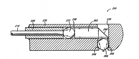
~eferring now to Fi~ure 7, there i~ ~hown another
o ~ odi~er~t of a la~er surgic~l probe 200. The probe 200
comprise~ a fiber holder sh~ft 226 ~nd ~ ~ad ele~ent 230.
The he~d element 230 is contoured for smooth insertion And
withdr~wal. The he~d el~oent 230 has a longitudinal tubul~r
cavity 238 ~nd ~ transverse cavlty 239. In this e~bodi~ent,
the diameter of the tran6ver6e cavity 239 is constricted ~t
the bottom relative to the rQ~ining portion of the c~vity
239, which is 6ubstantially tubul~r. ~hu6, ~ ledge 246 is
lS formed withln the trnnsver e c~vity 239.
The optical fiber 218 of thi~; embodi~nent i8 fit into the
head element 230 in a m~nner similar to that de~cribed above
in connection w$th Figur~ 4. The optical ~pparatus of this
embodiment comprises cepArate pieces, n~mely a collim~tor 273,
a diverter 262 and an lntensifier 266. Aç described in
connection with Figures 5-6, the collimator 273 and the
intensifier portion 266 compri6e mlcroballs, optionally coated
with an anti-reflective coating.
The diverter 262 comprises a capphire rod poli~hed at ~
2S 45- angle ~t it6 dictal end. The anglcd portion i~ coated
with ~ reflective coating and forms el reSlecting ~urface 268.
~he diameter of the rod i~ slightly ~maller than the diameter
of the longitudinal cavity 238 ~o that the diverter can be
in~erted therethrough.
~0 ~e intensifier aicrob~ll 266 reste on the l-dge 246 ~nd
the diverter 266 i6 inserted through the longitudinal cavity
238 6uch that the ~icrob~ll 266 ic held in the ~pace between
the ledge 246 and the diverter portion 268. Ihe colli~tor
microball 273, like the diverter portion 266, has ~ di meter
slightly smaller than the di~meter of the longitudinal c~vity
238, and is inserted proximally of the diverter portion 266.
. . . .. -: .
' ~ . ' '~
. .
, . . . . . . . . . . .
~Y2/1713~ 2~-~7 ~ PCT/US92/02871
Finally the optical fiber 218 within the shaft 226 i6 inserted
into the longitudinal cavity 238 The shaft 226 i~ held to
the proximal end of the h~ad element 230 ~ith U v c~rable
glue ~hu6, the diverter portion 266 and the collimator
microball 273 are held within the portion of the longitudinal
cavity 239 di~tal of the shaft 226 Preferably, there is
6ubstantially no ~paCQ betveen the intensifier ~icroball 266
and the diverter 262 or ket~een the diverter 2 62 and the
collimator microball 273
Referring now to Figure 8, there i~ ~ho~n ~till another
embodiment of a laser surgical probe 300 The probe 300
CompriEes ~n optical fiber 318, a head element 330 and a fiber
holder 358. T~e fiber holder 358 ~erve6 to pro~ide a grip for
the operator of t~e probe 300 ~nd also servec a~ ~ sleeve for
~S the opticAl fiber 318.
The head elemen~ 330 has a tubular longitudinal cavity
338 h~ving proxim~l, 2iddle and distal ~ection~ The middle
section has a constricted di~meter relative to the proximal
and distal ~ection~ of the c~vity 338. The proximal ~ction
of the cavity 338 i6 threaded to nccept a threaded portion 392
of the flber holder 358.
The optical apparatu6 of the probe 300 compri6e~ an
intensifier 366 and a diverter 362 The inten6ifier comprises
a ~pherical microball which i~ di~po~ed ln the di6tal ~ection,
and which re6t6 on a ledge 346 formed by the con6tricted
diameter of the middle ~ection of the lonyitudinal cavity 338
The diverter 362 co~price6 a ~apphire rod poli~hed at a
45 angle at it~ di~tal end The nngl-d portion i~ coated
with ~ reflective coating to foro a r-flecting ~urfac~ 368
The diameter of the rod i~ slightly ~maller than the diameter
of the longitudinal cavity 338 ~o that the diverter can be
inserted therethrough The diverter 362 can extend beyond the
di~tal end of the head element 330 as shown, or can be enca~ed
by the head element 330, with a hole at the point of emi6sion
of laser light energy If the di~erting portion extends
beyond the distal end of the head element 330, the exposed
~ . ~ , - . . '
: , ' . ' ' , ': ' - , ' ' .
: , ' ' ' '' ' , ':
--16-- ---
sapphire can op'cion~lly be co~ted with A protective over-coat.
The distance between the di6t~1 end of the fiber 318 and
the proximal portion oi~ the intensifier microball 366, and the
di6tance fro~ the distal portion of the inten~ifier microball
and the reflecting ~urface are selected ~o ~s to pro~ce a
focal point 379 a desired d1~t~mce (e.g., less th~n 1 ~u from
the bottom of the diverter 362. The distance fr~:~ the
intensifier microball 366 and diverter 362 can also be
m~nipulated to provide the de~;ired focal point 379. ~owever,
preferably, ~n input ~urface 370 of the diverter 362 i8
touching or almost touching the inten~;$fier microball 366 in
order to prevent _xial rovement of the ~icroball 366.
The diverter 362 can be held in place by providing
notch 364 on the he~d element 330 and crimping the notch 364
to the diverter 362. If de~ired, the input curface 370 of the
diverter 362 and the entire or proxi~al ~urf~ce of the
intensifier microball 366 may be coated with _n _nti-
reflective coatlng to mini~ize reflection.
In use, the probe 300 is in~erted into _n internal
portion of a mammal (e.g. an eye c_vlty) ~uch that the rod 362
i8 ~urrounded by ti~sue ~nd the portion of thc rod extending
from the housing is in cont~ct with the tissue, although in
the embodi~Dent disclo~ed, the light beam i~; redirected by
reflection, it ~ill be under~tood th_t, by eliminating the
reflective coating so that the light pas~ec t~rough the angled
output f_ce, redirection by refraction could be achieved.
Such refraction i~ due to differencQ~ $n refr~ctive index At
the angled output face. Redir-ction of the l$ght ~a~ by
refraction ~Day be ~imilarly ~chi-ved by utilizing a bare rod,
such a~; an optical fi~er, ~nd cl-aving the end of tbe f$ber of
~n angle (e.g. 45-) to cau~e the light output from the fiber
to be deflected. Nevertholess, use of the reflective coatir~
is preferred bec_u~e ~ greater ~ngle of deflection is
pos~ible.
3S Referring now to Figure 8a, there is hown a probe 400
that is a modiSication of the l~ser probe 300 of Figure 8. In
~ ~ .
";
WO92/17138 2~37~ PCT~US92/02871
-17-
thi~ modified probe 400, the microball 366 i6 pl~ced with its
center 395 At its focal distance (f) from the distnl ~nd of
the fiber 318 PlAcing the microball 366 in this po~ition
will achieve colli~ation of light incident thereon
S The focal di~tance (f) of a ~pherical l~ns can be
calculated ~s f ~ n r/2(n-l), ~here n i6 the ~ndex of
refraction and ~ i8 ~he radius 394 of the lQn~ For the
pr~ferred ~hodiment havlng a l S ~ di~eter ~pherical
~apphire len~ ~nd u6ing a l~ser pro~ucing l$ght ~nergy of
wavelength 2 94 ~, the index of refraction of the lens i6
1 72 Thus, for this preferred embodiment, f c
1 72(0 75)/2(1 72-l) ~ 0 896~
For a properly plac~d mlcroball 366 achieving colllmation
of light from the fiber 318, the focal distance (f) will be
~S egual to the length of the radius (R) 394 of the ~lcro~all 366
plu5 the gap (g) 396 between the ~icroball 366 nnd the di6tal
end of the fiber 318, i e f - g I R Thu~, ln the preferred
embodiment de6cribed above, the proper gap 396 can be
calculated a6 g ~ f - R - O 896 ~m - O 75 r~ - 0 146 ~m
In use of t~e probe 400, the colli~ated light travel~
along the longitudinal ~xi~ of the probe 400 until re~ting the
reflecting surface 36B The reflQcting curface 368 divert~
the light in a direction along an output AXiC 550 which i6 at
an angle relative to the longitudinal ~xi- of the probe 400
2S For ~any proc-dure6, cuch a~ an anterior cap~ulotomy, the
preferred angle of the output axi~ is perpendicular to the
longitudinal ~xis of thc probe 400 For other procedure~,
~uch ac certain proc-dur-s in the troatrent of glaucoma,
angles greater or l-~s than 9o ~r- pref~rred
In the modlS~ed la~er probe 400, the re~l~cting ~urface
368 is transparent ~nd requires no reflec~ive coating
Rather, reflection occurs due to the total internal reflection
achieved fror the differencec ln the indices of refraction
between the ~aterial of the rod 362 ~nd the m~terial
surrounding the probe 400 In the preferred embodiment, the
rod 362 i~ ~apphire, having ~n index of refraction (n) of 1 7
~,~ : . : ~ ,:, , - , . . . ..................... ..
::. , ~ , , , . , . , , -:
. , , . . ,, . ~, ,, ,,. :
~ d'~ ~ ~V L
- with light produced by the Erbium:YAG laser havir
wavelength of 2.~4~m.
The critical ~ngle (~c) of the reflecting ~urface 368
reguired to achieve tot~l internal reflection can be
determined according to Snell ~6 law, which can be ~tated as
follow~: nlcin~1 ~ n2sin~2. A~ ~tat-d above, in the preferred
e~bodiment, n1 ' 1.7. When the probe i~ u~ed in air, r~ ~ 1Ø
For total ~nternal reflection 82 ~ 90-- Tbus, ~c for the rod
of the preferred e~bodiment can be calculated from Snell's law
~o as follow~: l.Osin~c - 1.72sin90-. Accordingly, ~c ~ 35.5-.
This mean6 that total intern~l reflection will ~e achieved a~
long as the reflecting surfac~ 368 i~ at ~n angle greater than
or equ~l to 35.5- with re~p2ct to the collimated light
incident thereon.
~s The probe 400 functions to reflect l$ght in substanti~lly
the same manner in a fluid environment (e.g. H20) as in ~ir
because the refractive index difference between the fluid and
the 6apphire rod 362 $8 ~ufficiently great to cau~e total
internal reflection to occur at the ~urface 368.
As best seen in Figure 8b, ln the preferred ~mbodiment,
the rod 362 comprises a sapphire rod of circul~r cro~ ~ection
which i5 polished at a 45- angle at lts dlstal end to form the
reflecting surface 368. When the ~odifi-d la~er probe 400 of
this preferred embod$ment is used in air ~nd not in contact
with tissue, the curved bottom output surface 399 of the rod
362 acts as ~ cylindrical len~ to ~ocus the collimated light
into ~ ~ore linoar form. HowevQr, when the la~er pro~e 400 i~
used in ~ fluid environ~ent (e.g. E~O) or in contact with
tissue, the lens ~ction of the output ~urface 399 tend6 to be
neg~ted due to the higher index of rerraction of t~e fluid
and/or due to the cont~ct with ti~sue at the cylindrical
surface. Thus, wben the rodified l~ser probe 400 i~ used in
internal portions of ~ mammalian ~ody, such ~5 $n the eye, the
light exiting the probe will be in ~ roughly tubular form.
3S Advantageou~ly, where total internal reflection i~ used to
reflect the light within the probe 400 without the use of a
, . ~
.' .
,~ :
,' ' ' ,
. .
,
WO92/17138 2 ' ~ PCT/US92/02871
-19-
reflective co~ti~g, the probe i6 tran~pare~t to ~llo~ the user
of the probe 400 to view the point of cont~ct of the laser
light energy on ti~u~6 or oth~r ~atQriAls. Tbi6 i~
particularly advantaqeou6 in curgeries, ~uch afi anterior
S capsulotomie6, where the probe would otherwise ob~cure the
contact point of the light energy. Another ad~antage of the
probe 400 o~ Figure 8a i8 that the wedge-~haped end of the
probe, formed by the angled reflecting curface 368, can be
used as a tool to phy6ically ~an$pul~te tis~ue~ without the
~o need to insert an ~ddit$onal tool.
Although the 2.94 ~m wavelongth l$ght onergy of the
Erbium:YAG la~er $s not vi~$ble to the human eye, the point of
co~tact c~n generally be ceen due to the ~nergy rel-a~ed by
the ti6sue6 aSter com$ng lnto cont~ct wlth the l~cer energy
lS from the probe 400. The t$ssue ab~orbs the colli~ated $nfr~-
red light emitted from the probe along the output axi~ 550 and
causes ~ lnser-t$s6ue $nteraction to occur. Thi6 $nteract$on
generally resul~6 in the rele~se of light of A w$de ~pectrum,
including vi6$ble light, ~t many d$fferent ~nglc6. Thus, ~uch
of the l$ght relea~ed by l~ser-tlcsue interaction ~$11 strike
the reflect$ng surface 368 at an angle less than the critic~l
angle. Even ~ithout release of significant quantitie~ of
light by la~er-tissue $nteraction, the interaction cnn be seen
by the formation of an incision or oth-r o~fect on the ti~ue
by the laser. Accord$ngly, the operator of the probe can look
through the transparent reflecting curface 368 along a view$ng
axis 525 to view the vicible light from the t$ccue dur$ng
operation of the probe 400. The ~i~wing ~xic ic g nerally
perpendicular to the long$tudinal ~xic of the probe ~00.
In the pr ferred ~mbodirent of the la~er probe ~00, all
surfaces are pol$~hed w$th optical grade (e.g. 0.3 ~m) polish,
including the distal end of the fiber 318. Without th$s
poliching, ~pecular reflectionc c~n occur ~hich re~ult in
d$spercal of the energy pa6sing t~rough the probe ~00.
3S As discussed above, all ~urface6 should be generally
~mooth to prevent cnagging during in~ert$on or removal of the
. ... . .. . .. . . .... . .
,' .
2 ~ ~7 ` - -20-
probe 400. Thns, ~n t~e preferred embodi~ent, one or ~ :e
notch~s 364~ ~re provided on the rod 3~2 which will zllow the
rod 362 to be held to the head element 330 by crimping of the
head element at the position of each notch 364a.
S The 1~6er probe 400 can ~dv~ntageously be configured to
supply irr~gation fluid or v~cuum for ~6pirstion ~6 requently
employed with ~nown ph~coe~ul~ific~tion device~.
Alternatively, irrigation and~or ~piration can be 8uppl ~ ed
from ~eparate device~ in~erted into the rsgion of u~e of the
probe 400.
In the preferred embodiment, it i8 important that fluid
not enter the longitudinal cavity 338 of the head element 330
because the lense~ are configured for uce with air with an
index of refraction of 1.0 in the~e ~pace~. The entry of
fluid with ~ much higher lndex of refraction into the cavity
338 would prevent collim~tion of the light energy emanating
from the fiber 318. Thus, a sQalant 398, ~uch as epoxy glue
i8 preferably provided at the ~unction between the rod 362 and
the head elemen'; 330, to prevent entry of fluid into the
cavity 338.
Referring now to Figure 9, there i~ shown ~ variant 310
of the laser probe 300 of Figure 8. In this vari~nt, the head
element 330 i~ elongated to extend beyond the diverter 362,
~nd compri~e~ ~n open$ng 306 to ~llow l~er light energy to be
emitted out~ide the head lement 330 after it has been
diverted by the diverter 362. In the vari~nt 310, ~ cap 394
i6 inserted at the end of the head el~ment 330. The cap 394,
comprises ~ rod portion 395 ~nd ~ flange port$on 396. The
diameter of the rod portion 395 lc ~lightly maller than the
~0 diameter of the longitudinal cavity 338 co that the rod
psrtion 395 c~n be in~erted therethrough. The rod portion 395
i6 cut an angle which will compl-uent the angle of end the
diverter rod 362 to ~ub~tantially completely fill the
long$tudinal cavity 338 at its end. T~UC, if the diverter rod
362 is cut at a 45- angle, the rod portion 395 will al60 be
cut at a 4S- angle. The length of the rod portion i6 ~elected
,.,:"
i
WO92/17~38 2.1 ~ 7 ~ , S~ PCr/US92/02871
; .
--2 1--
to substantially completely fill the end of the longitudinal
cavity 338. The fl~nge portion 396 provides a smooth surface
for easy insertion ~nd withdrawal of the var~ant la~er probe
310. The c~p 394 1~ held in pl ~ by crimping t~e notchQ~ 364
S to the cap 394, thereby ~l~o pr~vQnting ~xial ~oVQment of the
diverter rod 362.
The laser prob~s lo, loo, 200, 300, 310, 400 ~re useful
in a wide variety of ~urgical pr~cQdur~s, ~ncluding procedures
cuch a6 dQscribQd by Berlin in U.S. Patent No. 4,846,172. The
use of the laser probQs 10, loo, 200, 300, 310, 400 i5
especially advantageous in procedurQs where it is dQ~ired to
operate a laser probe withi~ a tightly confinod ~pace, uch as
within bodily tis6ues or a tightly confinQd body cav~ty or
lumen. The probe allows a surgeon to direct la~er energy from
the 6ide of the probe, thereby ~llowing la~er ~nergy to be
directed around tight corner6.
Particular Qxamples of procedure6 in which the probe can
be ~pplied in a ~ammal ~nclude ophthal~ic proc~dures of ~any
type~. In thi~ regard, the probe can be used for cutting,
phacoemul6iflcation and phaco~blation. The~e and other
techniques u~ing the probe are believed to be ~specially
useful in corneal ~urgerie6, such as keratectomy or
keratoplastomy, in glaucoma eurgeries, such a~ filtration
procedures, trabeculopla~ty, iridectomies or iridotomies, in
2S cataract curgerie6 such a8 capsulotomy or cataract extraction,
in vitreous surgeries ~uch a8 cutting of the vitreous band6,
and in retinal surgery such as removal of retinal Dembrane or
repair of retinal teare. Non-ophthalmic procedures on a
mammal $n which the probe 1~ belie~-d to be useful include
surgery within a ~oint, cuch as a knee, and procedures w$thin
long narrow passages, such as can be found within t~e
cardiova~cular syste~ and the urethra.
The intensity of the light input to the probe is
regulated, depending on the procedure, to provide sufficient
intensity to achieve tbe desired result cuch as cutting,
welding, vaporization or coagul~tion o~ biotic ~aterial (e.g.
tissue). Where c~ooth cutting is desired, the freguency of
~ 22--
the pul~e ~hould be ln excess of 5 ~z, preferably lo ~z ~ 30
Hz or more. It 16 al~o preferable to u~e a laser light ~ource
with a relatively low energy thr~shold ln order to provide
smooth cutting. Prefer~bly the energy threshold is 5-10 mJ
for cuttin~ of the anterior capsule of the oye. Thu6, for
smooth cutting energy levels of 30 ~J per pulse are less are
preferred, ~ith ener~y l~vels ~ust above the ~nergy threshold
of S-10 ~J/pul~e being ~pecially preferred.
AB stated ~bove, one procQdure in which the probe is
particularly use~ul i6 ln anterior capsulotomy of the eye.
With reference to Figure 10, in ~hich a cro~ ~Qction of an
eye 510 is shown, a small roughly circular incicion 501
through one side of the ccler~ 502 of the eye 500 i6 first
made into the anterior chamber 516. Thi~ inci6ion i~ roughly
~5 2.0-3.5 ~m ln dia~eter. A~ discussed above, the probe i6
preferably contained within a housing having a di~meter of 2.5
mm or less. Thi6 i8 advantageous in anterior capsulotomies
and other procedures within the eye because larger probes
would require a larger incision. ~oreover, the use of large
probe6 also incr~ases the ri~k that the probe w$11 come into
contact the cornea 512, iris 515 or other d~licate ti~6ue6
within the eye 500, resulting in dnmage to thQse ti~6ue~.
In order to maintain the patency of the anterior chamber
516 during the procedure and to hold other ti~ues in
position, the cha~ber 516 can be ~$11ed ~ith a vi~coelastic
~aterial, ~uch as ~oalon~. The vi~co~la~tic ~aterial will
al60 hold tissue~ in position within the ~ye dur~ng the
procedure. Alternatively, irrigation fluid, ~uch ae ~alanced
~alt ~olution (BSS) can be continuou~ly lnfu~ed to naintain
patency of the chamber 516.
The probe 400 or other la~er probe c~n be lnserted into
the inci~ion 501 along wit~ the fiber 318 transmitting la~er
energy thereto. The probe i~ t~en ~anipulated to cut
circular incision (shown p~rti~lly formed ~t 560 in Figure 10)
3S around a portion of the anterior cap6ule 524 adjacent the
lens. As will be di~cus~ed in more det~il below, ~he laser
probe 400 advantageou~ly allow6 the operator to view the
,. : . ' : . ' . , ., :
.,- ~ ' ' . . ': . ~ :
W092/17138 PCTIUS92/02871
2 ~ 3 r~
-23-
energy released fro~ the surface of the cap6ule ~ la~er light
energy i6 applied.
~ variety of la~er light sources ~ay be used in the
procedure. However, it i~ preferred th~t the la~er light
source provide a ~ooth, non-uerrated cap~ular ~argin, in
order to enible a ~urgeon to aAke a clean circular cut on the
anterior cnp~ule. A percu~ive ~evice ~ould not be
appropriate, ~hich would punch ragged hole6 in the capsule.
T~us, the frequency o~ She lasar pulse ~hould be in ~xces~ of
lo 10 ~z, preferably 20 Hz - 30 Hz or ~ore, a8 discu~ed ~bove.
Preferably, a l~ser light ~ource producing light at a
wavelength readily absorbed by water 1~ provided. U~e of
wavelengths that are absorbed readlly by w~ter i8 u~eful for
ablation of ticsues. Al~o, use of ~uch wavelengthc ~er~es to
lS prevent unwanted transmis~ion and scatter of la~er ~nergy to
adjzcent or underlying tissues, resulting in ~inim~l thermal
damage to these tissues. It is alco preferred t~at t~e laser
light be deliver~ble by an optlcal fiber to allow the u~er of
the probe to deliver the laser light nergy by hand. Hand
delivery 18 important for ~llowlng delicate ~nipulations
within the eye and other tlghtly con~ln-~ tl~sues.
Thus, a preferred laser light ource iG an Erbium:YAG
laser wh$ch produces la6er light energy of w~velength 2.94 ~m,
a wavelength at which water hac an absorbance p~ak. Thu~, one
2S preferred wavelength range for the light ene,gy for use with
the probe 400 i6 the range from 2.8 ~ to 3.0 ~m. The
Erbium:YAG laser providec everal additional adv~ntages.
Firct, the ~nergy i8 non-ultraviolet, thereby allowing wor~ in
the eye with incr-a-ed af-ty, obv~ating the n~ed to UBe
blocking elementc or device to pr-~ont r~tinal toxicity.
Second, the Erbium:YAG light source can al80 be con~igured to
provide the pulse frequency greater th~n 10 ~z needed to
provide smooth cutting. ~hird, the high abQorbance by w~ter
makes the laser safer, more controll~ble, and more precise.
Fourth, the laser h~s a low thermal component, ~llowing for
precise sp~tial confinement of ene,~y deposition and reducing
-
, : .: . . . . : , .......... :. . . . . .
., .. . , . .. .. ~ , . . .
0 r~ /u~o
~ Q~ 2~-
thermal ~amage and charring of intraocular tissues. Finally,
the Erbium:YAG lsser i~ relstively $nexpen6i~e to manufacture
and maintain compared to certsin other l~sers.
La~er lig~t ~ource~ wh~ch produce Qnergy 8t other non-
ultr~violet absorb nce pe~ks of water, ~uch as 2.1 ym~ ~d aredeliver~ble by c~tic~l fiber provide ~dvantsges simi ~r to
those provided by the Erbium:YAG, ~er. Thu6, ~nother
preferred l~ser light source i8 the ~olmium:YAG lsser which is
h~nd deliverable and provide6 la~er light energy within the
~o range of 1.9 ~m to 2.2 ~m.
Delivering l~eer light energy ~y ~i~er 318 provides the
additional ~dvant~ge of allowing use of the bnre fiber through
removal of the probe tip. Thu6, ln the preferred e~bodi~ent,
the probe tip i6 removable by h~nd to expo6e the di~tal end of
~S the fiber 318, thereby enabling u~e of the bnre Slber end from
the 8 me hand held instru~ent. use of the bare fiber is
advantageous $n ~any procedure~, ~uch a~ the xci6ion of a
vitreous band w$thin the eye of ~ ~mmal.
j As Been in rigure 10, laser l$ght energy exits the probe
slong its output axis 550 to contact the t$~sue st a point of
contact 575. In order to view the point of contact 575
-through the viewing ~xis 525, the surgeon or operator of the
probe must look through the probe 400. Advantageously, when
the incision in thc c~psule 524 ls ~de wlth a transparent
laser probe having ~n unco~t-d, tr~n8p~rent re~lecting surface
368, such as the probe 400 of Figure 8a, the 8urgeon c~n look
through the surface 368 of the probe 400 to ~iew the incision
560 at the point of contact 575 ~hile tbe incision 560 is
being cut. Tbus, in order to vl-w tbe point of contact 575
the surgeon can look t~rough tbe corne~ S~2, pUpil 514 and the
transparent reflect~ng curface 368 along the vl-wing axis 525.
Preferably, the viewing axis S25 i~ generally parallel o the
output axi~ 550, ~nd in ~n e6pecially preferred emboc Dent,
the viewing ~xis 525 i6 collinear with the output axiL 550.
35 In many procedures, including anterior capsulotomies, it is
desirable to provide a ~icro~cope (not chown) for viewing the
.,:,; : . . - .
~092/17138 ~ ;~J'~ ' PCT/US92/02871
-25-
point of contact under ~gnif~cation.
Being able to view the point of contact 575 while the
incision 560 is belng cut, advantngeou61y allows the surqeon
to avoid sensitive areas and to ~ore ~aeily control the size
and shape of the incision 560. Advantageou~ly, viewing the
point of contact 575 also nllows the surgeon to more readily
cut a smooth inci6ion 560. Thu6, decentrat~on ~nd other
problems associated with l~s ~mooth incisions nre avoided.
After the ~mooth lncision 560 bas been made on the
~nterior capsule, the cutout portion of the ~nterior capsule
524 inside the ~ncision 560 ic removed to expo~e the
underlying lens 528. If neces~ary, thi6 cut out portion of
the lens 528 c~n ~e ~anipul~ted using the wedge for~ed by the
reflecting ~urface 368 on the probe 400. Advantageou61y, thi~
wedge can be ussd to m~nipul~te other ti6sues inside th~ sye
or elsewhere ~c well. The wedge has ~160 been found to be
adv~ntageous in re~dily allowing the user of the probe to
separate pl~nes of ti6sue, ~uch as in ~ep~rating fa~cia from
muscle or separating the anterior capsule 524 Srom the
underlying lens 528 w$thin the eye 500.
For insert$on of an intraocular lenc tI0L), the lens 528
can first be emulsified in a m~nner known to those of skill in
the art, such as through phacoemulsificat$on using an
ultrasonic device. Advantageously, as ~hown in Figure 11, the
laser probe 400 can al80 be used for emulsif$cation,
preferably using a higher energy level than u~d for lnc$sion,
e.g. 100 mJ/pulse. The probe ~00 can be u~ed to deliver light
energy to emu16$fy tbe len6 w$thin the co~pleted capsular
margin 600. Tbere iB le-c ne-d for high frequency o~ laser
light energy pulses during emuls$ficat$on, thu6 ~requencies of
5 ~z or les~ can be u~Qd. ~uring mul6iricAt$0n, tbe high
energy la6er light energy ~xits tbe probe ~long output ~xi6
5S0, and tbe len6 ti~cue w$tbin the ~argin 600 contacted by
the la6er light can be ~ie~d through the probe 400 as
~5 discussed above.
The emulsified lens ~aterial can advantageously be
re~oved using irrigation and aspiration supplied along with
,: .- . . . . . . . . . . . .
,., : : , , . . . . , : .. : .
~ 26-
the probe 400. The uge of irrig~tion ~eAnc 620 i~ ~how~-~n
Figure 11. The use of irrigatlon ~Qans 620 ~nd a8pir~tIon
~ean~ for thi~ purpo6e ~8 part of ~ l~ser probe ~6 well known,
and has been described, for axa~ple in ~.S. P~tent~ No6.
s 4,846,172 and 4,784,13Z, the di~clo~ures o whic~ ~re hereby
incorporated by reference. Irrigation ~nd/or ~6piration can
also be ~upplied ~ sep~rate components, a~ ~ well known to
those of ~kill in the ~rt. ,~ter ~nul~ificacion of the lens
528, the eye 500 i8 ready ~ ~plant~tion of the IOL.
It will be apprec~ated t. _ certain ~tructural vnriation6
~ay suggegt them~elves to tho~e ~killed in the art. The
, foregoing detailed de~cription i~ to be cl~arly underctood a6
; given by way of illuctration, the ~pirit ~nd ~cope of this
invention being li ited olely by tbe Append-d C1a1~G~
,;, . . , ~
~,, : . - . - . . . ............................ : - -
, . . - : - . - : -: . : :
. - .. . . . . . . . .. .