Note: Descriptions are shown in the official language in which they were submitted.
2~LO5~
FOCUSING ENDOSCOPE
This invention relates to endoscopes and more
particularly to electronic endoscopes.
Generally, endoscopes are instruments for
visualising the interior of an object, such as the
.
human body (e.g., an internal organ or anatomical body
passage). A typical endoscope includes an elongated
flexible or rigid outer tube within which a lens system
is disposed at a distal end. The image of the object
being viewed by the optical system is transmitted
through an optical system from the distal end to a
proximal end of the tube for viewing by the user or for
reception by a camera. Some endoscopes also carry fiber
optic cables for illuminating the area of observation
with light supplied by an external source.
In some endoscopes, the optical system includes a
bundle of fibre optic cables positioned immediately
proximally of a stationary objective lens assembly
located at the distal end of the tube. The lens
assembly focuses the image into the end of the fibre
bundle, which in turn transmits the image proximally.
An electronic endoscope typically includes an
electro-optic image sensor (such as a charge coupled
device or CCD) in place of the fibre optic bundle. The
CCD is positioned closely adjacent the objective lens
assembly and generates a video signal of the object
being observed. The video signal is transmitted by an
electrical cable to the proximal end of the endoscope
7 8 7 ~ ~
and is processed for viewing on a display such as a CRT
monitor.
This disclosure provides an endoscope with a
focusing mechanism having a focus control element at a
proximal end of the endoscope and a mechanical coupling
connecting the focus control element to an image
transmitting device mounted at the distal end of an
elongated insertion section of the endoscope for
causing the image transmitting device to move along at
least part of the length of the insertion section in
response to activation of th.e focus control element. In
a first embodiment there is an endoscope apparatus for
internal inspection of an object, comprising an
lS elongated insertion section having a distal end to be
inserted into said object and a proximal end to be
manipulated by a user,
a lens assembly disposed in a chamber in said
distal end,
an image transmitting device mounted in said
ch~her of said distal end, said image transmitting
device being movable along at least part of the length
of said insertion section, and
a focusing mechanism having a focus control
2S element at said proximal end and a mechanical coupling
connection said focus control element to said image
transmitting device for causing said image transmitting
device to move along at least part of the length of
said insertion section with respect to said lens
assembly for focusing in response to activation of said
focus control element.
~'~
~ ~ ~787~;
The image transmitting device may include an
electro-optical sensor. According to a second
embodiment, there is provided an endoscope apparatus for
internal inspection of an object, comprising
an elongated insertion section having a distal
end to be inserted into said object and a proximal end
to be manipulated by a user,
said distal end enclosing a chamber,
an electro-optical sensor mounted in said chamber
of said distal end, said sensor being movable along at
least part of the length of said insertion section and
a focusing mechanism having a focus control
element at said proximal end and a mechanical coupling
lS connecting said focus control element to said sensor
for causing said sensor to move along said at least
part of the length of said insertion section for
focusing in response to activation of the focus control
element.
2~.
The image transmitting devices may comprise one
or more optical fibres.
The focus control element may be a rotatable
manipulator accessible to a user. The mechanical
coupling may be a rigid actuator coupled between the
manipulator and the sensor, with the actuator moving
along at least part of the length of the insertion
section in response to rotation of the manipulator.
This arrangement allows an image viewed by
B
2 ~ 7 2
the endoscope to be easily focused while in use without
removal from the surgical site.
The mechanical coupling may include a helical
surface disposed on an inner sleeve which surrounds and
is rotatable around the insertion section. The rigid
actuator can then include a follower configured to
engage the helical surface and to more along the
helical surface in response to rotation of said
rotatable manipulator. The rotatable manipulator may
have an outer sleeve which surrounds the inner sleeve,
and the inner sleeve and the outer sleeve can be
connected by a coupling which may be selectively
engaged and disengaged by relative motion of said inner
and outer sleeves along the length of the insertion
section. As a result, the focusing mechanism can be
made easy to assemble and disassemble.
The outer sleeve of the rotatable manipulator
rotates smoothly with little physical effort and
preferably can be operated easily with one hand. The
mechanical coupling can include a slot provided along
and in parallel with a portion of the length of the
insertion section, with the follower riding within said
slot. The slot permits the rotatable manipulator to
focus the image while maintaining a non-rotating image
on an external display.
The manipulator can include an element that is
ferromagnetically coupled to the rigid actuator through
an external wall of the insertion section. This avoids
7 ~ ~
passing a mechanical element (such as the follower
discussed above) through the external wall, thereby
allowing the interior of the insertion section to be
more completely sealed to reduce the risk of foreign
material cominq into contact with the electrical and
optical components of the device.
The electronic endoscope can be disposable, and
the insertion section can be sterilised and packaged in
a sterile container together with a sterilised conduit
that is used to connect the insertion section to a
control unit; the conduit may carry an optical fiber
for transmitting light from the control unit to the
insertion section, and an electrical cable for carrying
electrical signals from the sensor to the control unit.
The endoscope and integral cable for providing
both electrical and illumination requirements to the
endoscope may have a single connector for connection to
a video processor (camera control unit). The endoscope
with integral cable may be provided in a sterile
container so that the endoscope is disposable after use
in a surgical procedure.
2S. According to a third embodiment there is a
disposable endoscope apparatus, for internal inspection
of an object, comprising
a sterilised elongated insertion section having a
distal end to be inserted into said object and a
proximal end to be manipulated by a user,
2 ~
an electro-optical sensor mounted at said distal
end,
a sterilised conduit connected to said proximal
end and having a free end for connection to a control~ - unit, said conduit carrying therein:
an optical fibre for carrying light generated by
said control unit along at least part of the length of
the insertion section to an illumination outlet at said
distal end; and
an electrical cable for carrying electrical
signals from said sensor along said at least part of
the length of the insertion section to said control
unit via said proximal end, and
a sterile container for enclosing and maintaining
said insertion section and said conduit in sterile
storage until said container is opened for exposure
during use of said endoscope.
According to a fourth embodiment there is an
endoscope apparatus for internal inspection of an
object, comprising
an elongated insertion section having a distal
end to be inserted into said object and a proximal end
to be manipulated by a user, said insertion section
~5 having a longitudinal axis,
an electro-optical sensor mounted at said distal
end,
an optical fibre for carrying light along the
length of the insertion section to an illumination
~0 outlet at said distal end,
an electrical cable for carrying electrical
a_
_ 7 - ~ 7 ~
signals from said sensor along the length of the
insertion section carried within said flexible conduit,
said flexible conduit having a central axis said
central axis being collinear with said longitudinal axis
of said insertion section at said proximal end.
More particularly in accordance with the
invention there is provided, an endoscopic apparatus
(10) for internal inspection of an object, comprising
an elongated insertion section (12) having a
distal end (14) to be inserted into said object and a
proximal end (19) to be manipulated by a user,
said distal end (14) enclosing a chamber,
an electro-optical sensor (24) mounted in said
chamber of said distal end (14), said sensor (24) being
movable along at least part of the length of said
insertion section (12) and
a focusing mechanism (17) having a focus control
element (80) comprising a rotatable-manipulator
accessible to said user and a mechanical coupling (68,
72, 74, 76, 78) connecting said focus control element
(80) to said sensor (24) for causing said sensor (24) to
move along said at least part of the length of said
insertion section (12) for focusing in response to
activation of the focus control element (80) wherein
said rotatable manipulator comprises an outer sleeve
which surrounds an inner sleeve wherein said inner
sleeve and said outer sleeve are connected by a coupling
which may be selectively engaged and disengaged by
relative motion of said inner and outer sleeves along
the length of the insertion section.
~,
- 7a - ~ 7 ~ ~
The present invention will now be disclosed by
way of example only with reference to the accompanying
drawings, whereas:-
Fig. 1 is a cross-sectional side view of a distal
region of an endoscope embodying the invention.
Fig. 2 is a cross-sectional side view of a
proximal rear end portion of the endoscope of Fig. 1.
Fig. 2a is an enlarged view of the region
enclosed by 2a-2a of Fig. 2.
Fig. 3 is a diagrammatic view of a lens assembly
of the endoscope of Fig. 1.
Fig. 4 is a top view of a main housing of the
endoscope of Fig. 2.
Fig. 5 is side view of a focus sleeve of the
endoscope of Fig. 2.
Fig. 6 is a diagrammatic view of the endoscope of
Fig. 1 in use.
Fig. 7 is a diagrammatic view of an alternate
embodiment of a disposable endoscope.
Fig. 8 is a cross-section of a portion of the
disposable endoscope taken along lines 8-8 of Fig. 7.
Fig. 9 is a cross-sectional side view of a distal
region of another embodiment of a disposable endoscope.
Fig. 10 is a cross-sectional side view of a
B
portion of the distal region of another embodiment of a
disposable endoscope.
Fig. 11 is a schematic representation of another
alternative embodiment of an endoscope embodying the
invention.
As shown in~Figs. 1 and 2, an electronic
endoscope 10 includes an elongated insertion section 12
for insertion into a body cavity or narrow body passage
102 (See Fig. 6) to observe an object therein.
Elongated insertion section 12 extends along a
longitudinal axis 13 of endoscope 10 from a distal end
14 to a handle 16 at the proximal end 19 of endoscope
10. Handle 16 permits the user to position elongated
insertion section 12 of endoscope 10 appropriately and
also houses a focus control mechanism 17 for endoscope
lo, which is described in detail below. A cable 18
extends from proximal end 19 for connection to a power
source and camera control unit 106 (Fig. 6). Images
observed at distal end 14 of endoscope 10 are processed
by the video processor for viewing on a display unit,
such as a colour CRT 108.
Elongated insertion section 12 includes an outer
2S tube 20 for housing an objective lens assembly 22, an
image transmitting device (which in this embodiment
includes an electro-optic module 23, having an
electro-optical sensor 24 (e.g. a charge-coupled device
(CCD)) for converting optical images of the received
light into electrical image signals), and light guiding
fiber optic elements 26 for illuminating the area being
21~7~72
observed. Outer tube 20 extends from distal end 14 of
endoscope 10 to a first end of a main housing 30 at
handle 16 where tube 20 is soldered within a
counterbore of main housing 30. Outer tube 20,
fabricated from anodised aluminium, has a length of
about 13 inches (33.02cm), an outer diameter of .379
inches (.963cm) and a wall thickness of approximately
0.010 (0.025cm) inches.
Elongated insertion section 12 further includes
an inner cylindrical tube 31 and a CCD tube 32, each
coaxially disposed within outer tube 20. Inner
cylindrical tube 31 extends proximally approximately
2.4 inches (6.09cm) from the distal end 14 of the outer
tube 20 to an enlarged proximal end 3la that receives
the distal end of CCD tube 32. Tube 31 is radially
spaced from outer tube 20 to provide a cylindrical
passage within which fiber optic elements 26 pass to
distal end 14. CCD tube 32 is shown having a first
cylindrical member 33 extending from a region directly
behind lens assembly 22 to a region partially within
main housing 30 and a second extension member 34
soldered to first cylindrical member 33 which extends
to focus control mechanism 17. The distal end 33a of
2S CCD tube 32 has a pair of enlarged diameter regions
29a, 29b as shown to receive electro-optic module 23
and a crimped end of cable 18, respectively.
Electro-optic module 23 is secured into region 29a of
CCD tube 32 with epoxy. Cable 18 has a woven ground
conductor surrounded by a band 21 which is crimped to
prove a snug fit within region 29b.
'~13~ 8'77
-- 10 --
Referring also to Fig. 3, an optical image from
the area being observed is provided to a front face 25
of CCD 24 by lens assembly 22 through a series of four
lenses. Images incident on lens assembly 22 pass
through a negative lens 35, a doublet lens 36, a
meniscus lens 38, an aperture stop 40, and a second
doublet 42, and are incident on face 25 of CCD 24.
Negative lens 35 has a flat distal surface 37. Distal
surface 37 being flat provides the advantage of being
easily cleaned and maintained. Doublets 36 and 42 are
fabricated from a pair of lens elements of glass
materials having different optical characteristics
(e.g. refractive index) that are joined with a thin
layer of epoxy. Lens elements 35-42 are spaced from
lS each other by air and are encased within a lens housing
41. Meniscus lens 38, aperture stop 40, and second
doublet 42 are supported by a spacer 43 (Fig. 1) to
assure their concentricity within lens housing 41. The
four-lens objective lens assembly 22 supported within
lens housing 41 has a physical length of about 16 mm, a
focal length of 4 mm, and an f number of 5. Lens
assembly 22 provides a field of view of approximately
75 degrees. Lens assembly 22 fits snugly within tube 31
and is recessed from the tip of outer tube 20 to
protect exposed distal surface 37 of lens element 35.
Lens assembly 22 and inner cylindrical tube 31 are
rigidly attached with epoxy.
Electro-optic module 23 is the solid-state image
sensor portion of a commercially available electronic
camera (lens objective removed) which is manufactured
ll -
by Panasonic Communications and Systems Company,
Secaucus, N.J., model No. GP-KS202. Electro-optic
module 23 has a diameter of about 0.5 inches (1.27cm)
and is sized to fit within distal end 33b of CCD tube
32, and includes a CCD 24 with its associated
preamplifier/driver circuitry (not shown). Images
generated by CCD 24 are transferred via the
preamplifier/driver circuit (not shown) to individual
wires 27 of cable 18. Cable 18 is inserted within CCD
tube 32 and supplies power to ele'ctro-optic module 23
and receives electrical image signals to be provided to
video processor 106 or camera control unit external to
endoscope 10 via wires 27.
15. Fiber optic elements 26 are disposed in a
cylindrical region between outer surfaces of both inner
cylindrical tube 31 and CCD tube 32 and the inner
surface of outer tube 20 along the length of insertion
section 12 extending from distal end 14 to main housing
30 such that in operation, a ring of light is provided
around the area viewed by lens assembly 22. During
assembly, fiber optic elements 26 are fed in an evenly
spaced manner within the region between tubes 20 and 31
(before inner cylindrical tube 31 is glued in place).
The loose ends of fiber optic elements 26 are allowed
to extend beyond the distal end 14. Fiber optic
elements 26 are potted in place with an epoxy and
portions extending beyond distal end 14 are then cut
and polished to ensure a clear epoxy-free glass surface
at the ends of each fiber optic element. Fiber optic
elements 26 are fabricated using a highly transmissive
21G~ ~7~
glass-like material, manufactured by Cuda Products
Corp., Jacksonville, FL, such that light provided from
an external light source 110 (See Fig. 6) to the
proximal end of fiber optic elements 26 is provided to
distal end 14 with minimal loss.
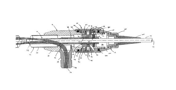
Referring again to Fig. 2, focus control
mechanism 17 allows a user to focus endoscope 10 by
adjusting the distance between objective lens assembly
22 and CCD 24 by + 1 mm. As mentioned above, objective
lens assembly 22 and CCD 24 are rigidly secured to
outer tube 20 and CCD tube 32, respectively. The
spacing between CCD 24 and lens assembly 22 is varied
by moving CCD tube 32 axially along longitudinal axis
13 of endoscope 10. As will be discussed below, front
face 25 of CCD 24 is moved by focus control mechanism
17 in response to rotation of focus ring 80 by the
user.
Main housing 30 has a through hole 46 extending
its length to receive CCD tube 32 and to allow cable 18
to pass through endoscope i0 (Fig. 4). A pair of oblong
slots 48, 50 are disposed through opposite sidewalls of
an end portion 65 of main housing 30 for respectively
2~ receiving a pair of cam screws 76, 78. Oblong slots
48, 50 have a length of about .35 inches (0.889 cm) to
provide the extent of forward and rearward motion of
CCD tube 32 and are parallel to axis 13 for purposes to
be described.
30.
A cylindrical actuator 62 is interposed between
7~-7~
- 13 -
main housing 30 and CCD tube 32 and is threaded to
receive the proximal end of CCD tube 32. A pair of
threaded holes 66, 67 orthogonal to axis 13 are
disposed through the walls of actuator 62 and are
aligned with oblong slots 48, 50 of main housing 30. A
lock nut 63 secures actuator 62 to the proximal end of
CCD tube 32.
Along an outer surface of main housing 30 is a
cylindrical focus sleeve 68 having a pair of
diametrically opposed helical grooves 70, 71 each of
which is aligned with a corresponding one of threaded
holes 66, 67 of actuator 62 and a corresponding one of
oblong slots 48, 50 (See Fig. 5). Each one of a pair of
cam bearings 72, 74 having through holes 75, 77 engages
a corresponding one of helical grooves 70, 71 and
contacts actuator 62 along diametrically opposite
helical surfaces of slots 70, 71. Each helical groove
70, 71 travels approximately 135~ around focus sleeve
68 and has a width slightly larger than the diameter of
cam bearings 72, 74. Cam bearings 72, 74 are made from
a polymer plastic such as Delrin , a product of Dupont
Co., Wilmington, DE. Cam screws 76, 78 are placed
within cam bearings 72, 74 and are screwed into
threaded holes 66, 67 of actuator 62.
.
A focus ring 80 surrounds main housing 30 and is
secured to focus sleeve 68 using a horizontally
disposed pin 82 that engages a hole 86 of focus sleeve
68 (Fig. 2A). An o-ring seal 98 is placed between main
housing 30 and focus ring 80. Main housing 30 has a
~10~7~
band 86 disposed on its outer surface that is proximal
to the edge of slots 48, 50 nearest insertion section
12. Band 86 provides a support surface 88 for a thrust
plate washer 90 placed between focus sleeve 68 and main
housing 30.
A rear housing 102 is threaded onto a rear
portion of main housing 30 with strain relief retainer
104. Retainer 104 holds strain relief 105 of cable 18.
A second seal 100 (a Quad Ring, Gallagher No.
AS568-014-559N) is positioned between main housing 30
and rear housing 102. Seals 98, 100 protect the inner
workings of focusing mechanism 45 from the environment
during surgical use and during sterilisation.
15.
Main housing 30 has an opening 92 along a side
portion for receiving a light post 94. Light post 94 is
a coupling member having a threaded end disposed in
opening 92 with its opposite end having a fitting 96
for receiving a cable having fiber optic elements 26.
The cable 18 is connected to a light source 110 (Fig.
6).
The mechanical configuration described provides a
focus control mechanism 17 that is easily assembled and
disassembled. For example, to disassemble focus control
mechanism 17, rear housing 102 is unscrewed from the
rear portion of main housing 30 and with retainer 104
is drawn along cable 18. Focus ring 80 axially
separated from focus sleeve 68 by applying a sufficient
force proximally along the length of hand 16 sufficient
2107~7~
to separate pin 82 from hole 86 of focus sleeve 68.
With focus ring 80 removed, cam screws 76, 78 are
exposed and can be unscrewed from actuator 62 allowing
cam bearings 72, 74 to be removed from through holes
75, 77 of focus sleeve 68. With cam bearings 72, 74
removed, focus sleeve 68 can be slid off of the end of
main housing 30.
Referring to Fig. 6, endoscope 10, operating as a
laparoscope, is shown inserted into a patient's abdomen
103 with a visual image 104 of the surgical site being
transmitted to a video pr~ocessor (i.e. camera control
unit) 106 for viewing on a CRT display 108. A user
focuses endoscope 10 by turning focus ring 80 in a
lS circumferential clockwise or counterclockwise
direction. With pin 82 firmly engaged within hole 86,
focus sleeve 68 is mechanically bound to focus ring 80
such that any rotation of ring 80 is transferred to
focus sleeve 68. Likewise, the transferred motion of
the focus sleeve is conveyed to CCD tube 32 through cam
screws 76, 78 and cam bearings 72, 74 causing CCD tube
32 to move linearly along axis 13 to change the spacing
between lens assembly 22 and front face 25 of CCD 24,
thereby focusing image 104. The helical path provided
by grooves 70, 71 would normally cause CCD tube 32 and
attached CCD 24 to rotate about axis 13, thus causing
the image 104 being viewed on CRT 108 to also rotate.
However, cam bearings 72, 74 are also captured within
oblong slots 48, 50 of main housing 30 which are
parallel with axis 13 so that CCD tube 32 is precluded
form rotating and maintains an upright image on CRT 108
2 ~ 7 2
- 16 -
as CCD tube 32 is moved with respect to lens assembly
22.
Other embodiments are within the scope of the
claims. For example, referring to Fig. 7, a disposable
electronic endoscope 200 is shown to include a
sterilised elongated insertion section 201 and an
integral sterilised cable 202 for providing both
electrical power to a CCD sensor and light to fiber
optic elements. In this embodiment, sterilised cable
202 has a connector 204 for connection to video
processor and light source 206 (camera control unit)
with the other end inserted directly into the rear
axial portion 208 of endoscope 200. The attachments
between cable 202 and disposable endoscope 200 is
connector-less, with both electrical wiring and fiber
optic elements being provided directly to an
electro-optic module (CCD) and illumination area,
respectively.
20.
In this embodiment, disposable endoscope 200
including sterilised cable 202 and connector 204 is
provided in a sterile container 210 (as indicated by
dashed lines). Container 210 maintains endoscope 200 in
sterile storage until container 210 is opened at the
time of use. When the endoscope procedure is completed,
endoscope 200 is discarded. Alternatively, the
endoscope 200 may be recycled by re-sterilising and
reenclosing endoscope 200 within a new sterile
container. Sterilisation procedures are described in
US. Patent No. 5,010,876 entitled "Arthroscopic
~ ~ 8 7 ~ ~
- 17 -
Surgical Practice", assigned to the present assignee.
As shown in Fig. 8, cable 202 includes a fiber optic
bundle 210 disposed in a center region of the cable 202
with electrical wires 212 for the electro-optic module
surrounding fiber optic bundle 210.
Referring to Figure 9, disposable endoscope 200
is shown having an alternative arrangement for
providing illumination to surgical site 103 (Fig 6). As
was the case with endoscope 10 of Fig 1, a sterilised
elongated insertion section 201 of disposable endoscope
200 includes an outer tube 220 and a coaxially disposed
inner tube 222. Inner tube 222 has objective lens
assembly 22 rigidly mounted to a distal end 224 with an
epoxy and electro-optic module 23 having a CCD 24 which
is movable along the length of inner tube 222 with
respect to lens assembly 22. (Lens assembly 22 may be
made from plastic lens elements or lens elements that
are hybrids of plastic and glass to reduce cost.)
Electro-optic module 23 has a rod 226 attached to a
rear portion 228 of module 23 which represents a
mechanical coupling extending to focus control
mechanism 17 (Fig. 2) at the proximal end of endoscope
200. A cable 230 is also connected to rear portion 228
of electro-optic module 23 to provide power and to
receive electrical image signals.
Inner tube 222 terminates at a proximal end 233
of insertion section 201, where a molded plastic light
connector 232 has proximal end surface 234 coupled to
21~7~72
- 18 -
fiber optic elements 26. Light connector 232 is formed
in a manner such that connector 232 wraps and surrounds
rod 226 and cable 230 in order to provide a light
emitting end ring surface 236. End ring surface 236 is
disposed within a cylindrical region 138 between outer
tube 220 and inner tube 222. Light emitted from end
ring surface 236 of connector 232 is propagated to
distal end 224 through cylindrical region 238 of
insertion section 201, via a glycerine 240, acting as a
liquid light guide. A plastic lens ring 242 seals
glycerine 240 within cylindrical region 238 at distal
end 224 and projects the transmitted light to the image
area. A thin metal membrane 241 is disposed within
cylindrical region 238 along an outer surface of inner
tube 222 to compensate for expansion and and
contraction of the volume of glycerine 240 due to
temperature variations. Glycerine 240 is injected
within cylindrical region 238 through a hole 243
provided from a proximal end 234 of connector 232 to a
region along the periphery of end ring surface 236 of
connector 232.
In this embodiment, outer tube 220 and inner tube
222 are fabricated from plastic to provide a relatively
2S low cost and light weight disposable endoscope 200.
Because tubes 220, 222 are made from plastic,
reflective metal surfaces 244, 246 are plated onto an
inner surface of outer tube 220 and an outer surface of
inner tube 222. Reflective surfaces 244, 246 prevent
3 light propagating through cylindrical region 238 from
escaping through the walls of tubes 220, 222.
~137~7~
Similarly, outer surfaces, texpecting end ring surface
236 of light connector 232), are plated with a
reflective layer 248.
Referring to Fig. 10, an alternative light
connector 250 is shown disposed at proximal end 233 of
insertion section 201. Light connector 250 has a pair
of holes 252, 254 to permit rod 226 and wires 256,
respectively to pass through connector 250 and connect
to electro-optic module 228 in the same manner shown in
Fig. 9.
As shown in Fig. 11, in another embodiment, an
electronic endoscope 300 includes a solid state image
sensor 302 (e.g. a charge-coupled device (CCD))
disposed at a proximal end 304 of endoscope 300. A
fiber optic bundle 306 extends from proximal end 304 of
endoscope 300 through an insertable portion 308 to a
region adjacent to lens assembly 310. Fiber optic
bundle 306 and CCD 302 are each couple to independent
focus control mechanisms 312 and 314, respectively. In
operation, fiber optic control focus mechanism 312 is
adjusted to move fibre optic bundle 306 axially with
respect to lens assembly 310, thereby focusing the
image of object 316 onto the distal end 318 of the
optical fibers 306. The image is conveyed to the
proximal end 320 of fiber optic bundle 306 for
conversion to an electrical signal by CCD 302. The
image at proximal end 320 is focused onto CCD 302 with
camera focus control mechanism 314 to provide a clear
image to a front face 322 of CCD 302.
7~7~ ~
- 20 -
In another embodiment, first cylindrical member
33 and second extensions member 34 (Figs. 1 and 2) of
CCD tube 32 can be formed as a single continuous tube.
In another alternative embodiment, focus ring 80
is coupled to CCD tube 32 magnetically rather than
mechanically. One example of such a magnetic coupling
technique is described in U.S. Patent No. 5,056,902,
entitled "Magnetically Coupled Actuator", assigned to
the present assignee. According to this technique, focus
ring 80 includes an element (such as a magnet) that is
ferromagnetically coupled to actuator 62 through the
external wall of housing 30. (Alternatively, the magnet
can be disposed on actuator 62.)
1~ .
In another embodiment, disposable endoscope 200
includes a plastic light transmission element
substituted for glycerine 240 in cylindrical region 238
to propagate externally provided light to distal end
224 of insertion section 201. The plastic light
transmission element may be a single piece and may
incorporate both molded plastic light connector 232 and
plastic lens ring 242. What is claimed is:
25.
30.
L~