Note: Descriptions are shown in the official language in which they were submitted.
WO 92/18053 PCT/GB92/00663
2~.a~1~~
-1-
TESTING IMPLANTS
The present invention relates to a method and
apparatus for testing an implant attached to a bone of a
human or animal subject. The use of implants involves the
insertion of a metal fixture into a prepared hole in the
bone. During the healing process, the surrounding bone
develops an intimate contact with tree implant surface and
after a suitable time a prosthesis may be attached to the
fixture. Such implants are frequently used in dentistry
and in cosmetic surgery.
There is a need for a means of clinically observing
the quality of the union between then bone and the implant
surface . Implant failures can be caused by errors in
placement, and premature or inappropriate loading. A non-
destructive test which could be used before loading the
implant would help to reduce failures of this type, and
would also enable periodic tests to be carried out on
implants which are in use to ensure that they are still
satisfactory. The test could also provide a quantitative
comparison between different implant systems.
X-rays are sometimes used to test the condition of
an implant, but they can only show the presence of gross
bone loss around the implant. It is also very difficult to
monitor the progress of integration over time with x-rays,
since it is difficult to reproduce the viewing position and
angle with sufficient accuracy. A different sort of test,
albeit a crude one, is to tap the structure attached to the
implant with a surgical instrument. This test can only
distinguish between satisfactory implants and the most
grossly defective systems.
J:
3'.'. ~;. ,.,
WO 92/18053 PCT/GB92/00663
-2-
It is therefore an object of the present invention
to provide a non-destructive test which is capable of
giving a reliable indication of the quality and/or extent
of the union between an implant and the bone to which it. is
attached.
Accordingly there is provided a method of testing
an implant attached to a bone of a human or animal subject,
the method comprising the steps of bringing a member into
contact with the implant; detecting at least one resonance
frequency of the member when it is in contact with the
implant; and interpreting the detected resonance frequency
in terms of the degree of the attachment of the implant
with respect to the bone.
The stiffness of the joint or interface between the
implant and the bone, and also the exposed length of the
implant, will affect the resonance frequency of the member.
Hence, monitoring this resonance frequency provides a means
of assessing the integrity of the joint.
Preferably, the member is releasably attached to
the implant.
According to one preferred arrangement, the member
comprises a cantilever beam. The implant often includes a
threaded bore by means of which the prosthesis, or a pillar
or post (called an abutment) intended to carry the pros-
thesis, is screwed to or into the implant. The abutment
or an associated fixing screw also usually has a threaded
bore by means of which the prosthesis is screwed to or into
the abutment. The cantilever beam, conveniently, can be
screwed to or into the implant, or abutment, using the
associated threaded bore in the latter.
The detected resonance frequency is conveniently
compared with one or more values for the resonance frequen-
cies of the same or similar members in contact with other
WO 92/18053
,,~. o
PCT/GB92/00663
-3-
implants. By comparing the detected resonance frequency
. with values obtained on other satisfactory or less satis-
factory implants, an indication of the degree of integra
g tion of the implant can be obtained. Furthermore, the same
implant could be tested when it is initially inserted, and
periodically thereafter, both during; the healing process,
when it is intended to attach the prosthesis, and thereaf
ter, and the various resonance frequency values compared,
to obtain an indication of the progress of the integration
process, whether and when a prosthe~~is or abutment should
be attached, and, subsequently, whether the condition of
the implant is still satisfactory.
The resonance frequency is conveniently detected by
exciting the member with an AC signal, detecting the re-
sponse of the member to the AC signal, and varying the
frequency of the AC signal until ths~ detected response of
the member is a maximum. Other methods of detecting the
resonance frequency are equally practicable.
The invention further resides in apparatus for
testing an implant attached to a bone of a human or animal
subject, the apparatus comprising a~, member adapted to be
releasably attached to the implant; and means for detecting
at least one resonance frequency of the member when it is
attached to the implant.
The apparatus conveniently includes means for
exciting the member with an AC signal., and a transducer for
detecting the response of the member to the AC signal, the
arrangement being such that the frequency of the AC signal
is varied; and the transducer detects when the response of
the member is at a maximum. The transducer preferably
comprises a piezoelectric element, and the means for excit-
ing the member may also conveniently comprise a piezoelec-
tric element driven by a variable frequency oscillator. The
WO 92/18053 PCT/GB92/00663
1~~145
-4-
detection and/or excitation means could alternatively com-
prise magnetostrictive or electromagnetic devices.
The invention will .now be further described, by way
of example only, with reference to the accompanying draw-
ings, in which:-
Figure 1 is a schematic diagram of one embodiment
of apparatus according to the invention;
Figure 2 is a graphical representation of a typical
frequency response curve of a cantilever beam attached to a
typical implant;
Figure 3 is a graphical representation of the
hypothetical change in the resonance frequency, over a
period of time, of a cantilever beam attached to a typical
implant; and
Figure 4 is a schematic view of a second embodiment
of cantilever beam.
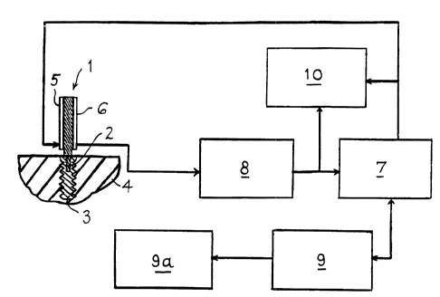
Referring to Figure 1, the apparatus comprises a
member in the form of a cantilever beam 1 attached by means
of a threaded section 2 to an implanted fixture, such as a
dental implant 3, in a section of bone 4, typically a human
jaw bone. The implant 3 may be any one of a number of
known types, formed from a metal, such as titanium, from a
ceramic material, or any other appropriate material. It
may, for example, be of the type suppled by Nobelpharma in
the U.K. Two transducers, such as piezoelectric elements
or strain gauges 5 and 6, are attached, for example bonded,
to opposite sides of the beam 1, gauge 5 being an exciter
gauge and gauge 6 a receiver gauge.
The exciter gauge 5 is driven by a variable fre-
quency oscillator, signals from which, for example in the
form of a sinusoidal excitation voltage, are fed to the
gauge 5 via an amplifier. The oscillator and amplifier
may be incorporated in a frequency response analyser 7.
PCT/GB92/00663
WO 92/18053
~!~: ,
-_5_
Signals detected by the receiver gauge 6 are amplified by a
charge amplifier 8 and applied as an input to the analyser
7. The output from the.analyser, which represents the
ratio of the response voltage to the excitation voltage,-is
fed to a processor such as a microprocessor 9, which is
used to vary the frequency output of the oscillator of the
analyser 7, and store the results in a data store 9a. The
results may be printed out, and/or displayed on an oscillo-
scope 10, and/or an AC voltmeter or t:he like.
In use the beam 1 is secured,, i.e. screwed, to the
implanted implant 3 with a ;predetermined torque, for exam-
ple using a Nobelpharma torque controller and counter tool.
The variations in resonance frequency with torque have been
found to be relatively small over a practical range of
torques, for example of the order of 10 to 15 Ncm, so that
such torque variations should not present a problem.
Constant amplitude, for example 1 volt, AC excitation
signals are then applied to the beam 1 via the gauge 5.
The frequency of the AC excitation signals is varied until
the amplitude of the signal displayed on the oscilloscope
is at a maximum. The resonance frequency is the fre-
quency at which the amplitude of the ratio of the response
voltage to the excitation voltage is a maximum. Figure 2
shows the data from a coarse sweep which is used to obtain
the resonance frequency roughly. A finer sweep around this
region is then used to identify thi:a frequency, typically
the first or fundamental frequency, more accurately. This
frequency is noted, and compared, for example, with the
data for other implants at similar stages of bonding.
It is expected that for a particular implant, the
resonance frequency will vary with time as depicted in
Figure 3. Thus by comparing the detected resonance fre-
quency with previously compiled data for similar implants,
WO 92/18053 PCT/GB92/00663
~1Q814~
-D-
an indication of the degree of attachment of the implant
can be obtained. With regard to Figure 3, the stiffness of
the interface may initially decrease following imn~.ant
placement because of acute inflammatory response. The
stiffness then recovers as integration occurs, and is ex-
pected eventually to approach, reach or exceed the initial
value.
The technique, which is based on detection and
comparison of resonance frequency shifts, rather than
amplitude changes, is effective to determine the quality of
the implant/tissue interface as a function of its stiff-
ness, and also in relation to any bone loss as a function
of the level or height of the marginal bone surrounding the
implant.
A currently preferred cantilever beam is illustrat-
ed in Figure 4. This beam 1 is generally L-shaped, having
base limb la with an aperture lb which locates over a boss
3a at the upper end of the implant 3. The beam is fixed in
place by a screw 11 screwed into the threaded bore in the
implant. The aperture lb and boss 3a may be non-circular,
for example hexagonal in cross-section, so that the beam
orientation about the longitudinal axis of the implant may
be accurately and repeatedly determined. Different readings
may be obtained for different angular orientations of the
beam relative to the implant, so as to determine the stiff-
ness/bone level at different positions around the implant
axis.
The beam 1 as shown in Figure 1 or 4, which will
preferably be of the same material as the implant, for
example titanium, is dimensioned so as to provide a reso-
nant frequency range of the system (placed implant and
beam) of the order of 1 to 20 kH, more specifically 5 to 15
kH, and preferably in the region of about 10 kH. For
WO 92/18053 PGT/GB92/00663
~~
example, in the embodiment of Figure 4, the limbs of the
beam 1 may be of approximately 5 to o mm square cross-
section, the upright limb.being approximately 2 cm high,
and the base limb being approximately 1.5 cm long.
It will be understood that various modifications
may be made without departing from the scope of the present
invention as defined in the appended claims.
For example, an additional pair of
excitation/detection transducers or gauges may be mounted
on the sides of the beam at 90° t~o the transducers or
gauges 5 and 6 shown, so as to provide readings at right
angles to the latter transducers, without the necessity of
re-orienting the beam on the implant. Additionally, or
alternatively, the beam and/or transducer system could be
adapted to turn relative to the implant.
Although the beam shown in l~igure 4 is L-shaped,
the upright limb could form a straight extension of the
base limb la so as to lie generally oparallel to the jaw or
mandible.
In practice, the prosthesis may be attached direct-
ly to the implant 3 using the threaded bore in the latter.
Alternatively, the prosthesis may be indirectly attached to
the implant via a separate pillar or post (called an abut-
ment). Such an abutment has means, such as an axial screw
passing completely through the abutment, which threads into
the implant bore, to fix the abutment: to the implant. The
upper end of the screw, or the abutment, has a threaded
bore for attaching the prosthesis. The beam 1 may be
attached, in the manner previously described, to the upper
end of the abutment. The beam may then be employed, not
only to assess the integrity of the i.mplant/bone interface,
but also the integrity of abutment/implant joint.
WO 92/18053 PCT/GB92/00663
:," .
~1~~~.4~
_g_
The transducers or gauges, and optionally also the
beam may be coated, for example with an air dry acrylic
material, to protect the transducers during sterilization
of the apparatus. The electrical connections or wires
connected to the transducers are arranged or adapted to
minimise their damping effect on the resonant structure.
The member may take a form other than a cantilever beam,
and/or the piezoelectric transducers could be replaced by
other receiver/transmitter elements, for example employing
sonic resonance. The beam, instead of being basically
straight, could be generally U-shaped, and connected to the
implant or abutment by its base. The transducers or equiv-
alent could be mounted on the same or opposite limbs.