Note: Descriptions are shown in the official language in which they were submitted.
WO92/18166 2 ~ 2 PCT/US92/03177
_l _
M~LANIN-BA8ED AGENTB FO~ IMAGE ENHANCEMENT
The present invention relates to image-enhancing
agents, contrast agents or spectral shift agents to
enhance tissue or organ images or nuclear spectra
obtained from live animals with magnetic resonance
imaging (MRI) or spectroscopy (MRS), radioisotope
scanning or ultrasound imaging.
I0
Magnetic resonance imaging is a medical procedure
that takes advantage of the magnetic spin properties of
nuclei to create an image fundamentally like that of the
more widely known X-ray procedure the CT scan. The
nucleus of an atom contains neutrons and protons. Atoms
which have an odd number of neutrons or protons have a
non-zero spin quantum number (I). these atoms can be
thought to behave like small spinning spheres. Because
the nuclei have positive charges, the spinning produces a
magnetic moment (U), analogous to an electric current in
a closed loop of wire. When exposed to a magnetic field
these dipoles align themselves with the magnetic field.
The spin (magnetic moment) vectors experience a torque
when subjected to a magnetic field. Due to this torque,
the nuclei process about the axis of the magnetic field
at a rate given by the Larmor relationship:
f = w/2~ = ~Bo/2~
f = resonance frequency in Hertz (Hz)
w = the angular frequency in radians per second
~ = magnetic gyric ratio
Bo = static magnetic field
~ = a nuclear constant characteristic of the
isotope
. W O 92/t8166 PC~r/US92/03177
2~ ~3~2 -2-
The resonance frequency of a nucleus is a function of its
magnetogyric ratio (a constant for the particular nucleus
being studied) and the strength of an applied magnetic
field (Larmor equation). The magnetogyric ratio of a
s nucleus relates the magnetic moment and the nuclear spin
quantum number (I). The spin quantum number depicts the
number of energy states that a nucleus can have; a
nucleus has 2I+l energy levels. The hydrogen nucleus (a
proton) has a nuclear spin of 1/2, and thus two possible
spin states. In nuclear magnetic resonance spectroscopy
(NMR), the magnetic moment is shown as revolving around a
fixed magnetic field (Figure 1) at a fixed frequency. In
a sample, large numbers of nuclei are revolving around
this field in one of the two possible spin states,
creating a "macroscopic magnetization~' M parallel to the
magnetic field. There are more nuclei in the low-energy
spin state when there is no outside influence (Figure
2).
Resonance is the induction of a transition between
two different energy states. The nuclei that lends
itself best to magnetic resonance imaging is the proton,
the major isotope of hydrogen. Hydrogen has a very large
abundance in biological systems in water (El023/cm3) as
well as in other biochemical molecules and has the
required magnetic moment. The energy necessary to
produce a transition between the two spin states of
hydrogen ~+~ and -~) is the difference in energy (oE)
between these spin states. In MRI, resonance occurs when
radiofrequency (RF) energy is applied at the Larmor
frequency, flipping the magnetic moments from their m =
+~ (lower energy) to their m z -~ (higher energy) states.
Magnetic resonance absorption can only be detected by
transverse magnetization (magnetization perpendicular to
Bo). Only the transverse component, Nxy, is time
dependent and therefore according to Faraday's law of
.
W O 92/18166 2 ~ PC~r/US92/03177
-3-
induction, only time dependent magnetization can induce a
voltage in a receiver coil. Transverse magnetization is
generated when a radiofrequency (RF) field of amplitude
B., rotating synchronously with the processing spins is
applied.
When the RF field acts in a direction perpendicular
to the main field, the effect is to rotate the
magnetization away from the rest state. The macroscopic
magnetization experiences a torque of the RF field,
forcing the magnetization to rotate about it. If the
duration of the B. field is such that the net
magnetization is rotated by an angle of 90, it will
become transverse or perpendicular to the stated field
lS (Figure 3). The angle of rotation q, the RF flip angle,
is given by the Ernst equation.
~ = ~B,r
Bl = amplitude of the FR field
~ = duration
Once the RF field is removed~ the magnetization is
subjected to the effect of the static magnetic field and
processes about it. With a detection coil positioned
with its axis along the y axis, the AC voltage induced in
the coil is given by
~ ~ Mxy cos w
Mxy = initial transverse magnetization following
a 90 degree RF pulse
r = time interval between the rotation
. - , - . .
.
, , ,
WO92/t8166 PCT/US92/03177
2 li3~3 ~2 -4-
The transverse magnetization decays to zero exponentially
with a time constant TZ .
Therefore:
~ ~ Mxy e~ cos w r
This equation represents a damped oscillation that
is called the induction decay or FID signal. As the
transverse magnetization decays to zero with time, the
longitudinal magnetization increases back to its
equilibrium value. This return to equilibrium values is
termed relaxation. RF stimulation causes the nuclei to
absorb energy transferring them to the excited state.
lS The nuclei can return to the ground state by transferring
energy to their surroundings, the so called lattice.
This method of relaxation is called spin-lattice
relaxation (Tl). (see Figure 4) Components of M, after
being rotated to the x'y' plane, (Figure 5) return to
their original magnetization values in a time (T2), the
"spin-spin" relaxation time. In the T2 relaxation
process, nuclei in the excited and ground state exchange
energy with each other. Thus T2 measures the amount of
time necessary for the nuclei to get out of phase with
each other and return to the original random state. Both
Tl and T2 relaxation times of a nuclei can vary widely
from milliseconds to minutes depending upon the nuclei
and the environment surrounding them. The primary task
of magnetic resonance imaging is soft tissue contrast and
the detection of low-contrast lesions. The detection of
lesions depends on the inherent difference in contrast
between lesions and surrounding normal tissues. The
tissue or media present around a resonating nucleus have
differential effects that can alter the Tl and T2
relaxation times. However, in general, organic
substances such as those found in the body (tissues,
.
: ~ .:. - -
.
. ~
WOg2/181~ PCT/US92/03177
2~ ~352
-5-
organs) have a fairly uniform effect on relaxation times,
largely due to the high percentage of water. MRI scans
of a substance take advantage of relaxation times (T1 and
T2) differences to generate an image of the object being
scanned in "slices." The difference in relaxation times
of, say, different organs in the body allows a visible
image to be formed. However, the largely uniform effect
on relaxation times of most parts of the body causes an
image that is very difficult to see due to lack of
contrast, hence the need for "contrast agents."
Inherent tissue contrast in MRI is determined by
differences in:
1) inherent spin density;
2) longitudinal relaxation time (T1);
3) transverse relaxation time (T2); and
4) flow.
Contrast agents can improve visualization of low
contrast organs and lesions. The most promising agents
affect signal by enhancing relaxation. A contrast agent
is a substance, either 1) a paramagnetic metal ion, 2)
free oxygen, or 3) a substance with free radicals
(unpaired electrons), that has a far different effect on
the proton relaxivity of tissue water. A good contrast
agent can be either directly injected into the target
area or tagged to an antibody or receptor against the
target area (e.g., a cancerous tumor) and thus provide
sharpened or altered contrast for aid in viewing the area
by MRI.
Pharmacologic basis for relaxation enhancement is
based on positive magnetic susceptibility (Bourdreaux,
E.A. and Mulap, L.N.: THEORY AND AppLIcATIoN OF MOLEC~R
PA~AGNETIsM, New York, 1976, John Wiley and Sons). When
a substance is placed in an external magnetic field,
induced magnetization in the substance is additive to
... . .
. . . . .
~ . .. .
,~ .
.
'- ,~',~ . . . .
: - - ~ . . .
: . ~ : .
WO92/181~ PCT/USg2/03177
, ,=
21083~2 -6-
that of the applied field. The magnetic susceptibility
of a substance is defined as the ratio of induced
magnetization to that of the applied field. Substances
can be categorized by their magnetic susceptibility (see
Table I).
TABL~ I (David Stark and William Bradley: MAGNETIC
RESONANCE IMAGING, St. Louis, 1988, Mosby)
Cr.a88 BA8I8 ~8C~PTIBII,ITY
Diamagnetic paired electrons - -lO~
no permanent spin
moment
Paramagnetic unpaired electrons +lO~
non-interacting
permanent moments
Superparamagnetic unpaired electrons- +lO+2
non-interacting
domains
Ferromagnetic unpaired electrons- +lO 2
interacting domains
Diamagnetic substances have negative
susceptibilities. Most organic and inorganic compounds
are diamagnetic and since all atoms experience induced
magnetic affects arising from electron orbital motion, a
diamagnetic component is present in all materials.
~iamagnetic effects are very weak and can be overwhelmed
in magnitude by relatively few unpaired electron spins.
Diamagnetic materials are generally of little interest as
contrast agents.
Paramagnetic, superparamagnetic, and ferromagnetic
substances are characterized by the predominant magnetic
: ,, .................. :
,. . ~ . . . . . .
WO92/18166 PCT/US92/03177
_7- 2 ~
effects of unpaired electron spins which produce positive
susceptibilities and positive induced magnetization.
Paramagnetism is characterized by independent action
of individual atomic or molecular magnetic moments.
Ferromagnetism is characterized by solid phase
microscopic volumes or domains in which unpaired electron
spins are permanently aligned. Multiple domains in bulk
can be isotropic (unmagnetized) or anisotropic
(magnetized).
Superparamagnetic materials can be regarded as
single domain particles. The susceptibilities per atom
or mole of these substances exceed those of corresponding
soluble paramagnetic species due to magnetic ordering.
Superparamagnetic and ferromagnetic susceptibilities
increase linearly with field strength. Superparamagnetic
substances unlike ferromagnetic substances are
characterized by restoration of induced magnetization to
zero upon removal of the external field.
Paramagnetic enhancement of nuclear relaxation was
first described in 1946 by Bloch and co-workers (Bloch,
F., Hansen, W.N., and Packard, P.: "The nuclear
induction experiment", Physiol. Rev. 70:474-485, 1946)
when they demonstrated a convenient practice of
shortening the time needed to observe water IH Tl by
adding ferric nitrate (a paramagnetic solute).
Positive susceptibility is necessary, but not
sufficient, for effective relaxation. The magnitude of
relaxation enhancement also depends on proximity and on
correlation time. A mathematical formulation of
3S paramagnetic enhanced solvent relaxation is described by
Bloembergen (Bloembergen, N.: "Proton relaxation times
.. .
W092/18166 PCT/USg2/~3177
2108362 -8-
in paramagnetic solutions", J. Chem. Phys. 27:572, 1957).
These equations imply that nuclear relaxation results
from several simultaneous mechanisms.
The paramagnetic contribution to nuclear relaxation
is proportional to
1) the paramagnetic concentration;
2) the distance (~); and
3) a time constant describing the dynamic nature
of electron-proton interactions (correlation
time).
The correlation time is dominated by the fastest
rate of paramagnetic tumbling, electron spin flips, or
chemical exchange. Due to more optimal correlation of
spin motion, nuclear T1 relaxation enhancement in
biologic systems is more effective with relaxation agents
of large molecular weight or asymmetric shape, that is,
materials possessing relatively long rotational
correlation times.
`
Solvent relaxation in the presence of
superpar~magn-tic particle~ chi-fly differs from that in
the presence of paramagnetic solutes due to much greater
weighting of the magnetic moment contribution. Compared
with paramagnetic solutes, superparamagnetic particulates
have
1) increased effective magnetic moment;
2) decreased freedom of molecular motion; and
3) decreased water IH exchange.
.
The much greater effective magnetic moment dominates
these factors and results in substantial T2 shortening
3S caused by long range effect from magnetic field
heterogeneity. Thus, although these types of contrast
'~ . .
- . . ., ~ ,,~ . ,
', ' '
' ` ~ , ',' '' , ' "', ' ' ' ' ' ' "
WO92/18166 PCT/US92/03177
21 ~8~62
g
agents also affect Tl, their primary influence is on T2
(e.g., T2 contrast agents).
The imaging of internal structures and organs of
live animals has been an important aspect of medicine
since the advent of X-ray usage for this purpose. Among
the techniques more recently developed for such imaging
are those involving scanning for emission of particles
form an internally located radioisotope. Such
radioisotopes preferably emit gamma particles and are
generally isotopes of metallic elements. One problem
common to the diagnostic usage of such gamma particle-
emitting radioisotopes concerns the localization of these
materials at sites of particular interest rather than to
have them randomly dispersed or rapidly excreted, by the
kidney, for example. Another problem of such
radioisotope mediated imaging concerns optimizing the
circulating half-life of radioisotopes, for example, by
preventing or accentuating their binding to serum
proteins (e.g., albumin), or by prior conjugation
(complexation) to polymeric carriers or receptor-binding
substances.
A second class of internal body imaging which is
undergoing a rapid growth in clinical use is ultrasound
imaging. This is based on the detection of differences
in the internal velocity (reflectivity) of directed,
high-frequency sound waves. Differences in image
brightness are produced at the interfaces between tissues
with different native densities and ultrasound
reflectivities. A present clinical problem is the
difficulty of visualizing lesions in the stomach, small
and large bowel, bladder, and cavities of the female
reproductive tract, due to similarities of ultrasound
velocity between these organs of interest and immediately
adjacent tissues. Diagnostic introduction of a dense,
: ~ , ,
: " , , , . ~;::~ . -
. . ~ ,. . -
wos2/1gl66 PCT/US92/03177
10-
nonradioactive metal element or ion at sufficient
concentrations can confer significant differences in
ultrasound reflectivity required to visualize otherwise
undetectable tumors and inflammatory lesions.
NMR intensity and relaxation images have been shown
in recent years to provide a third important method of
imaging internal structures and organs of live animals.
Clinical Magnetic Resonance Imaging (MRI) is a rapidly
growing, new form of brain and body imaging. Low-field
(proton) MRI detects chemical parameters in the immediate
environment around the protons because of differences in
body tissues (predominantly water protons because of
their relative abundance). Changes in these parameters
occur very early in disease and are independent of
physical densities detected by ionizing radiation. In
the brain and central nervous system, MRI has allowed
detection of tumors at an earlier clinical stage and with
fewer imaging artifacts than is possible with
computerized axial tomography (CA$~ (Runge et al., (1983)
Am. J. Radiol. V 141, p 1209). Under optimal conditions,
image resolution is in the submillimeter size range.
Seven factors are among those making it important to
develop nontoxic MRI image-enhancing agents analogous to
those available for CAT:
1. They increase the specificity of MRI diagnosis.
2. Smaller lesions can be identified earlier.
3. Image-enhancing agents enhance tumor masses
differently than surrounding edema fluid or abscesses.
This allows the extent and invasion of tumor to be
defined more precisely. Lesion with infiltrative-type
growth (e.g., certain metastatic carcinomas and
- .
;: - : .~. ,. . .. -
.: :. :. , ~ : .
....
: ~ -
.. ,... , . ,. - . . .
WO92/18166 PCT/US92/03177
-11- 2~ ~3~
glioblastomas) will require contrast agents for
demarcation between tumor and edema fluid (Felix et al.
(1985) Proc. Soc. Mag. Res. Med. V 2, p 831).
4. Image-enhancing agents improve the distinction
between recurrent tumor and fibrous tissue resulting from
surgery and radiation.
5. Image-enhancing agents can decrease the time
required per scan and potentially decrease the number of
scans required per procedure. This increases the volume
of procedures and decreases their expense.
6. Body imaging has a significantly lower resolution
(typically 0.5-1.0 cm) and sensitivity (decreased signal-
to-noise ratio) than brain imaging (Wesbey et al. (1983)
Radiology V 149, p 175). These differences result from
the greater inhomogeneity of the magnetic field; the
larger radio frequency coil; unequal phase-pulsing of
deep versus shallow nuclei; and motion artifacts produced
by rQspiration~ cardiac systole, gastrointestinal
peristalsis, and voluntary muscle movement; and contrast
agents can be succes~fully -ploy-d to improve image
resolution for specific structures and organs.
7. Advanced (polymeric and microsphere) forms of
contrast agents (see below) appear to be required for the
optimal acquisition and interpretation of blood-flow,
tissue-perfusion, and diffusion images and related
spectral (phase) information.
The discrete intensities of a two-dimensional,
Fourier-transformed image are described by the following
general equation (for spin-echo pulse seguences):
W092~181~ ~ PCT/US92/03177
,~ .
210~362
Intensity = N(H) . f (v) . exp(-TE/T2) . (1 - exptTE-
TR)/T1), where:
N(H) = number of protons in the
discrete tissue volume (spin
density);
f~v) = a function of proton velocity and the
fraction of protons which are moving
(e.g., due to following blood);
TE = time between the radio frequency (rf)
pulse and the detection of signal
~spin-echo);
TR = the interval between repetition of
the rf pulse;
T1 = the time interval associated with the
rate of proton enQrgy transfer to the
surrounding chemical environment
(spin-lattice or longitudinal
relaxation);
T2 = the time interval associated with the
rate of proton energy transfer, one
to other (spin-spin or transverse
relaxation).
The T1 and T2 times have reciprocal effects on image
intensity. Intensity is increased by either shortening
the T1 or lengthening the T2 or vice versa depending upon
whether proton density, T1-weighted or T2-weighted images
are desired. Tissue contrast occurs naturally and is
related to variations in the chemical environments around
water protons (major contributor) and lipid protons
(usually minor). Chemical agents have been used to
enhance this natural contrast. The one most widely
tested clinically is the paramagnetic metal ion,
gadolinium (Gd+3) chelated to an appropriate organic
chelate(Runge et al. (1983) Am. J. Radiol. V 142, p 619).
Although gadolinium shortens both the T1 and T2 times, at
the lower doses used for clinical imaging, the Tl effect
generally predominates and the image region affected
becomes brighter. Also, the rf pulse sequence can be
- ,: .. , ; . :
... . ; . ..
. ~. .: .
... . ~ . , ~- :.
.., . . ~
WO92/18166 PCTtUS92/03177
-13- 21 ~83fi2
programmed to accentuate T1 changes and diminish those
due to T2 (Runge et al. (9183) Am. J. Radiol. v 141, p
1209). Hence, "T1-weighted" enhancement can be achieved
by selecting the most favorable Gd dose and rf pulse
sequence.
The shortening of proton relaxation times by Gd is
mediated by dipole-dipole interactions between its
unpaired electrons and adjacent water protons. The
effectiveness of Gd's magnetic dipole drops off very
rapidly as a function of its distance from these protons
(as the sixth power of the radius) (Brown (1985) Mag.
Res. Imag. V 3, p 3). Consequently, the only protons
which are relaxed efficiently are those able to enter
Gd's first or second coordination spheres during the
interval between the rf pulse and signal detection. This
ranges from 105 t~ Io6 protons/second (Brown (1985) Mag.
Res. Imag. V 3, p 3). Still, because Gd has the largest
number of unpaired electrons (seven) in its 4f orbitals,
it has the largest paramagnetic dipole (7.9 Bohr
magnetons) and exhibits the greatest paramagnetic
relaxivity of any element (Runge et al. (1983) Am. J.
Radiol. V 141, p 1209 and Weinman et al. (1984) Am. ~.
Radiol. V 142, p 619). Hence, Gd has the highest
potential of any element for enhancing images. However,
the free form of Gd is quite toxic. This results in part
from precipitation at body pH (as the hydroxide). In
order to increase solubility and decrease toxicity, Gd
has been chemically chelated to small organic molecules.
To date, the chelator most satisfactory from the
standpoints of general utility, activity, and toxicity is
diethylenetriamine pentaacetic acid (DTPA) (Runge et al.
(1983) Am. J. Retail V 141, p 1209 and Weinman et al.
(1984) Am. J. Retail V 142, p 619). The first
formulation of this chelate to undergo extensive clinical
testing was developed by Schering Ag - Berlex Imaging
WO92/18166 PCT/US92/0317,
~,,
-14-
2~083~2
according to a patent application filed in West Germany
by Gries, Rosenberg and Weinmann (DE-OS 3129906 A 1
(1981). It consists of Gd-DTPA which is neutralized and
stabilized with the organic base, N-methyl-D-glucamine
(meglumine). The Schering-Berlex agent has completed
Phase III clinical testing at selected centers across the
United States and abroad. The results of this and
ongoing studies indicate that almost all human brain
tumors undergo significant enhancement (Felix et al.
(1985) Proc. Soc. Mag. Res. Med. V 2, p 831 and K.
Maravilla, personal communication). These include
metastatic carcinomas, meningiomas, gliomas, adenomas and
neuromas. Renal tumors are also enhanced satisfactorily
(Lanaido et al. (1985) Proc. Soc. Mag. Res. Med. V 2, p
877 and Brasch et al. (183) Am. J. Retail. V 141, p
1019). The Schering-Berlex formulation (MAGNAVIST) was
available for general clinical use by 1989 and has
received FDA approval.
Despite its satisfactory relaxivity and toxicity,
this formulation has four major disadvantages:
(1) Chelation of Gd mark-dly decreases its
relaxivity (by 1/2 an order of magnitude). This happens
because chelators occupy almost all of Gd's inner
coordination sites which coincide with the strongest
portion of the paramagnetic dipole (Koenig 1985) Proc.
Soc. Mag. Res. Ned. V 2, p 833 and Geraldes et al. (1985)
Proc. Soc. Mag. Res. med. V 2, p 860).
(2) Gd-DTPA dimeglumine, like all small
paramagnetic metal chelates, suffers a marked decrease in
relaxivity at the higher radio frequencies used
clinically for proton imaging (typically 85 MHz, 2T)
(Geraldes et al. (1985) Proc. Soc. Mag. Res. Med. V 2, p
860).
:: :,
, s ..:
.. ..
: , , .:
,, .:
W092/18166 PCT/US92/03177
2 ~ ~ ~ t~ '~ 2
--15--
(3) Due to its low molecular weight, Gd-DTPA
dimeglumine is cleared very rapidly from the bloodstream
(tln in 20 minutes) and also from tissue lesions (tumors)
(Weinman ~t al. (1984) Am. J. Radiol V 142, p 619). This
S limits the imaging window (to ca. 30 to 45 minutes);
limits the number of optimal images after each injection
(to ca. 2); and increases the agent's required dose and
relative toxicity.
(4) The biodistribution of Gd-DPTA is suboptimal
for imaging of body,(versus brain) tumors and infections.
This is due to its small molecular size. Intravenously
administered Gd-DTPA exchanges rapidly into the
extracellular water of normal tissues, as well as
concentrates in tumors and infections. This is
facilitated by an absence in body organs, of the "blood-
brain" vascular barrier which partly restricts the
exchange of Gd-DTPA into the extracellular water Of
nor,mal (versus diseased) brain. ~he result in body
organs, is a reduced difference in the concentration of
Gd-DTPA between normal and diseased regions of tissue,
and hence, reduced image contrast between the normal and
disQased regions o~ th~ organ. Al~o a disproportionatç
quantity (~90%) of Gd-DTPA is seguestered very rapidly in
~; 25 the kidneys (Weinman et al. (1984) Am. J. Radiol V 142, p
619). Of much greater interest to body MRI, are the
abdominal sites involved in the early detection and
staging of tumors, particularly the liver, and also the
sp}een, bone marrow, colon and pancreas, which can not be
probed successfully with Gd-DTPA.
Three approaches have been taken in attempts to
overcome these disadvantages:
(1) Alternative, small chelating molecules have
been tested. These make Gd more accessible to water
WO g2/1816C PCr/USg2/03t7~
2~ ~83~2
protons but still chelate the metal with a sufficient
affinity to potentially control its toxicity in vivo.
The most effective of these chelators is DOTA, the poly-
azamacrocyclic ligand, 1,4,7,10-tetraazacyclododecane-
N,N~,N"-tetraacetic acid (Geraldes et al. (1985) Proc.
Soc. Mag. Res. Med. V 2, p 860). Its relaxivity is
approximately 2 times greater than that of Gd-DTPA over a
wide range of Larmor frequencies. However, it is still
less active than free Gd.
1,0
(2) Gd and Gd-chelates have been chemically
conjugated to macromolecules, primarily the proteins,
albumin (Bulman et al. (1981) Health Physics V 40, p 228
and Lauffer et al. (1985) Mag. Res. Imaging V 3, p 11),
asialofetuin (Bulman et al. (1981) Health Physics V 40, p
228), and immunoglobulins (Lauffer et al. tl985) Mag.
Res. Imaging V 3, p 11 and Brady et al. (1983) Soc. Mag.
Res., 2nd Ann. Mtg., Works in Progress, San Francisco,
CA). This increases the relaxivity of Gd by slowing its
rate of molecular tumbling (rotational correlation time)
(Lauffer et ~1. (1985) Mag. Res. Imaging V 3, p 11).
This improves coupling of the energy-transfer process
between protons and Gd (Geraldes et ~1. (1985) Prôc. Soc.
Mag. Res. Med. V 2, p 860, Lauffer et al. 9198S) Mag.
Res. Imaging V 3, p 11 and Brown et al . ~1977)
Biochemistry V 16, p 3883). Relaxivities are increased
by multiples of S to 10 relative to Gd-DTPA (when
compared as R1=1/T1 values at 1 millimolar concentrations
of Gd) and by multiples of 2.5 to 5.0 (when compared as
the molarities of Gd reguired to produce a specified
decrease in the Tl relative to a control solution
(physiologic saline).
The reasons for using the latter method of
comparison are that 1) millimolar concentrations of Gd
are never achieved in vivo -- actual tissue
, .
.. . ............... , .,. .. . ... . ~ , .
.:
.
WO92/18t66 PCT/US9V03t77
2~3~
concentrations achieved in the usual image enhancement
are between 20 and 100 micromolar Gd; 2) the slopes of R1
graphs are frequently nonparallel for different enhancing
agents; 3) the second method allows agents to be compared
according to the more customary means of chemical
activity ratio, in other words, as the concentration
required to produce a specified percentage decrease in
the T1 (or T2) relaxation time. The second method is
considered preferable. A drawback of conjugating DTPA to
protein carriers for use in NMR image enhancement is that
it has been difficult to produce stable conjugates of
more than 5 DTPAs (and hence Gd's) to each albumin
molecule (Bulman et al . ( 1981) Health Physics V 40, p
228, Lau~er ~t al. (1985) Mag. Res. Imaging V 3, p 11
and Hnatowich et al. (1982) Int. J. Appl. Radiat. Isot. V
33, p 327 (1982)).
Comparably low substitution ratios (normalized for
molecular weight) have been reported for immunoglobulins
(Lauffer et al. (1985) Mag. RQS. Imaging V 3, p 11 and
Brady et al. (1983) Soc. Mag. Res., 2nd Ann. Mtg., Works
in Progress, San Francisco, CA) and fibrinogen (Layne et
1982) J. Nucl. ~ d. V 23, p 627). This r-sults from
the relative difficulty of forming amide bonds, the
comparatively low number of exposed amino groups on
typical proteins which are available for coupling, and
the relatively rapid hydrolysis of the D~PA anhydride
coupling substrate which occurs in the aqueous solvents
required to minimize protein denaturation during
~;~ 30 conjugation (Hnatowich et ~1. (1982) Int. J. Appl.
Radiat. Isot. V 33, p 327 (1982) and Krejcarek et al.
(1977) Biochem. Biophys. Res. Comm. V 77, p 581). The
overall effect of these suboptimal conditions is that a
~;; very large dose of carrier ~aterial is required to
;;~ 35 achievQ significant in vivo effects on MR images. At
` this high dose, the carrier produces an unacceptable
~:
WOg2/t8166 PCT/US92/03l77
.,
-18-
21~8~62
acute expansion of the recipient's blood volume by an
osmotic mechanism. Indeed, low substitution ratios have
generally limited the use of such protein-chelator-metal
complexes to the more sensitive (low-dose),
radiopharmaceutical applications (Layne et al. (1982) J.
Nucl. Med. V 23, p 627). Recent development of so-called
'non-ionic' Gd-chelates using amine ligands gives
contrast agents with improved osmolalities upon
administration to patients; however, there has been no
improvement in relaxivity over Gd-DTPA chelates.
An attempt to overcome this low substitution ratio
has been made by conjugating DTPA to the non-protein
carrier, cellulose (Bulman et al. (1981) Health Physics
40, p 228), however the chemi,cal method employed results
in suboptimal substitution of DTPA onto the carrier. The
nonbiodegradability of cellulose and its water-soluble
derivatives and the reported molecular aggregation which
results from organic-solvent conjugation (in
dimethylformamide) of CNBr-activated cellulose to the
diaminohexyl spacer groups which link the carrier to
DTPA, have rendered this class of carrier-conjugates
unacceptable for intravenous administration at the doses
required for MR image enhancement.
A very important consideration in the image
enhancement of solid tumors and inflammatory lesions by
polymeric contrast agents is that, in order for these
agents to extravasate (exit) efficiently from the
microcirculation into adjacent diseased tissues, they
must be completely soluble -- e.g., not be contaminated
by intermolecular or supramolecular microaggregates.
Optimal tumor access and localization requires that the
molecular size of such agents generally be less than
approximately 2,000,000 daltons (ca. 2 to 3 nanometers in
molecular diameter), and preferably less than 500,000
~ .
,.,
" , .. .
WO92/18166 PCT/US92/03t77
. . .
-19- 2~ ~? ~J ~
daltons (ca. 0.5 to 1.0 nanometers in molecular diameter)
(Jain (1985) Biotechnology Progress V 1, p 81). For this
reason, with rare exceptions the particulate and
microaggregate classes of contrast agents (which comprise
the liposomes, colloids, emulsions, particies,
microspheres and microaggregates, as described below) do
not concentrate efficiently in most solid tumors or
inflammatory lesions. Instead, following intravenous
administration, these supramolecular-sized agents: a)
are first circulated in the bloodstream for relatively
short intervals (225 minutes to 24 hours, depending on
size), potentially allowing direct image enhancement of
the blood pool (plasma compartment); and b) are
subsequently cleared by specialized (phagocytic) cells of
the reticuloendothelial tissues (liver, spleen and bone
marrow), potentially allowing selective enhancement of
these normal tissues, but producing indirect (negative)
enhancement of lesions within those tissues (due to
exclusion of the agents from the diseased regions).
Additionally, following installation into the
gastrointestinal tract and other body cavities, these
particulate and microaggregate classes of agents can
produce direct image enhancemQnt of the ~luid within
- these cavities, and thereby potentially delineate mass
lesions which encroach upon the lumens and cavities.
~; Both microspheres and microaggregates are supramolecular
in size. The microaggregate class of agents is produced
(intentionally or unintentionally) by either a) molecular
cross-linking of individual polymer molecules or b)
secondary aggregation of previously single (soluble)
polymers, as induced by charge attraction or hydrophobic
bonding mechanisms. It is distinguished from the
microsphere class of agents by virtue of its smaller
particle size, which ranges from approximately 2,000,000
daitons (ca. 2 to 3 nanometers in diameter) to 0.1
- micrometers (= lO0 nanometers in diameter). It is
.,.. . ...... ~ ;:. .
WO92/18166 PCT/US92/0317,
.
2~083~2
important to note that microaggregates are cleared by
reticuloendothelial phagocytes with significantly less
efficiency and rapidity than are microspheres. In
general, this property makes microaggregates a less
preferred class of agents for visualizing the liver,
spleen and bone marrow under the usual conditions of
clinical imaging, for which prompt post-injection
contrast enhancement is required.
(3) Gd-DTPA has been entrapped in liposomes
(Buonocore et al. (1985) Proc. Soc. mag. Res. med. V 2, p
838) in order to selectively enhance images of the
reticuloendothelial organs (liver, spleen and bone
marrow) and potentially the lungs. Liver clearance is
mediated by phagocytic (Xupffer) cells which
spontaneously remove these small (0.05 to 0.1 um)
particles from the bloodstream (Buonocore et al. (1985)
Proc. Soc. Mag. Res. Med. V 2, p 838). (Particles larger
than 3 to S um are selectively localized in the lungs due
to embolic entrapment in lung capillaries.) A recent
report indicates that the small-sized Gd-liposomes
produce effective decreases in liver Tl's (as determined
spectroscopically without imaging) (Buonocore et al.
(1985) Proc. Soc. Mag. Res. Med. V 2, 0838). Also,
insoluble Gd-DTPA colloids have recently been reported to
enhance MR images of rabbit livers under in vivo
conditions (Wolf et al . ( 1984) Radiographics V 4, p 66).
~owever, three major problems appear to limit the
diagnostic utility of these devices. The multilamellar,
lipid envelopes of liposomes appear to impede the free
diffusion of water protons into the central, hydrophobic
cores of these carriers, as assessed by the higher does
of Gd required for in vitro relaxivities eguivalent to
Gd-DTPA dimeglumine (Buonocore et al . ( 1985) Proc. Soc.
mag. Res. med. V 2, p 838). This increases the relative
toxicity of these agents.
WO 92/18166 PCI~/US92/03177
-21-
Even more importantly, these same lipid components
cause the carriers to interact with cell membranes of the
target organs in ways which lead to marked prolongation
of tissue retention (with clearance times of up to
5 several months) (Graybill et al. (1982) J. Infect. Dis. V
145, p 748 and Taylor et al., 1982 Am. Rev. Resp. Dis. V
125, p 610). Two adverse consequences result. First,
image enhancement does not return to baseline in a timely
fashion. This precludes re-imaging at the short
10 intervals (ca. 1 to 3 weeks) needed to assess acute
disease progression and therapeutic treatment effects.
Second, significant quantities of the liposomally
entrapped Gd-DTPA may be transferred directly into the
membranes of host cells (Blank et al. (1980) Health
Physics V 39, 913; Chan et al. (1985) Proc. Soc. Mag.
Res. Med. V 2, p 846). This can markedly increase the
cellular retention and toxicity of such liposomal agents.
The consequences for Gd toxicity have not yet been
reported. Protein (albumin) microspheres with entrapped
20 Gd and Gd chelates have been prepared and shown (Saini et
al. (1985) Proc. Soc. Mag. Res. Med. V 2, p 896) to have
only modest effects on Tl relaxivity in vitro. This is
because most o~ the Gd as wQll as other entrapment
materials (Widder et al. (1980) Cancer Res. V 40, p 3512)
25 are initially sequestered in the interior of these
spheres and are released very slowly as the spheres
become hydrated (with tl/2's of hours) (Widder et al.
(1980) Cancer Res. V 40, 3512).
Emulsions of insoluble gadolinium oxide particles
have been injected into experimental animals with
significant image-enhancing effects on the liver (Burnett
~t al. (1985) Magnetic Res. Imaging V 3, p 65). However,
these particles are considérably more toxic than any of
the preceding materials and are thus inappropriate for
human use. Because of the significant disadvantages of
- .
~: ,
... . .
WO92/18166 PCT/US9~03177
:
, ,.
21 0~'~62
existing MR image contrast agents, the present applicant
has formulated improved, second-generation prototype
agents with reduced toxicity, phenomenally increased
effectiveness, potentially increased selectivity of tumor
and organ uptake, as well as producing an agent with
significant potential for enhancing blood flow images.
Many of the advantages shown for the present
developments concerning NMR image-enhancing agents (also
referred to herein as NMR contrast agents or MR (magnetic
resonance) contrast agents) are also expandable to other
areas. Gadolinium and related agents can produce
characteristic changes in the NMR spectrum of adjacent
NMR-susceptible nuclei. These changes include:
modulation of resonance peak positions, widths,
intensities, and relaxation rates (which affect
intensity). Hence, perturbation of spectra by such
` chemical shift-relaxation agents can be used to localize
and identify the source of NMR signals with respect to
their location in organs, in tissue compartments
(intravascular versus extravascular), in cell types
within the tissue, and potentially, in specific metabolic
pathways within cQlls which are altered by drugs and
disease. Also in certain situations, ultrasound imaging
or body scanning of radioisotopic emissions is
particularly useful in achieving insight into internal
structures. The radioisotopic emissions most frequently
scanned are those of metallic radioisotopes emitting
gamma particles, however, positron emission tomography is
experiencing increased clinical use. The molecular
formulation and mode of administering these radioisotopic
metals will have significant consequences on the internal
localization and body half-life of these radioisotopes,
potentially leading to increased diagnostic usage of
these ultrasound images and emission scannings.
, .
., . ~ :- ,
WO92/18166 PCT/USg2/03t77
. ,
-23-
The present invention includes an image-enhancing or
spectral-shift agent comprising a biodegradable, water-
soluble or insoluble melanin polymer, synthetic or
derived from natural sources, and having either a signal-
s inducing (paramagnetic, superparamagnetic or
ferromagnetic) metal or metal particle incorporated
therein or a concentration of stable free radicals or
having both metal and stable free radicals.
Melanins are a group of pigments derived from
several different amino acid substrates in the natural
world. Different types of melanins are responsible for
much of the coloration in animals, plants, and bacteria.
Melanins are usually classified in one of three groups:
eumelanins, such as those found in hair, skin, feathers,
and as a part of the coloration in reptiles and fish;
phaeomelanins, which produce human red hair and the red
fur of foxes; and allomelanins, which are most often
present in bacteria and plants. An example of one of the
most striking effects of a combination of eumelanins and
phaeomelanins in the pattsrns found in the plumage of
certain tropical birds.
Melanins are created by the action of enzymes on any
of several amino acid precursors. For example, tyrosine
is the precursor of most eumelanins. However, the exact
process by which an amino acid is converted to melanin is
unknown, and indeed, seems to vary from one pigment to
another even when formed from the same substrate. In
general, blac~ pigments are formed from precursors
including 3,4-dihydroxyphenylalanine (DOPA), catechol,
dihydroxy indoles and various other dihydroxy-
substances.
- ;- . : .
;
;
- ~,, ~ .:.
': .:
WO92/18166 PCT/US92~03177
I; , ,.: .
2l a~ ~2 -24-
H ~ CH2 - CN - COOH HO
H NH HO NH
3,4-dihydroxyphenylalanine 5,6-dihydroxyindole
(DOPA)
~
catechol l,8-dihydroxynaphthalene
These substances have many active centers for
polymerization. Compounds with fewer active centers are
precursors to melanins of brown, reddish-brown, or
yellow-brown color.
Melanins in pure form are usually insoluble in water
as well as in most organic solvents, making them
difficult to work with. In part due to this, not all of
the chemical properties of melanins have been identified.
However, recent studies using electron spin spectroscopy
have identified free-radical properties of natural
melanins (Table l).
WO9V18166 PCT/US92/03177
2 ~
TAB~ 1
.
~ELANIN g-VALU~
CATECHOL-melanin 2.0038
1L-DOPA-melanin 2.0036
1D-DOPA-melanin 2.0038
L-Adrenalin-melanin 2.0040
Sepia-melanin 2.0030
Melanoma-melanin 2.0031
Human hair-melanin 2.0043
Potato-melanin 2.0040
Synthetic melanins of various molecular weights can
be created from all of the precursors used in nature and
from a wide variety of mono-, di-, or polyhydroxylated
aromatic compounds containing many different substituents
by using almost any chemical oxidant or free-radical
polymerizer, or, in some cases, merely leaving dissolved
substrate open to the air overnight. Figure lA
schematically designates some paths of melanin synthesis.
The present invention involves an image-enhancing
agent comprising water-soluble paramagnetic melanin or a
melanin polymer combined with a non-dissociable signal-
inducing (paramagnetic, superparamagnetic, orferromagnetic) metal and containing stable free radicals.
In one embodiment the agent preferably contains at least
about 0.l micromole metal per gram but can vary greatly
depending upon the synthetic conditions employed to
; 30 produce the agent. In an important embodiment the
melanin preferably contains no metal but has melanin-
incorporated stable free radicals which depend upon the
synthetic conditions employed to produce the agent.
.
'~' ''` ~'' -
: - . . ...
WO92/18166 PCT/US92/03177
,
-26-
~ ~83~2
In reference to the melanin-metal combination of the
present invention, the signal-inducing metal has an
association constant for its melanin combination of at
least about lO~, i.e., it is virtually non-dissociable.
Upon suspension or dissolution in water the metal remains
undissociated for an indefinite period. A preferred
signal-inducing metal is paramagnetic or
superparamagnetic, of course for magnetic resonance
imaging. Preferred paramagnetic or superparamagnetic
metals are gadolinium, iron, nickel, copper, erbium,
europium, praseodymium, dysprosium, holmium, chromium or
manganese. Gadolinium is a preferred metal. In one
aspect, paramagnetic melanin free of added metal of any
kind affects proton relaxivity more effectively than Gd-
DTPA (MAGNAVIST) because it contains stable freeradicals.
The metal is incorporated into the melanin in an
ionic, uncharged, or particulate form. Metals may be
utilized which are particularly useful to modify
ultrasound images by the enhancement of the image
obtained from emission and detection of high-frequency
soundwaves. Metals emitting gamma particles may also be
utilized to enhance images resulting from gamma particle
emission scanning. 5~Chromium, ~gallium, ~'technetium and
~indium are preferred metals for gamma particle scanning.
The best image-enhancing agents of the present
invention have preferred molecular weights between about
l,000 daltons and about lO0,000 daltons, although even
larger molecular weights may be appropriate for
particular uses. This agent may also utilize melanin of
the form phaeomelanin, eumelanin or allomelanin, each
depending on the particular melanin precursors utilized
for melanin synthesis in the presence or absence of a
signal inducing metal.
.
WO~2/18t66 PCT~US92/03177
-27- 2 ~
Although control of the synthetic parameters allows
preparation of soluble melanins, the water solubility of
the agents of the present invention can be enhanced or
altered. For example certain melanins (including natural
melanins) may be affixed to water-solubilizing agents
such as organic amines or acids through complexation or
derivatization. A preferred organic amine is N-
methylglucamine or triethylamine and a preferred acid is
glutamic acid, although there are many other suitable
water-solubilizing agents. The melanin or melanin-
signal-inducing metal combination of the present
invention may additionally be attached (coupled) to
biological site-directing moieties such as proteins,
peptides, receptors or antibodies having binding
specificity for particular biological sites of interest.
Of course, a method of preparing the above image
enhancing agents of the present invention constitutes an
important part of this invention disclosure. Such
methods involve forming melanin from melanin precursors
in the absence or presence of signal inducing metals at
different levels. Free radical sites on the melanins
will enhance the contrast to noise ratio of the image in
melanins without metal. In melanins prepared with metal
concentrations sufficient to form a melanin signal-
inducing metal combination with or without free radical
content there can be a synergistic enhancement of the
contrast of desired images. Melanin precursors generally
are those comprising a hydroxyphenyl or dihydroxyphenyl
moiety. These melanin precursors include, but are not
limited to hydroxyphenylalanine, catechol, dopamine,
tyrosine, 5,6-dihydroxyindole, 5,6-dihydroxyindole-2-
carboxylic acid, 5,6-dopachrome, 5,6-indolequinone,
dopaquinone, 3-aminotyrosine, and
dihydroxyphenylethylamine, each of which may be alone, or
in combination, or mixed with a thiol-containing
. . :, .
-~ . .
W O 92/t8166 P~r/US92/03t77
;:-`--`
2108362 -28-
substance such as glutathione or cysteine. The formation
of melanin from these precursors is induced by an
oxidizing or free radical-generating agent. Free
radical-generating agents are preferably azo- compounds,
persulfates or peroxides. Preferred free radical-
generating or oxidizing agents include but are not
limited to ammonium persulfate, azobisisobutyronitrile,
hydrogen peroxide, oxygen, sodium nitrite,
benzoylperoxide and t-butylhydroperoxide. Melanin is
also formed by the action of certain enzymes on
appropriate substrate melanin precursors. Such enzymes
may be utilized to incorporate signal-inducing ~etals in
melanin by allowing the enzymatic conversion of melanin
precursors to proceed in the presence of metal ions.
lS Although metal ions may inhibit certain of these enzymes
under some conditions, conditions may be readily
determined where such inhibition is not critical to
formation of melanin-signal-inducing metal combinations
and formations. These enzymes include polyphenol oxidase
tyrosinase, to name two of the most exemplary.
The present invention naturally includes methods of
imaging which utilize (paramagnetic) melanin or the
(paramagnetic) melanin-signal-inducing metal combination
of the present invention. Such methods include magnetic
resonance imaging, gamma emission scanning, and
ultrasound imaging, for example. Administration of an
agent of the present invention in an appropriate manner
such as parenteral, intravascular, enteral or other
methods prior to the imaging itself will enhance such
- imaging so that more accurate images with enhanced
contrast-to-noise will be obtained. Paramagnetism of
melanin is required when the agent is melanin alone,
preferred when a paramagnetic metal is .ncluded and non-
essential when an imaging method other than magnetic
resonance imaging is involved.
, ~
: : ., , - ~ . :
., . ............ : ;. :
,
WO92tl8166 PCT/US92/03177
2 ~
-29-
Figure lA schematically designates paths of melanin
synthesis twhere Nu=nucleophile).
Figure 1 illustrates a magnetic moment shown as
revolving around a fixed magnetic field at a fixed
frequency.
Figure 2 illustrates macroscopic magnetization (Mo)
where an excess of nuclei are in a low spin state but
aligned with the applied, Bo~ magnetic field.
Figure 3 illustrates the effect of an additional
magnetic field (B~) applied as an Rf pulse to the
resultant magnetization vector which causes M to move to
the y axis and then rotate (precess) in the x'y' plane.
Figure 4 illustrates the return of individual
magnetic moments to their original state after relaxation
(T~ and T2).
Figure 5 illustrates the partial return of Mo
components to their original values after a time (T2).
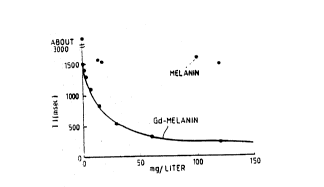
Figure 6 shows the effectiveness of a Gd-melanin
agent of melanin in the absence of gadolinium as having
less effect on the Tl relaxation time at high
concentration for Tl relaxation time alteration.
Figure 7 illustrates the effect of a particular
preparation (Figure 7A: synthesis 10; Figure 7B:
synthesis 12; Figure 7C: synthesis 14) of a melanin
gadolinium combination upon T~ relaxation times.
. .
.
~ ,
.... , ...... : . ~ : . :.
. .
:. ~ : . . :. -
WO92/18166 PCT/US92/0317~
, . , .. , ,-:
~ -30-
2108362
Figure 8 illustrates the effect upon T, relaxation
rate versus concentration of superparamagnetic iron-
melanin.
Figures 9-12 illustrate the effect of (L-DOPA)
melanin-Gd upon Tl relaxation rate versus concentration of
gadolinium for four different molecular weight ranges.
The lower line shown in all plots is the relaxivity of
gadolinium chloride in water as a reference.
Figure 13 illustrates the effects upon the T1
relaxation rate of melanin-Gd prepared from 3-amino-L-
tyrosine.
Figure 14 illustrates the effects upon the T2
relaxation rate of a melanin-superparamagnetic iron agent
produced by L-DOPA polymerization with
azobisisobutyronitrile.
Figure 15 illustrates the effect of an orally
administered (L-DOPA) melanin-gadolinium contrast
enhancing agent. Figures 15 A and B are coronal views of
a rat at time 0 and after a bolus of melanin agent,
respectively. Figures 14 C and D are of the same rat and
time points but different anatomical slice.
Figure 16 illustrates the effect of a (L-DOPA)
melanin-gadolinium contrast enhancing agent on the rat
bowel. Figure 16A is a coronal image taken 24 hours
after the rat was allowed to drink ad libitum of the
melanin solution. Figures 16B, 16C, and D represent 48,
78 , and 120 hours after ingestion to show clearance.
Figure 17 illustrates the effect of a 2 ml injection
of (L-DOPA) melanin-gadolinium contrast enhancing agent
i~to the rat tail vein. Figure 17A represents a coronal
' ;' . '
: .
., . ~.: .
., ; - ,.
:: .:: - . :: ; .
. . . . : : . . ~
.:. :. , - .
WO92/18166 PCT/US9~03177
-31- 2 ~ ~ ~3~j,?
- .
image taken 30 minutes post injection. Figure 17B is the
same animal 6 days after injection, again showing
clearance from the system. Figure 17C is a positive
photocopy of Figure 178.
Figures 18 A,B,C, and D illustrate the effect of a
typical dose (o.lmmole/Kg body weight) of MAGNAVIST given
IV to a rabbit. Figure 18A shows the region of rabbit
kidneys before administration of contrast. Figures
18B,C, and D are at times 2, 8, ard 13 minutes
- respectively post injection of contrast agent (read from
left to right top then bottom).
Figures 19 A,B,C, and D illustrate the effect of a
dose of gadolinium-melanin polymer (MW=1,0000 daltons) of
0.033 mmole/Kg body weight at the same times shown in
- Figure 18.
Figures 20 A,B,C, and D illustrate the e~fect of a
dose of gadolinium-melanin polymer (MW~50,000 daltons) o~
0.002~mole/Kg body weight at the same times shown in
Figures 18 and 19.
Figures 21 A,B,C, and D show a comparison of
MAGNAVIST- and a I,000 MW L-DOPA melanin-gadolinium
polymer. Figures 21A and 21B (top le~t and right) are
Magnavist at 0 and 3 minutes respectively. Figures 2lC.
and 2lD are the melanin contrast enhanced images at the
same times.
Figures 22A,B,C and D show a comparison of MAGNAVIST
and a 50,000 MW L-DOPA melanin-gadolinium polymer. Time
points are the same as in Figure 21.
,
.
. .. . .. .... . .
,
. : : : -.: . : ~, : ~ .::: .
-:- ~ , - .~ : . - .
..
, . ,. ~,
.. . -
W092/181~ PCT/US92/03177
-32-
21~83~2
Natural melanins, in general, are calcium-magnesium
salts that are amorphous, hydroscopic particulates and
extremely insoluble in aqueous solution. Only by
chemical degradation can these large, high molecular
weight particles be rendered partially soluble.
Treatment with solubilizing agents, such as meglumine, do
not impart sufficient solubility to these natural
melanins for them to be used as imaging agents. The
effect of natural melanins on T~ relaxation times in
melanomas was originally thought to be due to the free
radical content of the natural melanins. However,
Enochs, et al. (WS Enochs, WB Hyslop, HF Bennett, RD
Brown, SH Koenig, and HM Swartz, "Sources of the
Increased Longitudinal Relaxation Rates Observed in
Melanotic Melanoma, An In Vitro Study of Synthetic
Melanins," Investigative Radiology, 24, 794-804, 1989)
have concluded, based on their study, that the free
radicals in melanin are not responsible for the
re}axation rates found in melanomas. All the melanins
which they studied were particulate aggregates that were
examined in suspension rather than in solution. It has
been recently shown that solubilization of a melanin-
manganese system enhances the observed T~'s (S Aime, M
Fasano, E Terrono, C Saryanini, and E Mentasti, "An NMR
Study of the Interaction between Melanin Free Acid and
MN2+ Ions as a Model to Mimic the Enhanced Proton
Relaxation Rates in Melanotic Melanoma," Magnetic
Resonance Ima~ing, 9, 963-968, l99l). All of the melanin
contrast agents of the present invention at every
3 0 molecular weight range are soluble in aqueous solution,
contrary to the previous production of either synthetic
or natural melanins. In order to obtain contrast
enhanced images in a rabbit model (Gd-melanin; 50,000 MW)
a dose equivalent to l mg/ml in the total blood volume
3 5 was administered. This required a formulation dose of
l9.6 mg/ml of the agent administered as a bolus of one
. : --
WO92/18166 PCT/US92/03177
_33_ 2~3~2
ml. Natural melanins and synthetic melanins of
comparable molecular weight are particulate and insoluble
at these concentrations. Based upon the initial results
of the present invention a solution containing 1 mg/ml of
a melanin contrast agent appears to be required; however,
as relaxivity of the melanin agent is improved by design
(molecular weight, freé radical and/or metal content)
this quantity may be less than 1 mg/ml. Since natural
melanins tend to aggregate into larger particles, it is
unlikely that, without chemical modification, they can be
successfully employed at concentrations high enough to
avoid problems with the substantial changes in osmotic
pressure that would result from high volume doses. In
general, the term "water-soluble" as used herein
indicates at least 0.1 mg per ml.
Typical natural melanins have been shown to possess
an electron paramagnetic resonance (EPR) signal
characteristic of a material that contains unpaired
electrons (free radicals). The free radical content of
the natural melanins is very stable; howev-r, it is
affected by both oxidizing and reducing chemical agents
~hat alter the number of spins (free radicals) in the
material. the spin density on a melanin can be
quantitated by reference to a standard normally employed
a-diphenyl-B-picnyl-hydrazyl-DPPH). The melanins
possess a ¢haracteristic g value (Table 1) which is
dependent upon the environment in which the free radical
finds itself. In general the g-values of typical
melanins vary with the type of melanin, the degree of
conjugation in the melanin polymer, and whether a
paramagnetic metal is present (RA Nicolaus, MELANINs,
Hermann Publishers, Paris, 1968; L Chauffer, JJ Windle, M
Friedman, "Electron Spin Resonance Study of Melanin
Treated with Reducing Agents, n Biophysical Journal, 15,
565-571, 1975; PA Baldry, GA Swan, "Studies Related to
, ....... , . , . ~ . ... .. .. -
:. , . , ~ - ~.:
. .,.: . :: : .
, .,, ... . .. , ~ . : ,.
,., , ~ . :
,; : ~: ... . .
WO92/181~ PCT/US92/03177
, ,; ~;
-34-
2~ 2
the Chemistry of Melanins. Part 15. The Electron
Transfer and Free Radical Properties of DOPA-Melanin,"
Journal Chemlcal Society Portion II, 1346-1350, 1977).
~ypical spin densities for L-DOPA and/or other
natural melanins are approximately one free radical per
100-250 monomers (-1~) to one free radical per 1000
monomers (-0.1%). These melanin polymers are all
insoluble, high molecular weight particles. The EPR
signal is broad and lacks hyperfine splitting. This
indicates that the electrons may be localized in only one
or two aromatic rings of the melanin. The addition of
paramagnetic metals to a melanin cause various affects
including appearance of hyperfine splitting and a
reduction of the EPR signal intensity. The reduction in
apparent concentration of free radicals has been shown to
be due to a magnetic dipolar interaction between the
metal and a free radical (T Sarna, JS Hyde, HM Swartz,
"Ion-Exchange in Melanin: An Electron Spin Resonance
Study with Lanthanide Probes," Sc1ence, 192, 1132-1134,
1976). Because of the short spin-lattice relaxation
times of metals such as gadolinium the spin lattice
relaxation times of nQarby free radicals should also be
shortened; consequently, it is the rapidly fluctuating
magnetic dipole field seen by the free radicals that
provides a powerful spin-lattice relaxation mechanism.
By incorporating gadolinium and other metals into the
interior of the synthetic melanin polymers, contrast
agents with enhanced, synergistic affects between metal
content and free radical content have been produced.
Normal melanins can bind metal to these surfaces;
however, the meal is easily lost by dilution or other
chemical exchanges.
Alteration in the free radical content of melanins
used as contrast agents enhances the effectiveness of the
.. ,., ~ :
~ .,. . . , :
: . . ,; . .
.' ~
WO92/181~ PCT/US92/03177
_35_ 2~83~ ~
agent whether or not metal is present. Table II shows
some data from Chauffer et al. compared to initial data
from a 50,000 MW agent (see Table 4).
TABLE I~
E8R Data for Melanins
l ¦ Norm~lized E~R~
¦ M~lanin Sreat~ent Intensity
l , . ll
1 L-DOPA Autoxidation l0
l0 ¦ L-DOPAAutoxidation/Reduction 30
¦ L-DOPA Enzymatic l.2
~-DOPA No metal/50,000MW 15
L-~OPA Gadolinium/50,000MW 2s
¦ L-DOPAGadolinium/50,000MW oxidized
It is difficult to make quantitative comparisons because
the gadolinium lowers the free radical signal intensity
and we are comparing solid melanin to melanin in
solution; however, it is clear that by chemical treatment
the free radical content can be altered. Furthermore,
melanin without metal has a free radical content that
enhances T1 relaxation and this can be increased by adding
gadolinium in a non-dissociable forum. Also, the
presence of gadolinium without free radicals is not
sufficient for a melanin polymer to exhibit high
relaxivity.
The present disclosure discloses the formulation and
use of new contrast-enhancing agents particularly useful
for magnetic resonance imaging and magnetic resonance
spectroscopy. This new class of image-enhancing agents
relates to melanin alone and combinations of melanin and
signal-inducing metal ions containing various levels of
,. .-,,~... -
.
,, , , - ..
W O 92/18166 PC~r/US92/03177
2108362
free radicals. Paramagnetic melanin may be prepared with
ferromagnetically coupled complexes such as the
tetranuclear sulfido-bridged complex of Cr(III) described
in U.S. patent 4,832,877 (incorporated by reference
herein). This complex described as Cr4(~CO2) (L) 42+, where
R is an alkyl of 1 to 12 carbon atoms and L is a ligand
comprising oxygen, nitrogen, carbon or sulfur. Although
other imaging techniques and corresponding metals may be
likewise applicable, the primary focus of this invention
relates to magnetic resonance imaging and paramagnetic,
superparamagnetic, or ferromagnetic metal inclusions in
melanin as well as with melanins that contain free
radicals. The relaxation rates, T1 and T2 of nuclei
proximate to melanin-paramagnetic metal complexes appear
altered to a greater extent than with the more
tradltional paramagnetic metal-chelator complexes such as
gadolinium-DTPA. This affect is further enhanced by
increasing the free radical content of the melanins with
or without a metal. As utilized herein, the terms "free
radicals", "unpaired electrons" and "unpaired spins" are
generally viewed as eguivalent, each being preferred in
different situations or by different scientific fields.
This invention primarily involves the synthesis,
preparation and use of a class of melanin materials as
nuclear magnetic resonance relaxation agents for
effective contrast enhancement and/or spectral shifts in
magnetic resonance imaging and spectroscopy. Certain of
these new paramagnetic materials show greater than a
1000-fold increase in the NMR relaxation rate of protons
in water over the best readily usable relaxation
affecting agent so far known (gadolinium - DTPA chelate).
This observation has great potential importance in
contrast enhancement for magnetic resonance imaging since
contrast agents are becoming more necessary to
differentiate between iso-intense regions of normal and
... .
. .
:: . . .. .
- : .. ,. .. : . - ` -
..... .. .
.
"~
; - : , , ,
- , .~ ; ~
W092/18166 PCT/US92/03177
-37~ 2 ~ ~ 3 v~'~
diseased tissue. It is also noted herein that
paramagnetic melanin alone (i.e., without metal
inclusion) can have a greater relaxivity effect than the
current field standard, Gd-DTPA complexes.
Furthermore, these melanin contrast agents can be
affixed to monoclonal antibodies or to other target
(site) specific biochemical agents. These enable site-
directed MR imaging agents that allow site-directed MR
imaging and spectroscopy. This goal of site-specific
imaging agents has not before been completely successful
because, for example, antibodies normally function at
nanomolar concentrations and the minimum concentrations
of contrast agents, where effects on relaxation
lS properties can be seen are much higher (millimolar). The
melanin-~ased agents of the present invention either
containing gadolinium or superparamagnetic iron, function
in the micromolar or lower concentration range which is
much closer to the physiologic range of antibody action.
By incorporating other metals, a whole class of
contrast agents with different properties is accessible.
Likewise, the degree of polymerization of the melanin can
be synthetically controlled producing a molecular weight
range of agents that have quite different potential
applications (e.g., blood pool agents, oral contrast
agents, diséase-specific agents, tumor agents).
Alternatively, the effectiveness of the agent can be
adjusted by the amount of gadolinium (or other metal)
that is synthetically incorporated in the agent andtor
the free radical content. Also chemical derivatization
of the melanin-metal compounds could provide a lipophilic
agent for permeation of the blood-brain barrier or
provide other melanin agents with moieties for linking to
alternative types of site-specific materials ~liposomes,
peptides, enzymes, etc.). The choice of alternative
.
~: , .. :.. . .
: ~ , ~ . . . .
, ~, . . .
,......... : : , ;
.. . - -~: . . ~ ::
. . , : ~ - : :
:, . . -
:;
l ' , . ; . : -: ~
WO92/181~ PCT/US92/03177
2 1 0 8^3:6:~ -38-
melanin precursors and/or various reaction conditions
including catalysts, solvents and reaction times may be
manipulated to produce products having a wide variety of
clinical and physical properties for obtaining image-
enhancing agents that have desired biologicaldistribution and other properties to enhance detection or
disease or follow therapeutic treatments.
Finally, the use of these materials to produce in
vivo high resolution NMR chemical shift changes is a
largely unexplored research area. Lanthanide metal
complexes have been employed routinely for chemical shift
alterations in in vitro high resolution NMR for the
purposes of quantitation and identi~ication;
consequently, these new melanin-materials have opened an
entirely new area of in vivo magnetic resonance imaging
and spectroscopic applications.
The primary purpose and use of this invention is to
alter the nuclear magnetic resonance ~NMR) relaxation
rates, Tl and T2, of protons or other nuclei that are
proximate to the melanin material. If the Tl/T2
relaxation rates of the obsorved nuclei are changed
relative to an adjacent unaffected region, then the
magnetic resonance (MR) image or spectra acquired from
that region will show altered contrast or a particular
nuclei in the environment of the agent will exhibit an
altered chemical shift.
By altering the contrast of a specific region, that
area can be visualized, relative to other areas. There
are many tissue structures which have such similar or
iso-intense contrast in MR imaging that easy visual
evaluation or detection of lesions, tumors, or altered
tissue structures is prevented because the contrast-to-
noise ratio is not sufficiently large. By adding a
- - . . . .
:. . ~, ~ . .
.. ~ ,,.~ ~ -
: . ~ . ;.: ..
. - .. i , " . , . "
W092/18166 PCT~US92/03177
~39~ 2~ ~3~.~
melanin agent of the present invention, iso-intense
regions will become substantially altered allowing more
easy detection and identification due to an increased
contrast-to-noise ratio. The effect on the chemical
shift of specific NMR resonances, due to the paramagnetic
effect of the melanin agent, will also allow such r~gions
to be more easily detected, identified, and/or
quantitated when the melanin agent alters the
microenvironment surrounding the nuclei of interest.
An important purpose and use of this invention is in
the construction of specific contrast agents for in vivo
MR imaging applications. Employing the melanin matrix,
different additives (e.g. metals, and/or alterations in
free radical concentrations) can be included to alter
either the Tl, the T2 or both of a desired organ, tissue,
lesion, or structure to enhance its detectability.
Furthermore, the melanin matrix can be chemically
attached through covalent, ionic, hydrophobic or
hydrophilic bonding to monoclonal or polyclonal
antibodies, receptors, liposomes, membraneg, proteins,
enzymes, poly-peptides, and the like. These melanin-
containing materials ~ay be used as site ~target)
specific relaxation agents that will allow visualization
and/or identification of the target structure.
- ~ Consequently, disease states and healthy tissue could be
probed by MRI with enhanced specificity and
detectability.
The fol}owing aspects of this invention, among
others, are novel:
l. Melanin containing gadolinium ions or
superparamagnetic iron particles coupled with stable free
radicals shows the largest effect on NMR relaxation rates
~ that has ever been reported. Micromolar concentrations
:: _
W092/18166 PCT/US92/0317?
; ,.~ . .
-40-
2108362
(or less) of the melanin agent produce similar effects
that present relaxation agents achieve at millimolar
concentrations. Incorporation of other metals may
provide very specific agents designed for unique
applications such as chemical shift alterations using
metals of the lanthanide series. Alternatively other
paramagnetic metal ions may show relaxation effects
superior to the already improved agents containing
gadolinium or superparamagnetic iron when the free
radical or unpaired electron content of the polymer is
adjusted.
2. Melanin is a natural polymer that can be
synthesized from many relatQd materials (e.g. L-DOPA,
dopamine, catechol, tyrosine, other dihydroxy aromatics)
to produce different polymers that may have novel
paramagnetic properties or have different metal inclusion
and binding characteristics, free radical or unpaired
electron contents, different molecular weight ranges,
20 etc. These factors will influence the relaxation
enhancQment parameters; consequQntly~ a melanin agent can
be synthetically tailored for specific purposes by using
selected melanin precursors, selQctQd metal ions,
selected appendant functional groups, and selected
synthetic conditions designed to produce, for example,
different molecular weight agents with different free
radical contents.
3. Because the melanin is a polymeric substance with
multiple functional groups, the molecular weight ranges
of melanin agents could be tailored to produce an agent
of desired size and/or reactivity. Por example, low
molecular weight melanin agents can be synthesized with
lipophilic appendant groups for permeation of the blood-
brain barrier. Likewise, melanins of any desired size
,
-
, , ;;.;:; .. . .
WO92/18166 PCT/US92/03177
-41- 2 ~ 2
may be coupled to desired cell adhesion molecules or
critical portions thereof.
Bifunctional agents such as glutaraldehyde, water-
soluble carbodiimides and the like may be used to couplemelanins directly or through pendant additions to
carriers having desired binding specificity.
Simply changing the net external melanin
configuration by attaching positively charged groups,
negatively charged groups, water-soluble nonionic
moieties such as polyethylene glycol or the like will
alter in vivo distribution in manners which may be
selected for localization or avoidance of selected sites.
Higher molecular weight paramagnetic melanin-based
agents could be used as oral contrast agents or slowly
migratinq agents that allow observation at injected
sites. As the molecular weight increases above l,000
daltons, there appears to be a synergistic effect on
decreasing the relaxation times (Tl and T2) caused by the
free radical (unpaired electron) nature of the melanin
polymer when coupled with the metal (e.~., gadolinium)
being employed. The high degree of radical
delocalization, apparently due to extended conjugation in
the melanin polymers (as exemplified by their strong
W /VIS absorption and black color), is possibly
responsible for the enhanced relaxation caused by
coupling the metal to the melanin by inclusion into the
interior of the polymer. Thus, the hydrated melanin
surface appears to exhibit a higher concentration of
unpaired spins (free radicals) due to the delocalization
from the metal throughout the polymer. Consequently, the
agents are superior relaxation materials because a very
large amount of water is affected. Other mechanisms may
also be operative. The hypothesized mechanisms for the
: . -: . .......... ..
. ~ . : ; ~ .
,. : ~ ~ :.:: . - .
. . -
W O 92/18166 PC~r/US92/03177
.,
~ 8 3 6 2 -42-
effectiveness of the present agents as presented to
enhance comprehension of the present invention but may
not be entirely correct. These are not meant to limit
the deserved scope of the claimed invention.
Although melanins are known to act as ion-exchange
materials and bind metals on their external surface,
these metals are easily removed by exchange or dilution
(or dialysis). In fact both L-DOPA-melanins and L-DOPA
melanin-gadolinium-polymers bind gadolinium and exhibit
decreases in Tl and T2; however, upon dilution the L-DOPA
melanin returns to its intrinsically lower effect (based
on inherent free radical context) on Tl or T2 whereas the
included gadolinium incorporated during the synthetic
protocol returns a high level effect on Tl and T2.
Techniques normally used to dissociate metals from
chelates do not remove the metals included in the
melanin-metal polymers produced according to the present
invention.
4. A large number and type of metals can be
incorporated into melanin, resulting in different
materi~ls that are ~xpected to show equivalent but
somewhat different effects that potentially make them
very useful. For example, incorporation of
superparamagnetic iron creates an agent that affects T2
relaxation extremely strongly (e.g., T2 relaxivity =
32,815 (mmole/l) ~secl) while incorporation of gadolinium
affects Tl relaxation very strongly (Tl relaxivity z 30
(mmole/l)~sec'). Incorporation of europium praseodymium
or the like, very useful and potent NMR chemical shift
effectors, although only showing a moderate Tl relaxivity
effect of 19.8 and 18.6 (mmole/l)-~sec-~, may show enhanced
chemical shift changes in vitro as well as providing the
first case of an in vivo chemical shift agent for MR
spectroscopy.
. . . ~ - -
.
:, . ~ i, ..... , : ,
~ : :
~ ' '' . .
Wo92tl8166 PCT/US92/03t77
_43_ 2~ ~3~2
5. The synthesis of the metal-containing melanin
polymer must be accomplished in the presence of a metal
ion that is to ~e incorporated. Although prepolymerized
melanin can be placed in a solution of a metal ion or
exchanged into the melanin polymer, the exchange is not
very efficient. Contrast a~ents produced in the latter
manner are not as effective as agents polymerized in the
presence of a metal and, upon dilution or reaction with
amines or azide, lose their effect much more rapidly than
metal-containing melanins synthesized in the presence of
a metal, vide lnfra.
The addition of a metal ion to melanin (either by de
~QyQ synthesis or by exchange) often lead~ to a
precipitation of the melanin complex from aqueous
solution. Appropriate treatment with a suitable
solubilizer that is biocompatible, e.g. N-methyl-
glucamine, triethylamine, glutamic acid or the like,
provides a water soluble salt. Other techniques for
enhancing solubilization of recalcitrant melanins include
acidification of the polymer to eliminate salts formed
during the oxidative polymerization. The melanin free
acid, depending upon the monomer used, can be made more
soluble in aqueous solution as well as many other organic
solutions.
6. Chemical derivatization of the melanin-metal complex
is easily accomplished by a variety of well-established
reaction protocols for ionic and/or reactive functiona~
groups. Such derivatization could lead to melanin agents
that are highly lipophilic; are nonionic; are of specific
solubility; are attached to a liposome, m~mhrane,
receptor protein, enzyme, polypeptide, etc.; or are
attached to mono or polyclonal antibodies to be site-
specific contrast agents.
.,. ., -~. , . : ::
,. . ..
. , . ..
' .. :; : - ~:
- ,
WO92/18166 PCT/US92/03177
2108~62
7. Although toxicity and tolerance must be specifically
evaluated, natural melanin is ubiquitous and such a
natural or synthetic agent should be generally well
tolerated. Initial data obtained in both a rodent and a
rabbit model have shown no evidence of acute toxicity.
Although the metal ion content of the melanin-metal
agents can vary, it is anticipated that any metal
employed would not cause acute toxicity because of
protection by inclusion into the interior of the melanin
polymer and the inability of the metal to dissociate from
the melanin. The melanin agent is at least partially, if
not all, excreted in the urine of both the rodent and
rabbit.
Because the melanin agents of the present invention
exhibit such pronounced relaxation effects, they may be
the first agents that can be incorporated into monoclonal
antibodies and successfully show site specific contrast
affects at close to the nanomolar concentrations at which
antibodies normally function. This technique would
provide a protocol that could replace radionuclide
imaging and produce biochemical information that is
simi}ar to what is obtained from PET (position emission
tomography) but would potentially have much higher
resolution.
9. Relaxation parameters (Tl and T2) have been measured
at three different magnetic field strengths for a variety
of melanin-gadolinium agents and they all showed
substantially enhanced Tl and T2 relaxation rates as
compared to melanin synthesized without gadolinium ions
present. The paramagnetic melanin of the present
invention, even without gadolinium still exhibits a
relaxivity that is equivalent to Gd-DTPA (MAGNAVIST-) and
cou}d be used as an imaging agent. The results indicate
that the melanin agents are effective as contrast agents
.
.. - . ~ . . . . .
.
.
W O 92/18166 PC~r/US92/03177
-45_ 2 ~ 3 ~ r~ ~ ~
in the low micromolar concentration range. A melanin
agent containing superparamagnetic iron has been prepared
which has a T1/T2 effect approximately ten fold greater
than the best previous gadolinium agent. As Tables 2-5
S show, a number of metals and melanin polymers show
enhanced T1 relaxivity which is dependent upon both metal
ion content and free radical concentration.
Animal tests have shown good tolerance of
administered melanin-Gd. Preliminary results indicate
the melanin agent is an excellent oral contrast agent
when administered in the animals' water. Intravenous
injection of the agent was also well tolerated (no acute,
observable toxic effects). These preliminary results
suggest that the agent is also effective as a blood pool
contrast aqent. The circulatory system, kidneys, liver,
and bladder showed enhancement of contrast. Injection
into the rabbit tempomandibular joint (TMJ) has also been
accomplished and examined by MRI. Contrast was
substantially enhanced at the injection site; however the
area around the injection became hard with time and
evaluation of these results has not been completed.
Several well recognized synthetic methods can be
employed to prepare melanin agents containing in various
amounts of different metal ions being incorporated,
different molecular weights for the resulting products,
different concentrations of free radicals and different
solubilities of the final products (because of different
melanin precursors used) and other properties. The
following represents a typical preparation of a
gadolinium-containing L-DOPA melanin of approximately
11,000 molecular weight that shows substantially enhanced
relaxivity (Tl/T2).
The typical chemical reaction sequence is:
,
WO92/18166 PCT/US92/03177
~, .
i ; -46-
21 ~8362
1) Ammonium Persulfate/DMAP
or Azobisisobutyronitrile/Base
L-DOPA + GdCl3 Melanin + Gd+3
or other 2) Isolation and purification of
metal desired MW Range
3) Adjustment of free radical content
4) N-methyl-D-glucamine if required
A typical reaction to produce a melanin-Gd polymer
of about 15,000 MW involves mixing 1.25 g L-DOPA, 0.75
dimethyl-aminopyridine (DMAP), 0.25 g GdCl3 and 0.15 g
ammonium persulfate in 150 ml water. The reaction is
stirred for 30 minutes and is then either dialyzed
against a 14,000 MW cutoff dialysis membrane or 10 g N-
methyl-D-glucamine is added and the mixture heated at
50C overnight. After cooling, the solution of melanin-
Gd-meglumine is dialyzed against a 14,000 MW cutoff
(MWCO) dialysis membrane. Alternatively, the melanin
solution can be sized for MW by ultrafiltration or column
chromatography. The resulting aqueous solution can be
directly used or concentrated by evaporation. The
melanin can be reacted with oxidizing (e.g., H202) or
reducing (e.g., NaH,dithionite) agents to alter the free
radical content.
The attached graph (Figure 6) of T1 relaxation rate
versus concentration (g/1000 g H2O) of the melanin-Gd
agent shows the pronounced effect on the proton Tl of the
melanin-Gd solution. Note that melanin without
gadolinium also has a significant effect on T1, but for
this preparation there is not a concentration effect.
This melanin-Gd material can be further fractionated
according to molecular weight. The initial dialysis
solution can be evaporated to yield a low molecular
weight melanin-Gd (<14,000 MW). Using appropriate
ultrafiltration membranes the melanins can be sized to
any MW range. Similar reaction conditions can be used to
... .:: . ., , . ., . ;.; , . ~ .,
: ,. - ,- ... , ~ .. .; . ... .. .
. . .
w092/l8l66 PCT/US92/03177
~47~ 2 ~ 2
produce melanin with superparamagnetic iron or other
metals. By varying the catalyst, reaction time and
temperature, different molecular weight melanins are
produced. Furthermore, it is possible to take preformed
melanin (either synthetic or natural) and exchange
gadolinium or iron into the polymer. These materials;
however, show a smaller effect on the Tl/T2 relaxation
times, and this effect, insofar as it depends on the
presence of a signal-inducing metal, is lost more quickly
upon dilution or reaction with amines or azides. For
example, Figures 9-12 show the effect on Tl of several
different MW weight fractions MW ca. 1,000 to 50,000 of
an L-DOPA-melanin-gadolinium agent. Figure 13 can be
used to compare the effect of a different melanin
precursor (3-amino-tyrosine) at the same molecular weight
(ca. 50,000) to the melanin agent (Figure 12) produced
from the L-DOPA precursor. The effectiveness of all
these agents is compared to that of the highly toxic
gadolinium chloride (Figures 9-13) which shows
approximately a 3.5 fold decrease in T1 (17.5 (mmole/l)
Isec'). This compared favorably to the commercially
available DTPA-Gd (4.5 (mmole/l)-'sec-l).
The melanin-Gd product can be prepared with or
without the N-methyl-D-glucamine. Melanin agents with
good aqueous solubility are the best MR imaging contrast
agents and they can be easily derivatized or coupled to
an antibody or other carrier using carbodiimide or
alternative coupling technologies.
MRI provides a substantial range of tissue contrast
without enhancement; however, increasing numbers of
situations have demonstrated that contrast agents can
provide the physician with improved diagnostic
capabilities. The present invention provides a new class
of clinically useful contrast agents that exhibit
.
WO92~18166 PCT/US92/03177
~ 3 ~ 2 -48-
unusually strong influences on proton relaxation rates at
concentrations substantially lower than metal chelates
and polymeric agents currently available. Accordingly,
the present inventive agents serve to decrease the
effective agent concentration needed, have lowered
toxicity and make i~ more feasible to prepare tissue
and/or organ specific agents by linking contrast agents
to monoclonal antibodies or specific receptors.
The synthesis of a class of melanin polymers, which
are stable free radicals and either contain various added
paramagnetic metals or no added metals, is described
herein. Control of the synthesis provides a range of
agents with defined properties such as molecular weights
that show different proton relaxation properties. For
example, a high molecular weight (MW=50,000) gadolinium-
melanin polymer shows a T~ relaxivity of 2.45 x 10³
[mmole/l]~ sec' (n = 3) compared to Gd-DTPA (MAGNAVlST-)
of 4.5 [mmole/l]~~ sec'. This more than 500 fold effect is
dependent upon molecular weight (MW) of the melanin
polymer and the density of free radicals in the polymer.
For example, a gadolinium-melanin of intermediate MW
(14,000 daltons) has a T1 relaxivity of 9.5 x lo2
[mmole/l]~ sec~ whereas a low MW (8,000 daltons) agent is
26.6 [mmole/l]-l sec'. The choice of monomeric melanin
precursors and various metals for synthesis of the
melanin agents yield materials that vary in proton
relaxation effects. Superparamagnetic iron has also been
incorporated into a melanin polymer and exhibits
extremely fast Tl and T2 relaxation rates (see Figures 8
and 14).
Image data is presented herein to show the efficacy
of these new agents as both an oral contrast agent and a
blood pool agent in a murine model (Figures 15-17). An
image comparison of MAGNAVIST to both a l,000 and S0,000
-. . ~; .- , . .
,~` ~ , .. .. .
W O 92/18166 PC~r~US92/03t77
~49~ 210~362
MW melanin-gadolinium agent administered IV into a rabbit
ear vein shows the enhanced effectiveness of the melanin
agents over MAGNAVIST (Figures 19--23). In addition Gd-
melanin polymers that exhibit high Tl relaxivity, when
coupled to a monoclonal antibody raised against the
hormone, pancreatic polypeptide retain paramagnetic
activity. Gd-melanin has also been attached to IgG and
bovine serum albumin with retention of relaxivity. In a
rabbit model 'I~In incorporated into a melanin polymer has
successfully imaged the pancreas; however, the
selectivity is low and a high background is observed.
Melanins are macromolecules consisting of mixtures
o~ polymers that have a high degree o~ conjugation. This
conjugation produces and/or stabilizes many of the
properties that melanin possesses including its W /Vis
light absorption and its stable free radical properties.
Melanin, as a stable free radical, causes a relaxation
affect (acts as a contrast agent) by itself; however,
incorporating a paramagnetic metal or complex into the
melanin polymer enhances signi icantly it~ ability to
affect contrast in MRI. Natural in vivo melanins may
have some endogenous mQtals included but they do not
cause a relaxation affect similar to the melanin-
incorporated paramagnetic metal ions of the presentinventive agent~ The most likely biologic metals that
could be present are iron and manganese and these metals
included in a melanin polymer do enhance relaxivity but
not as greatly as several other metals and gadolinium
(see Tables 2-5). Furthermore, the biologic melanins are
generally produced has high MM particles (granules) that
are insoluble in aqueous solution. This lack of aqueous
solubility drastically decreases effective relaxivity.
The high toxicity of gadolinium makes it extremely
unlikely that it is available for incorporation into a
natural melanin produced in vivo.
.
.. .: . - . :
: - ., . ; ::-: : , :.
WO92/18166 PCTtUS9Z/03177
21~83~2
As has been known for a very long time, melanins may
combine with metal ions. Preferred synthetic and natural
melanins will bind metals such as gadolinium (or the
other lanthanides) and produce an agent that
substantially reduces the relaxation (T~ and T2) of a
solution. However, upon dilution (or dialysis) or
chemical treatment to remove the metal, this affect is
very rapidly lost due to dissociation of the metal ion
from the melanin. This is a reversible process and the
initial relaxivity of the preferred melanin may be
recovered.
The melanin-signal-inducing metal materials of the
present invention do not release significant gadolinium
(or other metals) upon dilution, upon extensive dialysis
or even upon treatment with azides and amines. This
leads to a consequent effectiveness at low concentrations
and great utility as image-enhancing agents.
Precursors for synthetic melanin include known
published precursors ~uch as catechol, L-DOPA (or DL-DOPA
or D-DOPA), dopamine, hydroxydopamine, tyrosine,
aminotyrosinej dihydroxytyrosines or phenylalanines, 1,8-
dihydroxynaphthylene, for example. L-DOPA, dopamine, and
aminotyrosine have primarily been used herein as melanin
precursors but certainly do not limit the choice of
melanin precursors. The polymerizations of the present
invention were preferably conducted in an aqueous solvent
or in aqueous solvent with an alcoholic comodifier;
however, other solvents including alcohols, acetonitrile,
tetrahydrofuran and the like may be used to tailor the
desired contrast agent polymer as to structure, molecular
weight, free radical content, metal content and final
solubility.
.
, .. - .
WO92/18166 PCT/US92/03t77
-51- 2~ ~u~ ~3~
Polymerization catalysts for such polymerizations of
these or analogous materials include hydrogen peroxide,
persulfates, peracids or peroxides, oxygen, sodium
nitrite and the like, i.e., essentially any strong
oxidizing or free radical generating agent. In addition
to these catalysts, azobisisobutyronitrile (a free
radical polymerization agent) has been used and found to
effectively produce a wide molecular range of polymers
with improved solubilities and adjustable free radical or
unpaired electron concentrations. Other such free
radical agents may also be used as catalysts or reaction
initiators but have not yet been defined in detail.
Reducing agents such as dithionite and the like may
also be used to induce melanin formation. To obtain
optimal free radical content, the amount of oxidizi~g,
reducing or free radical-producing melanin polymerizing
agent should be optimized for each set of conditions,
e.g., for particular melanin precursors used, desired
melanin size, etc. Photoinduction of free radicals by
agents such as riboflavin and the lik~ may be used to
initiate melanin formation, although the light absorption
of melanin and its prQcursors may limit their
effectiveness.
Melanin may also be prepared by enzymatic procedures
using a tyrosinase enzyme. While such enzymatic
catalyses have not yet been performed in the presence of
typical concentrations of paramagnetic metal ions used
herein, it is anticipated that the requisite metals will
inhibit tyrosinase activity and thus impede the enzymatic
catalysis. Metal ions, of course, must be present during
melanin formation to result in secure incorporation;
consequently, it is expected that a further research
effort will be required to determine proper conditions
allowing enzymatic synthesis of melanin-signal-inducing
. ..
., .: ~, .
:
,: ~
:
W092/18166 PCT/US92/03177
-
2 1b83 62 -52-
metal agents, and if it is practical to prepare them in
this manner. Typical melanins prepared enzymatically
without metals normally do not show significant
relaxivity (e.~. less than MAGNAVIST). However, by using
the appropriate oxidizing and/or reducing conditions, the
free radical content can be raised making them more
effective contrast agents.
Control of the free radical content of the preferred
synthetic melanins employ standard procedures for
oxidizing and/or reducing the melanin (+ metal) polymer
by adding or removing electrons. This is accomplished
electrochemically or by using chemical oxidants such as
peroxides (H202, etc.), ascorbate or other such oxidants
and chemical reducing agents such as sodium dithionate,
sodium hydride and the like. The quantity of spins (free
radicals) in the resultant polymer can be characterized
by electron paramagnetic resonance spectroscopy.
One solubilization method for the melanin agents
described herein, when need~d, may be outlined as
follows. After molecular weight fractionation separates
the melanins into various molecular weight ranges, it has
been observed that some of the higher molecular weight
materials (>lOO,OOOMW) tend to be insoluble in every
solvent tested thus far (this is dependent upon
polymerization conditions and precursors). A general
procedure has been developed which produces a stable
paramagnetic melanin or paramagnetic melanin-metal amine
salt. For example an N-methylglucamine (meglumine) salt
of the melanin will allow high molecular weight
gadolinium-melanin to be easily brought into solution.
This observation is completely general and significant.
If one has an acidified melanin, an amine salt may be
made that will allow production of a final product with
solubility characteristics adapted to particular
, . . ............ . . .
, : . -; .; ~: ,
. , . . - : - -
- ~. . -.: - .
W O 92/18166 PC~r/US92/03177
i~ -
~53- 21~3~2
conditions such as full aqueous solubility or solubility
in more lipid-like environments. For example, choosing a
hydrophobic amine should produce a melanin that is
soluble in non-polar materials. This technique has been
used extensively for enhancing drug delivery. Control ~f
melanin synthesis ensures full aqueous solubility for the
L-DOPA melanins; however, other precursors may show
poorer aqueous solubilities and require this type of
solubilization.
The formation of an amine salt of the melanin
produces a soluble material that may exhibit poor
characteristics upon administration into a patient due to
the high osmolality of the melanin-salt. This was a
perceived problem with CT contrast agents which lead to
the development of non-ionic CT contrast agents. Similar
concerns for the melanin MRI agents are eliminated by
derivatization of the L-DOPA melanin. Melanins produced
from other precursors can be similarly derivatized.
Derivatization of the carboxyl groups in L-DOPA through
ester or amide formation is straightforward and well
established. Choice of the ester or amide groups added
to the melanin will enhancQ aqueous solubility or
increase lipophilicity, or serve as a linkage area for
attachmént to an antibody or receptor.
A large number of metals have been employed in the
syntheses of the present MRI agents and found to produce
a melanin metal agent that affects the relaxation in a
positive way for contrast alteration. Some are better
than others but all are better than melanin alone. The
list of utilized metals includes gadolinium; manganese;
superparamagnetic iron; iron; praseodymium; ytterbium;
dysprosium; and europium. That the particular metal is
important is demonstrated by the fact that magnesium or
zinc can be substituted and the resulting melanin-metal
, . , , , . ~
- .: ,.
- .. : .
.. . - ~
.- ,
. : , . ,
.
wos2/18lh6 PCT~US92~03177
21~8362; ~54~
.... ;
product does not show increased relaxation as compared to
melanin without a metal. Magnesium is not paramagnetic
and should not influence relaxation rates. In none of
these melanin-metal combinations prepared according to
the present inventive procedure has the metal been
removable by dialysis or treatment with azide ~ollowed by
dialysis. The metal is an integral internal component of
the melanin polymer.
Using other precursors, such as, e.g., dopamine or
aminotyrosine, for the melanin synthesis provides for
ready synthetic manipulation of these agents. For
example, such agents may be more readily coupled to
antibodies, receptors or other substances than L-DOPA-
based materials. This does not mean that they are
necessarily better materials, only that they may be more
versatile in certain contexts.
Control of melanin-metal molecular weight may be
achieved by choice of polymerization catalyst and by
reaction conditions and time of polymerization. While
certain differences in relaxation rates of low molecular
materials compared to high molecular weight materials
have been found, the total amount of gadolinium or other
paramagnetic metal is sometimes an important factor.
However, the extent of incorporation of the metal into
the conjugated polymer matrix is also important. The
ability to obtain a series of molecular weight materials
of different solubilities allows design of an agent
specifically for a type of application. General blood
pool agents should be of high molecular weight to prevent
their entrance into certain capillary structures; lower
molecular weight materials could be made permeable to the
blood-brain barrier, for example. The procedures for
separation of a crude synthetic melanin (+ metal) into
various molecular weights can be readily accomplished by
; . . - . .. . .. ~- ,, :-: . , .
WO92/18166 PCT/US92/03177
.
2 ~ 2
several standard techniques which can be optimized for
each desired melanin polymer. Dialysis of the crude
reaction solution can quickly separate the reaction into
broad molecular weight ranges by a choice of dialysis
membranes. An analogous technique using a series of
cutoff membranes is ultrafiltration which can produce
relatively narrow molecular weight ranges. Further
separation into narrower MW ranges is accomplished by gel
permeation chromatography (gravity or HPLC).
Attachment of melanin-metal polymer to an antibody
has been successful; however, the pilot antibodies chosen
do not facilitate imaging studies since they were not
specific for a biological site. Nevertheless, these
preliminary studies established the feasibility of the
process - to produce.a melanin-metal polymer coupled to
an antibody and which has retained both a strong
relaxation effect as well as antibody activity.
Experiments are currently in progress with a coupled
antibody to a specific disease process to more clearly
illustrate the efficacy of the antibody-melanin agents.
Preliminary imaging experiments have shown that
gadolinium-melanin is effective as both an oral contrast
agent and a blood pool agent in animal models.
Experiments are currently underway to further delineate
the abilities of all the melanin-metal agents thus far
prepared to act as in vivo contrast agents. Placement of
these agents in specific organs and tissues to evaluate
organ imaging effects (e.g. heart imaging; liver imaging,
etc. ) are foreseen.
Several of the lanthanides have been used for many
years as chemical shift agents in high resolution NMR
spectroscopy (dysprosium, europium, etc. ) . The effect on
imaging is not the only effect these agents may have;
W O 92/18166 PC~r/US92/03177
21Q8362 -56-
they may also affect chemical shift position in vivo.
This has the potential to open a new area for in vivo
diagnostic application. Melanin-metal combinations may
be particularly useful as chemical shift agents.
Table 2 compares relaxivity of various prior and the
present MRI agents.
TABL~ 2
AGENT R~LAS~VITY t~mol/l]lsac~
_., ._ _ _ _ _ . _ _ _
Gadolinium - DTPA (Magnavist) 4.5
Gadolinium - DOTA 3.8
(in liver) 6.7
Manganese - DPDP 2.8
(in liver) 21.7
Gadolinium - Melanin (L-DOPA)
MW = 50,000 2450.0
Melanin (L-DOPA)
MW + 50,000 4.8
Table 2 illustrates the relative relaxivity induced
by various gadolinium chelates as compared to gadolinium-
melanin and illustrates the superiority of the latter.
It further shows that a paramagnetic melanin without
metal can exhibit a relaxivity equivalent to Gd-DTPA
(MAGNAVIST). This is due to the free radical content of
this melanin.
Melanin was synthesized from L-DOPA, 3-amino L-
tyrosine, dopamine, and catechol incorporating various
paramagnetic metal ions and varying temperature,
W O 92/18~66 PC~r/US92/03177
~57~ 2~33~2
catalyst, and reaction time in an attempt to determine
the most effective metal ion-melanin polymer for use as a
contrast agent in magnetic resonance imaging. It was
found that L-DOPA melanin-gadolinium synthesized with
S azobisisobutyronitrile (ABN) as catalyst produced a
soluble, effective contrast agent with a prominent effect
on T~ relaxation times at concentrations less than 1
mg/ml. Gadolinium-labeled L-DOPA melanin synthesized
with ammonium persulfate as catalyst were more effective
at lowering T~ relaxation times, but that effect was
greatly diminished in the effort to optimize solubility.
Metals such as europium, praseodymium, and ytterbium also
had an effect on relaxation time; however, that effect
was not as notable as that produced by gadolinium.
Superparamagnetic iron oxide had a dramatic effect on
both ~r, and T2 relaxation times; however, for use as a
contrast agent a distinct ratio between Tl and T2 effect
is necessary unless MRI equipment can take advantage of
extremely short T2 times (typical clinical imaging
equipment cannot yet routinely employ fast scan, fast
imaging techniques, e.g., echoplanar imaging.)
The following example~ are presentQd to illustrate
embodiments of the present invention and are not intended
to limit the claimed scope of this invention unless
specifically so stated in the appended claims.
... ,.. ~ .
, . . . . .
; .. , .- , : ~ ., . ~
" . . , .. ,. : .. . ..
.. . . . ..... .. . .. .
wos2~t8l66 PCT/US92~03177
.. ..
~ 58-
2~08362
BSAMPL~ 1
8YNT~TIC, ~Rys~A~I~aTIoN AND SOL~BILIZATION
PROTOCOL8 UTILIZBD HBREIN
PROTOCOL A: Synthesis of Gd-labeled melanin from L-DOPA
¦Reagent: Amount: ¦Molecular Moles:
¦ L - DOPA 1. 2s g 197 .0063s
¦GdCl3 0.25 g 371 .000625
10 ¦DMAP 0.75 g 122 .00635
ammonium persulfate 0.15 g 228 000625
Procedure:
1. Dissolve DMAP (dimethylaminopyridine) in 100 ml
distilled water
2. Add L-DOPA (L-dihydroxyphenylalanine), add
additional water if necessary to dissolve
3. Place in flask (2x volume minimum)
4. Add GdCl3 and dissolve
5. Add ammonium persulfate (catalyst)
6. React for various times to achieve desired
molecular weights and yield
7. Size melanin according to molecular weight.
8. Measure/adjust free radical content.
:
-.. : ~ . ; . . ;. .
. . .. ; .. . : . ~.
W O 92/18166 PC~r/US92/03177
;' '
-59- 2~ 0~3~
,
PROTOCOL B: Synthesis of melanin from DOPA
¦Reagents Amount: Molecular Moles:
Weight: _
L-DOPA (or DL-DOPA) 6.25 g 197 0.0317
DMAP 3.75 g 122 0.0307
absent 2.49 g 371 0.0067
Azobisiso- 0.50 g 164 0.00305
butyronitrile
NaOH 0.05m, 20%=
MQOH 600ml
Procedure:
1. 600 mls of 0.05M NaOH, 209~ methanol ~pH 12. 6)
is placed in a reaction ~lask and heated to
70C with stirring.
2. DOPA is added and allowed to completely
; dissolve.
3. DMAP and ABIN are added and allowed to
completely dissolve.
4. GdCl3 is added to reaction vessel
5. React for various times to control molecular
` ~ weight and yield
6. Size melanin according to molecular weight
7. Measure/adjust free radical content.
PROTOCOL C: Alternative synthesis of melanin from DOPA
As PROTOCOL B except NaOH (0.05M) and 20~ methanol
(pN-12.6) are not included in the reaction mixture.
~:
.
: . .: . :, . : .......... . . . . . .
. : - . , . ~ .. . ... .. .
WO92~18166 PCT/USg2/03177
~ -60-
21'083'6~' `'
PROTOCOL D: Synthesis of melanin from 3-amino L-tyrosine
(also other precursors)
Reagents: Amount: MM: Moles:
¦3-amino L-tyrosine1.81 g 289 .0062S
DMAP - O.75 g 122 .00635
GdCl3 0.25 g 371 .000625
¦ammonium persulfate 0.15 g 228 000625 (10%)
Procedure
Same as for Protocol A. Other precursors (e.g.
dopamine, catechol, ~tc.) follow this general protocol.
PRO$0COL E: Solubilization of melanin
¦Reag-nt Amount
l I
lS Synthesized melanin 100-150 ml
¦N-methyl D-glucamine 5 g
Procedure:
1. Add 5 g NMEG (N-methyl D-glucamine) to melanin
2. Heat in round-bottom ilask at 50C overnight
: ~ .
PROTOCOL F: Recrystallization of catechol
Dissolve catechol into heated toluene and allow to
2S cool, recrystallizing catechol. Filter catechol from
toluene solution.
PROTOCOL G: Derivatization of melanin
In order to produce a melanin that has altered
lipophilic properties or can be more easily attached to
an antibody or receptor; advantage can be taken of the
functiona} groups on the melanin to derivatize the
WO92/18166 PCT/USg2/03177
, , .:
. ,
21~62
paramagnetic melanin without altering its relaxivity
properties. This is a typical protocol but a wide
variety of coupling agents (e.g., carbodiimides and the
like) and linkers can be used to couple through the
phenolic hydroxyls, the carboxylic acid or amine
f~nctional groups, depending upon the melanin type.
~ . .... _ . . ..... I
Reagant Amount:
Melanin-Gd (SOK daltons) ll.6mg
triethylamine excess
isobutylchloroformate 40mmoles
Procedure:
l. Form the triethylammonium salt by heating at
65C overnight in dimethylformamide (DMF)
2. Evaporate and redissolve in 5 ml DMF.
3. Cool to 0C while protecting from moisture.
4. Add isobutylchloroformate (IBCF) and let
solution stir at 0C for two hours.
5. Add the IBCF-actuated melanin dropwise (by
syringe) to l ml, cold (4C) ethylene diamine
over two minutes.
6. Purify melanin by ultrafiltration to remove low
molecular weight byproducts:
PROTOCOL H: Ultrafiltration scheme for melanin sizing
and purification
Amicon Ultrafiltration Scheme for Nelanin Sizing and
Purification
Melanin reaction is precipitated with HCl, and the
filtrate decanted. The melanin is then washed with
methanol (or methanol:H2O) twice, and the filtrate
: ~ :
-
:
. . .. , ~ ., . - ,
`'~'' . '
WO92/181G6 PCT/US92/03177
21083~2 -62-
decanted. Transfer melanin to Amicon Stir Cell with
Buffered H2O (Tris, PBS, Carbonate . . .), pH 9. The
following is a typical scheme.
Melanin vs. 100,000 MWCO (YM 100) filter
/ \co
\L ~
diluted >100,000 MW (Saved)
Melanin < 100,000 MWCO vs. 50,000 MWCO (YM 50) filter
cO
~ ~n~
diluted >50,000 MW (Saved)
Melanin < 50,000 MWCO vs. 10,000 MW (YM 10) filter
cO
~ \~JnC
diluted >10,000 MW (Saved)
Melanin < 10,000 MWCO vs. 1,000 (YM 1) filter
cO
C
diluted >l,OOO MW (Saved)
Melanin < 1,000 MWCO
Other filter sizes are used to obtain the desired
molecular weight ranges.
~A~PL~ 2
Synthesis 1, 2, 3, 4
Melanin was synthesized from catechol in an attempt to
optimize conditions for highest yield. Catechol was first
purified by recrystallization (See Protocol C). Benzoyl
peroxide (7g) to be used as a catalyst was added to 175 ml
stirred acetonitrile preheated to 55C. Recrystallized
catechol (5g) was added to the above solution.
~riethylamine 2 ml in 25 ml acetonitrile was then added.
. . - .
. . .
",. .. :
... . .
W O 92/18166 PC~r/US92/03177
-63- 21~362
The solution was alIowed to react overnight. Following the
reaction, the solution (now a dark brown color) was
rotoevaporated to remove the acetonitrile. The remaining
product was precipitated with ether and, the supernatant
decanted, and the residue allowed to dry. The precipitant
was dissolved in methanol and re-precipitated with ether:
the ether was again decanted and the precipitant was dried
(by vacuum) and saved. The ether insoluble material of
both precipitations was dissolved in methanol and saved.
This procedure yielded 0.51 g precipitate. Synthesis 2 was
a repeat of this procedure and yielded 0.441 g precipitate.
Synthesis 3 was modified by using 20 ml triethylamine
rather than 2 ml. As a result of this modification, the
sample was difficult to precipitate. S-nce the
triethylamine was not all removed with the acetonitrile in
rotoevaporation, the ~xcess amine remained in the sample.
It was necessary to wash the non-ether soluble material
(dissolved in methanol) prior to precipitation with 0.12 M
HCl solution, which fully precipitated the melanin while
leaving the triethylamine in solution. The mixture was
then filtered and the filtrate was wa~hed repeatedly with
water. The filtrate was then allowed to dry. Synthesis 4
was a repeat of synthesis 3 éxcept the reaction was run at
room temperature. The non-ether soluble precipitate was
disso}ved in chloroform rather than methanol prior to being
~ washed with HCl and water. This procedure yielded 1.033 g.
:: .
.~
Synthesis 5
Melanin was synthesized from dopamine with ammonium
persulfate as catalyst. Dopamine ~lg) was dissolved in 100
ml water. Ammonium persulfate (0.1184g) was added to the
dopamine solution. The solution was allowed to react
overnight. The resultant melanin was insoluble in water as
well as methanol and dimethylformamide. Due to this
insolubility nothing further has been done with this
.
.
W092/181~ PCT/US92/03177
,
21~836~ -64-
product. Modification of this synthesis using ABIN as the
catalyst produces a soluble dopamine-melanin-Gd polymer.
Synthesis 6
Melanin was synthesized from dopamine with ammonium
persulfate as catalyst. The primary difference between
this and the previous reaction is the addition of 0.5 ml
benzoyl chloride in 4.5 ml methylene chloride to the
reacting solution. The resultant samp~e was still
insoluble in water and several organic solvents.
~-AMPL~ S
Synthesis 8
Melanin was synthesized from L-DOPA in an attempt to
incorporate gadolinium ions (Gd~') for use in contrast
agent studies. Dimethylaminopyridine (DMAP) (3g) was
dissolved in 300 ml water. L-DOPA (5g) was added to the
solution and stirred until completely dissolved. The L-
DOPA was somewhat difficult to dissolve. GdC13 (lg) was
added to the solution. The GdCl3 did not completely
dissolve and turned the solution a cloudy green color upon
its addition. Ammonium persulfate (0.6) (catalyst) was
added to the solution. A reaction occurred almost
immediately after the catalyst was added and was allowed to
run for 2 weeks. The reacted solution was then dialyzed
against water (4L) with 6-8X molecular weight cutoff (MMCO)
Spectrapor dialysis tubing for 1 day to remove excess
catalyst, DMAP, and unreacted reagents. The dry weight of
10 ml of the solution indicated a concentration of 7.7
mg/ml. Tl relaxation time of a 1:100 dilution of the
sample was 488 msec, although the solution was not 100%
soluble and the solid matter might have introduced error
into the results.
- . . - , - . .
.
,
W O 92/t8166 P~r/US92~03177
-65- 2 ~ ~ 3~ 2
~SAMPL~ C
Synthesis 10
Melanin was synthesized from L-DOPA while
incorporating gadolinium ions under varying reaction
conditions. The basic procedure noted in Synthesis 8 was
used, with reagents in quantities as noted above. The
reaction was allowed to run 1 day. The solution was first
dialyzed agains$ water (4L) with 6-8K MWCO dialysis tubing.
This fraction was then dialyzed against water with a 50,000
MWCO dialysis tubing. This and other Gd-melanin solutions,
even if soluble immediately following reaction, tended to
become more insoluble with time. While Synthesis 8
material was already partially insoluble immediately after
the reaction, the material from the pre~ent synthesis was
at first soluble, probably due to decreased reaction time.
Dry weight of a 10 ml solution sample indicated a
concentration of 3.55 mg/ml. The 50,000 MW sample was
particularly effective at high concentrations for T1
relaxation time (see Figure 7A). Relaxivity of this sample
is 2,470 [mmole/l]~secl.
Synthesis ll
Melanin was synthesized from L-DOPA as a control for
Synthesis 10. The conditions were identical to those in
Synthesis 10, but this solution contained no gadolinium.
Melanin without gadolinium incorporated tended to be more
soluble than that with metal ions. It also tended to be
lighter in color than Gd-melanin. Dry weight of a 10 ml
sample indicated a concentration of 1.75 mg/ml. Relaxation
measurements of melanin without gadolinium have much less
effect on Tl relaxation times. The relaxivity of the
melanin control without gadolinium is 4.95 [mmole/l]lsec~.
This relaxivity compares favorably to gadolinium chloride
(17.5 [mmole/l]'sec') and to Gd-gadolinium-DTPA
(Magnavist)) (4.5 (mmole/l)isec-~.
.. . .
.. . ...
.
WO92/18~ PCT~US92/03177
~.
-66-
21~83~2
~AMPL~ 8
Synthesis 12
Melanin was synthesized from L-DOPA incorporating
gadolinium ions under different conditions. The procedure
follows that in Protocol A but with the following reagent
quantities: DMAP (1.5g), L-DOPA ~2.5g), GdCl3 (0.5g), and
ammonium persulfate (0.3 g). The solution was heated in a
round-bottom flask with an oil bath set at 50C overnight.
The solution was first dialyzed against water (4L) with 6-
8K MWCO (molecular weight cutoff) dialysis tubing and thensequentially with 50,000 MWCO tubing. Dry weight of a 10
ml sample indicated a concentration of 1.2 mg/ml. This
sample also had a significant effect on Tl relaxation
times, although not as prominent as that in Synthesis 10
(see Figure 7B). Decreased run time might have accounted
for this discrepancy; however, it is also possible that,
given the amorphous nature of the melanin polymer, the
reaction is not precisely repeatable. Relaxivity of this
sample was estimated at least 1,918 (mmole/l)-~sec-l since
the exact molecular weight was not known.
~SAMP~E 9
Synthesis 13
Melanin was synthesized from L-DOPA as a control for
Synthesis 12. Procedure was identical to that of Synthesis
12, except that the sample contained no GdCl3. Dry weight
of a 10 ml sample indicated a concentration of 1.2 mg/ml.
Tl relaxation times indicate some small effect at high
concentrations; however, it is noted that the effect seems
to level off at about 2400-2500 msec. Since this is the
approximate relaxation time of water, it is presumed that
in all extremely dilute samples, the relaxation time would
appear at about the same point. The equation of the line
present in most rate graphs indicates an essentially
constant y-intercept of 0.4 reciprocal seconds, or about
the relaxation rate of water, as is expected (relaxation
- , . . . . .
, . .. .. .
;, - .~ . . . :
.. . . . . ..
.
WO92/18166 PCT/US92/03177
-67- 2~ ~3~.
rate = 0.47 concentration (~g/l) + 0.42). The molecular
weight of this sample is approximately four times the
molecular weight of control synthesis ll; consequently, the
relaxivity is ca. 5.8 (mmole/l)~sec~~.
~AMP~E l0
Synthesis 14
Melanin was synthesized from L-DOPA incorporating
gadolinium ions under varying conditions. The procedure
0 follows that in Protocol A with the following reagent
quantities: DMAP (2.9g), L-DOPA (4.95g), GdCl3 (lg), and
ammonium persulfate (0.6g). The reaction was allowed to
run 4 weeks. The sample was dialyzed against water (4 L)
with 6-8X MWCO dialysis tubing. The sample had almost
completely precipitated out by the end of dialysis. Dry
weight of l0 ml of the sample indicated a concentration of
9.3 mg/ml. The slope of the Tl relaxation rate vs.
concentration line is almost identical to that in Synthesis
l0 (see Figure 7C); presumably, the sample (if allowed to
react long enough) reaches a saturation point beyond which
the on~y effect is to alter yield and solubility.
Synthesis lS
Melanin was synthesized from L-DOPA as a control to
Synthesis 14. Procedure was identical to that in Synthesis
14, including the reagent quantities except that the sample
contained no GdCl3. The resultant material was also almost
completely insoluble in water. Dry weight of l0 ml of the
sample indicated a concentration of 8.l mg/ml. The effect
on Tl relaxation time is almost negligible. It is possible
that this diminished effect over other samples of melanin
without metal ions was due to the nature of the synthesis.
Two ways to synthesize melanin are by oxidation or by free-
3S radical induced polymerization. The free-radical element
of the melanin is that part which itself may affect
. ...
,-
W092/181~ PCT/US92/03177
2108362 -68-
relaxation time. Ammonium persulfate is both a free-
radical polymerizer and an oxidant. It is thought that
free-radical polymerization occurs faster than oxidation,
so the initial segment of the reaction is probably
primarily by that process. However, the melanin polymer is
highly malleable; it alters from a state containing free-
radicals to a completely oxidized state rather easily. If
the reaction is left running past a certain point, the
oxidizing nature of the catalyst may alter the majority of
the free-radicals to a more stable state, thus lessening or
eliminating the free-radical component of the polymer and,
coincidentally, the polymer's effect on relaxation time.
(See also Syntheses 28, 29.)
~AMP~ 12
Synthesis 16
Melanin was synthesized from L-DOPA incorporating
gadolinium ions under different conditions. Procedure
followed Protocol A with the following reagent quantities:
20 DNAP tl.5g), L-DOPA ~2.5g), GdCl3 (0.5g), and ammonium
persulfate (0.3g). The solution was placed in a round-
bottom flask in an oil bath and heated to reflux (100C).
The rQaction was allowed to run 1 day. The solution was
dialyzed against water (4 L) with 6-8K NWCO dialysis
tubing. Dry weight of 10 ml of the sample indicated a
concentration of 2.5 mg/ml. T1 relaxation time of a 1:10
dilution of the sample was 389 msec.
~SA~P~ 13
Synthesis 17
Nelanin was synthesized from L-DOPA as a control to
Synthesis 16. The procedure followed was that of Syntheses
16 exactly except that the sample contained no GdC13. Dry
weight of 10 ml of the sample indicated a concentration of
; 35 1 mg/ml.
- - . .
. ..
. i .
- : - -; -
- .:
,' ~ .
W092/18166 PCT/US92/03177
-69- '~ 3 ~ 2
~SAMPLE 14
Synthesis 18
Melanin was synthesized from L-DOPA incorporating
gadolinium ions under the following conditions. The
procedure followed Protocol A with reagents as follows:
DMAP (l.Sg), L-DOPA (2.5g), GdCl3 (O.Sg), and ammonium
persulfate (0.6g) (20% catalyst rather than the previous
10%). The mixture was allowed to react 1 day. The
solution was dialyzed against water (4L) with 6-8K MWCO
dialysis tubing. Dryf weight of 10 ml of the sample
indicated a concentràtion of 0.8 mg/ml. The extra ammonium
persulfate might have inhibited the reaction, causing the
decreased yield and ~ree radical content. A greater than
tenfold decrQase in effect on the relaxivity (35.2
(mmole/l)lsec') was observed. The Gd-labeled melanin
polymer showed a diphasic plot, with a decreasing slope at
very low concentrations.
~ '
20Synthesis 19
MQlanin was synthesized from L-DOPA as a control for
Synthesis 18. Procedure was identical to that in Synthesis
18 except that the 5~mple contained no GdCl3. Dry weight
of 10 ml of the sample indicated a concentration of 1.4
~25 mg/ml. The relaxation rate of this sample showed a
~characteristic effect on relaxation measurements; that is
to say, little to none. The relaxation measurements were
in a scattered pattern, probably as a result of the
difficulties encountered in measuring long relaxation
times. The relaxivity of the control was 1.6 (mmole/l)lsec
' .
~ L6
Synthesis 20
35Melanin was synthesized from L-DOPA incorporating
gadolinium ions under the following conditions. The
~ .. . .. .. .....
.. , ,,
.
, ..
. ~ , , . " .,. :.
WO~2/181~ PCT/US92/03177
:~ ,
21~8362 -'-
procedure followed Protocol A with reagents as follows:
DMAP (0.75g), L-DOPA (1.25g), GdCl3 (0.25g), and ammonium
persulfate (0.15g). The reaction was run at room
temperature for 30 minutes. The solution was dialyzed
5 against water (4L) with 3500 MWC0 dialysis tubing, and then
dialyzed against water (lL) with 12-14R MWC0 dialysis
tubing, saving both molecular weight ranges. Dry weight of
10 ml of the two samples indicated a concentration of 2.1
mg/ml for both the 12-14K MW dialyzed material and the 3500
MW fraction. The 14,000 MW material had a relaxivity of
211 (mmole/l)~sec'.
E~AMPLB 17
Synthesis 21
Melanin was synthesized from L-DOPA as a control for
Synthesis 20. Procedure followed that of Synthesis 20
exactly except for the absence of GdCl3. Dry weight of 10
ml of the two samples indicated a concentration of 0.7
mg/ml for 12-14K MW dialyzed material and a negligible
amount of material between 3500 and 12-14K MW. The 14,000
MW material had a relaxivity of 8.6 (mmole/l)~sec~.
Shorter reaction times appear to yield a greater free
radical content in the resulting polymer whether metal is
pre~ent or not.
~A~PL~ 18
Synthesis 22
Melanin was synthesized from 3-amino L-tyrosine
incorporating gadolinium ions. Procedure followed Protocol
D. The solution was allowed to react 4 days at room
temperature. The substance was then dialyzed against water
(4L) with 6-8K MMCO dialysis tubing. This reaction ran
neither guickly nor effectively. Dry weight of 10 ml of
the sample indicated a concentration of 0.4 mg/ml. Tl
relaxation time of a 1:10 dilution of the sample was 1266
.
,
~ ' :
.. . ..
,:
WO92/181~ PCT/US92/03177
-71- 2~ ~3~2
msec and the relaxivity of the material with a MW of 8,000
was 30.7 (mmole/l)~sec~l. (See Figure 13A).
~AMPL~ 19
Synthesis 23
Melanin was synthesized from 3-amino L-tyrosine as a
control for Synthesis 22. Procedure followed that of
Synthesis 22 exactly except that the sample contained no
GdCl3. Dry weight of 10 ml of the sample indicated a
concentration of 0.2 mg/ml and the relaxivity was 1o.s
(mmole/l)-lsec'.
~SAMPL~ 20
Synthesis 24
Melanin was synthesized from 3-amino L-tyrosine
incorporating gadolinium ions as described in synthesis 22.
In an effort to improve yield and thus relaxation data, the
amount of catalyst was increased to 0.75 g ammonium
persulfate (50% catalyst). The solution was allowed to
react 1 day at room temperature. The solution was then
dialyzed against water (4L) with 6-8K MWCO dialysis tubing.
The increased catalyst resulted in more insoluble material;
howev~r, dry weight of 10 ml of the soluble sample
indicated a concentration of 2.2 mg/ml, a much higher yield
than that obtained in Synthesis 22. Relaxivity of the ~8R
MW fraction was 187 (mmole/l)-' sec-'.
pSAMP~ 21
Synthesis 25
Melanin was synthesized from 3-amino L-tyrosine as a
control for Synthesis 24. Procedure followed that for
Synthesis 24 exactly except for the absence of GdCl3. Dry
weight of 10 ml of the sample indicated a concentration of
3.1 mg/ml and the relaxivity was 39.8 (mmole/l)-lsec-~ for
the 8,000 MM fraction.
.
. ~ ~
WO92~18166 PCT/US92/03177
-72-
21 083~2
~AMPL~ 22
Synthesis 26
Melanin was synthesized from L-DOPA incorporating
gadolinium ions and N-methyl D-glucamine was then used to
S ensure complete solubility. Procedure followed Protocol A.
The solution was allowed to react 210 minutes. The
solution was then placed in a round-bottom flask in an oil
bath at 50C. 5 g N-methyl D-glucamine (NMEG) was added
and the solution was heated overnight. The solution was
then dialyzed against water (4L) with 6-8X MWC0 dialysis
tubing. The dialysate was rotoevaporated and re-dialyzed
with 3500 MWC0 dialysis tubing. The solution may continue
to react while being solubilized; to prevent this, it may
be necessary to dialyze the ~olution prior to
solubilization with NMEG. Dry weight of 10 ml of the
sample indicated a concentration of 4.42 mg/ml of a 14,000
MW sample that had a relaxivity of 20.6 (mmole/l)~sec~.
EraMpL~ 23
Synthesis 27
Melanin wa~ ~ynthQsiz-d from ~-DOPA as a control for
Synthesis 26. Procedure followed that of Synthesis 26
exactly exccpt that the sample contained no GdCl3. Dry
weight of 10 ml of the sample indicated a concentration of
7.21 mglml and the 14,000 MW ~ample had a relaxivity of
0.46 (mmole/l)-lsec~.
:
~AMP~ 2~
Cleaving of Samples from Syntheses 26 and 27 with
hydrogen peroxide was done as follows. A sample from both
Synthesis 26 and Synthesis 27 was cleaved with 10% hydrogen
peroxide in an attempt to obtain low molecular weight
material. A 10 ml sample of #26 was placed in a round-
bottom flask. Equipped with a condenser and magnetic stir
bar, 10 ml 10% hydrogen peroxide was added dropwise. The
solution was allowed to react for 6 days and was then
,. .. .. . .
",.
.
,
...... ~ ~
- . . .
:, .: , -
WO92/18166 PCT/US92/03177
~73~ 21~36~
dialyzed against water (lL) with 12-14K MWCO dialysis
tubing. The process was repeated with a 10 ml sample from
#27. Relaxation measurements before and after dialysis
- indicated considerable loss of relaxivity. The substance
also appeared bleached; it was, e.g., more yellow in color
than unreacted melanin. These results are consistent with
the removal of free radicals from the polymer.
~x~mDle 2~
Synthesis 28
Melanin was synthesized from L-DOPA while
incorporating gadolinium ions and using sodium nitrite as
catalyst. The procedure followed both Protocol A and
Protocol E (solubilization). However, 43 mg sodium nitrite
(10%) was substituted for ammoniu~ persulfate. The soluble
mixture was allowed to react for 30 minutes and was then
dialyzed against water (4L) with 3500 MWCO dialysis tubing.
It was next dialyzed against water (lL) with 12-14X MWCO
dialysis tubing but the dialysate was not saved since only
a negligible amount of material dialyzed out. Dry weight
of 10 ml of the dialyzed sample indicated a concentration
of 3.4 mg/ml. The sample had a MW of 14,000 and exhibited
a relaxivity of 149.8 ~m~ole/l)lsec-'.
~ Dl- 25
Synthesis 31
This reaction was incidentally noted. DMAP (0.75) was
dissolved in 100 ml water. L-DOPA (1.25g) was then added
to the DMAP solution. The resulting solution was covered
and left overnight. The L-DOPA reacted to form a melanin
without catalysis other than that by oxygen dissolved in
the water and from the surrounding air. The solution was
then dialyzed against water (4L) with 6-8K MWCO dialysis
tubing. Relaxivity of the 8,000 HW material was 28.0
(mmole/l)-lsec'. The reactions of catechol and L-DOPA with
oxygen by constant bubbling through basic solutions of
' . , .
` ' ' ' , ~ ' "
' .' ' .,' '' . ,. "
WOg2/18166 PCT/US92/03177
21 08362 ~74~
these precursors efficiently produces melanin as described
above.
~SAMP~ 26
Synthesis 32
Melanin was synthesized from L-DOPA while
incorporating gadolinium ions in an attempt to obtain low-
molecular weight, soluble material without use of NMEG.
The procedure followed protocol A, except 0.015 g ammonium
persulfate (1%) was used instead of the usual 10% catalyst.
The solution was allowed to react 10 minutes at room
temperature and was then dialyzed against water (4L) with
3500 MWCO dialysis tubing for 4 days. It was next dialyzed
against water (lL) with 12-14K MWCO dialysis tubing;
however, a negligible amount of material dialyzed out,
possibly because any substance that was under 12K MW was
insoluble. The solution was solubilized overnight with 5
ml triethylamine at 60C. The solution was then re-
dialyzed against water (4L) with 6-8K MWCO dialysis tubing.
The relaxivity of the 8,000 MW material was 54.5 (mmole/l)
, IgQc~l.
Synthesis 33
Melanin was synthesized from L-DOPA as a control for
Synthesis 32. Procedure followed that for Synthesis 32
exactly but contained no GdC13. Relaxivity was 2.92
(mmole/l)~sec~.
:: :
~U~P~ 28
Synthesis 34
Melanin was synthesized from L-DOPA incorporating
gadolinium ions in an attempt to obtain low-molecular
weight Gd-melanin. The procedure followed Protocol A.
0.015 g ammonium persulfate (1% catalyst) was substituted
for the standard amount. The reaction was allowed to run
~, ~
~:. . .. . . .
WO92/t8166 PCT/US92/03177
_75_ 2~ ~3~2
10 minutes. After 10 minutes, 1~ potassium iodide was
added to the solution to halt the reaction. The solution
was then dialyzed against water ~4L) with lOOO MWCO
dialysis tubing and 6-8,000 MWCO tubing and the fraction
<6,000 was evaluated. The relaxivity of the sample was
22.2 (mmole/l)-'sec~'.
L~AMPL~ 29
Synthesis 35
- 10 Melanin was synthesized from L-DOPA as a control for
Synthesis 34. Procedure followed that for synthesis 34
exactly but the sample contained no GdC13 and the
relaxivity was 1.8 (mmole/l)-lsec-~.
E~MPLB 30
Synthesis 50
Melanin was synthesized from L-DOPA incorporating
superparamagnetic iron oxide. Procedure followed Protocol
A but 2 ml of 50 mg/ml superparamagnetic iron oxide was
substituted for GdCl3. The solution was allowed to react 3
days. The ~olution was then solubilizQd with 10 g NMEG.
The solution was dialyzed initially against 4L water with
3500 MWCO tubing. A Tl relax~tion time of 16.6 (mg/ml)~sQc-
I and a T2 of 76.3 (mg/ml)~sec~ was measured for the
; 25 superparamagnetic iron-melanin agent fraction that had a MM
ca. 50,000. (See Figures 8 & 14).
-, , ~.- . ~ -
.. ~.,,
WO 92/18166 PCI-/VS92/03t77
: , . .
--7 6--
21 03362
TABL~ 3
su~ar~ of cert~in 8y~the~e~ an~ Yie1~
Synthesis' Run Time metal precursor catalyst2 soluble3 Yield
8 2 wks. Gd+++ L-DOPA 1 none 7.7 mg/ml
1 day Gd+++ L-DOPA 1 none 3.55 mg/ml
11 1 day none L-DOPA 1 none 1.75 mg/ml
12 1 day Gd+++ L-DOPA 1 none 1.2 mg/ml
13 1 day none L-DOPA 1 none 1.2 mg/ml
14 4 wks Gd+++ L-DOPA 1 none 9.3 mg/ml
4 wks none L-DOPA 1 none 8.1 mg/ml
16 1 day Gd+++ L-DOPA 1 none 2.5 mg/ml
17 1 day none L-DOPA 1 none 1.0 mg/ml
18 1 day Gd+++ L-DOPA 2 none 0.8 mg/ml
19 1 day none L-DOPA 2 none 1.4 mg/ml
30 min. Gd+++ L-DOPA 1 none 2.1 mg/ml
21 30 min. none L-DOPA 1 none 0.7 mg/ml
22 4 days Gd+++ 3-NH2L-Tyr' 1 none 0.4 mg/ml
23 4 days nonc 3-NH2L-Tyr' 1 none 0.2 mg/ml
24 1 day Gd+++ 3-NH2L-Tyr4 3 none 2.2 mg/ml
1 day none 3-NH2L-Tyr4 3 nonc 3.1 mg/ml
26 30 min. Gd+++ L-DOPA 1 1 4.42 mg/ml
27 30 min. none L-DOPA 1 1 7.21 mg/ml
28 30 min. Gd+++ L-DOPA 4 1 3.4 mg/ml
32 10 min. Gd+++ L-DOPA 5 2 1.30 mg/ml
33 10 min. none L-DOPA 5 2 1.23 mg/ml
34 10 min. Gd+++ L-DOPA 5 none 3.24 mg/ml
10 min. none L-DOPA 5 none 1.18 mg/ml
36 2 days Gd+++ 3-NH21-Tyr~ 4 none 1.47 mg/ml
37 2 days none 3-NH2L-Tyr4 4 nonc 0.36 mg/ml
38 2 days Gd+++ dopan~ine 4 1~ 2.64 mg/ml
~ ~ .
....... . .. ... , ~ ,,
..
.. ..
- ~ . .~ .. ...
WO 92J18166 PCI/US92/03177
_77_ 2 1 ~ ~ 3 ~ 2
Synthesis' Run Time metal precursorcatalyst2 soluble3 Yield
39 2 days none dopamine 4 1~ 0.47 mg/ml
2 days Gd+++ 3-NH2L-Tyr4 4 none 1.38 mg/ml
42 2 days Gd+++ L-DOPA 6 none 6.47 mg/ml
43 2 days none L-DOPA 6 none 5.08 mg/mi
44 2 days Gd+++ L-DOPA 6 none 6.80 mg/ml
2 days none L-DOPA 6 none 2.75 mg/ml
3 days FeO L-DOPA 1 1 9.05 mg/ml
51 30 min. Fe+++ L-DOPA 1 1 2.80 mg/ml
52 30 min. Fe++ L-DOPA 1 1 1.00 mg/ml
54 3 days Gd+++ L-DOPA 7 none 2.21 mg/ml
3 days none L-DOPA 7 none 0.55 mg/ml
56 30 min. Pr+ '+ L-DOPA 1 1 2.63 mg/m
57 30 min. Eu+++ L-DOPA 1 1 3.64 mg/m.
58 30 min. Yb ' ++ L-DOPA 1 1 2.92 mg/ml
2 number catalyst
10% ammonium persulfate
2 20% arnmonium persulfate
2 o 3 10% sodium nitnte
4 1% ammoniumpersulfate
10% azobisisobutyronitrile (ABN)
6 100% benzoyl pero~cide
7 10% t-butyl hydropero~ide
3 number method of solubilization
5g N-methyl D-glucaa~ine overnight e~50Oc
2 5 ml triethylamine ovemight
none indicates melanin polymer was soluble
4 3-amino L-tyrosine
. ~ . .,. :
-- . . . .
,;: , : ... .
WO92/181~ PCT/US92/03177
, ~
21~83,~2 ` "
BXAMPL~ 31
Generalized Procedure for Melanins from L-DOPA Using
Persulfate or Azobisisobutyronitrile.
A primary solution was prepared as follows: l.Z5 gr
(0.006 moles) L-DOPA and 0.75 gr (0.006 moles) of
dimethylaminopyridine (DMAP) were dissolved in water ~300
ml) with stirring. Ammonium persulfate or
azobisisobutyronitrile, 0.15 g (0.007 moles) was added as
a solid and the solution stirred for another 30 minutes.
When Zn2+/L-DOPA-melanin was being prepared 1.80 g
ZnSo~ was added to the primary solution. When Mn2+/L-
DOPA-melanin was being prepared 0.124 g MnCl2 was added to
the primary solution. When Cu2+/L-DOPA-melanin was being
prepared 0.156 g CuS04 was added to the primary solution.
Other lanthanides including praseodymium, europium,
ytterbium and dysprosium were likewise incorporated in
melanin polymers.
Solubilization procedure:
All melanin-metal compounds do not require
solubilization. Differences in precursor, catalyst, or
25 ~ molecular weight can cause insolubility. The following
procedure was used to solubilize otherwise insoluble L-
DOPA melanins whenever necessary:
5 gr N-methy}giucamine or other amine is added to
acidified and washed metal/L-DOPA-melanin solutions and
heated at 50C overnight (12-18 hrs). Other melanins, if
solubilization is necessary, can be solubilized through
this salt formation or other well-known procedures.
Dialysis and molecular weight fractionation procedure:
~ , .
, -, , : :
,.: . .. :
- - - ,: :: , .
: ., . .. ,.. :
~ . . .-
- ~ , - . . .
WO92/18166 PCT/US92/03177
,. ,
,9 2~ ~352
L-DOPA-melanin or metal /L-DOPA-melanin solutions,
with or without solubilizing agent were dialyzed using a
series of molecular weight cutoff membranes to first
remove unreacted starting materials and low molecular
weight contaminants, and then to fractionate the material
into several broad molecular weight ranges. ~he
molecular weight cutoff dialysis membranes (Spectra/POR3)
were 1,000; 3,500; 8,000; 14,000; and 50,000 giving
molecular weight ranges for the melanin of 1,000-3,000;
3,500-8,000; 8,000 - 14,000; 14,000-50,000; and >50,000.
Alternatively a faster procedure using ultrafiltration
technigues and membranes of similar molecular weight
cutoffs can be used to obtain molecular weight ranges of
the melanins.
Paramagnetic melanin samples sized into ranges by
dialysis or by ultrafiltration have been analyzed and
further sized to narrow homogeneous molecular weights
through gel permeation chromatography using either
standard column techniques or high pressure liquid
chromatographic procQdures. For sxample, paramagnetic
melanin can be purified on a Sephadex LH-20 column (1.0 x
115 cm, 90 ml; quilibrated in 85~ methanol containing
25mM tetrabutyl - ammonium dihydrogen phosphate (TBADHP).
The melanin sample is applied in a sodium phosphate
buffer (pH-8). The column is eluted with 1 bed volume
(90 ml)~ followed by a linear gradient of 85% methanol
containing 25mmol TBADHP to 60% methanol containing 25 mM
TBADHP. In a typical preparation three primary
~paramagnetic melanin bands are separated corresponding to
~; 1,000 MW, 10,000 MW, and 50,000 MW melanins.
Pigures 9-12 show the effect of different molecular
weights on the relaxivity of gadolinium-~-DOPA-melanin
amples. The relaxivity for gadolinium chloride is
plotted on the same Figures for comparison.
, ~
~.............. . . . . . ... .. . .
. . . : :,, . . ,., . :- .:: . . . ..
, , . ,. - ~ . ~ ,, :" . , -:
wos2/18l66 PCT/USg2/03177
.; , . , ,:
2108362 -80-
Experimental protocol relating to data of Table 4
(and also elsewhere herein) were as follows. Nelanin and
melanin-gadolinium polymers were sized according to
molecular weight and the amount of gadolinium was
measured by atomic absorption and x-ray fluorescence
techniques. Serial dilutions were prepared with
characterized and weighed samples that were dissolved in
water and solubilized as required, producing aqueous
solutions of known concentrations of paramagnetic melanin
I0 or melanin-metal polymers. Spin-lattice (T1) and spin-
spin (T2) relaxation measurements were done at 0.25T
(lOMHz) on sample volumes of about 10 ml using a pulsed
FT Praxis II. Tl, and T2 measurements have also been
performed at 2T and 7T on selQcted polymers.
Table 4 clearly shows that there is not a simple
relationship between the amount of metal included in the
melanin and the relaxivity of the melanin. For example
compare two L-DOPA melanins, both 50,000 MW and with
comparable gadolinium metal contents (12.7 to 10.2 mmole
Gd/mole polymer) yet their relaxivitiQs, 148 to 2
respectively, are very different. This result is due to
the concentration of unpaired spins (fre- radical~) in
the melanin. The low relaxivity melanin (2) does not
exhibit an EPR signal, so even though it has a
paramagnetic metal it is not su~ficient for good
relaxivity. This is also consistent with the gadolinium
being secluded in the interior of the melanin polymer and
unaccessible to surface water. Doubling the gadolinium
content to 24.8 mmole increases the relaxivity by > 10
fold. This melanin has a higher concentration of free
radicals than the sample which shows a relaxivity of 148.
Finally, a paramagnetic melanin with no added metal shows
a relaxivity equivalent to MAGNAVIST .
~ ~ .
,~, ,
WO92/181~ PCT/US92~03177
-81- 2~ 2
TABLE 4
GADOLINIUM CONTENT OF SELECTED POLYMERS
I
MELANIN MOLECULAR GADOLINIUM CONTENT RELAXIVITY
S TYPE ~1EIGHT /~molo Gd/mu,umolo Gd//~molo1 /1~mmolo/L~-oc
L-DOPA 1,000 <0.001 <0.001 0.24
L-DOPA 1,000 <0.001- ~0.001 0.86
L-DOPA 1,000 1.000 1.000 6.85
L-DOPA 1,000 0.500 0.500 4.26
L-DOPA 6,000 0.855 5.090 107.00
3-AT 50,000 0.062 3.110 30.00
L-DOPA 50,000 0.00S 0.240 0.21 ¦
L-DOPA 50,000 0.157 7.840 221.00
L-DOPA 50,000 <0.001 ~0.01 5.91
L-DOPA 50,000 0.085 4.230 3.55
L-DOPA 50,000 0.255 12.700 148.00
L-DOPA 50,000 0.204 10.240 2.01
L-DOPA 50,000 0.495 24.800 1550.21
MACNAVIST938 0.745 1.000 6.31
GdCl,6H~ 372 2.690 1.000 19.20
: 25
,
: . ~ ,~. , ." - : : - `.
:` ~:: :: `:, ;
-~ ` :. -` . , -
WO92/18166 PCT/US92/03177
. ~
-82-
2las~62
Table 5 represents a comparative study of the relaxivity
of a variety of agents that shows the efficacy of the
paramagnetic melanins and the melanin-metal contrast
agents.
TA~L~ S
AG~NT RBLa2IVITY l/~mM/L) (~ec)
3-NH2-LTyr-Melanin l.4
Manganese-DPDP 2.8
Dopamine-Melanin 3.6
Gadolinium-DOTA 3.8
Gadolinium-DTPA(MagnavistO) 4.5
L-DOPA-Melanin 4.8+2.5 (n~8)
Gadolinium-DOTA (in liver) 6.7
Gadolinium Chloride 17.5
L-DOPA-MELANINS
Ferric Iron 14.3
Praseodymium 18.6
Europium l9.8
Ferrous iron 22.3
Ytterbium 33l.0
Superparamagnetic iron 830.0
Gado}inium 2470.0
3-NH2-LTyrosine-Melanin-Gd 39.8
D-opamine-Melanin-Gd 325.0
~2AMPL~ 32
Coupling of Gd-melanin to antibody
G-n-ral Proc-dur-s
~; The following is a brief outline of work to date
concerning the coupling of Gd-melanin to antibodies. The
main reactions (synthesis of melanin and coupling of
:
. :, . - ., .
-.
,
... ..
WO92/181~ PCT/US92/03177
2 ~
-83-
melanin to protein) and deductive support work provide a
major portion of the following section.
A General Coupling Reaction involves the following
reagents and procedures:
Reagents:
Melanin or Gd-Melanin lOml
0.33 M EDC (l-(3-dimethylaminopropyl)-3-
ethylcarbodiimide 40~1
O.89 M DMAP (4-dimethylaminopyridine) 36~1
Protein (control protein or antibody) 25 mg
Procedure:
Prepare sand or water bath by warming to 35C.
Mix melanin, EDC, and DMAP in reaction flask.
Stir at 35C for 30 minutes.
Remove from bath and cool to room temperature
(26oc)-
Add protein (including IgG) to reaction flask.
Stir at room temperature overnight.
Variations in scale of this procedure were performed
utilizing a wt:wt ratio. This was necessary due to the
inability to determine molar ratios for IgG solutions.
Clone BSA-33 contains 30.5 mg/ml total protein, of which
3.7 mg/ml is IgG~. The other proteins of this material
are serum albumins and interstitial fluid proteins of
indeterminate size.
.. -: . , , , :
- ,
. .
.. , .,- : ..
,, ,,~
: :- : , . . -
WO92/18166 PCT/US92/03177
2i083~2 -84~
General Melanin Synthesis Reaction (No metal)
R~aqQnt~:
Precursor 0.00634 moles
S DMA* 0.00614 moles 122.17 MW
ABN (azobisisobutyronitrile)
o.00061 moles164.21 MW
H20 6.94444 moles 18.02 MW
: 10
Procedure:
Prepare oil bath for reaction by warming to 70C.
Add 125 ml of water to a 250 erlenmeyer flask.
Add 0.75 g DMAP to flask and stir until completely
in solution
: 20 Add melanin precursor to flask and stir until
completely in solution.
Add O.lg of ABN (azobisisobutyronitrile) to flask
and stir at 70C until completion (solution will get
progressiYely darker with time; stir until no change
; ~ is apparent).
General Gd-Melanin Synthesis Reaction
Reagents:
"~
~:: Precursor 0.00634 moles
: DNAP 0.00614 moles122.17 NM
- ABN 0.00061 moles164.21 MW
~ : ,
:: ~ 35 GdCi3 hexahydrate 0.00067 moles 370.70 MM
H20 6.94444 moles18.02 MW
Procedure:
Prepare oil bath for reaction by warming to 70C.
.
.. - . - ~: : :' ''' : :.
., : , . , ,- -- :
wo 92/18166 PCltUS92/03177
i
-85- 2~ ~3~i~
Add 125 ml of water to a 250 erlenmeyer flask.
Add o.75 g DMAP to flask and stir until completely
in solution.
Add precursor to flask and stir until completely in
solution.
Add 0.25 g GdCl to flask and stir until completely
in solution.
Add 0.1 g of ABN to flask and stir at 70C until
completion (solution will get progressively darker
with time; stir until no change is apparent).
A coupling reaction of bovine serum albumin with Gd-
melanin was carried out as follows.
Reagents:
20 Gd-Me1anin #32-S
(synthesis, (see Example 26) 10 ml
O.33 M EDC 40 ~1192 MW
0.89 M DMAP 36 ~u1122 MW
BSA 25 mg67,000 MW
Note: Gd-Nelanin of Synthesis #32 was a > 6,000 MW
fraction produced from synthesis of Gd-Melanin with
1% ammonium persulfate.
This reaction solution (R~) was used for TLC
analysis.
Rx 503: Synthesis of Melanin from L-DOPA and
Rx 504: Synthesis of Gd-Melanin from L-DOPA.
These reaction solutions were evaluated for Tl and
T2 relaxation times.
,, .. . . . ........... .. - - .- . . . : . .
` ! . '. ~ ' . ~ ' . ' ' .
. ' ' " ' ' ' ` `'';: '"'. '' " ', ~' ' . '' . '' ' ' ' ~
WO92/181~ PCT/US92/03177
_.
2 1a83 6 2 -86-
Synthesis of Gd-Melanin (#32.S) Control solution for
TLC.
This solution was produced in accordance with the
General Coupling Reaction procedure, except no
protein was added to the solution. This was used as
a TLC control solution.
More specifically, the coupling Reaction of Gd-
Melanin (#32.S) to Mouse IgG was as follows:.
Reagents:
Gd-Melanin 0.4ml
EDC 1.6~1
DMAP 1.4~1
IgG 1 mg
Reaction~carried out according to general procedure,
except for volume adjustments.
The coupling Reaction of Gd-Melanin (#32.S) with
Monoclonal Mouse Anti-BSA IgG2a (Clone BSA-33;
Sigma) was carried out as follows.
Reagents:
Gd-Melanin 6.iml
EDC 24.4~1
DMAP 22.0~1
BSA-33 0.5ml (0.01525g of IgG)
Procedure:
Reaction was carried out according to general
coupling reaction procedure with the noted exception
of vol:vol and wt:wt ratios. After the product was
allowed to stir overnight, it was diluted to 500ml
in TRIS dilution buffer solution at pH 7.4. This is
,:: ~,... . .
.. ... ~: . :'
.. , , . ., . ,
.; . ....
; ,. ,; ;.. ... : . .. - .
.
,,.. , , .:: ~ .
W O 92/18166 PC~r/US92/03177
. ~:
-87- 2 1~ 83 ~ 2
equivalent to the 1:1000 solution recommended by
Sigma.
nzyme I u~oassay (EIA) for IgG
EIA Developed for coupled IgG and may be described
as follows. This is a basic assay using
concentrations recommended by the literature. A
microtiter plate was labeled with appropriate
controls (-BSA, -Coupled IgG, -Conjugate). These
reagents were to be omitted on the row of wells so
labeled.
Procedure:
Add 0.2ml BSA Stock Soln. to wells. 0.2 ml Bovine
Serum Albumin, lO~g/ml, in 0.02 M TRIS Dilution
Buffer pH 7.4. BSA is the antigen for Sigma
Monoclonal Mouse Anti-BSA IgG~.
Incubate overnight at 5C.
Remove BSA from wells.
Wa~h 6 time6 with 0.2ml of TRIS Wash Buffer w/Tween.
TRIS Wash Buffer is TRIS Dilution Buffer + 0.5ml/L
of ~ween 80.
2.4g TRIS
8.0g NaCl
0.2g KCl
0.5 ml ~ween 80
Q5 to 1 liter and adjust pH to 7.4 with lMHCl
Add 0.2ml of Coupled IgG Soln. to wells. 0.2 ml of
Coupled IgG soln. = This is the Sigma Monoclonal
Mouse Anti-BSA IgG2A + Gd-Melanin diluted 1:1000 in
.02M TRIS Dilution Buffer from Example 32d of
.
~, : :; : , - -, . . .
, ~ , .
, , . :
WO92/181~? PCT/US92/03177
~ -,
.
2108362
outline. It functions as the test antibody in the
EIA procedure.
Incubate at room temperature for 1 hour (1/2
reco~mended time interval).
Remove Coupled IgG Soln. Remove the Sigma
Monoclonal Mouse Anti-BSA IgG~A attached to Gd-
Melanin from the test wells after incubation.
Wash 3 times with Wash Buffer.
Add 0.2 ml of Conjugate Soln. to wells. Bio-Rad
Go~t Anti Mous??e IgG AlkalinQ Phosphatas~ Conjugate
diluted 1:3000 in 0.02 M TRIS Dilution Buffer. This
is a Goat antibody directed against Mouse IgG, and
coupled to Alkaline Phosphatase enzyme. Presence of
the conjugate is revealed by the enzyme~s convasion
of pNPP to p-Nitrophenol.
i:: , : . .
Incubate at room temperature for 1 hour (l/3
recommended time interval).
Remove Conjugate Soln. Removal of unbound Goat
anti-Mouse IgG? Alkaline Phosphatase Conjugate from
~'?~ the test wells. ~
Wash 3 times with Wash Buffer.
.
.
0 Add Enzyme Substrate Soln. p-Nitrophenyl phosphate
- ~ (pNPP): as follows:
.
; 1 M ~NPP Soln.
pNPP 15mg
Diluent Stock 15ml (lOmM diethanolamine, 0.5mM
~ MgCl2, pH 9.5)
. .; . - ., .,,.,,.;,.. .. . . .
; . . ~ -- -. . -
, . . . . .
.. , . . , ~
W O 92/18166 PC~r/US92/03177
-89- 2~ ~3~2
One unit of NZ activity corresponds to the hydrolysis of
1.0 ~mole of pNPP per minute to p-Nitrophenol and organic
phosphate, absorbance can be read at 405 nm
(qualitatively there is a yellow color change).
s
Incubate 30 minutes and check for color change.
Stop reaction with Stop Soln. 0.1 M EGTA. EIA #l
performed. Results indicated binding activity.
Stopping Soln. did not work. EIA #2 performed.
This was an attempt to duplicate first EIA
conditions. Duplicate results were obtained.
EIA Reagent Soln. Optimization scheme developed.
This is an EIA procedure using 4 BSA concentrations,
4 Conjugate concentrations, and 4 Coupled IgG
concentrations; each soln. concentration vs. each
soln. concentration (64 combinations) in duplicate
(128 wells) to determine the optimum concentration
for each soln.
EIA Buffer and Stabilizer Optimization Procedure
developed. This is an EIA procedure using 2 Wash
Buffers of differing Tween concentration and 3
different stabilizing (inert proteins to prevent
non-specific binding); each stabilizer vs. each wash
soln. and the necessary controls to determine the
optimum Buffer and Stabilizer for future procedures.
All coupled melanin BSA or antibody products have
shown positive EIA results indicating e~fective coupling
and retention of antibody reactivity.
.
..
.
WO92t181~ PCT/US92/03177
21~g362
~ANPL~ 33
Mouse-i~Aging e~peri~ents ~ith Gd-melanin
IMAGING ~EA8~REMENT8: Tl weighted images (TR = 450 msec,
TE = 30 msec) of the anesthetized rat were acquired on a
GE CSI 2T/45 MRI using a 7 cm imaging coil.
1. A forced feeding experiment was conducted where a 10
ml bolus of a sugar water solution containing gadolinium-
I0 L-DOPA-melanin (MW ca. 50,000) at 0.04 mg/ml, at two
intervals thirty minutes apart, was orally administered.
Images were acquired over time until the agent was
cleared.
Figure 15 A represents a coronal image of a rat
before oral administration of the contrast agent (see
Figure 15B for positive photocopy). Figure 15C
represents a coronal image in the same plane as Figure
15A after the second 10 ml bolus oral administration of
the melanin contrast aqent (see Figure 15D for positive
photocopy). The areas of increased intensity on Figure
15C show the location of the agent in the intestines and
stomach of the rat. Note that the positive photocopy
(Figure 15D) represents the same intensified areas as
; 25 darkest or of most decreased intensity because of the
method used to produce an accurate, observable photocopy.
As shown in these comparisons, (15A to C or 15B to D) the
MRI of stomach and gastrointestinal region was greatly
enhanced by melanin-Gd.
2. Feeding experiment 50 ml agent fed overnight
In a feeding experiment a rat was allowed to drink
; ad libitum only a sugar water solution which contained
gadolinium L-DOPA-melanin-Gd (MM ca. 50,000) at 0.04
mg/ml. The volume consumed during 12 hours prior to
~ . ~
'. `, .
W092/18166 PCT/US9V03177
~ -91- 2~3~3~2
imaging was about 60 ml. Images were acguired over time
until the agent was cleared.
Figure 16A represents a coronal image of a rat
allowed to drink the contrast solution ad libitum for 12
hours prior to imaging. See Figure lSA for a typical rat
control image. The areas of increased intensity shows
the location of the agent in the intestines and stomach.
Figure 168 is the companion positive photocopy that
represents the same regions as dark due to the method of
reproduction. Further observations have shown that the
agent clears the stomach and intestines and the images
return to control images.
As shown in these comparisons, bowel MRI is greatly
enhanced by Gd-melanin.
3. Rat tail vein injection - circulatory system at 30-
60 min.
A 2 ml tail vein infusion of the above melanin-Gd
solution was conducted in less than five minutes. Images
were acquired over time until the agent was cleared.
25~ Figure 17A represents a coronal image of the rat 60
minutes after infusion clearly showing the location of
; the~agent in the circulatory system (e.g. kidney,
spleen). Figure 17B shows the rat after six days,
indicating clearance of the agent from the system.
Figure 17C is a positive photocopy of Figure 17B.
S~PLS 3~
Rabbit-imaging Experiments with Two Molecular Weight
G~-~ lan~s ComD~r-~ to ~AGN~VI8
IMAGING EXPERIMENTS: T1 weighted images
(TR-SOOmsec, TE-30msec) of the anesthetized rabbit were
, .
WO92/181~ PCT/VS92/03177
~1~83~2
acquired on a GE CSI 2T/45 MRI using a 15 cm volume
imaging coil. Rabbits averaged about 2.5 Kg in body
weight.
l. An ear vein IV was used to introduce a total
volume of 0.92 ml of standard MAGNAVIST (Gd-
DTPA) solution at 0.1 Mmole/kg into an
anesthetized rabbit. Images of a plane through
the kidneys were acquired over time until the
agent cleared.
2. An identical protocol to the above was used to
introduc~e either a Gd-L-DOPA-melanin cf 1,000
MW (0.94 ml, 0.033 mmole/kg) or 50,000 MW (0.94
ml, 0.15 mmole/kg) into anesthetized rabbit.
An equivalent image plane through the kidneys
was examined over time until the agent cleared.
Figures 18 to 22 show the results of this study
which compared MAGNAVIST ~MW~938, relaxivity of 6.31) to
melanin-Gd agents of 1,000 MW and 50,000 MW having
relaxivities of 6.85 and 1,550 respectively. The dose of
MAGNAVIST was 0.1 mmole/Kg body weight which is the
typical, recommended dosage level. Table 6 compares the
imaging parameters.
TA~LE 6
Comparison of Imaging Paru~et-rs for MAGNaVI8~- and
M~lanin
MELANINMOLECULARRELAXIVITY RELATIVE AMOUNT*
TYPE WEIGHT~n~MMoLE~)sEcl USED IN IMAGING
L-DOPA 1,000 6.85 33.3
L-DOPA 50,000 1,550.00 < 0.2
MAGNAVIST938 6.31 100.0
* standard dose for MAGNAVIST is 0.1 mmole/kg
W092/18t66 PCT/US92/03t77
f.
~93~ 2 ~ 3 2
Figures 18, 19, and 20 show the time course of o
min. (before contrast agent administration - top left
image), 3 min. (top right), 8 min. (bottom left), and 13
min. (bottom right) of MAGNAVIST , 1,000 MW melanin-Gd
agent, and 50,000 MM agent respectively. All show
increased intensity (contrast) in the rabbit kidney with
time; however, as expected the high molecular weight
agent reaches a maximum intensity more slowly and also
clears from the rabbit system more slowly. The 50,000 MW
agent also shows greater definition in the renal
collecting ducts than observable with MAGNAVIST .
Figures 20 and 21 show side by side comparisons of
MAGNAVIST (top: O min., 3 min.) and the 1,000 MW agent
(bottom: O ~in., 3 min.) and the 50,000 MW agent (bottom:
O min., 3 min.). these results have been obtained in
different animals and they clearly show the superior
efficacy of the melanin agents. As Table 6 shows, the
amount of the 1,000 MW melanin agent which causes better
contrast than MAGNAVIST is one-third of the dose. This
means that less potentially toxic gadolinium would be
required to be administered to a patient. The dose of
the 50,000 MW agent was about 1/600 of the MAGNAVIST-
dose, yet superior contrast is obtained. On a mass basis
this dose is one-tenth of the MAGNAVIST dose.
Consequently, only a very small amount of gadolinium has
been administered to the rabbit.
No acute toxic effects of melanin or melanin-Gd have
been observed in either the rodent or rabbit experiments;
however, complete toxicology has yet to be performed.
The effectiveness of the paramagnetic melanins strongly
indicates that they may be successful site-directed
imaging agents. Furthermore, they can be tailored by
molecular weight and/or chemical modification to serve in
. .
W092J18166 PCT/USg2/03177
~'. !,.
-94-
~ 2108362
a variety of disease specific and therapeutic imaging
applications.
Note that the vasculature and especially the spleen
is greatly enhanced by the presence of Gd-melanin in the
blood stream.
The references.mentioned in the above description
are incorporated in pertinent part by reference herein
for the reasons cited.
., ~ . , - . .
,, . ~