Note: Descriptions are shown in the official language in which they were submitted.
WO 92/18618 PCT/GB92/00589
' . 2108777
MODIFIED PLANT VIRUSES AS VECTORS
This invention relates to the use of viruses as carriers (vectors) for the
production
or presentation of foreign peptides. More particularly, the invention relates
to the
genetic manipulation of viral nucleic acid by incorporation of foreign nucleic
acid
sequences which are expressed as peptides in the virus particle (virion). In
this
specification the term "foreign", as applied to a peptide or to the nucleic
acid
encoding therefor, signifies peptide or nucleic acid sequences which are not
native to the plant virus used as a vector. Such sequences can be
alternatively
described as exogenous or heterologous sequences. The term "peptide" includes
small peptides and polypeptides.
The use of viruses as carriers of foreign peptides has been extensively
explored
in the field of composite virus vaccines. Such vaccines are based on chimeric
viruses which are hybrids of different animal virus components. Usually the
major component of such hybrids is derived from a virus which is or has been
rendered harmless and the minor component is a selected antigenic component
of a pathogenic virus. For example, a pox virus such as vaccinia or an
attenuated poliovirus may be used as a vector for immunogenic components of
other animal viruses including human viruses.
However, the above technique has several disadvantages. Such vaccines are
produced from viruses grown in cell culture systems which are expensive to
design and run. The composite virus approach involves genetic manipulation of
live, animal-infecting viruses, with the risk that mutations may give rise to
novel
forms of the virus with altered infectivity, antigenicity and/or
pathogenicity. The
animal virus used as the vector is often a virus to which the animal may
already
have been exposed, and the animal may already be producing antibodies to the
vector. The vector may therefore be destroyed by the immune system before
the incorporated antigenic site of the second virus induces an immune
response.
pg777
WO 92/18618 PCT/GB92/00589
-2-
The present invention avoids the above-mentioned disadvantages by the use of
a radically different type of virus component in the design of chimeric
viruses
expressing foreign sequences. Moreover, although the invention has particular
relevance to the solution of problems encountered in the production of virus
vaccines, it is much wider both in concept and field of application as
indicated
hereinafter.
The present invention utilises plant viruses as vector systems for the
expression
of foreign nucleotide sequences ie nucleotide sequences (RNA or DNA) which
are not present in plant viruses, as found in Nature, and which in consequence
code for peptides not normally found in any naturally occurring plant virus.
The present invention comprises assembled particles of a plant virus
containing
a foreign peptide. The plant viruses of the present invention are therefore
modified forms of the native viruses and for convenience will be referred to
as
modified viruses.
The foreign peptides which may be incorporated into plant viruses according to
this invention may be of highly diverse types and are subject only to the
limitation that the nature and size of the foreign peptide and the site at
which it
is placed in or on the virus particle do not intertere with the capacity of
the
modified virus to assemble when cultured in vitro or in vivo. In broad
concept,
modified viruses may be formed from any biologically useful peptides (usually
polypeptides) the function of which requires a particular conformation for its
activity. This may be achieved by association of the peptide with a larger
molecule eg to improve its stability or mode of presentation in a particular
biological system. Examples of such peptides are peptide hormones; enzymes;
growth factors; antigens of protozoal, viral, bacterial, or fungal origin;
antibodies
including anti-idiotypic antibodies; immunoregulators and cytokines eg
interferons
and interleukins; receptors; adhesins; and parts or precursors of any of the
foregoing types of peptide. The peptide preferably contains more than 5 amino
acids.
-3- 2 1 0 8 7 7 7
Among the broad range of bioactive peptide sequences bound to plant virus
vectors
in accordance with the present invention special importance attaches to the
antigenic
peptides which are the basis of vaccines, particularly animal (including
human) virus
vaccines. For vaccine applications the invention provides an especially
attractive
epitope presentation system. When used for such applications the antigenic
peptide
component will be sited appropriately on the virus particle so as to be easily
recognised by the immune system, for example by location on an exposed part of
the
coat protein of the virus. As applied to the latter, therefore, the present
invention
comprises assembled particles of a modified plant virus containing an antigen
derived
from a pathogen e~c an animal virus, incorporated in an exposed surface of the
coat
protein of the plant virus. The invention also comprises the use of such
assembled
modified plant virus particles as the immunogenic component of a vaccine.
In US patent 4,407,956, European application Publication No. 0 067 553
(National
Research Council of Canada) published December 22, 1982, European Application
Publication No. 0 194 809 (Lubrizol Genetics Inc.) published September 17,
1986,
European Application Publication No. 0 221 044 (Monsanto Company) published
May
6, 1987, and PCT applications WO 87/06261 and WO/90/00611 it has previously
been
proposed to use the DNA and RNA of certain plant viruses as vectors for the
introduction of exogenous material into plants either to confer new properties
on the
plant itself or to enable certain valuable compounds (eg plant metabolites) to
be
recovered. In such prior proposals interest focuses entirely on the modified
DNA or
RNA of the plant virus and its use in the phenotypic transformation of plants.
Most
of these prior specifications are of a theoretical and speculative nature and
none
discloses the isolation of whole particles (virions) of a plant virus having a
modified
coat protein or the use of such particles for the preparation of an animal
virus vaccine,
or for any other purpose. In Febs Letters (1990) 269, 73-76 Takamatsu et al
describe
attempts to express a foreign peptide of 5 amino acids fused to the carboxy-
terminus
of the coat protein of tobacco mosaic virus. However, no modified plant viral
particles
were produced in the experiments described.
A
-~'- 2 1 0 8 7 7 7
To produce modified plant virus particles in accordance with this invention
the plant
viral nucleic acid is modified by introducing a nucleotide sequence coding for
the
foreign peptide eg an animal virus antigen at the part of the plant viral
'A
WO 92/18618 PCT/GB92/00589
2108777
-4-
genome which codes for an exposed portion of the coat protein, infecting
plants
or plant cells with the modified viral nucleic acid, and harvesting assembled
particles of the modified virus. This procedure is best carried out by direct
manipulation of the DNA of the virus in the case of DNA viruses or by
manipulation of a cDNA corresponding to the RNA of an RNA virus. In the case
of an RNA virus, an RNA transcript of the modified DNA is usually prepared for
inoculation of plant cells or preferably whole plants so as to achieve a
multiplication stage prior to the harvesting of assembled particles of the
modified
virus. In the case of a DNA virus, the DNA itself is introduced into the
plant.
In this way, the foreign peptide is initially expressed as part of the capsid
protein
and is thereby produced as part of the whole virus particle. The peptide may
thus be produced as a conjugate molecule intended for use as such.
Alternatively, the genetic modification of the virus may be designed in order
to
permit release of the desired peptide by the application of appropriate agents
which will effect cleavage from the virus particle.
In order to produce modified virus on a commercial scale, it is not necessary
to
prepare infective inoculant (DNA or RNA transcript) for each batch of virus
production. Instead, an initial inoculant may be used to infect plants and the
resulting modified virus may be passaged in the plants to produce whole virus
or viral RNA as inoculant for subsequent batches.
For the purposes of the present invention a particularly valuable group of
viruses
for use as vectors are those in which the nucleic acid coding for the capsid
is
a separate moiety from that which codes for other functional molecules and
whose coat proteins have a ~i-barrel structure. An advantage of the use of
viruses which have this structure is that the loops between the individual
strands
of (3-sheet provide convenient sites for the insertion of foreign peptides.
Modification of one or more loops is a preferred strategy for the expression
of
foreign peptides in accordance with the present invention. This group includes
the comoviruses such as cowpea mosaic virus and bean pod mottle virus, and
the nepoviruses such as tomato ringspot virus and strawberry latent ringspot
virus. An advantage of the comoviruses is that their capsid contains sixty
copies
_5_
2108777
,
each of 3 different p-barrels which can be individually manipulated. Other
virus
groups with similar 3-dimensional structures but a single type of ~-barrel
include
the tombusviruses and the sobemoviruses. Other groups of plant and animal
viruses which share structural similarities but whose coat proteins do not
have
a ~3-barrel structure may also be modified in accordance with this invention,
for
example the plant and animal rhabdoviruses.
The foreign RNA or DNA may be inserted into the plant virus genome in a
variety of configurations. For example, it may be inserted as an addition to
the
existing nucleic acid or as a substitution for part of the existing sequence,
the
choice being determined largely by the structure of the capsid protein and the
ease with which additions or replacements can be made without interference
with
the capacity of the genetically-modified virus to assemble in plants.
Determination of the permissible and most appropriate size of addition or
deletion
for the purposes of this invention may be achieved in each particular case by
experiment in the light of the present disclosure. The use of addition inserts
appears to offer more flexibility than replacement inserts in some instances.
In accordance with this invention, multiplication of modified virus and
production
of significant yields thereof in plant hosts is an important part of the novel
strategy of the invention to produce antigens for vaccines and other types of
peptide in an advantageous manner. As indicated above, the inserted
heterologous nucleotide sequence may include those coding for amino acids
which are readily cleaved so that, after the multiplication stage, the desired
material may be separated from the virus particles. As an alternative to total
cleavage of the peptide, it may be possible and desirable in some cases to
release the peptide in a form in which it remains intact within a major part
of the
capsid but separated from the viral nucleic acid.
In one aspect, the present invention provides assembled particles of a plant
virus
containing a predetermined foreign peptide as part of a coat protein of the
virus, the
particles having been assembled in whole plants or in plant cells.
~~2108777
In another aspect, the present invention provides the use of a vaccine
according to
the present invention for protecting animals (including humans) against
pathogens by
administration of said vaccine to an animal.
Included among the many epitopes which. can be expressed on the surface of the
CPMV capsids are those from picomaviruses such as foot-and-mouth disease virus
(FMD~, poliovinrs, human rhinovirus (HRH and Hepatitjs A vinrs (HAV), epitopes
associated with either gp41 or gp120 of human immunodeflciency virus
A
WO 92/18618 PCT/GB92/00589
- 210777
(HIV) and the epitope derived from the major coat protein of human
papillomavirus (HPV).
As applied to the preparation of vaccines, the present invention has many
advantages over conventional vaccines, recombinant vaccines based on animal
viruses, and peptide vaccines, for example:
1. Lower production costs, as very high yields of pure virus are obtainable
from infected plants, and no tissue culture production step is necessary.
2. Improved safety, as plant viruses are incapable of infecting and
replicating
in animals, and thus will not be able to mutate into virulent forms, as may
be the case with conventional and recombinant animal virus vaccines.
3. Some plant viruses, particularly comoviruses, are exceptionally stable, and
purified preparations can be dried and stored for many years at room
temperature without losing infectivity. This property will allow the
development of slow-release vaccines, reducing the number of injections
required to maintain immunity.
4. Animals are unlikely to have been exposed to plant viruses, and therefore
will not already have antibodies to the vector, thus increasing the
effectiveness of the composite vaccine.
5. The plant viruses, being smaller than most of the animal viruses which have
been previously used as vectors eg vaccinia, allow the introduction of
chimeric genes by in vitro manipulation as contrasted with homologous
recombination in vivo (transfection).
To demonstrate this system, the plant virus cowpea mosaic comovirus (CPMV)
was chosen. The three-dimensional structure of the CPMV has been solved at
atomic resolution which has enabled identification of sites suitable for
modification
without disruption of the particle structure.
WO 92/18618 PCT/GB92/00589
-
2108777
To demonstrate the wide applicability of this invention, antigenic sites of
three
different animal viruses were used. Two were viruses belonging to the
picornavirus group of animal viruses - foot and mouth disease virus (FMDV) and
human rhinovirus (HRV). There are several important pathogens in this group,
particularly FMDV, poliomyelitis (polio) and hepatitis A.
The third virus selected was human immune deficiency virus (HIV) which bears
no similarity to any known plant virus, and for which no successful vaccines
are
currently available.
The invention will now be further described with reference to the following
accompanying drawings:
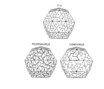
Figure 1 depicts a comparison of the structures of simple T=3 virus,
picornavirus
and comovirus capsids. In each case, one trapezoid represents one ~3-barrel.
Thus the large (L or VP37) capsid protein of comoviruses consists of two
covalently linked ~3-barrels which are equivalent to the C and B-type subunits
of
a T=3 virus or VP2 and VP3 of picornaviruses. The small (S or VP23) capsid
protein of comoviruses contains a single ~3-barrel which corresponds to the A-
type subunits of a T=3 virus or VP1 of a picornavirus.
Figure 2 depicts the secondary structure and connectivity of a canonical ~i-
barrel. The individual strands of p sheet are labelled B through G and the
amino- (NH2) and carboxy- (COOH) termini of the protein are indicated.
Figure 3 depicts the plasmids (A) pPMM2902 and (B) pBT7-123. The stippled
regions represent the CPMV-specific regions of the plasmids with the coding
regions being indicated by the wider portions on which the various virus-
encoded
proteins are marked. Relevant restriction enzyme sites are indicated. Details
of
the construction of the plasmids are given in Hotness et al (1989) and Dessens
and Lomonossoff (1991 ).
WO 92/18618 PCT/GB92/00589
2 1 0 8 7~. 7 7
_g_
Figure 4 depicts the region of CPMV M RNA which encodes the amino-terminal
40 amino acids of VP23. The numbers below the nucleotide sequence refer to
the M RNA sequence and the position of the unique Nhe1 site is indicated. The
amino acids involved in forming the ~3B and (3C strands of VP23 are indicated
above the amino acid sequence of the protein which is shown using the standard
one-letter code.
Figure 5 depicts (A) the nucleotide sequence of the oligonucleotides used in
the
construction of pFMDV together with the amino acid sequence encoded by the
top (positive) strand and (B) the structure of VP23 after insertion of the
FMDV-
specific oligonucleotides. The arrowed region indicates the extent of the
inserted
FMDV epitope. The Nhe1 site not restored during the cloning is indicated by
xNhel. The diagnostic Bg111 site present in the inserted sequence is also
indicated.
Figure 6 depicts the construction of plasmid pFMDV. The representation of the
various CPMV-specific regions is as in Figure 3. The FMDV-specific region
which is inserted into VP23 is shown as the black segment in the CPMV-specific
coding region.
Figure 7 depicts plasmid pMT7-601. The representation of the various CPMV-
specific regions is as in Figure 3. Relevant restriction sites are indicated.
Figure 8 depicts the construction of a "substitution" vector by site-directed
mutagenesis. The asterisk indicates the T residue that is changed to a C by
site-directed mutagenesis, thereby creating a novel Aatl1 site.
Figure 9 depicts (A) the nucleotide sequence of the oligonucleotides used in
the
construction of pMT7-HIV together with the amino acid sequence encoded by the
top (positive) strand and (B) the structure of VP23 after insertion of the HIV-
specific oligonucleotides. The arrowed region indicates the extent of the
inserted
HIV epitope. The diagnostic Pvu1 site present in the inserted sequence is also
indicated.
WO 92/18618 PCT/GB92/00589
2108777
Figure 10 depicts (A) the nucleotide sequence of the oligonucleotides used in
the construction of pMT7-HRV together with the amino acid sequence encoded
by the top (positive) strand and (B) the structure of VP23 after insertion of
the
HRV-specific oligonucleotides. The arrowed region indicates the extent of the
inserted HRV epitope. The diagnostic CIa1 site present in the inserted
sequence
is also indicated.
Figure 11 depicts the construction of plasmids pMT7-HIV and pMT7-HRV. The
representation of the various CPMV-specific regions is as in Figure 3. The HIV-
and HRV-specific regions inserted into VP23 are shown as the black segment
in the CPMV-specific coding region.
Figure 12 depicts a "Western blot" of protein extracts of the leaves of five
cowpea plants (lanes A to E) inoculated with a mixture of pMT7-FMDV-1 and
PBT7-123 transcripts. The blot was probed with serum specific for the FMDV
epitope. Lanes "mock" and "wt" contain extracts of leaves which were either
mock-inoculated or inoculated with wild-type CPMV RNA, respectively. The lane
marked "virus" contains purified wild-type CPMV. The sizes of marker proteins
are shown on the right-hand side of the blot.
Comoviruses
Comoviruses are a group of at least fourteen plant viruses which predominantly
infect legumes. Their genomes consist of two molecules of single-stranded,
positive-sense RNA of different sizes which are separately encapsidated in
isometric particles of approximately 28nm diameter. The two types of
nucleoprotein particles are termed middle (M) and bottom (B) component as a
consequence of their behaviour in caesium chloride density gradients, the RNAs
within the particles being known as M and B RNA, respectively. Both types of
particle have an identical protein composition, consisting of 60 copies each
of a
large (VP37) and a small (VP23) coat protein. In addition to the nucleoprotein
WO 92/18618 2 ~ ~ $'~ '~ ~ PCT/GB92/00589
-10-
particles, comovirus preparations contain a variable amount of empty
(protein-only) capsids which are known as top (T) component.
In the case of the type member of the comovirus group, cowpea mosaic virus
(CPMV), it is known that both M and B RNA are polyadenylated and have a
small protein (VPg) covalently linked to their 5' terminus. More limited
studies
on other comoviruses suggest that these features are shared by the RNAs of all
members of the group. Both RNAs from CPMV have been sequenced and
shown to consist of 3481 (M) and 5889 (B) nucleotides, excluding the poly (A)
tails (van Wezenbeek et al. 1983; Lomonossoff and Shanks, 1983). Both RNAs
contain a single, long open reading frame, expression of the viral gene
products
occurring through the synthesis and subsequent cleavage of large precursor
polypeptides. Though both RNAs are required for infection of whole plants, the
larger B RNA is capable of independent replication in protoplasts, though no
virus
particles are produced in this case (Goldbach et al., 1980). This observation,
coupled with earlier genetic studies, established that the coat proteins are
encoded by M RNA.
A 3.5A electron density map of CPMV shows that there is a clear relationship
between CPMV and the T=3 plant viruses such as the tombusviruses, in
particular tomato bushy stunt (TBSV) and the sobemoviruses, in particular
southern bean mosaic (SBMV). The capsids of these latter viruses are
composed of 180 identical coat protein subunits, each consisting of a single
~3-barrel domain. These can occupy three different positions, A, B and C,
within
the virions (Figure 1 ). The two coat proteins of CPMV were shown to consist
of three distinct ~3-barrel domains, two being derived from VP37 and one from
VP23. Thus, in common with the T=3 viruses, each CPMV particle is made up
of 180 ~i-barrel structures. The single domain from VP23 occupies a position
analogous to that of the A type subunits of TBSV and SBMV, whereas, the N-
and C- terminal domains of VP37 occupy the positions of the C and B type
subunits respectively (Figure 1 ).
WO 92/18618 ~ ~ ~ ~ ~ ~ ~ PCT/GB92/00589
- 11
X-ray diffraction analysis of crystals of CPMV and another member of the
group,
bean pod mottle virus (BPMV) shows that the 3-D structures of BPMV and
CPMV are very similar and are typical of the comovirus group in general.
In the structures of CPMV and BPMV, each ~3-barrel consists principally of 8
strands of antiparallel ~i-sheet connected by loops of varying length. The
connectivity and nomenclature of the strands is given in Figure 2. The flat
~3-sheets are named the B,C,D,E,F,G,H and I sheets, and the connecting loops
are referred to as the ~iB-~iC, ~3D-~iE, ~iF-~3G and ~3H-ail loops.
The comoviruses are also structurally related to the animal picornaviruses.
The
capsids of picornaviruses consist of 60 copies of each of three different coat
proteins VP1, VP2 and VP3 each one consisting of a single ~3-barrel domain.
As in the case of comoviruses, these coat proteins are released by cleavage of
a precursor polyprotein and are synthesised in the order VP2 - VP3 - VP1.
Comparison of the 3-dimensional structure of CPMV with that of picornaviruses
has shown that the N- and C- terminal domains of VP37 are equivalent to VP2
and VP3 respectively and that VP23 are equivalent to VP1 (Figure 1 ). The
equivalence between structural position and gene order suggests that VP37
corresponds to an uncleaved form of the two picornavirus capsid proteins, VP2
and VP3.
One of the principal differences between the comoviruses and picornaviruses is
that the protein subunits of comoviruses lack the large insertions between the
strands of the ~3-barrels found in picornaviruses though the fundamental
architecture of the particles is very similar. The four loops (~iB-~3C, ~3D-
(iE, ~3F-
~iG and ~iH-ail - see Figure 2) between the ~3-sheets are not critical for
maintaining the structural integrity of the virions but, in accordance with
this
invention, are used as sites of expression of foreign peptide sequences, such
as
antigenic sites from animal viruses.
210877
WO 92/18618 PCT/GB92/00589
-12-
Modification of CPMV virus
In order to make insertions into the coat protein of CPMV, it is necessary to
have a means of manipulating the genome of the virus. A full-length cDNA
clone of CPMV M RNA (pPMM2902) in the transcription vector pPMI was
available (see Figure 3A) (Ahlquist and Janda, 1984, Hotness et al. (1989) and
Hotness (1989). We have shown that transcripts from pPMM2902 can multiply
when electroporated in cowpea mesophyll protoplasts in the presence of highly
purified virion B RNA, therefore allowing modifications to be made to the
viral
coat proteins without affecting the multiplication and assembly of the virus.
In view of the possible danger that B RNA purified from virions to provide the
proteins required for viral replication with pPMM2902 might be cross-
contaminated
with wild-type M RNA, we have constructed a full-length cDNA clone of B RNA,
pBT7-123 (see Figure 3B). The full-length copy of B RNA is immediately
downstream of a modified T7 promoter. Following linearisation with the
restriction
enzyme MIu1, transcripts identical in size to natural B RNA can be synthesised
by T7 RNA polymerase. A mixture of transcripts from pPMM2902 and pBT7-123
gives rise to a full virus infection when electroporated into cowpea
protoplasts,
and therefore replaces the use of natural B RNA.
We have selected the (3B-(3C loop in VP23 for the insertion of foreign
peptide.
This loop is clearly exposed on the surface of the viral particle and computer
modelling has shown that even large loops inserted at this site are unlikely
to
interfere with the interaction between adjacent subunits responsible for
capsid
structure and stability. This loop has a unique Nhe1 site at position 2708 of
the
M RNA-specific sequence where foreign sequences may be inserted (see Figure
4).
The principle antigenic sites of the picornavirus foot and mouth disease
(FMDV)
and human rhinovirus (HRV), and the lentiretrovirus human immune deficiency
virus (HIV) were used to illustrate the use of this invention in the
production of
vaccines to animal viruses.
.VO 92/18618 2 ~ ~ g ~ ~ 7 PCT/GB92/00589
-13-
Design and construction of pFMDV, a full len4th cDNA clone of CPMV
M RNA containing a DNA insert codin4 for a se4ment of FMDV loop
rotein
To insert the "FMDV loop" into the (3B-~3C loop of VP23 of CPMV, two
complementary oligonucleotides, both 81 residues long, were chemically
synthesised. Their sequences are given in Figure 5A. The positive sense
oligonucleotide contains the sequence encoding amino acid residues 136-160
from VP1 of FMDV serotype O, strain BFS 1860. The nucleotide sequence of
oligonucleotides was designed to take account of the codon usage preference
found in CPMV and includes a Bg111 site in the middle of the sequence to
facilitate screening. When annealed, the oligonucleotides give a double-
stranded
DNA sequence with Nhe1-compatible ends. Thus the oligonucleotides can be
inserted into the unique Nhe1 site of pPMM2902. The effect of such an
insertion
on the sequence of the VP23 is shown in Figure 5B. To facilitate the insertion
of the FMDV loop, the FMDV-specific oligonucleotides were initially ligated
into
an M13 sub-clone of pPMM2902 which contained the sequence encoding VP23.
This was done to enable clones harbouring the FMDV-specific sequence to
readily be identified by sequence analysis. All the standard DNA manipulations
were carried out according to Maniatis et al (1982). Details of the
construction
of pFMDV are given below and are shown diagrammatically in Figure 6.
STEP 1. The plasmid pPMM2902 was digested with the restriction enzyme Sst1
which cuts twice within the CPMV M RNA-specific sequence at positions 2296
and 3423 but does not cut within the sequence of the plasmid pPMI. Following
agarose gel electrophoresis, and both the large (6.Okb) the small (1.1 kb)
fragment were purified by electroelution from the gel. The 1.1 kb Sst1
fragment
was ligated into the Sst1-cut, phosphatase-treated replicative form DNA from
the
bacteriophage M13mp18. The ligation mixture was used to transform E.coli
strain JM101 using the calcium chloride procedure. Plaques containing the 1.1
kb
Sst1 fragment from M RNA were identified by the Lac complementation assay
and DNA sequence analysis and one, M13-JR1 was selected for further work.
WO 92/ 18618 ~ ~ ~ ~ ~ '~ '~ PCT/GB92/00589 .
-14
STEP 2. The double-stranded replicative form DNA of M13-JR1 was isolated
from infected E. coli strain JM101 cells by the method of Birnboim and Doly
(1979). The purified DNA was linearised by digestion with the restriction
enzyme
Nhe1 and the linearised plasmid treated with calf intestinal phosphatase. The
two oligonucleotides with Nhe1-compatible termini encoding amino acid residues
136 to 160 of VP1 from FMDV were phosphorylated with ATP using
polynucleotide kinase and annealed to each other by boiling and slow cooling.
The annealed oligonucleotides were ligated into Nhe-1-digested M13-JR1, the
ligation mixture used to transform E. coli strain JM101 and the transformation
mixture plated out on a lawn of JM101. A large number of plaques were found
on the plates, 20 of which were selected for sequence analysis. Bacteriophage
were propagated in JM101 and the single-stranded DNA isolated exactly as
described by Sanger et al (1980). The nucleotide sequence of the region of the
bacteriophage DNA around the Nhe1 site was determined by the dideoxy method
as modified by Biggin et al (1983), using an l8mer,5'
AGT-TAC-TGC-TGT-AAC-GTC-3', complementary to nucleotides 2735-2752 of the
M RNA sequence, as primer. Of the plaques analysed, one, designated M13-
ushal, had a single copy of the desired sequence in the correct orientation.
STEP 3. M13-ushal was propagated in E.coli strain JM101 and the replicative
form DNA was isolated from the infected cells by the method of Birnboim and
Doly (1979). The DNA was digested with Sst1 and the 1.1 kb fragment purified
by agarose gel electrophoresis. This fragment was ligated to the large (6.Okb)
Sst1 fragment from pPMM2902 (see above) which had been treated with calf
intestinal phosphatase. The ligation mixture was used to transform E.coli
strain
JM83 using the calcium chloride method. The transformation mixture was plated
out on L-agar plates containing 100 pg/ml carbenicillin and the plates
incubated
overnight at 37°C. 12 carbenicillin-resistant colonies were selected
for further
study. The colonies were grown up as 1 ml cultures in L-broth, plasmid
"minipreps" prepared and analysed by restriction enzyme digestion. From the
patterns obtained by digestion with the enzymes Sst1, Bgl11 and EcoRV, it was
possible to deduce that 4 colonies consisted of full-length clones of CPMV
containing the sequence of the FMDV-specific oligonucleotides in the correct
-15- 2108777
orientation. One of these, pFMDV, was subsequently propagated on a large
scale and the plasmid DNA was isolated by the method of Birnboim and Doly
(1979) and further purified by centrifugation using caesium chloride/ethidium
bromide gradients (Maniatis et al (1982).
Properties of pFMDV transcripts
1. Purified pFMDV DNA was linearised by digestion with EcoR1 and transcribed
using E.coli RNA polymerase exactly as described for pPMM2902 by Hotness et
al (1989). Electrophoresis of the products of formaldehyde-containing agarose
gels (Lehrach et al., 1977) revealed the presence of transcripts which
co-migrated with authentic viral M RNA.
2. Following treatment with DNAsei and lithium chloride precipitation to
remove
the template DNA, the transcripts were translated in vitro in the
message-dependent rabbit reticulocyte system (Pelham and Jackson 1976) in the
presence of 35S-methionine. The products were examined by electrophoresis on
polyacrylamide gels containing SDS (Laemmli, 1970) and visualised by
autoradiography of the dried-down gel. The autoradiographs revealed the
presence of two proteins of 105 and 95kDa which co-migrated with the
translation products of natural M RNA.
3. The ability of transcripts of pFMDV to be replicated in plant cells was
examined as follows: Cowpea mesophyll protoplasts were prepared as described
by de Varennes et al (1985). Transcripts from pFMDV were mixed with
transcripts from pBT7-123 (the pfasmid containing a full-length copy B RNA)
and
electroporated into the protoplasts as described by Hotness et al (1989). As
control, a sample of the same preparation of protoplasts were electroporated
with
a mixture of transcripts from pBT7-123 and pPMM2902. 72 hours
post-electroporation the protoplasts were harvested and the nucleic acids were
extracted as described by de Varennes et al (1985). Samples of the RNA were
electrophoresed on formaldehyde-containing agarose gels (Lehrach et al. 1977)
and the nucleic acids blotted on to Hybond* N membranes (Amersham
* Trade mark
WO 92/18618 PGT/GB92/00589
-1s- 2 1 0 87 77
International). The nucleic acids were cross-linked to the membranes by
irradiation with u.v. light. The membranes were probed for M RNA sequences
using a Hind111 fragment form pPMM2902 corresponding to nucleotides
482-2211 of the M RNA which had been labelled with 32P as described by
Feinberg and Vogelstein (1983). Samples from protoplasts electroporated with
pBT7-123 and either pPMM2902 or pFMDV-1 revealed the presence of M
RNA-specific sequences confirming that the presence of the sequence encoding
the FMDV loop did not prevent the transcripts from replicating. To confirm
that
the progeny of pFMDV replication retained the sequence encoding the FMDV
loop, replicate membranes were probed with the positive-sense FMDV
oligonucleotide which had been "oligo-labelled" (Feinberg and Vogelstein,
1983)
to give a (+) sense-specific probe. The sample from protoplasts electroporated
with a mixture of pBT7-123 and pFMDV transcripts gave a clear signal at the
expected position for M RNA, a signal which was absent from the pPMM2902
control.
4. To establish that protein subunits containing the FMDV loop assemble into
virions, extracts from infected protoplasts were examined for the presence of
virus particles by immunosorbent electron microscopy. Samples of protoplasts
electroporated with a mixture of transcripts from pBT7-123 and pFMDV were
lysed by repeated passage through a 23 gauge needle. The extracts were
centrifuged in an Eppendorf microfuge and supernatant retained for
examination.
microlitre samples of the supernatants were incubated with gold electron
microscope (em) grids which had been coated with anti-CPMV antiserum. After
washing and staining with uranyl acetate, the grids were examined using a JEOL
1200 electron microscope. Particles of diameter 28nm could be seen which had
the characteristic appearance of CPMV virions. This demonstrates that the
presence of the FMDV loop in VP23 does not prevent virus assembly.
The foregoing description establishes that plant viruses modified in
accordance
with this invention can multiply and assemble into virus particles when
electroporated into plant protoplasts. To produce modified plant viruses on a
large scale it is necessary to prepare a construct which can be inoculated
directly
WO 92/18618 ~ ~ ~ ~ ~~ PCT/GB92/00589
-17-
onto whole plants, and which will replicate and assemble into virus particles
as
in the above described protoplast system. We have therefore modified
pPMM2902 in such a way that the resulting transcripts incorporate a "cap"
structure at their 5' ends, and RNA synthesis is driven by a more efficient
promoter. The steps in the modification of pPMM2902 to produce pMT7-601
(Figure 7) are described in detail below.
Development of a system capable of infecting whole cowpea plants
Construction of pMT7-601
1. 1 st strand cDNA to purified CPMV M RNA was synthesised exactly as
described by Lomonossoff et al (1982), using pdT,2_,8 as a primer. 2nd strand
synthesis was primed using the following oligonucleotide:
Pst1 T7 Promoter ~. 5' end M RNA
5'CCCTGCAGTAATACGACTCACTATAGTATTAAAATCTTAATAG
Conditions for synthesis were as described in Lomonossoff et al (1982) and
Shanks et al (1986).
2. The double-stranded cDNA was digested with the restriction enzymes Pst1
and BamH1 (which cleaves the M RNA sequence at position 1504) and the
l.5kb Pst1/BamH1 fragment ligated into Pst1/BamH1 digested M13mp18. The
ligation mix was used to transform E.coli strain JM101. Recombinant phage
harbouring inserts were identified by the lac complementation assay and
checked
for the presence of the correct insert by "T-track" analysis (Sanger et al.,
1980)
as modified by Biggin et al (1983). One clone, M13-MT7-6, was selected for
further analysis and the sequence of the 5' terminal 200 nucleotides of M RNA
specific sequence was determined as described by Biggin et al (1983) and
shown to be identical to the equivalent sequence in pPMM2902.
WO 92/18618 PCT/GB92/00589
0 ~,'~'~ "~ _ 1 s _
3. The double-stranded, replicative DNA was isolated from E.coli JM101 cells
infected with M13-MT7-6 by the method of Birnboim and Doly (1979). The
double stranded DNA was digested with Pst1 and Bg111 (which cuts the M RNA
sequence at position 189) and the 200bp fragment released was purified by
electrophoresis on and electro-elution from an agarose gel (Maniatis et al
(1982).
4. The plasmid pPMM2902 (Hotness et al 1989) was digested with Pst1 and
Bgl11 to produce two DNA fragments of 1.1 and 6.Okb. The smaller (1.1 kb)
fragment contains the sequence of the E.coli promoter linked to the first 189
nucleotides of the sequence of CPMV M RNA while the larger (6.Okb) fragment
the rest of the sequence of M RNA linked to pUC9. The digest was treated with
calf intestinal phosphatase, the two fragments separated by agarose gel
electrophoresis and the 6.Okb fragment recovered by electro-elution.
5. The 200bp Pst1/Bg111 from M13-MT7-6 and the 6.Okb fragment from
pPMM2902 were ligated together (Maniatis et al, 1982) and the mixture used to
transform E.coli strain JM83. A number of carbenicillin-resistant colonies
were
identified and one, pMT7-601, was shown to have the desired structure.
Large-scale quantities of plasmid pMT7-601 were therefore prepared as
described
for pFMDV.
6. After linearisation with EcoRl, plasmid pMT7-601 could be transcribed using
T7 RNA polymerase to give RNA which was identical in size to natural virion M
RNA when analysed on formaldehyde-containing agarose gels (Lehrach et al.
1977). The yield of transcript was approximately 1 ~g of full-length M
transcript
per pg of linearised template DNA.
7. When a mixture of T7 transcripts from pMT7-601 and pBT7-123 was
electroporated into cowpea mesophyll protoplasts, Northern blot analysis of
progeny RNA revealed that transcripts from pMT7-601 are biologically active.
The methods used for protoplast isolation and nucleic acid analysis were
identical to those used to analyse the biological properties of pFMDV.
WO 92/18618 2 Z ~ g ~ ",~ ~ PGT/GB92/00589
_19_
Infectivity of a mixture of capped p8T7-123 and pMT7-601 transcripts on
cowoea plants
Samples of pBT7-123 and pMT7-601 were linearised with MIu1 and EcoR1
respectively. Portions of the linearised templates were transcribed using T7
RNA
polymerase in the presence of GpppG essentially as described by Ziegler-Graaf
et al (1988). The transcription reactions contained 0.1 mg/ml linearised DNA
template, 40mM Tris-HCI pH 8.0, 25mM NaCI, 8mM MgCl2, 2mM spermidine
hydrochloride, 0.5mM each of UTP, ATP and CTP, 0.025mM GTP, 0.5mM
GpppG, 0.05mg/ml BSA, lOmM DTT, 200 units/ml RNAguard and transcription
was initiated by the addition of T7 RNA polymerase to a final concentration of
1400 units/ml. Incubation was at 37C for 2 hours. At 30, 60 and 90 minutes
portions (5p.1 per 1 ml transcription reaction) of a 5mM solution of GTP were
added. Following transcription, EDTA was added to lSmM final concentration
and the integrity of the transcripts was checked by electrophoresis on
formaldehyde-containing agarose gels. The transcription mixtures were
extracted
with 2 volumes of phenol/chloroform (1.1 v/v) and the nucleic acids
precipitated
twice with ethanol. The nucleic acids were harvested by centrifugation, washed
with ethanol and dried under vacuum. The nucleic acids were dissolved in
50mM Tris-phosphate, pH 8.0 for inoculation on to plants.
The primary leaves of 10 day-old cowpea (Vi4na un4uiculata var. California
blackeye) were dusted with carborundum and a 1:1 (w/w) mixture of transcripts
derived from pMT7-601 and pBT7-123 were applied to the leaves with gently
rubbing. A variety of transcript concentrations were used but in all cases the
final inoculum volume was 50p.1. The results obtained showed that when a total
of 5p.g of each transcript was applied per primary leaf, 100% of plants
inoculated
routinely developed symptoms characteristic of a CPMV infection. The presence
of CPMV-specific sequences in both the inoculated and upper leaves of such
plants was confirmed by "Dot blot" analysis. Samples of the inoculated and
trifoliate leaves were taken using a number 10 cork borer and macerated and
extracted with 0.4 mls of lOmM sodium phosphate. The samples were
centrifuged and 5pl of the supernatant was applied to nitrocellulose filters
pre-
WO 92/18618 ~ PCT/GB92/00589
_ 20 -
wetted with 20XSSC. The nucleic acid were cross-linked to the membranes by
irradiation with u.v. light and probed for M RNA-specific sequences using a
'2P
"oligo-labelled" (Feinberg and Vogelstein, 1983) probe consisting of
nucleotides
482-2211 of the M RNA sequence. The conditions for hybridisation and washing
of the filters were as described by Maniatis et al (1982). After drying, the
filters
were autoradiographed. A strong hybridisation signal indicated the presence of
CPMV-specific sequences.
Construction of pMT7-FMDV-I, pMT7-HIV and pMT7-HRV
To construct pMT7-FMDV-I, pMT7-601 and pFMDV were both digested with
restriction enzyme Sst1, the digest from pMT7-601 being subsequently treated
with calf intestinal phosphatase. Sst1 cuts each plasmid twice at positions
2296
and 3423 of the M RNA-specific region to release a 1.1 kb fragment. As
discussed previously this Sst1 fragment contains the region of VP23
encompassing the ~iB-~iC loop where the FMDV loop insertion has been made.
Following electrophoresis on an agarose gel, the 1.1 kb fragment from pFMDV
and the 5.1 kb fragment, encompassing the vector sequence and all the rest of
the M RNA specific sequence, from pMT7-601 were recovered by electo-elution.
The two Sst1 fragments were ligated together and the mixture transformed in
E. coli strain JM83. A number of carbenicillin-resistant colonies were picked,
"minipreps" made and the plasmid DNA examined by restriction enzyme digests
to identify recombinants containing the FMDV loop. One such clone was
identified, designated pMT7-FMDV-I and grown up on a large scale. All the DNA
manipulations were as described for the construction of pFMDV and pMT7-601.
Both pFMDV and its derivative pMT7-FMDV-I have a straightforward insertion
into
the ~3B-~3C loop of VP23. To limit the increase in size of the loop upon
insertion
of a foreign sequence, a replacement vector was designed where the foreign
sequence would replace the natural (iB-~3C loop in VP23 rather than be added
to it. In the nucleotide sequence of the region of the CPMV genome encoding
VP23 a single silent base change (U to C) at position 2740 creates a unique
WO 92/18618 ~ ~ ~ ~ ~ ~~ PCT/GB92/00589
-21 -
Aatl1 site at amino acid valine 27. The change in the sequence of M RNA is
shown in Figure 8. The creation of the Aatl1 site enables the nucleotide
sequence encoding the six amino acids from the native ~iB-~iC loop in CPMV to
be removed by digestion with Nhe1 and Aatll. The sequence can then be
replaced by any sequence with Nhe1- and Aatl1-compatible ends.
Two different sequences were designed to be substituted for the sequence
between the Nhe1 and Aatl1 sites of the mutated M RNA sequence. The first
sequence to be substituted into VP23 consisted of oligonucleotides encoding
residues 735-752 from the transmembrane glycoprotein gp41 from human
immunodeficiency virus (HIV-1 ). This sequence was selected as a synthetic
peptide for this region is recognised in enzyme-linked immunosorbent assays
(ELISA) by antisera from seropositive AIDS patients and is capable of inducing
antibodies which neutralise a range of HIV-1 isolates (Kennedy et al, 1986;
Chanh et al, 1986; Dagleish et al, 1988). The second sequence consists of the
nucleotide sequence encoding residues 85-99 from VP1 of human rhinovirus 14
(HRV14). In both cases, the oligonucleotides were designed to contain
restriction
enzyme sites to facilitate screening. The sequences of the oligonucleotides
and
the effect of the substitutions on the amino acid sequence of VP23 are shown
in Figures 9 and 10. The steps in the construction of pMT7-HIV and pMT7-
HRV are given below and are shown diagrammatically in Figure 11.
STEP 1. M13-JR-1 (see Figure 6) was propagated in E. coli strain CJ236 and
dU-containing single-stranded DNA isolated as described by Kunkel (1985). The
T to C mutation at position 2740 of the M RNA sequence was made by
oligonucleotide-directed mutagenesis of dU-containing single-stranded M13-JR1
DNA using the primer CTG-CTG-TGA-CGT-CTG-AAA-A as described by Kunkel
(1985). This resulted in the construction of clone M13-JRAatll. The mutation
was confirmed by dideoxy sequence analysis of single-stranded DNA (Biggin et
al, 1983) and by restriction enzyme digestion of the double-stranded
replication
form DNA.
STEP 2. The replicative form DNA of M13-JRAatl1 was isolated and digested
WO 92/18618 PCT/GB92/00589
~.~a~~~ _ 22 _
with Nhe1 and Aatl1 and treated with calf intestinal phosphatase. The pairs of
oligonucleotides shown in Figures 9 and 10 were phosphorylated with ATP using
polynucleotide kinase, annealed together by boiling and slow cooling and
ligated
into Nhe1/Aatl1-digested M13-JRAatll. Recombinant M13 clones harbouring the
inserted sequences were identified by sequence analysis of the single-stranded
bacteriophage DNA exactly as described previously for pFMDV. Two clones,
M13-HIV and M13-HRV containing the required sequences were identified and
the double-stranded replicative form DNA was isolated shown to give the
expected pattern of fragments on restriction enzyme digestion.
STEP 3. Replicative form DNA from M13-HIV and M13-HRV was digested with
Sst1 and the l.2kb M RNA-specific fragment recovered by electro-elution after
electrophoresis of the digest on an agarose gel. The l.2kb fragments were
ligated into the large Sst1 fragment from pMT7-601 as previously described for
production of pMT7-FMDV-I. The ligation mixture was used to transform E. coli
strain JM83 and carbenicillin-resistant colonies selected. Two clones,
designated
pMT7-HIV and pMT7-HRV, were shown to contain the desired structure by
restriction enzyme mapping and nucleotide sequence analysis.
For transcription, pMT7-HIV and pMT7-HRV, were linearised by digestion with
EcoRl. Transcription using T7 RNA polymerase was carried out exactly as
described for pMT7-601 and pBT7-123. The resulting transcripts were identical
in size to natural virion RNA.
Demonstration of the ability of pMT7-FMDV-I and pMT7-HIV transcripts to
replicate in cowpea protoplasts
1 O~g samples of the in vitro transcripts from either pMT7-601, pMT7-FMDV-I or
pMT7-HIV were mixed with 15~g samples of transcripts from pBT7-123 and the
mixtures used to electroporate 106 cowpea mesophyll protoplasts. Samples were
either taken immediately (0 hour) or after incubation of the protoplasts for
72
hour in the light at 25°C. Nucleic acids were extracted from one
quarter of each
PCT/G B92/00589
WO 92/18618
-23-
sample and electrophoresed on a 1 % formaldehyde-containing agarose gel as
previously described. The nucleic acids were blotted on to Hybond N, cross-
linked to the membrane by u.v, irradiation and probed for CPMV M RNA-specific
sequences as previously described. In each case a strong hybridisation signal
corresponding in position to M RNA could be detected in the 72 hour but not
the
0 hour incubation samples, demonstrating that the transcripts from all four
constructs can multiply in cowpea protoplasts.
The remaining three quarters of each protoplast sample was lysed as previously
described and applied to electron microscope grids coated with anti-CPMV
serum. The grids were then examined using a JEOL 1200 electron microscope.
Large numbers of particles could be seen in the 72 hour samples of protoplasts
electroporated with pMT7-601, pMT7-FMDV-I and pMT7-HIV transcripts. These
results show that the modified coat proteins encoded by pMT7-FMDV-I and
pMT7-HIV can assemble into virions.
Ability of pMT!-FMDV-I and pMl7-HIV transcripts to replicate in whole
cowpea plants
To demonstrate the ability of transcripts from pMT7-FMDV-I and pMT7-HIV to
replicate in whole cowpea plants in the presence of transcripts derived from
pBT7-123, transcripts capped with GpppG were prepared as previously described.
6 groups, each consisting of 5, 10 day old, cowpeas, were inoculated with the
transcripts using the method previously described. In each case, the amount of
transcript refers to the amount applied to an individual leaf.
Group 1. Mock-inoculated with 50mM Tris-phosphate, pH 8.0
Group 2. Inoculated with 1.5p.g of natural CPMV virion RNA
Group 3. Inoculated with 5pg each of pMT7-601 + pBT7-123 transcripts
Group 4. Inoculated with 5~g each of pMT7-FMDV-I + pBT7-123
transcripts
Group 5. Inoculated with 5~g each of pMT7-HIV + pBT7-123 transcripts
WO 92/18618 PCT/GB92/00584
-24
Symptoms were scored on a daily basis and samples of leaf tissue from each
plant were taken 11 days post-inoculation for "dot blot" analysis which was
carried out as described previously. The rest of the leaf tissue from all the
plants in groups 4 and 5 was harvested and frozen for future use.
Results
None of the plants in group 1 (mock-inoculated) developed any symptoms up to
11 days post infection (P.I.) and no CPMV-specific nucleic acids could be
detected in the leaf tissue by "dot blot" analysis. This shows that no
accidental
infection of the cowpea plants with CPMV had occurred during the experiment.
All plants in groups 2 and 3 (inoculated with either virion RNA or a mixture
of
pMT7-601 and pBT7-123 transcripts) showed strong symptoms on both the
inoculated and systemic leaves by 7 days P.I. "Dot blot" analysis of leaf
tissue
showed the presence of large amounts of virus-specific RNA in both the
inoculated and systemic leaves of all plants. This confirms that the plants
used
in the experiment were fully susceptible to infection with CPMV using either
virion
RNA or a mixture of wild-type transcripts.
By 11 days P.I. the inoculated leaves of all the plants in group 4 (inoculated
with
pMT7-FMDV-1 transcripts) developed a mottled appearance distinct from that
normally associated with a wild-type virus infection. This result shows that
the
transcripts from pMT7-FMDV-I can multiply and spread from cell-to-cell in
whole
cowpea plants.
4 out of 5 of the plants in group 5 (inoculated with pMT7-HIV transcripts)
developed symptoms on their systemic leaves by 11 days P.I. "Dot blot analysis
showed that plants showing symptoms had substantial quantities of virus-
specific
sequences in both the inoculated and systemic leaves. This result shows that
transcripts from pMT7-HIV can multiply and spread within whole cowpea plants.
-25- 2 1 0 87 77
Further investigation of the results obtained with pMT7-FMDV-I and pMT7-HIV
pMT7-FMDV-I: To demonstrate that modified viral capsid proteins were
synthesised in the inoculated leaves of the group 4 plants, samples of the
frozen
leaf tissue were finely ground and extracted with 1 X Laemmli sample buffer.
The
extracts were electrophoresed on 15% polyacrylamide-SDS gels and the proteins
transferred to nitrocellulose membranes using a Biorad* semi-dry transfer
cell.
The membranes were probed either with serum raised against whole CPMV virus
particles or with a serum raised against the synthetic oligopeptide,
VPNLRGDLQVLAQKVARTLP(CG), corresponding to residues 141-160 of VP1 of
FMDV strain O,. This sequence corresponds to the epitope which was inserted
into VP23 in pMT7-FMDV. Both antisera were raised in rabbits. Western blot
analysis was carried out using alkaline phosphatase-conjugated goat anti-
rabbit
IgG as the second antibody. The protein extracts of all five group 4 plants
were
found to react with the anti-CPMV serum indicating that the virus coat
proteins
were synthesised in the inoculated leaves of the group 4 plants. When similar
blots were probed with the anti-FMDV oligopeptide serum, a single band lit up
in the extracts from each of the group 4 plants (Figure 11 ). This band
migrated
with an apparent molecular weight of 24kDa, which is exactly the size expected
for VP23 carrying the FMDV loop. No product of similar size could be seen
when extracts from mock-inoculated or wild-type CPMV-inoculated leaves were
analysed (Figure 11 ). Likewise, purified wild-type CPMV coat proteins did
react
with the FMDV-specific antiserum. Furthermore, pre-treatment of the anti-FMDV
serum with the peptide which was used to raise it, abolished the reaction with
the extracts from the group 4 plants demonstrating the specificity of the
immunological reaction. These results demonstrate that the inoculated leaves
of
the group 4 plants contained CPMV coat proteins harbouring the FMDV loop.
pMT7-HIV: As discussed above, the "dot blot" analysis of both the inoculated
and systemic leaves from the Group 5 plants indicated that transcripts from
pMT7-HIV can multiply and spread in whole plants. The levels of signal
obtained
and the fact that the infection went systemic show that the progeny RNA is
encapsidated. To prove that the HIV-specific insert was retained in the
progeny
* Trade mark
WO 92/18618 PCT/GB92/00589
_26_
RNA, "dot blots" of extracts from the group 5 plants were probed with a HIV-
insert specific probe. This was made by "oligo-labelling" the positive sense
oligonucleotide used in the construction of pMT7-HIV (see Figure 9). The
results
obtained showed the presence of the HIV sequence in extracts of the inoculated
leaves of the four plants which showed symptoms.
EXTENSION OF RESULTS WITH CPMV HARBOURING THE FMDV EPITOPE
To extend the previous findings obtained with transcripts derived from pMT7-
FMDV-I, five groups of five cowpea plants were inoculated with capped
transcripts, prepared as previously described, as follows:
Group 1: Mock-inoculated with 50mM Tris-phosphate, pH 8.0
Group 2: Inoculated with 0.5pg of natural CPMV virion RNA
Group 3: Inoculated with 5pg GpppG capped pMT7-601 + pBT7-123
transcripts
Group 4: Inoculated with 5pg GpppG capped pMT7-FMDV-I +
pBT7-123 transcripts
Symptoms were scored on a daily basis. 13 days post-inoculation triplicate
leaf
disk samples were taken from one inoculated and one trifoliate leaf of each
plant.
The samples were treated as follows:
Sample 1 (Crude homogenate): Homogenised in 0.4m1s lOmM sodium
phosphate buffer, pH7.0, centrifuged and the supernatant recovered.
Sample 2 (RNA extract): Frozen in liquid nitrogen, finely ground and the
nucleic
acids extracted with phenol/chloroform. After ethanol precipitation, the
nucleic
acids were finally resuspended in 0.1 mls of water.
Sample 3 (Protein extract): Frozen in liquid nitrogen, finely ground and the
powder dissolved in 0.1 ml 1 X Laemmli sample buffer.
WO 92/18618 ~, ~ $ ~ ~ ~ PCT/GB92/00589
-27-
"Dot blots" were prepared from 5~,1 aliquots of samples 1 and 2 and were
probed
with either a probe specific for nucleotides 482-2211 of CPMV M RNA (CPMV-
specific probe), prepared as described previously, or with a probe specific
for
FMDV-specific insert. The latter was prepared by "oligo-labelling" the
positive
sense oligonucleotide shown in Figure 5. Western blots were prepared from
aliquots of sample 3 and probed for FMDV-specific epitopes as described
previously. ISEM was carried out on aliquots of sample 1.
Results
No symptoms developed on any of the group 1 (mock inoculated) plants. Dot
blots of crude homogenates or RNA extracts revealed no CPMV-specific or
FMDV-specific sequences were present in extracts from either the inoculated or
trifoliate leaves. ISEM of the crude homogenates using electron microscopy
grids
coated with anti-CPMV serum showed no virus particles were present. Western
blot analysis of the protein extracts using the FMDV-specific serum showed an
absence of any FMDV epitopes. These results provide the negative control for
the rest of the experiment.
Symptoms developed on both the inoculated and trifoliate leaves of all group 2
(virion RNA-inoculated) and group 3 (pMT7-601 + pBT7-123) -inoculated plants
by 7 days PI. By 11 days PI the lesions on the primary leaves had expanded
to a diameter of 2-3 mm. Dot blots of both the crude homogenates and RNA
extracts revealed the presence of CPMV-specific but not FMDV-specific
sequences. ISEM using grids coated with anti-CPMV serum revealed the
presence of copious numbers of CPMV particles in crude homogenates from both
the inoculated and trifoliate leaves. Western blot analysis of the protein
extracts
showed an absence of any FMDV epitopes.
Small lesions (approximately 1 mm in diameter) developed on the inoculated
leaves of the group 4 (pMT7-FMDV-I + pBT7-123)-inoculated plants by 11 days
PI. Dot blots of the RNA extracted from the leaves (sample 2) showed the
presence of both CPMV- and FMDV-specific sequences in the inoculated of 3 out
WO 92/18618 ~ ~ ~~~'~ ~ PCT/GB92/00589
-28-
of the 5 group 4 plants. ISEM using grids coated with anti-CPMV serum
revealed the presence of CPMV-like virus particles in the crude homogenates
from the inoculated leaves of 4 out of the 5 group 4 plants. Western blotting
of
the protein extracts (sample 3) revealed the presence of the FMDV epitope on
the small coat protein in extracts from all group 4 plants. These results
confirm
that transcripts from pMT7-FMDV-I can multiply in whole cowpea plants and show
that virus particles are produced in such plants.
PREPARATION OF VIRUS FROM TRANSCRIPT-INOCULATED PLANTS
To isolate virus particles from pMT7-FMDV-I-infected leaf tissue the following
method was developed:
22 grams of primary cowpea leaves which had been inoculated with 5p.g each
of pBT7-123 and pMT7-FMDV transcripts were harvested 16 days post-
inoculation. The leaves were homogenised in 2 volumes (approximately 50m1s)
of 0.1 M sodium phosphate, pH 7.0 at 4°C. The sap was filtered through
two
layers of muslin, centrifuged at 15,OOOg for 15 minutes and the supernatant
retained. The pellet was re-extracted with a few mls of 0.1 M sodium phosphate
buffer pH 7.0, re-centrifuged. The supernatants were combined and centrifuged
in a Beckman type 30 rotor at 27,000 rpm for 4 hours at 4°C. The
resulting
pellet was resuspended overnight at 4°C in 3.5m1s of 0.1 M sodium
phosphate,
pH 7.0 and subsequently centrifuged in an Eppendorf centrifuge for 10 minutes.
The supernatant was taken and made up to 4 mls with 0.1 M sodium phosphate
pH 7.0 and 1 ml of a solution containing 1 M NaCI, 20% PEG 6000 was added
and the mixture incubated for 2 hours at room temperature. The resulting
precipitate was collected by centrifugation in an Eppendorf centrifuge for 10
minutes, resuspended in 0.25m1s of lOmM sodium phosphate, pH 7.0 and the
solution clarified by re-centrifugation in an Eppendorf centrifuge for 10
minutes.
The supernatant, which contains the virus particles, was then removed and
stored at 4°C. It was estimated spectrophometrically that the virus
concentration
in the final suspension was approximately 1.5 mg/ml. Western blot analysis of
-29- 2 ~ ~ 87 77
the virus using FMDV-specific antiserum revealed the presence of FMDV antigen
associated with the small coat protein subunit of the chimaeric virus
particles.
PASSAGING MODIFIED RNA
In order to produce large quantities of chimaeric virus as efficiently as
possible,
RNA extracted from transcript-inoculated leaves was passaged in plants. 5p,1
samples of the RNA extract from a pMT7-FMDV-I-inoculated leaf were diluted
to 50p.1 with Tris-phosphate pH 8.0 and were inoculated on to the primary
leaves
of a batch of 5 cowpea plants. All the plants developed symptoms typical of a
CPMV infection and at 23 days PI the primary leaves from the plants were
harvested. The leaves were homogenised in 0.1 M sodium phosphate buffer and
virus extracted as described above except that the initial high speed
pelleting
step was omitted. A total of 3.0 mgs of virus at a final concentration of
0.5mg/ml in lOmM sodium phosphate pH7.0 was isolated in this way. This
preparation was finally concentrated in a Centriprep* concentrator (Amicon) to
a
final concentration of 1.4 mg/ml and has been designated P1.
Samples of P1 were examined by electrophoresis on SDS gels and Coomassie
Blue staining and shown to contain the expected pattern of coat proteins.
Western blot analysis using anti-FMDV serum showed the small coat proteins
contained the FMDV loop. RNA extracted from the virus particles was of the
expected size for M and B RNA of CPMV. This demonstrates that chimaeric
virus can be produced by passaging the RNA derived from transcript-inoculated
leaves.
An experimental vaccine was prepared from virus preparation P1 by dispersion
in sterile phosphate buffered saline (PBS) at a final concentration of 1
mg/ml.
Guinea pigs were injected with 40 ~g of the P1 vaccine on days 0 and 28.
Preliminary results indicate that the animals produce antibodies against the
FMDV
loop. a response not seen when wild-type virus is injected.
r~~ * Trade mark
-CVO 92/18618 PCT/GB92/00589
-3°- 2 1 0 87 7~'
REFERENCES
Ahlquist, P., and Janda, M. (1984). Mol. Cell Biol. 4, 2876-2882.
Biggin, M.D., Gibson, T.J. and Hong, G.F. (1983). Proc. Natl. Acad. Sci. USA
80, 3963-3965.
Birnboim, H.C. and Doly, J. (1979). Nucleic acids Res. 7, 1513-1523.
Chanh, T.C., Dreesman, G.R., Kanda, P., Linette, G.P., Sparrow, J.T., Ho, D.D.
and Kennedy, R.C., (1986). EMBO J. 5, 3065-3071.
Dalgleish, A.G., Chanh, T.C., Kennedy, R.C., Kanda, P., Clapham, P.R. and
Weiss, R.A. (1988). Virology 165, 209-215.
De Varennes, A. and Maule, A.J. (1985). Virology 144, 495-501.
Dessens, J.T. and Lomonossoff, G.P. (1991 ). Virology 184, 738-746.
Feinberg, A.P. and Vogelstein, B. (1983). Analytical Biochem. 132, 6-13.
Goldbach, R., Rezelman, G. and van Kammen, A. (1980). Nature 286, 297-
300.
Hotness, C.L. (1989). PhD Thesis, University of Warwick.
Hotness, C.L:, Lomonossoff, G.P., Evans, D. and Maule, A.J. (1989). Virology
172, 311-320.
Kennedy, R.C., Henkel, R.D., Pauletti, D., Allan, J.S., Lee, T.H., Essex, M.
and
Dreesman, G.R. (1986). Science 231, 1556-1559.
WO 92/18618 ~ PCT/GB92/00589
2108777
-31 -
Kunkel, T.A. (1985). Proc. Nat. Acad. Sci. USA 82, 488-492.
Laemmli, U.K. (1970). Nature 227, 680-685.
Lehrach, H., Diamond, D., Wozney, J.M. and Boedtker, H. (1977). Biochemistry
16, 4743-4751.
Lomonossoff, G.P. and Shanks, M. (1983). EMBO J. 2, 2253-2258.
Lomonossoff, G.P., Shanks, M., Matthes, H.D., Singh, M. and Gait, M.J. (1982).
Nucleic Acids Research 10, 4861-4872.
Maniatis, T., Fritsch, E.F. and Sambrooke, J. (1982). Molecular Cloning. A
Laboratory Manual. Cold Spring Harbor Laboratory.
Pelham, H.R.B. and Jackson, R.J. (1976). Eur. J. Biochem. 67, 247-256.
Sanger, F., Coulson, A.R., Barrell, B.G., Smith, A.J.H. and Roe, B.A. (1980).
J.
Mol. Biol. 143, 161-178.
Shanks, M., Stanley, J. and Lomonossoff, G.P. (1986). Virology 155, 697-706.
van Wezenbeek, P., Verver, J., Harmsen, J., Vos, P., and van Kammen, A.
(1983). EMBO J. 2, 941-946.
Ziegler-Graff, V., Bouzoubaa, S., Jupin, I., Guilley, H., Jonard, G. and
Richards,
K. (1988). J. Gen. Virol. 69, 2347-2357.