Note: Descriptions are shown in the official language in which they were submitted.
. 0 2~ 03837
i . ~
--1--
,..
APH~RESIS METHOD AND D m CE
F~ e! ~ ~ f ~he Tnv~n~t or~ ~
The present invention pertains generally to fluid
procesolng egulpment ana more ~artl¢ularly to a method and
devtce for effectlng apheres~ 8 proceaure~.
Rl~rounA of ~h~ Tnv~nt~ on
In current ~ractlce, there exi~t numerous situatlons
in which it 18 de~lrable to efficiently separate fluids
such as whole blood lnto two or more 6pec~fic ¢omponents
(e.g. plas~a, red blood cells, leukocytes, platelets,
etc.). Tn co~mer¢lal appl~cations, it is often nece~sary
~to separate whole blood into two or more constituents in
order that a speciflc blood const~tuent may be harvested
and utilized for the preparation of medically useful blood
derlvative~ or preparation~ ~e.g. packed red blood cells,
fresh frozon pla~a, specific blood factor~, etc.). Also,
20 ln th-rapeutlc ~ettl~gs lt is often d~slrable to separate
whole blood into two or ~ore constltuentg for purposes of
- treatlng or removlng a speciflc con~tituent(~) of the blood
ln accordance with cortaln thorapeuti~ protocols.
~ n al~o~t ail blood constituent separation procedures,
whothor com~erclal or therapeutic, guantities of whole
blooA are witharawn fro~ a hu~an sub~ect, the whole blood
i~ the~ ~eparatQd lnto two or ~ore con~tituent fract~on~
an~ ~at l~a-t one of the con~tltuent fracttons i8
~ubs-guently transfusea back into the human ~ub~ect. The
0 nonrelnfu8ed constltuont fraction~s) ~ay be retA1ne~ for
u~ i~ the ~ep-rAtlon of varloug blood pla¢ma product~
(e.g. fr--h froz-n pla-~a, albu~in, or Factor VIII) or, ln
~' the th-r4peutlc applloatlon~, may be di~carded and replaced
by pla~a fron a hoalthy donor or may be sub~ected to
0 !~ ~ 3 7
physical pharmacologic or radiologic treatment and
sub~e~uently returned to the human sub~ect.
The ~eneral term ~apheresis~ used to describe three-
step proeedures wherein whole blood is a) withdrawn, b)
separated into fractions and c) at lea~t one of the
fraetions i~ retransfused into the human sub~ect. Speeifie
types of apheresis proeedures inelude: ~plasmapheresi6~
(for the eollection of blood plasma), ~leukapheresis~ (for
the collection of leukocytes), ~thrombocytapheresis~ (for
the eolleetlon of platelets), therap~utie plasma exchange
(wherein a portion of the subject~ 8 blood plasma i8
replaced with other fluids, such as plasma obtained from
another human), and therapeutie plasma processing wherein
-~ a portion of the subject's plasma is separated, treated or
- 15 processed and then returned to the sub~ect.
Prior to the 1970'8, when it was desired to separate
~: whole blood into ~pecific blood c~nstituent(s), it was
generally neeessary to draw, on a unit by unit basis,
quantitles of whole blood from a human donor. Eaeh unit of
,~ .
whole blood withdrawn was manually eentrifuged to effeet
separatlon of the desired blood constituent or component
and, there~fter, the remaining portions of the blood were
manually reinfused into the donor. It was typically
-necessary to repeat such a procedure, on the ~ame donor,
'~ 25 several tl~es (i.e. unit after unit) until the maximum
~-~ allowable ~olume of pla~ma or other blood con~tituent had
been eoileoted.
More recently, automated apheresi~ maehines been
developed to minimize the degree of manual endeavor
required whèn separating and eolleetlng speeifie blood
eonstituents. These automated apheresis maehines typieally
eompri~e a eentral eomputer eleetrieally oonnected to, and
programmed to control, a ~ystem of tubes, vessels, filters
and at least one blood separation device. The blood
. ~
~-35 separation device is typically a rotating centrifugal
~J
~ , .
.:
h
e
~lV9837
--3--
filter or membrane which operates to separate the desired
specific blood constituent(61 (e.g. plasma, cellfi,
platelets, etc.). ~he typical automated apheresis maQhines
of the prior art incorporate one or more ~peristaltic
pumps~ or ~tubing pumps~ for moving blood, blood
con6tituents and/or reagent solutions through the machine.
Such ~peristaltic pumps~ or ~tubing pumps~ generally
consist of a 6eries of rotatlng rollers or cams over which
a length of plastic tubing i~ stretched. Rotation of the
cams or rollers then serve~ to dynamically compress regions
of the tubing 80 as to move the desired fluids through the
tubing at a desired rate. The u~e of such peristaltic
pumps is particularly suitable in automated apheresis
eguipment because the mechanical working components of such
pumps do not come in contact with the blood or other fluids
being pumped, thereby preventing contamination of such
~ fluids. Moreover, the use of peristaltic pumps permits
lntermittent disposal and replacement of the attendant
tubing, as i8 commonly done to maintain sterile and
hygienic oonditions during each blood donation procedure.
These peristaltic pumps are, however, given to a great deal
of uncertainty or ~drift" in calibration. Such uncertainty
~; or ~drift~ in the pump c~ ration occurs because of
variations in the size and material consistency of the pump
tubing, variations in the rotational speed of the pump aam
or rollers, otretching and/or wear of the pu~p tubing, etc.
The re8ultant varlations in the throughput of the
peri8t~1tic pu~ps complicates the operation of automated
apheresi8 machines because 8uch variation~ in pump
throughout render lt dlfficult to accurately control volume
- of bIood or blood constituents collected ~n a particular
;~ procedure. Strict aontrol of the volumes of blood or blood
-~ constituent~ withdrawn ~8 required by governmental
re~ulation intended to prevent inadvertent or purposeful
3S over-withdrawal of blood or specific blood constituents
o
~lu9837
-4-
from.the human sub~ect, as may result ln in~ury to the
human ~ub~ect. Furthermore, variations in throughput of
the pumps is problematic because many steps ~n automated
apheresis procedures reguire precise knowledge of âctual
S fluid flow rates. Also, certsin system com~one~ts, such as
the separator device 20 reguire pres~ure and flow control
in order to operate safely and efficiently.
In view of the above-stated shortcomings of the prior
- art automated apheresis machines, there exists a-need for
new apheresls machines and/or methods whlch minimize the
expense and/or complexity of apheresis procedures, without
any prohibitive diminution in the ability t~ monitor and
- maintain accurate control of the c~l~hration ana throughput
of the blood a~d other fluids being extracted from the
human su_ject and processed by the aphere~is machine.
~:
~ry of ~hol T.~ ol~
The present invention comprises a simplified fluid
separation method and device.
In aocordance w$t_ the prQgent invention, there is
7~ provlded a fluid separation or apheresis ~ethod wherein at
least one pump is utilized to draw fluld ~e.g. blood) from
a source (e.g. a human subject) and to move such fluid into
a fluld separation device. Thereafter, the separation
de~ice ls utilized to separatQ the fluid (è.g. blood) into
at least a~first ~l~o~ fractisn (e.g. cell concentrate) and
a secon~ hl~oA ~raotion (e.g. pla~ma). A single welghing
device ls operati~ely connected to a flrst fluld fractlon
~con~D-r (e.g. a cell bag) and a ~econd fluid fract$on
cont~t~r (e.g. ~ plasm~ ~essel) 80 ~8 to measure the
combined we~ght o~ such first fluia fraction cont~ner and
econd fluld fractlon cont~ne~ along with the contents
thereof. Inltlally, the weight on the weighing device is
that of the empty first fluid fraction container and the
empty second fluid fraction container, and such weight may
CA 02109837 1997-12-22
--5--
be recorded or stored. After the fir~t and second
fluid fractions have been collected in the
respective containers, a second weight on the
weighing device may be recorded. Such second weight
includes the first and second fluid fraction
containers as well as the first and second fluid
fractions contained therein. Thereafter, the first
fluid fraction is removed from the first fluid
fraction container and reinfused into the human
subject. Following such reinfusion, a third weight
on the weighing device (i.e. the weight of the empty
first blood fraction container and the weight of the
second blood fraction container plus its contents)
may be recorded. The weights recorded on the
weighing device may then be utilized to calculate
new flow constants for the pump(s) utilized in
drawing and/or reinfusing the fluid and/or fluid
fraction(s). The calibration of the pump(s) may
then be adjusted in accordance with the newly
calculated flow constants.
Another aspect of this invention is as follows:
An apheresis method comprising the steps of:
(a) fluidly connecting a blood separation
device to the vasculature of a human subject;
(b) operating at least one pump to withdraw
whole blood from the human subject and to move said
whole blood into said separation device;
(c) providing a single weighing device having a
first blood fraction container and a second blood
fraction container po~itioned thereon, such that
said weighing device will measure the combined
weight of the said first blood fraction container
and said second blood fraction container, along with
any material contained therein;
(d) recording an initial weight on said
weighing device when said first blood fraction
container and said second blood fraction container
are empty;
~ CA 02109837 1997-12-22
-5a-
(e) operating said separation device to
fraction the whole blood into at least a first blood
fraction and a second blood fraction;
(f) recording a second weight on said weighing
device after said first blood fraction and said
second blood fraction have been collected in said
first blood fraction container and said second blood
fraction container;
(g) providing a fluid connection between said
first blood fraction container and said human
subject;
(h) operating at least one pump to reinfuse
said first blood fraction, through said fluid
connection, into said human subject; and
(i) recording a third weight on said weighing
device after said first blood fraction has been
removed from said first blood fraction container
reinfused into said human subject.
Further in accordance with the invention,
weights recorded by the single weighing device may
be continuously or periodically used to monitor the
flow of first fluid fraction during reinfusion. The
monitored weight, or change in weight, is then
compared to an n expected" weight based on the
expected throughput of the pump being utilized to
effect such reinfusion. If the monitored weight, or
change in weight, is found to differ more than an
allowable amount from the "expected" weight, such is
taken to be an indicator of either (a) depletion of
the first blood fraction from the first blood
fraction container or (b) a malfunction in the
system. At such point, the reinfusion pump(s) is
stopped.
Still further in accordance with the invention,
there is provided an automated fluid processing or
apheresis machine having at least one pump, a fluid
or blood separator and a single weighing device with
separate fluid fraction collection vessels
(e.g. a plasma vessel and a
2109837
flexible eell coneentrate bag) positioned thereon. This
automated maehine may be utilizedeto oarry out the method
of the present invention as described herein.
Still further in aeeordanee with the invention, an
automated apheresls maehine may comprise a plurality of
pump~ te.g. a whole blood pump and a eell eoncentrate pump)
whieh operate, in eombination, to effeet the withdrawal,
~eparation and reinfusion of the blood and/or blood
eomponents. A 8ingle weighing deviee is utilized to
simultaneously weigh at least two of the separated blood
eomponents, at various points in the proeedure. The
weights reeorded by the ~ingle weighlng device may,
thereafter, be utilized to ealculate actual flow constants
for the pumps and/or to monitor and verify quantities or
dynamies of fluid movement(s) within the machine.
Rrl Qf De~er~p~on of ~he nraw~gs
Figure 1 i8 a sehematie diagram of a plasmapheresis
method and device of the prior art, during a typieal
eolleetion eyele;
Flgure 2 18 a ~chematic diagram $11ustrating a
plasmapheresls method and device of the present inventlon,
during a typioai eolleetion eyele;
Figure 3 i~ a sehematie diagram illustrating a
pla~mapheres;is method and deviee of the prior art during a
~;~ typieal reinfu~ion eyele;
Pigure~ 4 i8 a sohematie diagram illustrating a
plasmapheres1~ method and deviee of the pre6ent~ invention
during a typleal reinfusion eyele;
- 30 Pigur- Sa i6 a flow diagram illu8trating a
plasmaph-re'sls methoa in aeeord~nee with the pre~ent
- invention;
Figure Sb is a continuation of the flow diagram of
Figure Sa;
'~ ~
'
~i~9837
, -7-
Figure 6 is a frontal perspective view of an automated
plasmapheres~ B machine of the prior art;
' Flgure 7 i8 a frontal perspective view o~ an automated
-. plasmapheresis machine of the pre~ent invention; -~~
! 5 Figure 7a is a frontal per~pective view of an
,~ automated plasmapheresis machine of the present invention,
? with darkened areas showing the portions of the machine
which contain fluid during the initiat~on of a priming
cycle;
Figure 7b i~ a frontal per~pective view of a
plasmapheres~s machine of the present invention with
darkened areas showing the portions of the machine which
contain fluid at the end of a priming cycle;
Figure 7c is a frontal perspective view of a
plasmapheresi~ machine of the present invention with
darkened aseas showing the portions of t~.e machine which
~ contain fluid during the beg~ nni ng of a collection cycle;
: Figure 7d is a frontal per6pective view of a
plasmaphere~is machine of the present invention with
~: ~ 20 dar~e~e~ areas 8howing the portions of the mach~ne which
contaln fluid at the end of a collectlon eycle;
Figure 7e is a frontal perspective view of a
: plasmapheresis machine of the present invention with
~a r~e~e~ areas ~howing the portions of the ~achine which
:25 contain fluid during the begin~ing of a reinfusion cyele;
and
:Figure 7f i8 a frontal perspeetive view of a
@ ~::plasmapheresis ~achine of the present invention with
darkened areas showing the portions of the machlne which
oontain fluid at the end of a relnfusion cyele.
Figure 8 i~ a perspective view of a presently
preferred blood fllter/bubble trap usable as a component ~n
the deviee of the pre8ent invention;
Figure 8a i8 a perspectlve view of a portion of the
-35 blood filter/bubble trap shown in Pigure 8;
. . . ~, . ..
- ,. . .
o
i 1)9837
--8--
Figure 8b is a partial longitudinal sectional view
through line b-b' of Figure 8;
Figure 8c is a cross-sectional view through line c-c'
of Figure 8;
Figure 9a is an illustration of that whieh eonstitutes
the ~DRY TARE~ measurement taken in aceordance with the
method of the present invention;
Figure 9b is an illu8tration of that which constitutes
the ~PRIMED TARE~ (fir~t eycle~ measurement taken in
aceordanee with the method of the present invention;
Figure 9e is an illustration of that which eonstitutes
the UEMPTY CELL BAG TARE~ measurement taken in aeeordanee
with the method of the presént invention;
Figure 9d i8 an illustration of that whieh eonstitutes
the UPRIMED TAREU (later eyeles) measurement taken in
aceordanee with the method of the present invention;
Figure 10 i8 an illustration of that whieh constitutes
the ealeulated predieted plasma weight (Ppr~) in aeeordanee
with the methoa of the present invention;
Figure 11 i8 an illustration of that which eonstitutes
the POST CQT-T-FCTION WEIGHT determined in aeeordanee with
the method of the present ~nvention; and
~:~ Figure 12 i8 an illustration of that whieh eon8titutes
~; the POST REINFUSION WEIGHT determined in aecordance with
the method of the pre~ent invention.
a~l~A r~cr1~ nn of l-h~ Tl ~ rA1~1v~
i. The 8yste~ of the Present I~ on
The followlng detailed de~eription and the
a¢oompanying drawlngs are provided for purposes of
llu6trating eerta~n em~odiment6 of the present invention
and are not intended to limit the ~cope of the invention ~n
any way.
:~:
~ ~ .
.
o
"'
~109S37
~ he present invention i8 particularly applicable to
automated plasmapheresis equipment and, thus, will be
described herein with particular reference to
plasmaphere~ls procedures. It will be appreciated,
h~e~er~ that the lnvention 18 egually appllcable to other
fluld processing and apheresis procedures, includ~ng but
not limited to, leukapheresis, thrombocytapheresis,
therapeutic plasma exchange, therapeutic plasma processing,
etc.
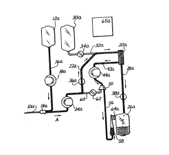
Flgures 1 through 4 are comparative, schematic
illu~trations of a prior art apheresis method and device
tFigure~ 1 and 3) and an embodiment of the method and
device of the present invention (F$gures 2 and 4).
- Generally, the apheresis systems of the prior art and lS those of the present invention incorporate certain common
: components. A venipuncture needle 10, lOa is
percutaneously insertable into a peripheral vein of a human
plasma donox. A bag or other cont~ner of anticoagulant
. ~olution 12, 12a is fluidly connected, by tube 16, lSa, to
: 20 a ~ixing chamber 14, 14a which i~ proximal to needle 10.
: An antlcoagulant pump 18, 18a is positioned on tube 16, 16a
to draw anticoagulant solution from bag 12, 12a through
tube 16, 16a, into the mixing chamber 14, 14a.
Anticoagulant ~olution entering the mixing chamber 14, 14a
~:: 25 will ~oin with, and will become dispersed in, blood which
has been extr~cted proximally through nes~e 10.
: ~ blood ~eparation apparatus 20, 20a is fluidly
oonn r ~ea to the mixing chamber 14, 14a by tube 22, 22a.
A bidirectional blood pump 24, 24a, preferably a
~:~ 30 peri~t~ltic pump, i~ positioned on tube 22, 22a for
alternate withdrawal of blood and infusion of cell
'concentrate through needle 10, lOa. Movement of the blood
pump 24, 24a in a clockwise direction will move blood in
the direction of arrow A (withdraw), while movement of
blood pump 24, 24a in a counter-clockwise direction will
r ~ 10 ~ 9 ~ 3 7
i~ move fluids (e.g. cell concentrate from line 60) in the
F~ direction of arrow B, back to the human subject.
A cell pump 44, 44a is positioned on line 42, 42a to
move cell ooncentrate out of the ~eparation dev~ce 20, 20a
5 at a controlled rate. Close control of the calibration of
: the cell pump 44, 44a is critical in that there exists
strict limits on the amount of oxygen transporting red
blood cells which may be held in the extracorporeal circuit
at any point in time. Thus, close control of the amount of
10 cell concentrate being pumped by the cell pump 44, 44a is
necèssary to ensure that such limits are not exceeded.
Also, the calibration and throughput of the cell pump
directly affects the transmembrane pressure within the
separ~tion device 20, 20a. If the calibration and
15 throughput of the cell pump 44, 44a is not closely
controlled, errant pressures within the separation device
20, 20a may result in hemolysis of the blood cells,
incomplete separatlon of the blood and~or an automatic
error signal and shut down of the machine. A plasma
20 cont~ne~ 26, 26a ~ oonnected to t~e plasma outlet port of
blood separator 20, 20a by way of tube 28, 28a. A s~ e
bag or container 30, 30a is connected to blood line 22, 22a
-~; at a point near the inlet port of blood separation device 20, 20a. A saline valve 34, 34a is alternately
25 positionable in an open po~ition whereby flow through line
; 32, 32a, ls permltted ana a closed po~ition whereby flow
through llne 32, 32a ~s prohibited.
A blood valve 36, 36a ~ positioned on blood line 22,
22a. Blood valve 36, 36a ~s alternatQly pos~t~onable in an
open poslt~on whereby flow through line 22, 22a is
perm~tted, ~nd a closea posit~on whereby flow through line
22, 22a is blocked.
A plasma valve 38, 38a is po~itioned in line 28, 28a.
The plasma valve 38, 38a ls alternately positionable in an
open position whereby flow through line 28, 28a is
,'
.. . .
8 3 7
--1 1--
permitted and a closed position whereby flow through line
28, 28a i# prohibited.
In the typical apheresis maeh~ne of the prior art
(Figures 1 and 3), a cell eone~ntrate reservo~r io ls
loeated remotely from th~ ~eparate plasma vessel 26.
Separate, diserete systems are employed to monitor the
relative weights and/or volumes of a) ~ell eoncentrate
colleeted in the eell reservoir 40 and b) plasma eollected
in the plasma vessel 26. As shown, the plasma vessel 26 is
attached to weighing device 64, sueh as an electronic
balance, 80 as to continuously monitor the weight of the
plasma eontainer 26 and. its .eontents.. The level of eell
eoneentrate in the eell reservoir 40 is, on the other hand,
often mea~ured by a series of eleetronie sensors or other
measuring device(s) loeated in or ad~aeent to the eell
reservoir 40. Thus, the weighing deviee 64 and the sensors
or other measuring device(s) associated to the eell
: reservoir 40, are ~eparately eonneeted to, and provide
..separate si~nal~ to a eentral eomputer 65, 65a. The
.:~ 20 eomputer 65, 65a may inelude an eleetronie mieroproees~or,
~: t~ing and logie eireuits, program memory, eo~munieation
busses and power supply eonnections.
The eell eoneentrate re~ervoir o~ the prior art
maehine 40 (Figures 1, 3) is fluidly eo~neeted to the eell
eoneentrate output port of the blood separation deviee 20
by way o~~a flexible tube 42. A eell pump 44, sueh as a
peri~taltle pump, is positioned on tube 42 80 as to pump
-' the eell eoncentrate from the eell eoneentrate outlet port
::~ of the blood separation device 20 through l~ne 42 into the
eell eoneentrate reservoir 40. The outl~t port of eell
:eoneentrate re:servoir 40 i~ eo~ns~ed to the lower portio~
:~ of the blood line 22 by way of a flexible tube or line 46.
Cell eoneentrate valve 48 is positioned on line 46. The
eell concentrate valve 48 is alternately pos~tionable in an
;~
.' '~lOg837
~ -12-
open position whereby flow through line 46 is permitted, a
clo~ed po~ition whereby flow through line is prohibited.
As ~hown ln the diagrams .of Figs 2 and 4, the ~ystem
of the present invention differs from the prior art system
shown in Flgures I and 3 in that the concentrate outlet
port of the blood 6epar~tion device 20a i8 connected to the
top inlet port of a blood filter/bubble trap 50 by way of
a flexible tube or line 52. ~he cell pump 44a i8
po8itioned on line 42a to pump cell concentrate -from the
cell concentrate output port of blood separator device 20a
into the top port of blood filter/bubble trap 50. Another
~ flexible tube or l~ne 56 conne¢ts the right--side bottom
port .of blood filter/bubblé trap 50 to a bottom fill port
of cell bag 58. A left side bottom port of cell
filter/bubble trap 50 i~ connected to a point on line 22a,
as shown, by way of a flexible tube or line 60. A cell
concsntrate valve 62 i8 positioned on line or tube 60.
Cell concentrate valve 62 i8 alternately positionable in an
open po8ition whereby flow t~rough line 60 i~ permitted,
~20 and a closed position whereby flow through line 60 i8
: blocked.
The darkened tubes and components (shown in Figures 1
and 2) lndicate the respective flow paths of fluids w~thln
a typical prior art apheresis ~ystem during collection
(Figure 1) and reinfu~ion lFigure 3).
As speci~10ally lllustrated in Figure 1, the
collection of plasma by a prior art plas~aphere~i~ machine
was generally accompllshed with valves 36 and 38 in their
open po~itions and valves 34 and 48 in their closed
position8. Antiooagulant pu~p 18, blood pu~p 24 and cell
:pump 44 are conco~itantly actuated during colle¢tion, so a~
to pump flulds ln the dlre¢tion~ indicated by the arrows of
Figure 1. Specifi¢ally, an anticoagulant pump 18 turns in
a clockwise direction to pump dilute anticoagulant sol~tion
from anticoagulant reservoir 12, through line 16, into the
,:
~'
~ "
-13- 21~37
mixing chamber 14 which is positioned proxlmal to
venipuncture ~eedle ~0. Blood pump 24 rotates in a
clockwise direction and operates to withdraw blood through
needle 10 ~uch th~t blood will become mixed with
anticoagulant solution as the blood is drawn through the
mixing chamber 14. Whole blood (mixed with anticoagulant
solution) i~ then withdrawn by blood pump 24, through line
22, lnto tbe separation device 20. The separatlon device
substantially separates blood plasma from a cell
concentrate which contains the formed elements of t~e blood
(i.e. red cells, white cells and platelets). The cell pump
44 operates to withdraw the cell concentrate from the cell
concentrate outlet port of blood geparation device 20,
through 11ne 42 and deposits the cell concentrate in cell
lS concentrate reservoir 4~. Since valve 48 is in its
closed~ position, the cell concentrate is prevented from
moving past valve 48 when the devlce is in the depicted
collection mode. Air di~placed from the interior of the
reser~oir is vented through a hydrophobic filter/vent port
41 formed in the top of the reservoir 40. Blood plasma
flowing from the plasma outlet port of the blood separation
device 20 is permitted to drain through line 28 lnto plasma
colleotion vessel 26.
In the device of the present invention (~igures 2 and
4) a i~n~!e we~g~n~ ~ev~ce 64a, uch ~as an electronic
~l~nce or load cell, is utilized to concomitantly weigh
~ a) ~he plasma conta~er 26a and its contents, and b) the'
-- cell conce~trate bag 58 and its contents. The use of this
~ingle weighing dev~ce 64a for both the plasma cont~ner
68a ~na tbe cell bag 58 eliminate6 the need for a separate
system for colleqtin~ and measuring the cell concentrate at
a locatlon remote from the pla~ma contA~ner. Also, the use
- of the ~ingle weighing device 64a, in accordance with the
method of the present invention, provides ~or highly
accurate measurement of the throughput, of the blood pump
~10983~
-14-
24, 24a and cell pump 44, 54, thereby permittlng accurate
and frequent recalibratlons thereof. Addltionally, this
invention enables continuous, redundant monitorlng of the
blood/cell concentrate flow during withdrawal ~ and
S reinfusion by providing a continual lndicatlon of flow rate
~ based on the changes of weight being recorded by the single
; weighing device 64a as the withdrawal or reinfu~ion occurfi.
'5 The change in weight or rate of change in weight recorded
by welghing device 64a is then continuously or periodically
compared to the calculated flow rate or actual rotations of
pump 44a. If the actual or expected flow through pump 44a
differs more than a certaln amount (e.g. 25%) from the flow
rate indicated by the chan~e in weight being recorded by
the weighing device, such will indicate a problem with the
sy~tem, such as a tubing leak, vessel fracture or
improperly rigged or malfunotioning pump. Thus, this
redundant, comparative flow monitoring capability provided
~y the single weighing device 64a, is also an advantage of
the present invention. Additionally, the invention
provides for the use of an inexpensive pla~tic cell
,~ concentrate contA~er bag 58 and inexpensive blood
filter/bubble trap S0 a~ opposed to the more expensive
components u~ed in ~ome prior art devices, such as the
~~ rigid, vented cell reservoir 40 with attendant electronic
(LED) volume monitoring used in the prior art ~ystem shown
in Figures 1 and 3.
The general method by which the aphere8is system of
the present invention operatès i8 shown in Figures 5a-Sb.
This method i8 more fully de~cribed herebelow with specific'
reference to the ~chematic diagrams of Figures 2 and 4.
ii. The Nethod of the Pre8ent Invention
Initially, the empty plasma reservoir 26a and cell
concentrate bag 58 are placed on a ~ingle weighing device
- 35 84a. A ~DRY TARE" is then measured by the weighing device
~, .
~,
0
~1~983
-15-
64a. The ~DRY ~ARE~ value is communicated to the computer
65a wherein the ~DRY TARE~ value i8 stored. The ~DRY TAREU
rvalue is the combined weight of a) the empty plasma
container 26a, and b) the empty cell bag 58. This ~DRY
5 TARE~ step ifi carried out at the beg~nn~n~ of the
procedure, prior to the initial priming of the system, as
illustrated in Figure 9a. The ~DRY TARE~ value is the
combined weight of the empty plasma vessel 26, ~6a, 234,
234a and the empty cell bag 58, 237. In subseguent cycles
10 after the initial cycle, an ~EMPTY CELL BAG TARE~ 105 is
determined and stored instead of the UDRY TARE" determined
and stored at initiation o~ the first cycle. The ~M~-Y
CELL BAG TARE~ 105 differs from the ~DRY TARE~ in that it
includes the weig~t of plasma collected in previous
15 collection cycles, as illustrated in ~igure 9c
Thereafter, a portion of the system (e.g., the blood
tube 22a, b~ood ~eparator device 20a, tube 52, blood
filter/bubble trap 50, tube 56 and blood bag 58 ) is
initially ~rimed witk a guantity of anticoagulated whole
20 hlooA withdrawn through ven$puncture needle lOa. Such
prim~ng of the system 110 will typically result in a small
amount of whole blood being disposed in the bottom of the
cell concentrate bag 58. At thls point, a ~PRIMED TARE~ i8
measured ~12 by the weighing device 64a. ~he ~PRIMED T~RE~
25 value i~ communicated to the computer 65a wherein such
~PRIMED TARE~ value is stored. The ~PRIMED TARE~ value is
the comb~ed weight of the a) empty plasma cont~i~er, and
b) cell bag containing the small amount of pri~ing blood as
illu~trated in Figure 9b.
After the ~PRIMED TARE~ has been recorded 112, an
lnitial collection cycle is begun 114. During such
collection cycle, the blood valve 36a is in its ~openU
position, the infusion yalve 62 is in its ~closed~
position, plasma valve 38a is in it~ ~open~ posit~on and
blood pump 24a and cell pump 44a are operated in their
- ' ~ o
~ lVt~37
-16-
rQ~pective, clockwise and counter-clockw~se directions, at
specifically controlled rates, as dictated by the program
of the computer 65a. The ~et rates of the pumps 24a and S4
are calculated by the computer 65a on the basls of the
desired pressures to be maint~e~ within the attendant
tubing 22, 52, 28a and the blood separation device 20a.
The rate of the blood pump 24a i8 also determined, to some
degree, in view of the volume and pressure of blood
available to be withdrawn from the blood vessel of the
human subject.
The total volume of blood to be withdrawn into the
extracorporeal circuit in any given collection cycle is
controlled by preeetting the number of rotations to be made
by the cell pump 44a during the next collection cycle. The
numbers of rotations that the pumps 24a and 44a will
undergo, in each given collection cycle, is controlled by
computer 65a on the basis of a preset Upump flow constant"
for each pump (BP and CP). The de~ired number of rotations
~ for any given collection cycle i8 ~enerally determined on
the basis of the following eguation:
~.~
t;o~ No. 1
Weight of Material Counted
Pltm~ ~m~~r of Pl-m~ Rev~. (Rev.)
8p.Gr. of Material (g/ml) Flow Constant (Rev./ml)
~ .
~
To control the volume to be pumped during the f ~ rst or
~tart-up collectlon cycle (step 114-116), the desired
- rotatlons for the cell pump 44a wlll be preset by th~
computer 65a on the basi~ of an ~initial~ flow con~t~nt for
each pump. Thereafter, for each repetitive collection
cycle, an ~ad~u~ted~.flow constant will be determined and
stored in the computer 65a. Each such ~adjusted~ flow
g837
-17-
constant will be based on actual measurement~ made during
the prev~ou~ collection cycle. Such frequent ad~ustment of
the desired rotations of the blood pump and cell pump helps
to insure that accurate fluid volumes are maint~ined
throughout the procedure.
The collection is accomplished by r~nn~n~ the blood
pump 24a and cell pump 54 in their respective ~collection~
directions or modes. Typically, such will require that the
blood pump 24a be rotated in a clockwi~e direction while
the cell pump 54 be rotated in a counter-clockwlse
direction. Typically, the cell pump 44a is utilized to
precisely gage and control the amount of red cells
withdrawn in a single collection cycle and the blood pump
24a continues to run in conjunction with the cell pump 44a
until the cell pump is stopped (i.e. where it has undergone
a present number of rotations. Thus, in any collection
cycle prior to the final collection cycle of a given
procedure, the cell pump 54 will undergo a predetermined
number of rotations as preset in the computer 65a or as
selected or overridden by the operator. The present number
of rotations will achiove a precalculated guantity of cell
concentrate pumped by cell pump 44a. Such precalculated
quantity of blood cell concentrate withdrawal is generally
related to a ~pecific weight of cell concentrate cont~ne~
within the cell bag 58 and 18 below the maximum allowable
-~- extracorporeal red cell volume permitted by applicable
government regulations.
In order to insure that the maximum allowable plasma
collection is not exceeded, it i8 desirable to continuously
or periodically calculate the current predicted or
- calculated plasma wt. (Pp~) and to continuously, or at
di~crete time points during each colloction cycle, compase'
such predi¢ted plasma volume to the maximum allowable
volume of~plasma withdrawal (P~x) 116. The Pm~X~ in most
instances, is determined from generally published data
~,
. ~ .
~109837
-18-
tabies or nomograms, based on the height and/or weight of
a generally healthy blood donor and in accordance with
governmental regulations. In certain therapeutic
instances ~ however, the Pm~ will be determlned and ~et by
the operator or medical practitioner t~ g ~nto account
the general health of the patient and~or other facts
relating to the therapeutic procedure being performed.
In a preferred embodiment of the present invention,
the computer 65a continuously monitors the P~" in
compari~on to P~. The predicted plasma (Pp~ determined
by the following formula:
F.~1A t~o~ No.2
Primed Cell Bag ~ z Primed Tare - Empty Cell
Bag Tare
~ t~ on No . 3
Pp" = Current Weight - Dry Tare- Primed Cell Bag
- Current Cell ~.~ Coast Bias
When P~" i8 determined to equal Pm~X, the collection i8
immediately terminated by the computer 65a and the device
moves directly.into the final reinfus~on cycle of the
procedure, as will be fully described hereinafter.
In a typical prefinal collection cycle (a full
¢ollection cycle whlch yields a final volume of plasma
collectea which 18 less than P~l) prlor to the flnal
collection cycle dur~ng which the procedure is terminated,
the end of collection will be marked by a weight of red
cell con¢entrate w~t~in the cell bag 58 and an attenAant
weight of separated plasm~ wlthln the plasma contalner 26a.
After the particular collection cycle has been ended 118,
the weigher 64a will take a ~post-collectlon weight~ 122,
as illustrated in Figure 11 and will transmit such weight
~, . . .
.. ..
o
-19- ~1~9837
to the computer 65a wherein it will be stored. The "post-
collect~on weight~ 122 is the combined weight of a) the
p~asma cont~e~ plus all pla6ma contained therein, and b)
the cell bag plu8 all cell concentrate (and any priming
S blood) collected therein plu8 any pri~ing blood, pr~i~e~
cell bag ~, 324, contained therein.
After the ~post-colleotion weight" has been recorded
122, the blood valve 36a will move to its ~closed~ position
and reinfusion valve 62 will move to its ~opened~ positlon.
The blood pu~p 24a will then be operated in its counter-
clockwise direction to effect reinfusion of the cell
concentrate (and/or any priming blood) from the cell bag
58. through~tube 56, through blood filter/bubble trap 50,
through tube 60, through mixing chamber 14a, and distally
through needle lOa, into the blood vessel of the human
donor. It is desirable that such reinfusion cycle ef~ect
complete reinfusion of all cell concentrate (and/or priming
~ blood) contained in the cell bag 58a. Thus, the computer
j 65a may be capable of continuously or periodically
monitoring the flow of fluid through the reinfusion system
: in order to detect when the cell bag 58a has been fully
emptied and to automatically stop the counter-clockwise
movement of the blood pump 24 at such point. The actual
number of revolutions made by the blood pump 24 during each
reinfu~ion of cell concentrate i~ counted 128 and stored in
co~puter 65a. If a subseguent collection cycle is to be
co~pleted, (i.e. if the volume of plasma collected thus far
has not reached P~), then the weighing device 64a wi 11
determine and ~tore 134 a ~post-re~nfugion weight~. ~he
~po~t-reinfusion weight~ is the combined wetght of a) the
plasma oon~n~r plu8 all pl~sma cont~n~d t~ere~n, and b)
- the empty cell bag~
After the ~pogt-reinfugion weightU has been ~tored 134
in the oomputer 65a, the computer 65a will proceed to
calculate the ~weight of cells reinfused~ 136. The ~weight
8 3 7
-20-
of cells reinfused" is determined on the basis of the
' following formula:
~uat~on No. 4
Wt. of Cell Concentrate Reinfusedtg) -
, (Post-Coll.Wt.(g~ - Post-Reinf.Wt.(g)~
t ~dditionally, the computer will calculate the "weight
10 f actual plasma collected" 138 as of the end of the ~ust-
ended collection cycle. The "we~ght of actual plasma
collectedN, ~wt. of blood pu~ped during collectionl' and the
wt. of cell concentrate pumped during collection~ are then
calculated by the following equations nos. 5, 6, and 7:
~uation No. 5
Wt. of Pla6ma = Post-Reinfusion W~.(g)
Collected (g)
- EMPTY CELL BAG TARE (g)
.
~ t~on No. 6
Wt. of Blood = Post-Collection Wt.(g)
Pumped dur$ng ( see Fig. 11)
Collection
- Cycle (g) - PRIMED TARE (g)
(see Fi~. 9d)
- 30
; ~uat~on No. 7
Wt. of Cell Concentrate - Po6t Collection
Pumped During Collection Wt. (g)
Cycle.(g) (see F$g. 11)
'r ' ~ Post Reinfusion
Wt. (g)
t8ee Figure 12)
- PRIMED
CELL BAG
' ~ 2109837
-21-
The eomputer 65a will also ealeulate new eo~lect
flow eonstants for the blood pump 24a and eell pump 44a.
Also, the eomputer 65a will automatieally, on the ba~ls of
sueh new flow eon~tants, reset the deslred number of
rotations for the blood pump ~nd eell pump for the next
eollection eycle. Sueh resetting of the desired pump
rotation~ prior to each eollection cyele serves to ensure
that during the next eolleetion eyele, there ~ill be
aeeurate eontrol of the volumes of fluids pumped by the
blood pump 24a and cell pump 44a.
Thevealeulation of the collectlon flow constants for
the blood pump and cell pump are ba#ed on the following
equations nos; 8 and 9:
~u~t~o~ No. 8
Blood Pump No. of Pump
Colleetion Flo.Con. - S~.~r. of Rloo~ t~/mlL X revs. during
(Rev/ml) Wt of Blood eollection
20Pumped (g) (Re~s)
R~-~t~o~ No, 9
Cell Pump No. of Pump
Colleetion Flo.Con. = ~.Rr. of Cel~ (~/mlL X rev~. during
Rev/ml) Wt. of Cell collection
Coneent. Pumped(~) (Revs)
,
~he welght of eell~ reinfused will subsequently be utilized
n~the ealeulation of a rev~ed ~e~n~.~Q~n flow eonstant
for the blood pump 24a by applieation of Equation l and the
newly ealeulated reinfus1on flow eonstant for sueh pump
~-~ will be re8et in the computer for subsequent reinfusion
, ~ -
~- ~ 35 ey~les.~
The ealeulation of the relnfusion flow eonstant for
the blood pu~p lc based on the following formula:
~ ~ .
: ' 210~837
;
--22--
P~-~tl oll No . 10
Blood Pump Sp.Gr. of Cell No. of Pump
- Reinfus. ~lo.Con. = Co~c. (~ml ) X revs. during
(Rev/ml) Wt. of Cell Reinfu~ion (Revs)
Conc. Reinfused(g)
;
After the new flow constants have been calculated and
~tored in computer 65a, and, the desired number~ of
rotations of the cell pumps 44a has been ad~ustea (steps
140 and 142), a new collection cycle i8 begun. Steps 105-
142 are repeated until ~uch time as the computer 65a
determines, during step 116 (i.~. monitoring of Pp" versus
Pm~) that, the Ppr~ is equal to P~x~ When it i8 determined
that Pp~ eguals P~, the collection i8 automatically
terminated by the computer 65a, and the final reinfusion
~tep i8 carried out.
After the fi~l reinfusion step has been completed,
the actual total amount of pla~a collected will be
etermined by the weighlng device 65a. Such Total Plasma
Collected (Actual) will be stored by the computer 65a. The
Total Plasma Collected (Actual) is determined by the
following formula:
'
uat~on No. 11
Total Pla~ma Collected (Actual) (g?
(Post-Reinfu~ion Wt. (g) - D~Y TARE (g))
f
lii. A 8pecific Plasmapheresis H~ch~e
~ah~l~ent of The Present Invention
In accordance with the genesal ~y~tem and method
:
described above, the following detailed description of a
- specific plasmapheresi~ machine embodiment of the present
invention is provided.
~ '
:
f~ .
i ~
~ 2~9837
,.
-23-
A blood line 180, 180a is fluldly connected to a
venipuncture needle which resides within a perlpheral voin
of a human donor (not shown). The proximal end of the
blood llne 180, 180a bifurcates into a left ~enous pres~ure
transducer line 182, 182a and a right blood pump tube 184,
184a. The left venous pressure transducer line is
connected to a venous pressure transducer located within
the hou~ing 200 80 as to provide to the computer (not
shown) cont~ or di~crete monitoring of the pos-itive or
negative pressure within the blood line 180, 180a. The
blood pump tube 184 is operatively positioned within a
peristaltic blood pump 186, 186a. The opposite end of
blood pump line 184a is concom~tantly connected, by way of
a Y connector, to a reinfusion line 188, 188a and a first
6eparator feed line 190, 190a bifurcates into a second
separator feed line 192, 192a and a transmembrane pressure
transducer line 194, 194a. The transme~brane pressure
transaucer line 194, 194a is connected to a transmembrane
pre~sure transducer (not shown) which, in turn, is
cQ~ ec~ed to the system computer (not ~hown) ~uch that the
computer may continuously or discretely monitor the
junction of the first ~eparator feed line 190, 190a and the
second separator~feed line 192, 192a.
A presently preferred, automated plasmapheresis
mao~e of the present invention is shown in Flgures 7-7f.
Figur- 6 shows a similar ~achine of the pr~or art, which
do-s not incorporate the ~ethod or aev~ce of the prefient
invention.
Referring to Figures 6 and 7, the prior art machine
(Figure 6) and the machine of the pre~ent invent~on (Figure
7) ~hare certain common components~ Both of these machines
comprise a housing 200, 200a wherein a central computer~,
wiring, electrical connections and other general components
of the device (all not ~hown) are ~ounted. On the frontal
; 35 surface of the housing 200, 200a, there is provided a
~ ,~ ,
~ ' ~ 21~837
-24-
~y~tem of tubes, pumps, reser~oirs and components for
effecting the desired a) withdrawal, b) ~eparation, and c)
; reinfu~ion of blood and~or blood component~. Generally, a
6aline line 202, 202a leads from an attendant bâg or
cont~ner of phy~ologlcal 0.9% ~aline solutlon and an
antieoagulant line 204, 204a leads from an attendant bag or
eGntA~ner of antieoagulant solution. The ~aline line 202,
202a passes through a power actuated elamp 206, 206a and is
eonnected to a Y adaptor 208, 20Ba. The oppo~lte side of
the Y adaptor 208, 208a i~ concomitantly eonnected to the
inlet port 210, 210a of a blood ~eparation device 212,
212a. ~he blood 6eparation device may eonsist of any type
of device capsble of effectuating the de6ired 6eparation of
blood eonstituents. In a preferred embodiment, ~eparation
device 212, 212a eomprises a disposable, rotational plasma
separator having an internal rotatable membrane which i~
driven rotationally by an external magnetic motor drive
(not shown). Sueh rotation of the inner membrane causes
blood plasma to geparate from the eell eoneentrate (a
eombination of red blood cells, blood whlte eell~,
platelets and a small amount of plasma). The eell
eoneentrate flows out of the separation device 212, 212a
through eell eoneentrate outlet port 214, 214a. The plasma
flows out of the separation device 212, 212a through plasma
25 outlet port 216, 216a.
- A eoneentratea eell llne 220, 220a i8 eonneeted to the
eell eoneentrate outl~t port 214, 214a of the blood
separation deviee 212, 212a. The eoneentrated eell line
220, 220a is mounted within a peristaltie eell pump 222,
~: 30 222a. the p~rista:ltic cell pump 222, 222a may be
ub~tantially identieal to the previously des¢ribed blood
pump 186, 186a, or ~ay eompri6e any other type of pump
eapable of effeeting the desired movement of eell
coneentrate through eoneentrated eell line 220, 220a.
~09837
In the prior art device (Figure 6), the concentrated
cel~ line ~20 carries cell concentrate from the blood
separation device 212, through cell pump 222 and into the
inlet port 224 of a rigid cell collection reservoir 226
having a cap~city of approxim~tely 300 milliliters. Such
300 ml capacity allow~ adequate extra space in the cell bag
237 when a usual collection amount limit of 180 ml of cell
concentrate is observed. ~ cell concentrate outlet 228 is
located at the bottom of the ce~l concentrate re~ervoir
226. The cell concentrate reinfusion line 188 i~ connected
to the cell concentrate outlet 228 of the cell concentrate
re~ervoir 226 80 as to permit reinfusion of the cell
concentrate into the human donor when the clamp 189 is
open, clamp 191 is closed and the blood pump 186 is
operated in its ~reinfusion~ direction (counter-clockwise).
Also on the device of the prior invention (Figure 6) a
pla~ma line 230 extends downwardly from the plasma outlet
port 216 of the blood ~eparat~on device 212, passing
through plasma cla~p 232 and le~ng directly into the top
of pla~ma collection ve~sel 234.
~In contrast, the device of the present invention
;(F1gure 7) is configured 80 a~ to eliminate the need for a
rigla cell reservoir and to collect the cell concentrate in
- a low co~t flexIble cell bag 236 which hangs from the same
we~ghing ~evice 235a as the plasma collection ve~sel 234a.
Also, in the device of the pregent invention~(Figure 7) the
;cono-ntrated cell line 220a~ i~ conneoted to one of the
~nlet/outlet ports of a blood filter/bubble trap 240. The
~loo~ filter/bubble trap 240 contains a ~creen or quantity
of fibrous filtration ~aterial so as to trap bubbles,
foreign ob~ects, e~boli, etc. (A specifio preferred
e~bo~ment of the blood fllter/bubble tra~ 240 i~ ~hown in
Flgure~ 8a through 8d and will be ~ore fully described
hereinafter.)
~ .
~::
~1()9837
--26--
,' Al80 fluidly connected to the blood filter/bubble trap
,S, 240, opposite the inlet of the concentrated cell line 220a
is a lower cell line extension 242. Such lower cell--line
extension 242 fluidly connects the blood filter/bubble trap
240 to the inlet/outlet port 244 po~itioned at the bottom
of the cell collection bag 237.
A preferred mode of operation of the device shown in
~igure 7 is illustrated in Figures 7a through 7f.
Specifically, Figure 7a shows a preferred plasmapheresis
machine of the present invention during the initial priming
of the system. Such priming of the system is effecting by
closing clamp l91a, opening, clamp 189a and operating blood
pump 186a in it8 ~collectionU direction (clockwise) while
anticoagulant pump 205a operates relatively slowly in its
operative direction (clockwise). The combination of such
,~: will result in withdrawal of whole blood (containing a
mall amount of anticoagulant) through the blood l,ine 180a,
: ~ blood pump line 184a, opening clamp 189a, through blood
~ filter/bubble trap 240, down the lower cell line 242 and
:: 20 into the very bottom of the cell bag 237. This initial
~:~ pri~ing ~tep i8 illustrated by the darkened and shaded
areas shown in Figure 7a. Generally, it is predetermined,
- ~ based on the calculated dead space of the tubing and
comr~nents, that approximately 32 ml of whole blood mu~t be
pumped by~the bloo~ pu~p ln order to ef~ec~ this ini~ial
~ primlng step;ana to bring whole blood through to the bottom
'.~: of the c~ll bag 237. ..... Thu8, the computer (not shown)
signals tl~e blood pump 186a to rotate ~n a cloclcwise
diré¢tion. The blood pu-np 186a stops after a mass of 12
30 - gram- i~ a-t-ctea on'the welghing aevi¢e 235a, a~ generally
provides for initial priming of the lower portlon of the
~: system as ~hown in Fiçlure 7a.
~; After the initial priming step has been completed, the
device moves on to a secondary priming step known as the
~' 35 "filter prime~ he "filter prime~ step is illustrated by
~109837
-27-
the darkened and shaded areas in ~igure 7b. During the
filter prime step, the clamp l91a i8 opened, clamp 189a is
allowed to remain open, and the blood pump 186a is operated
in its ~collectionU direction tclockwise) for a sufficient
5 number of rotations to pass whole blood upwardly through
line 192a and to generally fill the concentrated cell line
220a, and the remainder of blood filter/bubble trap 240.
'This will also result in the flow of some additional whole
sblood into the lower concentrated cell line 242 and the
10 entry of a slight additional amount of blood into the
bottom of the cell bag 237. Based on the initial,
.. .
empirically determined or otherwise chosen pump ~low
constants, the blood pump 186a and the cell pump 222a are
commanded by the computer (not 6hown) to pump sufficient
amounts of blood to fill the tubes, blood separator and
blood filter/bubble trap, as shown in Figure 7b. ~he
computer (not shown) permits the blood pump 186a to undergo
a preset nu~ber of revolutions determined to deliver that
decired volume of blood and thereby effecting the desired
filter prime without aspirating more than the necessary
amount of blood from the patien~.
After the "filter primeU step has been completed, the
~PRIMED TARE~ ~tep 112 as lllustrated in Figure 9d, is
carried out. Thereafter, the ~nit~al collection cycle 114
is begun.
The collection 6tep, as appl$ed to the presently
preferred device, is illustrated in Figure 7c. Dur~ng
collection, the anticoagulant pump 205a, blood pùmp 186a
and cell pump 222a are all operative in their ~collection"
directions. Valve 191a is opened and valve 189a i8 closed.
Whole blood, along with a small amount of anticoagulant
solution, is drawn by blood pump 186a, through the
attendant tubing, into the blood separation device 212a.
Plasma çlamp 232a is opened and cell pump 222a
operates to withdraw cell concentrate 220a from the blood
,'
,,
.,, . . ~ " . . .
' ~la~s37
-28-
; separation device 212a. The cell concentrate passes
through blood filter/bubble trap 240, down the lower cell
concentrate line 242 and is collected ~n the cell bag 237.
It will be appreciated that, while the collection process
is cont~n~ g, the computer may continually monitor the
plasma predicted (P~") versus pla~ma maxlmum (P~x) in
accordance with step 116 of the inventive method (Flgure
3a). If, at any point, the Ppr~ becomes equal to P~,~, the
computer will immediately stop the blood pump 186a,
anticoagulant pump 205a, and cell pump 222a, thereby
terminating the collection at P~x~ The device will, upon
detection of Pp" eguals P~,~, move into reinfusion mode in
accordance with step 124 of the inventive method (Figure
3a). However, if Ppr~ does not become egual to P~x during
the collection cycle, that collection cycle will be
permitted to continue to full completion (e.g. collection
of 180 milliliters of cell concentrate) where the cell pump
222a has undergone its preset number of rotations based on
the precaloulation of necessary rotations to obtain the
desired amount (e.g. approximately 180 milliliters) of cell
~- ~ concentrate in the cell bag 237. When the cell pump 222a
has undergone lts preset number of rotations, the computer
will stop the movement of all pumps 184a, 205a, 222a,
thereby ~n~ ~ ng that collection cycle. Of course, during
the collection, the computer will continually monitor the
instant predlcted plasma volume (P~) and will contlnuously
or periodically compare Ppr~ to the maximum allowable plasma
volume, in accordance with ~tep 118 of the inventive method
(Figure 3a).
The end of the collection cycle is lllu8trated ln
Figure 7d.
Prior to beg~ nn1 n~ reinfusion, the weighing device
235a will measure the ~post-collection weight~ and ~uch
value will be stored in-the computer. Thereaf~er, the
~ ~ '
. .
'~109837
-29-
device will begin relnfusion of the cell concentrate into
the donor.
Reinfusion of the ¢ell concentrate i6 effected by
opening cla~p 189a, closing clamp l91a, and r~)nn~ng the
blood pump 186a in it~ ~reinfusionU direction (counter-
clockwise) until the entlre amount of cell concentrate
contained in the cell bag 237 has been reinfused into the
human donor. In a preferred embodiment, the computer will
~onitor the flow of cell concentrate through the device ln
order to determine when the dynamics of reinfusion flow
indicate that the entire volume of red cell concentrate
(approximately 180 ~l) has been reinfused. Thls may be
achieved by cont~m~lly monitoring the rate at which the
weight on weighing device 235a change~ with respect to
lS blood pump flow rate and determining from the detected
change in weight on weighing device 235a, when the cell bag
237 has been emptied by applying the function, such as:
2g < Mag ¦Current weight on - Past weight on < 6g
¦weighing device (g) weighing device (g)
where1n: ~past weight~ i8 the weight which
~ was on the weighing device at the
- ti~e when the expected ml. of
pump flow was 4 ml. less that the
pre8ent expected ml. of pump
flow.
Additionally, during both collection and reinfusion, the
co~puter will cont~u~lly verlfy the functionlng of the
pump~ by app}ylng a function #uch a~ the above-~et-forth
funct~on~ and, lf at any point, the magnitude of difference
between current wt. and past wt. exceed~ the allowable
range, the device will ~hut down and the operator will be
'" . ~ 30
~ 2~98~7
.. . .
signaled to check for possible malfunctions (e.g. leaks in
the system). Detecting an empty cell bag can be
distin~~ e~ from a system malfunction b~D~ upon a
predicted expected time o~ ence of the emptying.
;
During the reinfusion, the computer will count and store
the number of rotations undergone by blood pump 186a in its
"reinfusion" direction. This number will be subsequently
-utilized in recalculating and adjusting the reinfusion pump
(i.e. reverse direction) flow constant of the blood pump
186a, in accordance with the method of this invention.
At the end of reinfusion, the cell bag 237 will be
completely empty as shown in Figure 7f. At ~hat point, the
weighing device 235a will obtain the post-reinfusion weight
; in accordance with step 134 of the method (Figure 3b).
Thereafter, the computer will calculate the a) weight of
cell concentrate reinfused (step 136), b) weight of actual
plasma collected (step 138), c) collection flow constants
for th~e blood pump and cell pump (step 140), and d) a
reinfusion flow constant for the blood pump (step 142).
The desired number of cell pump rotations for the next
collection cycle will be recalculated by the computer on
the basi~ of the newly calculated flow constants and, the
~r--~t number of cell pump rotations will be accordingly
reset for the next collection/reinfusion cycle.
The blood filter/bubble trap 240 of the device may
consist of any type of outer housing or shell having
positioned therein one or more materials operative to
effect filtration of the blood and/or trapping of h~hhles
as the blood p~ 's through the blood filter/bubble trap
240.
iii. A Preferred 8100d Filter/Bubble Trap
U~able in the Device of the ~ nt Invention
One presently preferred type of blood filter/bubble trap
~- is shown separately in Figure 8. This preferred blood
filter/bubble trap 300 comprises an outer plastic shell 302
.. ,, . . .. , . . ., . ~ .. ... .. . . . .
~1~9~337
-31-
of generally cylindrical configuration. The shell is
compressed to a flat, closed configuration at its top end
304 a~d bottom end 306. A filtration bag formed of a
material approved for use in blood pathway and ~lood
processing, (e.g. certain fabrics, filtration media or fine
mesh materials, such as a nylon mesh) is positioned inside
the shell 302. The opening size or mesh size of the mesh
material or fabric or filtration material is preferably
about 220 microns. Second 312 and third 314 inlet tubes
10 pa88 through the closed bottom end 306 of the shell 302.
A ~tand pipe 314 is fluidly connected to the third input
tube 312 and extends upwardly therefrom with the confines
of the shell 302.
' In its preferred embodiment, the filter 300 is
approximately 12 centimeters in lengthffrom the top edge
304 of the shell to the bottom edge 306. The stand pipe
314 is approximately 2 centimeters in length.
In normal operation, the preferred blood filter/bubble
trap device shown ln Figure 8 is mounted in the device of
the present lnvention (Figure 7) such that the cell
concentrate line 220 i8 connected to the fir~t inlet tube
308, the reinfusion line 188 ls connected to the second
inlet tube 312 and the lower cell concentrate line is
connected to the third inlet tube 314. When 80 mounted in
the device of the present invention, the filter bag 310
; will operate to strain or f~lter c~ll concentrate flowing
into the blood filter~bubble trap 300 from the blood
s~paration device 212a. Additlonally, the pre~ence of the
stand pipe 314 within the blood filter/bubbl~ trap 300 will
- 30 lnsure that a guant~ty of blood or cell concentrate pools
-in the bottom of the inner chamber of the blood
filter/bubble trap 300 before such blood or cell
concentrate begins to flow down the lower cell concentrate
line 242. The opening of the second inlet tube 312 which
is connected to the reinfusion line 188a is generally flush
,
.
~109837
.,
-32-
with the inner floor or bottom of the interior of the ~hell
302. Thus, the opening into the seeond inlet tube 312 will
routinely be mainta~ned below an approximate 2 eenti~eter
head of blood or eell eoneentrate. By this arrangement,
eell eoneentrate flowing through the f~ltor bag 310 will
fall lnto the bottom of the ehamber and will rise to the
level of the top of the stand pipe 314 before flowing down
the lower eell concentrate line 242. This will help to
prevent turbulent eell eoneentrate eontA~n~ aberrant
bubbles from entering the lower cell concentrate line 242.
Sueh pooling of the cell eoneentrate in the lower 2
centimeters of the blood filter/bubble trap 240 will a'llow
the eell eoneentrate an opportunity to dega~ before
~, beg~nn~ng to flow down the lower cell concentrate line 242.
Sueh will help to prevent the introduction of air or
~: bubbles into the cell bag 237.
The foregoing detailed deseription has discussed only
several illustrative embodiments or examples of the present
lnvention. ~hose 6killed in the art will reeognize that
numerou~ other emho~ents, or additions, modlficatlons,
deletions and variations of the deseribed embodiment, may
be made without ellminating the novel and unobvious
features and advàntages of the present invent~on. It is
:; intended that all sueh other embodiments, modifications,
deletions and variations be ineluded within the seope of
the following o1aims.
:: ~
::
,