Note: Descriptions are shown in the official language in which they were submitted.
~~ 1 t n,~~%/~~p
1.n ~.
1
Clinical measurements of arterial blood flow
are used for both diagnosis of disease severity and for
assessing the success of interventional and surgical
procedures. Both the average flow rate and the degree of
waveform pulsatility are of clinical interest. Because of
this interest, several techniques have been developed to
measure blood flow and velocity using Doppler ultrasound
and magnetic resonance imaging. Although these
techniques show great promise, they have yet to displace
conventional x-ray angiographic techniques in clinical
practice, although they may be used in addition to x-ray
angiography to assess stenosis severity.
X-ray angiographic procedures still remain the
"gold standard" for determining anatomical information,
such as lumen boundary. Since angiography is in common
clinical use, there has been considerable interest in the
development of x-ray techniques to measure blood-flow
rate. Quantitative angiographic flow measurements have
the potential to provide additional functional
information with little additional risk to the patient.
Previous x-ray angiographic techniques to
measure blood flow have involved the injection of
iodinated contrast agent while recording the passage of
the bolus through the vessel of interest, usually with
digital-subtraction angiography (DSA) at the highest
frame rate possible. The resulting image sequsnce can
then be processed by one of several quantitative
techniques which analyze the passage of contrast agent
through the vessel. These techniques include analysis of
the dilution curve by Stewart-Hamilton's formula,
transit-time analysis, and cross-correlation techniques,
including statistical cross-correlation.
Pulsed-injection of contrast agent has been
proposed to improve the accuracy and precision of
quantitative radiographic flow measurement techniques.
CA 02110240 2004-06-23
2
Pulsed injection with multiple boli provides an improved
signal, since more features are presented within the
vessel, for the same volume of injected contrast agent.
Techniques have been developed using a pulsed-injector
system which showed promising results in vitro tests.
However, an injector with improved bolus definition at
high pulsing speeds is required to measure the entire
range of flow rates found in the vasulature.
According to the present invention, there is
provided apparatus supplying a pressurized source of
contrast agent to a vessel and a control valve to control
flow of the agent from the source into the vessel. The
control valve is a rotary valve that aligns a supply port
with an outlet port as the body of the valve rotates.
Flow to the vessel is thus pulsed. The resulting
distribution of contrast agent within the vessel provides
a strong radiographic signal for quantitative flow and
velocity analysis, using techniques such as cross-
correlation.
In a further aspect of the present invention
there is provided an injection apparatus for quantitative
angiographic blood flow measurements comprising a
pressurised source of contrast agent to be injected into
a fluid transporting vessel, a control valve to control
flow of said agent between said source connected to an
inlet port of said valve and a catheter extending from an
outlet port of said valve to said vessel to convey
contrast agent therebetween, said control valve having a
valve body rotatable within a housing and including a
passageway operable upon rotation of said valve body to
connect periodically said inlet and said outlet and allow
fluid flow therebetween.
An embodiment of the invention will now be
described with reference to the accompanying drawings, in
which
Figure 1 shows a schematic diagram of the
pulsed injector system;
CA 02110240 2004-06-23
2a
Figure 2 shows on an enlarged scale a section
of a rotary valve shown in Figure 1;
Figure 3 shows a perspective view of a catheter
used with the apparatus of Figure l;
Figure 4 shows the use of the apparatus of
Figure 1 in the collection of data;
Figure 5 shows the change of flow rate at the
output from the valve in Figure 2 compared with a
commercially available valve;
Figure 6 shows the change of flow rate of the
contrasting agent at the outlet to the valve in Figure 2
..
0
3
Figure 7 shows a sample image obtained from the
apparatus of Figure 4;
Figure 8 shows the passage of a bolus of agent
along a vessel: and
Figure 9 shows the results obtained by
correlating successive values of the curve of Figure 8.
Referring to Figure 1, a pressurized air supply
is regulated over the range 40 to 100 Kpa and
introduced into a low-pressure cylinder 12 through valve
10 14. The low-pressure cylinder 12 (inside diameter 38 mm)
drives a piston in the high-pressure cylinder 16 (inside
diameter 9.8 mm), resulting in a factor of 15 increase in
pressure. This allows the production of the high
pressures needed to drive contrast agent through small
diameter catheters, while retaining compatibility with
low-pressure medical air sources commonly found in a
hospital environment. Air supply 12 is also connected to
a heated reservoir 18 of contrast agent that supplies the
cylinder 16 through a valve 20.
The high pressure cylinder 16 is connected by a
supply duct 22 with a rotary valve 24 described in
further detail below. Flow between the cylinder 16 and
valve 24 is controlled by a valve 26. When the valve 26
is opened, the high-pressure cylinder drives contrast
agent through supply duct 22 to rotary valve 24 to
produce a series of pulses of contrast agent at an outlet
28 connected to a catheter 30. When the high-pressure
cylinder has been emptied of contrast agent, it is
refilled quickly by closing valve 26, opening valve 20,
and venting valve 14 to atmospheric pressure. This
sequence of operations causes 10 ml of pre-heated
contrast agent to flow from the reservoir 18 into the
high-pressure cylinder 16, in preparation for the next
infection sequence.
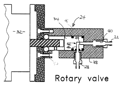
The rotary valve 24 is shown in more detail in
Figure 2. Valve 24 is driven by a DC servo-motor 32
under the control of a servo drive 33. The motor 32
a;1 I (~ ;' ~l: (l
4
includes a driveshaft 35 connected to a cylindrical
teflon valve body 34. Body 34 is rotatably mounted
within a cylindrical bore 36 provided in a brass housing
38 that is connected through base plate 39 to the motor
32 so as to allow relative rotation between the valve
body 34 and housing 38. The servo drive 33 is operable
to control the rotational speed of the motor 32 in a
conventional manner.
Supply duct 22 is connected to an end port 40
located in the housing on the axis of rotation of the
valve body 34. An internal passageway 42 is provided in
the valve body 34 and has an axial leg 44 aligned with
end port 40 and a radial leg 46. The radial leg 46 is
axially located to be aligned with outlet 28 in the
housing 38 which is connected to catheter 30.
As the valve body 24 rotates, the radial leg 46
comes periodically into alignment with the outlet 28 of
the housing 38 and contrast agent is able to flow through
the valve 24 from port 40 to outlet 28. At other times
during the rotation of the valve body 34, the housing 38
blocks flow through the duct 42 and prevents flow out of
the outlet 28. The duty cycle of the valve 24, i.e. the
fraction of each cycle that the valve is open, is thus
determined by the diameter of the radial leg 46 and the
circumference of the body 34. In a preferred design, the
body circumference is 80 mm and the radial leg diameter
is 3.2 mm, resulting in a duty cycle of 8%. 0-ring seals
48 are provided on the valve body 34 to prevent contrast
agent from leaking past the valve body 34 and thrust
bearings 50 are located between the base plate 39 and the
body 34 to prevent excessive thrust loads on the DC
servo-motor 32.
The objective of the pulsed-injection technique
is to produce compact boli of contrast agent within the
vessel. Thus, it is important that the contrast agent
not be spread excessively along the vessel when it is
ejected from the catheter 30. Catheter 30 includes an
F~
elongate body 51 with an internal duct 53. The tip 52 of
catheter 30 as shown in Figure 3 has been found to
provide a reduced dispersal of contrast agent. The end
face 54 of the tip 52 of the catheter 30 is sealed so as
5 to be leak-tight. Three pairs of apertures 56 are formed
in the wall of the catheter 30 to provide a total of six
apertures. Each pair of apertures 55 is axially and
circumferentially spaced from the adjacent pair so that
two pairs are aligned on the diameter of the catheter and
l0 the intermediate pair is disposed at 90° to the other
two. It has been found that an aperture diameter of 0.7
mm and an axial spacing of 2 mm between adjacent pairs of
apertures has been appropriate with a catheter having an
internal duct with a diameter of 1 mm. This hole pattern
not only produces an exit orifice with a total area which
is three times larger than that obtained with an end-hole
alone but also reduces the initial extent of the bolus
along the vessel to lass than 1 cm in length. The
increased exit area also avoids jetting of the agent and
consequent impingement against the side wall of the
vessel.
To verify the performance of the pulsed
injector with a digital angiographic system, tests were
performed ,~ vitro with an x-ray image intensifier (XRII)
62 coupled to a linear photodiode array (PDA) 64 as shown
schematically in Figure 4. X-rays from source 66 pass
through the vessel of interest and are detected by the
XRII 62 after processing by image intensifier 62. The
optical output signal from the XRII 62 is transferred to
a 1024 element PDA 64 (Reticon RL 10245). The PDA 64 is
positioned such that its long axis is aligned with the
long axis of the vessel of interest, and magnification
factors are chosen such that the entire diameter of the
vessel is recorded by the PDA 64. In this manner, the
PDA 64 provides instantaneous integration of the signal
across the vessel diameter, with the PDA elements
recording this information at each of 1024 points along
st ~ ~.
:r
6
the vessel simultaneously. The output from the PDA 64 is
digitized with a 12-bit ADC 68 at a line rate of 60 Hz.
Thus, a distance-density curve was recorded by the
acquisition computer 70 every 16.6 ms, for a total
acquisition time of up to 8 s.
Simulated blood flow for the ,~ v o
experiments has been provided by a computer-controlled
flow simulator which consists of a piston driven within a
450 ml glass housing by a micro-stepping motor. This
produces steady and pulsatile flow waveforms to within
~ 1%, with waveform shape specified completely from
flow-rate values tabulated in a data file. Such a device
is described more fully in an article entitled "Computer
Controlled Positive Displacement Pump for Physiological
Flow Simulation" by D.W. Holdsworth et al. published in
Med. Bio. Eng. & Computing, Vol. 29, at pages 565-570
(1991). For the ,~ vitro experiments described below,
the simulator was used to produce steady flow over the
range of 5 to 30 ml s'~, as well as simulated human
carotid and femoral flow waveforms.
As noted above, rotation of the valve body 34
within housing 38 produces a pulsed flow of agent at the
output 28. Figure 5 shows the form of the pulse produced
as flow rate versus time from which it can be seen that
sharp (vertical) leading and trailing edges are produced.
By comparison, a pulse form produced by a commercially
available unit available from Viamonte-Hobbs under model
number 2000 is shown in chain dot line on Figure 5.
Figure 6 shows the pulse forms as the
rotational speed of the body 34 within housing 36 is
increased under the control of servo drive 33. As can be
seen, the leading and trailing edges remain well defined
with a reduction in the maximum flow rate as the
frequency, i.e. the rotational speed, increases.
A typical set of results fram the apparatus of
Figure 4 is shown in Figure 7 with the passage of a bolus
of agent along the vessel being identified as dark (x-ray
~a 1 a ;i ;: ~ ()
absorbing) streaks proceeding from left to right. The
slope of the streaks represents the velocity of flow in
the vessel. The high definition of the pulses produced
by the valve 24 enhances the definition of the passage of
the bolus on the image shown in Figure 7.
A further representation of the passage of the
bolus along the vessel is shown in Figure 8 which
represents a plot of transmission through the vessel
versus position along the vessel at different times.
Traces obtained at different times are indicated by
different line formats. It can be seen that the minimum
transmission, corresponding to the bolus position, is
well defined and progressively moves along the vessel.
The enhanced definition of the bolus produced by the
valve 24 and catheter 30 is clearly seen from the
correlation of the results of Figure 8 represented on the
curve of Figure 9 with a well defined minimum value
displaced from the origin by a distance equivalent to
movement of the bolus through the vessel in the interval
between the correlated samples. This provides an
indication of the average velocity of fluid at a given
location of the vessel. Flow rate can then be determined
if the cross-sectional area of the vessel is known.