Note: Descriptions are shown in the official language in which they were submitted.
2113089
UNIVERSAL DONOR CEL.LS
The U.S. Government has rights in this invention by
virtue of Grants Nos. GM40924, 5 K11 HC02351, ROl HL 28373, and
9T32 DK07556 awarded by the National Institutes of Health,
Bethesda, Maryland.
Background of the Invention
This invention relates to genetically engineered endothelial
cells and, in particular, to endothelial cells which have been modified to
resist lysis by complement and evade the host's immune mechanisms for
removing foreign cells, when inserted into a non-autologous host.
Endothelial cells are specialized cells which form the lining
of the heart and the blood vessels. Because of their direct contact with
the circulating blood, a number of proposals have been made to
genetically engineer these cells and use them as "in vivo" drug delivery
systems, for example, by Culliton, B. J. 1989. 'Designing Cells to
Deliver Drugs," Science 246:746-751; and Zwiebel et al., "High-I.evel
Recombinant Gene Expression in Rabbit Endothelial Cells Transduced by
Retroviral Vectors," Science 243:220-222 (transfer of a human adenosine
deaminase gene and a rat growth hormone gene to aortic endothelial cells
using a retroviral vector and demonstration of the secretion of rat growth
hormone from such cells after seeding onto a synthetic vascular graft).
Natural endothelial ctlls play important roles in normal
physiology. In particular, these cells constitute the interface between the
blood and the vessel wall and the organs of the body. As such,
endothelial cells secrete various natural products directly into the blood
stream, maintain an antithrombotic surface on the inside of the vessel,
restrict leukocytes from penetrating the vessel wall, regulate various of
the biological properties of smooth muscle cells, and participate in the
control of vessel wall tone. Therefore, loss of endotheliai cells results
~r
WO 93/02188 2113 0 8 9 PCT/US92/05920 ~
-2-
in the loss of these normal physiological processes and ultimately leads
to pathological conditions such as coronary artery disease, organ
transplant rejection and vasculitis.
Accordingly, in addition to their use as a medium for the in
vivo administration of therapeutics, there is a need to provide genetically
engineered endothelial cells to replace natural endothelial cells which
have been lost due to disease or surgery.
In the past, proposals and/or efforts to use endothelial cells for
either administration of therapeutics or cell replacement have generally
been limited to autologous cells, i.e., cells derived from the organism
undergoing treatment. Alternatively, the patient must be
immunosuppressed, which is costly and leaves the patient vulnerable to
infection.
This approach has suffered from a number of problems. For
example, it is difficult to harvest healthy endothelial cells from the
individual to be treated in significant quantities. The procedures for
doing so require removal of a section of vasculature and then scraping
or otherwise dislodging the endothelial cells from the walls of the
vessels. As a result, to be useful for either the administration of
therapeutics or cell replacement, a large number of autologous endothelial
cells must be grown in culture. To be of practical use, especially in the
case of cell replacement, this culturing must take place quickly.
Unfortunately, the cell doubling time for endothelial cells is on the order
of at least 24 to 48 hours, leading to time periods on the order of a week
or more before sufficient quantities of endothelial cells are available for
genetic engineering or cell replacement. In addition, under normal
physiological conditions, the cell doubling time for natural endothelial
cells in vivo is also prolonged, making naturally occurring cell
replacement in vivo following endothelial cell loss or damage highly
inefficient.
WO 93/02188 21.18 0 8 9 PC.'T/US92/05920
-3-
When a foreign cell is transplanted into a host, the immune
system of the host rapidly mobilizes to destroy the cell and thereby
protect the host. The immune system attack on the foreign cell is
referred to as transplant rejection. The organism's first line of defense
is through either lytic destruction or the activation of procoagulant and
prothrombotic properties of the donor endothelial cell that may result
from activation of the host's complement system and is generally known
as the "hyperacute rejection response" or simply the "hyperacute
response."
Several studies have demonstrated that the hyperacute response
to transplants of either xenogeneic (from different species) and allotypic
(from different individuals of the same species) organs is mediated by
antibody-dependent activation of the complement system at the surface of
the donor endothelium, as discussed, for example, by Platt et al. , 1990
"Transplantation of discordant xenografts: a review of progress"
Lnmunology Today 11:450-456. That is, the complement system attacks
the endothelial cells lining the vessels of the transplanted organ.
The complement system is a complex interaction of plasma
proteins and membrane cofactors which act in a multistep, multi-protein
cascade sequence in conjunction with other immunological systems of the
host organism. The classic complement pathway involves an initial
antibody recognition of, and binding to, an antigenic site on a target cell.
This surface bound antibody subsequently reacts with the first component
of complement, Clq, forming a Cl-antibody complex with Ca2+, Clr,
and Cls which is proteolytically active. Cls cleaves C2 and C4 into
active components, C2a and C4a. The C4b,2a complex is an active
protease called C3 convertase, and acts to cleave C3 into C3a and C3b.
C3b forms a complex with C4b,2a to produce C4b,2a,3b, which cleaves
C5 into C5a and C5b. C5b forms a complex with C6 and this complex
interacts with C7 in the fluid phase thereby exposing hydrophobic
domains within C5b and C6 that stabilize the C5b,6,7 ternary complex
2113089
-4-
in the cell membrane. C8, which is in the fluid phase, then binds to the
CSb, 6, 7 ternary complex and this complex may contribute to the
development of functional membrane lesions and slow cell lysis. Upon
binding of C9 to C8 in the C5b-8 membrane complex, lysis of foreign
cells is rapidly accelerated.
U.S. Patent No. 5,135,916, issued August 4, 1992, assigned to the Oklahoma
Medical Research Foundation discloses that the human complement regulatory
protein CD59 can be used to protect non-hunsan endothelial cells, for
example, porcine endothelial cells, from attack by hunian complement,
either when provided in solution with the cells or expressed in genetically
engineered cells. See also Zhao et al., 1991 "Amplified gene expression
in CD59-transfected Chinese Hamster Ovary cells confers protection
against the membrane attack complex of human complement' J. Biol=
Chem, 266:13418-13422. The homologous complement inhibitory
activity of CD59 resides in its species-specific interaction with the
terminal complement components C8 and C9, as further reported by
Rollins and Sims, 1990 "The complenient inhibitory activity of CD59
resides in its capacity to block incorporation of C9 into membrane
C5b-9" J. Immunol. 144:3478-3483.
Although the use of CD59 does successfully address the
problem of hyperacute rejection as a result of canplement attack, it does
not protect the coll against the overall immune atta+k of the host
organism against foreign endothelial cxlls.
In stimulating iminune respooses, antigens elicit many
molecular and cellular changes. Lymphocytes recognize antigens as
foreign and are responsible for initiating both cellular aad humoral
responses against the presenting antigen. B lymphocyte cx11s respond to
antigen by the production of antibodies against the presenting antigen; T
lymphocytes respond by initiating a celhilar response to the presenting
antigen. The two major subsets of T cxll: are T. calls, involved in
~
WO 93/02188 21130$ 9 PCT/US92/05920
-5-
processing of antigen for presentation to B cells, characterized by the
presence of a cell-surface glycoprotein called CD4, and cytolytic T
lymphocytes (CTI-s), involved in recognition of antigen on cell surfaces
and lysis of cells recognized as foreign, characterized by the presence of
a cell-surface glycoprotein called CD8. T cells recognize peptide
fragments in conjunction with one of the two main classes of cell-surface
glycoproteins of the major histocompatibility complex (MHC): either
class I(1VHC-I) or class II(MHC-II) proteins. CD8 + T cells recognize
antigens in conjunction with MHC-I, whereas CD4+ T cells recognize
them in conjunction with MHC-II.
The MHC contain three major classes of genes. Class I genes
encode the principal subunits of MHC-I glycoproteins, called human
leukocyte antigens in humans, the principle ones being HLA-A, B, and
C. These are present on virtually all cells and play a major role in
rejection of allografts. They also form complexes with peptide fragments
of viral antigens on virus-infected cells: recognition of the complexes by
CD8+ CTLs results in destruction of virus infected cells. Recognition
of the complexes is by a single receptor on the T cells which recognizes
antigen in combination with MHC.
Class II genes, the major classes in humans being known as
DP, DQ (subclasses 82, a2, and Blal) and DR (subclasses Bl, 92, B3
and al), encode cell-surface glycoproteins that are expressed by antigen-
presenting cells, principally B cells, macrophages and dendritic cells.
Together with peptide fragments of antigens, the class II proteins form
the epitopes that are recognized by T helper cells (CD4+). Class III
genes encode at least three proteins of the complement cascade and two
cytotoxic proteins, tissue necrosis factor and lymphotoxin, which are
involved in diverse immune reactions that destroy cells.
T-cell mediated immune reactions can be organized into three
sequential activation steps. First, CD4+ and CD8+ T lymphocytes
WO 93/02188 2113089 PCT/US92/05920
-6-
(T-cells) recognize the presence of non-autologous MHC class II and
class I proteins, respectively, on the surface of the foreign cell.
Second, the T-cells are activated by interaction of a ligand
with the T cell receptors and other accessory stimulatory molecules, so
that activation depends upon a variety of variables including humoral
signals such as cytokines received by protein receptors on the surface of
the cells. Most important is the interaction between the antigen specific
T cell receptor and ligand, a complex of MHC and antigenic peptide on
the antigen presenting cell (APC). Other receptors present on the T cell
must also be contacted by their ligands on APC to insure activation.
Once activated, the T-cells synthesize and secrete interleukin-2 (IL-2) and
other cytokines.
The cytokines secreted by the activated T-cells lead to the
third, or effector, phase of the immune response which involves
recruitment and activation of lymphocytes, monocytes, and other
leukocytes which together lead to cell lysis, as reviewed, for example,
by Pober et al., 1990 "The potential roles of vascular endothelium in
immune reactions" Human Immunol. 28:258-262.
Historically, attempts to interrupt the T-cell immune response
have generally met with limited success. For example, several strategies
have tried to use reagents of various types, including antibodies and
blocking proteins, to interfere with adhesion between T-cells and foreign
cells. Lider et al., 1988 "Anti-idiotypic network induced by T cell
vaccination against experimental autoimmune encephalomyelitis" Science
239:181 reported on the use of T-cell vaccines; Owhashiand et al., 1988
"Protection from experimental allergic encephalomyelitis conferred by a
monoclonal antibody directed against a shared idiotype on rat T cell
receptors specific for myelin basic protein" J. EU. Med. 168:2153,
reported on the use of T-cell receptor blocking antibodies; Brostoffand
et al. 1984 "Experimental allergic encephalomyelitis: successful treatment
in vivo with a monoclonal antibody that recognizes T helper cells" L
WO 93/02188 21130g g PCT/US92/05920
-7-
Immunol. 133:1938 reported on the use of antibodies to CD4; and
Adorini et al., 1988 "Dissociation of phosphoinositide hydrolysis and
Ca2+ fluxes from the biological responses of a T-cell hybridoma"
Nature 334:623-628, reported on the use of blocking peptides that occupy
T-cell receptors. These strategies have generally resulted in immune
responses to the reagents, rather than the desired interruption of T-cell
binding.
It would clearly be advantageous if one could decrease the
probability of T-cell mediated reaction against transplanted cells, as well
as complement-mediated attack and lysis of the cell.
It is therefore an object of the present invention to provide an
improved method and compositions for constructing endotheli.al cells that
are resistant to both complement and cellular attack when transplanted
into a foreign host.
It is a further object of the present invention to provide
genetically engineered cells that are not recognized as foreign when
implanted into a foreign host and therefore evade attack by the immune
system.
It is still further object of this invention to provide genetically
engineered cells which after transplantation can resist
complement-medi.ated attack and evade lymphocyte-mediated lysis,
specifically CD4 + T-lymphocytes, and preferably CD8 + T-lymphocytes.
It is another object of the invention to provide a mechanism
for selectively killing such genetically engineered cells when their
presence in the host is no longer desired.
It is still another object of the present invention to provide a
biological vehicle for delivery of therapeutic products.
Summary of the Invention
Genetically engineered cells are provided which include a
DNA sequence which is expressed by the cell and which codes for a
WO 93/02188 211 30 89 PC'I'/US92/05920
-8-
protein having complement inhibitory activity that is not normally
expressed in the cell. These cells may also be engineered so that they
do not express on their cell surfaces functional proteins encoded by the
class II major histocompatibility complex (MHC) genes, the HLA DP,
DQ, and DR genes in human cells, or their equivalent in cells of a
different species. Alternatively, the genetically engineered cells do not
express on their cell surfaces the proteins encoded by the class I MHC
genes, the HLA A, B and C genes in human cells, or their equivalent in
cells of a different species, or they do not express either the class I and
class II MHC genes. In some embodiments, the cells include a genetic
(DNA) sequence which is expressed by the cell and which codes for a
protein which in the presence of a selected agent results in death of the
cell.
The genetic sequence which codes for a protein which has
complement regulatory activity protects the cell from hyperacute rejection
through attack and lysis resulting from activation of the complement
system. The removal of the cell surface proteins encoded by the class
I (for example, HLA A, B and C) and class II(for example, IiI.A DP,
DQ, and DR) MHC genes makes the cells substantially unrecognizable
by the host's CD8+ and CD4+ T-lymphocytes, respectively. The
genetic sequence which codes for a protein which can produce cell death
provides a mechanism for eliminating the genetically engineered cells
from the host when their presence is no longer desired.
The cells are modified in culture using standard in vitro
transfection techniques, or can be derived from transgenic animals
modified as embryos. These modified cells can serve as universal donor
cells for administering therapeutic agents to the host or as replacements
for natural cells which have been damaged or lost. In the most preferred
embodiment, the cells are dissociated endothelial cells.
-WO 93/02188 2113 0 8 9 pCr/US92/05920
-9-
Brief Description of the Drawings
Figure 1 is a graph of the induction of CD59 antigen in CHO
cells transfected with plasmid containing human CD59 cDNA, '25I-1F1
bound CD59 (molecules/cell x 10*') versus methotrexate ( g/ml).
Chinese hamster ovary cells were transfected with a plasmid containing
the pFRSV vector and cDNA for human CD59. After subcloning and
selection, the cells were maintained in medium containing methotrexate
and surface antigen measured by the specific binding of monoclonal
antibody 'uI-1F1 (10 g/ml) against CD59. All data were corrected for
nonspecific binding measured for control (nontransfected) CHO cells
grown in the absence of inethotrexate (or.igin). Data denote means f
S.E. of three measurements made on separate days.
Figure 2 is a graph of the removal of cell-surface CD59 by
phosphatidylinositol-specific phospholipase C(PIPLC), plotting cell
number versus mean fluorescence. CD59-transfected CHO cells
amplified by growth in 1 mg/ml methotrexate were suspended at 2 x
106/ml in HBSS and incubated for 1 h at 370C with either 0(. ...) or 1
(--) unit/ml phosphatidylinositol-specific phospholipase C. Cell-surface
CD59 was then measured by flow cytometry using monoclonal antibody
1F1 (10 g/m1), which was detected with FITC anti-mouse IgG (67
1&g/m1). Histograms denote mean fluorescence per cell on logarithmic
scales. Also shown is background cell fluorescence ineasured in the
absence of 1F1 (-).
Figure 3 is a graph showing protection of CD59-transfected
CHO cells from human serum complement, dye release ( 9b ) versus
human serum ( 9b ). CD59-transfected CHO cells were induced to express
various amounts of CD59 antigen by growth in methotrexate-containing
media, molecules CD59 expressed/cell x 19s: 0.0 (dark circles), 1.9
(open circles), 2.8 (dark diamonds), 6.8 (open triangles), 7.2 (dark
squares), 13.0 (open squares), and 31.3 (dark diamonds).
WO 93/02188 PCT/US92/05920
2113089
-10-
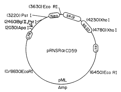
Figure 4 shows the structure of the retroviral vector used in
Example 2. This vector was constructed from a defective Moloney
murine leukemia virus. The SV40 promoter was excised and replaced
with the SRalpha promoter. A 500 bp cDNA fragment containing the
CD59 coding sequence was cloned into an XhoI site and verified by
restriction analysis. The resulting plasmid was designated
pRNSRaCD59. Ecotropic retrovirus was produced by transfecting Psi-
2 cells with polybrene and selecting in the toxic aminoglycoside G418.
Amphotropic virus stocks were prepared by infecting the amphotropic
packaging cell line Psi-AM with the ecotropic virus, were added directly
to endothelial cell cultures in the presence of polybrene, and transfectants
were selected with 400 g/ml G418.
Figure 5 is a graph of cell surface expression of human CD59
on porcine aortic endothelial cells (PAEC) as detected by anti-CD59
antibody and analyzed by flow cytometric analysis. The solid line
represents the fluorescence intensity of PAEC infected with retrovirus
shown in Figure 4 carrying only the control neomycin resistance gene.
The dashed line, small dotted line, and larger dotted line represent the
fluorescence intensity of CD59-expressing PAEC cell clones 2, 9, and 1,
respectively.
Figure 6 shows a scanning electron micrograph of CD59-
expressing PAEC attached to a synthetic Gortex""' graft. Figure 6a is the
control Gortei', Figures 6b, c, and d are Gortex' with CD59-
expressing cells implanted thereon.
Figure 7 is a bar graph showing the protection of human
CD59-expressing PAEC from lysis by human complement. The solid bar
represents the percentage of cell lysis of PAEC expressing human CD59.
The cross-hatched bar represents the percentage of cell lysis of PAEC
expressing only the control neomycin resistance gene while the stippled
bar represents the percentage of cell lysis of control (noninfected) PAEC.
2113089
-11-
Figure 8A shows a restriction digest map of the gene targeting
vector for the mouse invariant chain gene cloned into pBS (Bluescript)*
The targeting vector contains the neomycin gene (neo). Figure 8B shows
a partial restriction digest map of the endogenous mouse invariant chain
gene and Figure 8C shows a restriction digest niap of the disrupted
invariant chain gene achieved by homologous recombination. Arrows
indicate the direction of transcription in all three panels. The recognition
region for the radiolabeled invariant gene probe used for the Southern
blot shown in Figure 9 is indicated by a solid bar below Figure 8C.
Figure 9 is a Southern blot showing the restriction digestion
pattern for two independent neomycin resistant mouse embryonic stem
cell clones where the endogenous invariant chain gene has been disrupted
by homologous recombination and replaced with a mutated form of the
gene. The two clones are designated 11.10.93 and 11.10.128. Size
markers are indicated on the left side. The DraIII restriction pattern of
the parental cells is indicated in the far right lane and is clearly different
from the restriction pattern of the two clones carrying the modified
invariant chain gene.
Detailed Description of the Invention
I. Protection From Hyneracute Rejection
The lysis of cells by complement has been detennined to
typically require only the terminal complement components, in contrast
to previous reports that it may be essential to interrupt complement
activation at the C3 stage, as described, for exainple, in PCT application
WO 91/05855.
Sequential addition of C6,7,8, and 9 to C5b leads to the
formation of a membrane attack complex (MAC), a pore-like complex
which, when inserted into the plasma membrane of the target cell,
increases membrane penneability to calcium and other ions.
Consequently, lysis of the plasma membrane ensues and the cell is either
Denotes trade mark
WO 93/02188 21 13 0 8 9 PC'T/US92/05920 -~
-12-
destroyed, or alternatively, there is a non-lytic alteration of specific cell
functions affecting vascular hemostasis. In the case of human endothelial
cells exposed to human serum complement, membrane deposition of the
C5b-9 complex initiates a variety of procoagulant and prothrombotic
changes in the cell that are expected to accelerate blood clotting and
thrombus formation, as described, for example, by Hattori, et al., 1989
"Complement proteins C5b-9 induce secretion of high molecular weight
multimers of endothelial von Willebrand Factor and translocation of
granule membrane protein GMP-140 to the cell surface" J. Biol. Chem.
264:9053-9060; Hamilton, et al., 1990 "Regulatory control of the
terininal complement proteins at the surface of human endothelial cells:
Neutralization of a C5b-9 inhibitor by antibody to CD59" Blood 76:2572-
2577; and Hamilton and Sims 1991 "The terminal complement proteins
C5b-9 augment binding of high density lipoprotein and its apoproteins A-
I and A-II to human endothelial cells" J. Clin. Invest. 88:1833-1840.
These responses appear to depend upon insertion of C9 into the plasma
membrane of the target cell and therefore can be prevented by interfering
with assembly of the C5b-9 complex.
Membrane proteins inhibiting complement.
Specific membrane proteins which exhibit potent inhibitory
activity for the complement cascade have been isolated and molecularly
cloned.
In particular, with regard to the human complement system,
protection against the pore-forming activity of the C5b-9 complex can be
conferred on non-primate cells by transfection of such cells with a cDNA
encoding the human complement regulatory protein CD59.
The capacity to stably express CD59 in Chinese hamster ovary
(CHO) cells has enabled direct evaluation of the C5b-9 inhibitory activity
conferred when CD59 is selectively expressed in mammalian cells that
normally express neither CD59 nor HRF. The results demonstrate that
the inhibitory activity of human blood cells toward the membrane attack
WO 93/02188 2113089 PCT/US92/05920
-13-
complex of human serum complement can be transferred to a non-human
mammalian cell by transfection with the CD59 cDNA and demonstrate
that the C5b-9 inhibitory function of this protein correlates with the
amount of newly expressed surface CD59 antigen.
The existence of these proteins and the studies detailed below
indicate that a deletion or inactivation of these cell surface components
increases the risk of vascular thrombosis and lead to a decreased storage
time for platelets and platelet rich plasma (PRP), and perfused organs
and transplanted tissue. Accordingly, the survival and hemostatic
efficacy of platelets, the survival and function of hematopoietic progenitor
cells, such as CFU-S, CFU-GEMM, and CFU-L, and their progeny,
such as BFU-E, BFU-MK, and CFU-GM, as well as the mature blood
cells, including erythrocytes, platelets, monocytes, granulocytes, and
lymphocytes, that may derive from these progenitor cells after bone
marrow transplantation, as well as the survival of organs and tissue for
transplant, which are collected and stored in vitro, can be increased by
addition of the C5b-9 inhibitor to the storage buffer or perfusate and/or
by the introduction and expression of the gene encoding CD59 in the
cells to be protected. Autoinunune disorders and other disease states that
involve C5b-9 mediated platelet activation, including lupus, rheumatoid
arthritis, and additional types of immuno-vasculitis, can also be treated
by the intravascular administration and/or transfection and expression of
an effective amount of the inhibitor or a functionally active polypeptide
thereof to suppress C5b-9 activity in a patient requiring such treatment.
Similar uses of the inhibitor can be applicable for cell culture in human
blood derived culture media.
The data shown in the examples below are evidence that
transfection with the gene for CD59 can be used to confer protection
against the membrane attack complex of complement to cells that do not
normally restrict activation of the human C5b-9 proteins. These data
confirm by DNA transfection the C5b-9 inhibitory function that has
WO 93/02188 2113 0 8 9 PCT/US92/05920
-14-
previously been attributed to CD59 antigen present on human
erythrocytes and exclude the possibility that the activity found associated
with this protein reflects the . presence of another membrane constituent
with complement inhibitory activity that copurifies with CD59 antigen.
Despite apparent differences in glycosylation, the C5b-9 inhibitory
function observed for recombinant CD59 expressed in CHO cells exhibits
specificity for human C8 and C9 (within C5b-9), analogous to that
observed for the human erythrocyte membrane and for purified
erythrocyte CD59 antigen. This capacity to confer species-selective
protection against the human C5b-9 proteins by transfection of a non-
human cell with cDNA encoding the CD59 sequence establishes
unequivocally that this 18-21 kD protein functions as a homologous
complement restriction factor on human blood cells and is consistent with
the observation that the syndrome of paroxysmal nocturnal
hemoglobinuria can be associated with an isolated deficiency of
erythrocyte CD59.
As illustrated by the following examples, the complement
inhibitory activity of recombinant CD59 was found to saturate when the
expression of surface antigen was ampliSed to greater than or equal to
1.3 X 10⁶ molecules/CHO cell. Assuming a spherical diameter of
approximately 25 m for the CHO cell, this is equivalent to greater than
or equal to 600 molecules of CD59 antigen/ m2 of plasma membrane
surface. By comparison, human erythrocytes, which are highly resistant
to activation and lysis by human complement, express approximately 2.5
X 10' molecules of CD59 antigen/cell, which is equivalent to
approximately 200 molecules/ m2 of membrane surface. Extrapolating
from this data, 1 x 10³ molecules CD59/cell or greater than or equal to
1 molecule of CD59 antigen/ m2 of plasma membrane surface should be
effective in inhibiting complement mediated activation and lysis.
The data also demonstrate that recombinant CD59 expressed
in CHO cells exhibits the species-selective recognition of human C5b-
WO 93/02188 2113089 PC,'i'/US92/05920
-15-
9 characteristic of CD59 in human erythrocytes despite apparent
differences in N-linked glycosylation. These data indicate that the species
selectivity exhibited by CD59, which includes recognition for human C8
(within C5b-8) and human C9 (within C5b-9), is conferred by the core
protein, independent of its carbohydrate, or that the relevant carbohydrate
structures are conserved in the recombinant protein when expressed in
CHO cells. As used herein in the compositions and methods for the
prolongation of platelet and organ survival and enhancement of
therapeutic efficacy or suppression of complement mediated disorders,
"C5b-9 inactivator" refers to any CD59 molecule, including the 18 kDa
protein on erythrocyte membranes, peptide fragments thereof having C5b-
9 inhibitory activity, preferably containing a membrane binding domain,
whether isolated from naturally produced materials or recombinantly
engineered sequences. The term 'also includes cells infected or
transfected with, and expressing, the gene for CD59 or a biologically
functional portion thereof, as well as cells in transgenic animals in which
the gene in combination with a promoter such as the murine K' MHC
class I promoter has been stably introduced into an embryo of the animal
using a technique such as microinjection. All molecular weights are
determined by SDS-PAGE under non-reducing conditions.
Other complement inhibitors which have been identified and
can be used alone or in combination with CD59 include:
(1) CD46, also known as membrane cofactor protein
(MCP), as described by Purcell, et al., 1990 "The human cell surface
glycoproteins HuLy-m5, membrane cofactor protein (MCP) of the
complement system, and trophoblast leucocyte common (1'LX) antigen,
are CD46" J. Immunol. 70:155-161; and Seya and Atkinson, 1989
"Functional properties of membrane cofactor protein of complement"
Biochem. J. 264:581-588. This inhibitor functions by binding to
complement component C3b thereby activating molecules that cleave C3b
into inactive fragments preventing accumulation of C3b and, therefore,
WO 93/02188 2 113 p89 PCT/US92/05920
-16-
its contribution to the formation of the MAC. See also White et al.
1991 "Protection of mammalian cells from human complement-mediated
lysis by transfection of human membrane cofactor protein (MCP) or
decay accelerating factor (DAF)" Int. Meeting on XenotransFlantation
---(recombinant human CD46 shown to provide protection of
non-primate cells from lysis by human complement).
(2) CD55, also known as decay accelerating factor (DAF),
described by Nicholson-Weller et al., 1982 "Isolation of a human
erythrocyte membrane glycoprotein with decay-accelerating activity for
C3 convertases of the complement system" J. Immunol. 129:184;
Lublin and Atldnson, 1989 "Decay accelerating factor: Biochemistry,
molecular biology, and function" Annu. Rev. Immunol. 7:35; Lublin et
al., 1987 "The gene encoding decay-accelerating factor (DAF) is located
in the complement-regulatory locus on the long arm of chromosome 1"
J. Exp. Med. 165:1731; and Medof et al., 1987 "Cloning and
characterization of cDNAs encoding the complete sequence of decay
accelerating factor of human complement" Proc. Natl. Acad. Sci. USA
84:2007. This inhibitor is a membrane bound protein of approximately
70 kD in molecular mass which interferes with the assembly of C3
convertase. See also White et al., 1991, reporting that recombinant
DAF provides protection of non-primate cells from lysis by human
complement.
The relative contributions of CD46, CD55, and CD59 in
providing protection from complement-mediated lysis has been assessed
in human amniotic epithelial cells (HAEC) by the use of specific blocking
antibodies, as reported by Rooney et al., 1990 "Protection of human
amniotic epitheli.al cells (HAEC) from complement-mediated lysis:
expression on the cells of three complement inhibitory membrane
proteins." Immunoloev 71:308-311. The results demonstrated that
CD59 provides the most protection against complement attack, as
compared with CD46 and CD55. Additionally, a patient with
~ WO 93/02188 2113089 PCT/US92/05920
-17-
paroxysmal nocturnal hemoglobinuria, a rare disorder caused by an
unusual susceptibility of erythrocytes to the lytic action of complement,
was described as having an inherited deficiency of CD59 without a
deficiency of CD55, by Yamashina et al. 1990 "Inherited complete
deficiency of 20-kilodalton (CD59) as a cause of paroxysmal nocturnal
hemoglobinuria" New EnQI. J. Med. 323:1184-1189.
By contrast to the intravascular hemolysis observed for this
patient reported to be deficient in CD59 but normal for decay
accelerating factor (and presumably normal for other complement
inhibitors), individuals with inherited defects or deficiencies in
erythrocyte CD55 (Decay Accelerating Factor) generally do not exhibit
intravascular hemolysis, as reported by Daniels, G. 1989 "Cromer-
related antigens-blood group determinants on decay accelerating factor.
Vox. Sane. 56:205; Holguin, et al. 1992 "Analysis of the effects of
activation of the alternative pathway of complement on erythrocytes with
an isolated deficiency of decay accelerating factor. J. Immunol. 148:498-
502, suggesting the CD59 is necessary and sufficient to protect these
cells from the cytolytic effects of complement in human plasma.
Cells suitable for tiansplantation into a foreign host are
protected from complement-mediated lysis by introducing into the cell
DNA encoding a protein, or combination of proteins, inhibiting
complement-mediated lysis, for example, CD59, CD55, CD46 and/or
other inhibitors of C8 or C9. CD59 is the preferred inhibitor, introduced
into the cells by transfection or infection with a vector encoding the
CD59 protein, and expressed on the surface of the transfected/infected
cells. The inhibitor is preferably of the same species of origin as the
host into which the cells are to be transplanted.
The gene encoding the complement inhibitor can be introduced
into a cell of a different species of origin, for example, a human CD59
gene can be introduced into a porcine cell so that the cell resists attack
when transplanted into a human, or the gene can be introduced into a cell
WO 93/02188 21 13 0 8 9 PCT/US92/05920
-18-
of the same species of origin so that increased amounts of the protein are
expressed on the surface of the cell. For example, the gene can be
placed under the control of a promoter enhancing expression of the gene
which is then inserted by homologous recombination into the host cell
chromosome at the site where the gene is normally located, but under the
control of the promoter which enhances expression, or can be inserted
into the chromosome at another locus on the chromosome.
DNA sequence information for CD46, CD55 and CD59 has
been reported in the literature.
The sequence reported by Lublin et al., 1988 "Molecular
cloning and chromosomal localization of human membrane cofactor
protein (MCP): Evidence for inclusion in the multi-gene family of
complement-regulatory proteins" J. Exn. Med. 168:181-194, for CD46
is shown below (Sequence I.D. No. 1).
HUMCD46 cDNA Sequence Acquired from GenBank: HUMCD46Q
GAATTCGGGGATAACAGCGTCTTCCGCGCCGCGCATGGAGCC
TCCCGGCCGCCGCGAGTGTCCCTTTCCTTCCTGGCGCTTTCCT
GGGTTGCTTCTGGCGGCCATGGTGTTGCTGCTGTACTCCTTCT
CCGATGCCTGTGAGGAGCCACCAACATTTGAAGCTATGGAGCT
CATTGGTAAACCAAAACCCTACTATGAGATTGGTGAACGAGT
AGATTATAAGTGTAAAAAAGGATACTTCTATATACCTCCTCTT
GCCACCCATACTATTTGTGATCGGAATCATACATGGCTACCTG
TCTCAGATGACGCCTGTTATAGAGAAACATGTCCATATATACG
GGATCCTTTAAATGGCCAAGCAGTCCCTGCAAATGGGACTTAC
GAGTTTGGTTATCAGATGCACTTTATTTGTAATGAGGGTTATT
ACTTAATTGGTGAAGAAATTCTATATTGTGAACTTAAAGGATC
AGTAGCAATTTGGAGCGGTAAGCCCCCAATATGTGAAAAGGT
TTTGTGTACACCACCTCCAAAAATAAAAAATGGAAAACACAC
CTTTAGTGAAGTAGAAGTATTTGAGTATCTTGATGCAGTAACT
TATAGTTGTGATCCTGCACCTGGACCAGATCCATTTTCACTTA
--- WO 93/02188 211 3 0 8 9 PC.'T/US92/05920
-19-
TTGGAGAGAGCACGATTTATTGTGGTGACAATTCAGTGTGGAG
TCGTGCTGCTCCAGAGTGTAAAGTGGTCAAATGTCGATTTCCA
GTAGTCGAAAATGGAAAACAGATATCAGGATTTGGAAAAAAA
TTTTACTACAAAGCAACAGTTATGTTTGAATGCGATAAGGGTT
TTTACCTCGATGGCAGCGACACAATTGTCTGTGACAGTAACAG
TACTTGGGATCCCCCAGTTCCAAAGTGTCTTAAAGTGTCGACT
TCTTCCACTACAAAATCTCCAGCGTCCAGTGCCTCAGGTCCTA
GGCCTACTTACAAGCCTCCAGTCTCAAATTATCCAGGATATCC
TAAACCTGAGGAAGGAATACTTGACAGTTTGGATGTTTGGGTC
ATTGCTGTGATTGTTATTGCCATAGTTGTTGGAGTTGCAGTAA
TTTGTGTTGTCCCGTACAGATATCTTCAAAGGAGGAAGAAGAA
AGGCACATACCTAACTGATGAGACCCACAGAGAAGTAAAATT
TACTTCTCTCTGAGAAGGAGAGATGAGAGAAAGGTTTGCTTTT
ATCATTAAAAGGAAAGCAGATGGTGGAGCTGAATATGCCACT
TACCAGACTAAATCAACCACTCCAGCAGAGCAGAGAGGCTGA
ATAGATTCCACAACCTGGTTTGCCAGTTCATCTTTTGACTCTAT
TAAAATCTTCAATAGTTGTTATTCTGTAGTTTCACTCTCATGAG
TGCAACTGTGGCTTAGCTAATATTGCAATGTGGCTTGAATGTA
GGTAGCATCCTTTGATGCTTCTTTGAAACTTGTATGAATTTGG
GTATGAACAGATTGCCTGCTTTCCCTTAAATAACACTTAGATT
TATTGGACCAGTCAGCACAGCATGCCTGGTTGTATTAAAGCAG
GGATATGCTGTATTTTATAAAATTGGCAAAATTAGAGAAATAT
AGTTCACAATGAAATTATATTTTCTTTGTAAAGAAAGTGGCTT
GAAATCTTTTTTGTTCAAAGATTAATGCCAACTCTTAAGATTA
TTCTTTCACCAACTATAGAATGTATTTITATATATCGTTCATTGT
AAAAAGCCCTTAAAAATATGTGTATACTACTTTGGCTCTTGTG
CATAAAAACAAGAACACTGAAAATTGGGAATATGCACAAACT
TGGCTTCTTTAACCAAGAATATTATTGGAAAATTCTCTAAAAG
TAAAGGGTAAATTCTCTATTTZITGTAATGTGTTCGGTGATTTC
AGAAAGCTAGAAAGTGTATGTGTGGCATTTGTTTTCACTTTTT
AAAACATCCCTAACTGATCGAATATATCAGTAATTTCAGAATC
WO 93/02188 2>>308 7 PCT/US92/05920
-20-
AGATGCATCCTTTCATAAGAAGTGAGAGGACTCTGACAGCCAT
AACAGGAGTGCCACTTCATGGTGCGAAGTGAACACTGTAGTCT
TGTTGTTTTCCCAAAGAGAACTCCGTATGTTCTCTTAGGTTGA
GTAACCCACTCTGCCCGAATTC
The sequence reported by Medof et al., 1987, for CD55 is
shown below (Sequence I.D. No. 2).
Human DAF cDNA Sequence Acquired from GenBank HUMDAF;
HUNIDAFC 1
TTCTCTCTACAGTCAGTCTGGAGTAATCCCAAAGTGGTGTC
TTTCGTAAATAAGGAGAACCCGGGTGAAGAAAATGACTCCC
ACCCGAACAAGGCATGAAC AATGTTCACTCCCTACTGTGTT
ATTCAAC
CTGTTTC C C CAGGTCTCTGTITrCACATTAGAGAGTGTTCT
AGGAGATGACG CCCTTCCTCCTTAGTTATTTCCCCACCCTC
GTGCTGGCCTTTGACAGACCTCCCAGTAGAGGGCCCAAGA
CGCGGGTAGAGCACCGCGTCTCAGCGCCTGAGTCTCAGCC
CCCGAACTCCACCGCACCTCGAGGTCCCCTTGGCACGACTC
AAGCGCGGGGATGCTCCGCTTAGACGAACTCACGTGCGGG
CAGCAAGGCCTGCGATACTTGAGCACCCCTCCCCCTCTCCC
GTTTACACCCCGTTTGTGTTTACGTAGCGAGGAGATATTTA
GGTITCTAGAAGGCAGGTCATCGCAGGCCCCACCCAGCAG
TGGAGAGAGTGAGTCCAGAGGGTGTTGCCAGGAGCTCCTC
CTCCTTCCCCTCCCCACTCTCCCCGAGTCTAGGGCCCCGGG
GTATGACGCCGGAGCCCTCTGACCGCACCTCTGACCACAAC
AAACCCCTACTCCACCCGTCTTGTTTGTCCCACCCTTGGTG
ACGCAGAGCCCCAGCCCAGACCCCGCCCAAAGCACTCATTT
AACTGGTATTGCGGAG
C CAC GAGGCTTCTGACTTACTGCAACTCGCTCCGGCCGCTG
GGCGTAGCTGCGACTCGGCGGAGTCCCGGCGGCGCGTCCT
-.WO 93/02188 21 1 3089 PCT/US92/05920
-21-
TGTTCTAACCCGGCGCGCCATGACCGTCGCGCGCCGAGCGT
GCCCGCGGCGCTGCCCCTCCTCGGGGAGCTGCCCCGGCTGCTG
CTGCTGGTGCTGTTGTGCCTGCCGGCCGTGTGGGGTGACTGTG
GCCTTCCCCCAGATGTACCTAATGCCCAGCCAGCTTTGGAAGG
CCGTACAAGTTTTCCCGAGGATACTGTAATAACGTACAAATGT
GAAGAAAGCTTTGTGAAAATTCCTGGCGAGAAGGACTCAGTG
ACCTGCCTTAAGGGCATGCAATGGTCAGATATTGAAGAGTTCT
GCAATCGTAGCTGCGAGGTGCCAACAAGGCTAAATTCTGCATC
CCTCAAACAGCCTTATATCACTCAGAATTATTTTCCAGTCGGT
ACTGTTGTGGAATATGAGTGCCGTCCAGG3TACAGAAGAGAA
CCTTCTCTATCACCAAAACTAACTTGCCTTCAGAATTTAAAAT
GGTCCACAGCAGTCGAATTTI'GTAAAAAGAAATCATGCCCTAA
TCCGGGAGAAATACGAAATGGTCAGATTGATGTACCAGGTGG
CATATTATTTGGTGCAACCATCTCCTTCTCATGTAACACAGGG
TACAAATTATTTGGCTCGACTTCTAGTTT'ITGTCTTATTTCAGG
CAGCTCTGTCCAGTGGAGTGACCCGTTGCCAGAGTGCAGAGA
AATTTATTGTCCAGCACCACCACAAATTGACAATGGAATAATT
CAAGGGGAACGTGACCATTATGGATATAGACAGTCTGTAACG
TATGCATGTAATAAAGGATTCACCATGATTGGAGAGCACTCTA
TTTATTGTACTGTGAATAATGATGAAGGAGAGTGGAGTGGCCC
ACCACCTGAATGCAGAGGAAAATCTCTAACTTCCAAGGTCCC
ACCAACAGTTCAGAAACCTACCACAGTAAATGTTCCAACTAC
AGAAGTCTCACCAACTTCTCAGAAAACCACCACAAAAACCAC
CACACCAAATGCTCAAGCAACACGGAGTACACCTGTTTCCAG
GACAACCAAGCATITfCATGAAACAACCCCAAATAAAGGAAG
TGGAACCACTTCAGGTACTACCCGTCTTCTATCTGGGCACACG
TGTTTCACGTTGACAGGTTTGCTTGGGACGCTAGTAACCATGG
GCTTGCTGACTTAGCCAAAGAAGAGTTAAGAAGAAAATACAC
ACAAGTATACAGACTGTTCCTAGTTTCTTAGACTTATCTGCAT
ATTGGATAAAATAAATGC AATTGTGCTCTTCATTTAGGATGCT
TTCATTGTCTTTAAGATGTGTTAGGAATGTCAACAGAGCAAGG
WO 93/02188 2113U S 9 P(T/US92/05920
-22-
AGAAAAAAGGCAGTCCTGGAATCACATTCTTAGCACACCTGC
GCCTCTTGAAAATAGAACAACTTGCAGAATTGAGAGTGATTCC
TTTCCTAAAAGTGTAAGAAAGCATAGAGATTTGTTCGTATTAA
GAATGGGATCAC GAGGAAAAGAGAAGGAAAGTGATTTTTTTC
CACAAGATCTGAAATGATATTTCCACTTATAAAGGAAATAAAA
AATGAAAAACATTATTTGGATATCAAAAGCAAATAAAAACCC
AATTCAGTCTCTTCTAAGCAAAATTGCTAAAGAGAGATGACCA
CATTATAAAGTAATCTTTGGCTAAGGCATTTrCATCTTTCCTTC
GGTTGGCAAAATATTTT'AAAGGTAAAACATGCTGGTGAACCA
GGGTGTTGATGGTGATAAGGGAGGAATATAGAATGAAAGACT
GAATCTTCCTTTGTTGCACAAATAGAGTTTGGAAAAAGCCTGT
GAAAGGTGTCTTCTTTGACTTAATGTCTTTAAAAGTATCCAGA
GATACTACAATATTAACATAAGAAAAGATTATATATTATTTCT
GAATCGAGATGTCCATAGTCAAATTTGTAAATCTTATTCTTTT
GTAATATTTATTTATATTTATTTATGACAGTGAACATTCTGATT
TTACATGTAAAACAAGAAAAGTTGAAGAAGATATGTGAAGAA
AAATGTATTTT'TCCTAAATAGAAATAAATGATCCCAT=ITGGT
BOLD TEXT = HUMDAFC 1 (Promoter and 5' end of Exon 1,
genomic Sequence)
PLAIN TEXT = HUMDAF cDNA
The amino acid and nucleic acid sequences reported by
Philbrick, W.M., et al., 1990 Eur. J. Immunol. 20, 87-92, for CD59 are
as follows (Sequence I.D. No. 3).
The amino acid sequence for the protein is:
LQCYNCPNPTADCKTAVNCSSDSDACLITK
AGLQVYNKCWKFEHCNFNDVTTRLRENEL
CA 02113089 2004-03-26
WO 93/02188 PCT/US92/05920
-23-
TYYCCKKDLCNFNEQLENGGTSLSEKTVLL
LVTPFLAAAWSLHP.
A cDNA sequence encoding the CD59 protein is (Sequence
I.D. No. 4):
CTGCAGTGCTACAACTGTCCTAACCCAACTGCTGACTGCAAAA
CAGC CGTCAATTGTTCATCTGAT'ITTGATGCGTGTCTCATTACC
AAAGCTGGGTTACAAGTGTATAACAAGTGTTGGAAGT'ITGAGC
ATTGCAATTTCAACGACGTCACAACCCGCTTGAGGGAAAATG
AGCTAACGTACTACTGCTGCAAGAAGGACCTGTGTAACTITAA
CGAACAGCTTGAAAATGGTGGGACATCCTTATCAGAGAAAAC
AGTTCTTCTGCTGGTGACTCCATTTCTGGCAGCAGCCTGGAGC
CTTCATCCCTAAGTC.
L
Matching oligonucleotide prirners can be readily designzd and
then used to obtain full length cDNA sequences for these proteins by
performing a polymerase chain reaction arnplification on human CDNA.
The oligonucleotide primers are preferably designed with specific
restriction enzyme sites so that the full length CDNA sequences can be
readily subcloned into vectors for use in transfecting/infecting the target
donor ceIls.
Introduction of DNA encoding the Complement Inhibitors
into the Endothelial Cells.
DNA encoding the complement inhibitors can be introduced
into the cells in culture using transfection or intn embryos for production
of transgenic animaLs expressing the complement inhibitors on the surface
of their cells.
Introduction into cells in culture.
As known in the art, tiansfection can be accomplished by
electroporation, calcium phosphate precipitation, a lipofectinTM-based
procedure, or microinjection or through use of a 'gene gun". In each
case, CDNA for the inhibitory protein, such as CD59, is subcloned into
WO 93/02188 2 113 08 9 PC'T/US92/05920 '
-24-
a plasmid-based vector which encodes elements for efficient expression
in the genetically engineered cell. The plasmid-based vector preferably
contains a marker such as the neomycin gene for selection of stable
transfectants with the cytotoxic aminoglycoside G418 in eukaryotic cells
and an ampicillin gene for plasmid selection in bacteria.
Infection, which for endothelial cells is preferred, is
accomplished by incorporating the genetic sequence for the inhibitory
protein into a retroviral vector. Various procedures are known in the art
for such incorporation. One such procedure which has been widely used
in the art employs a defective murine retrovirus, Psi-2 cells for
packaging the retrovirus, and the amphotropic packaging cell line Psi-
AM to prepare infectious amphotropic virus for use in infecting the target
donor cells, as described by Kohn et al., 1987 "Retroviral-mediated gene
transfer into mammalian cells" Blood Cells 13:285-298.
Alternatively, rather than a defective Moloney murine
retrovirus, a retrovirus of the self-inactivating and double-copy type can
be used, such as that described by Hantzopoulos et al., 1989 "Improved
gene expression upon transfer of the adenosine deaminase minigene
outside the transcriptional unit of a retroviral vector" Psoc. Natl. Acad.
Sci. USA 86:3519-3523.
Introduction into Embryos for production of
transgenic animals expressing complement inhibitor
on the surface of their cells.
A variety of methods are known to those slolled in the art for
making transgenic animals expressing a complement inhibitory protein on
the surface of the cells for use as a source of modified cells for
transplantation. Examples of particularly useful animals include rabbits
and pigs, although transgenic mice, rats, rabbits, pigs, sheep, and cattle
have been made using standard techniques. The most well known
method for making a transgenic animal is by superovulation of a donor
female, surgical removal of the egg and injection of the genetic material
CA 02113089 2004-03-26
-25-
in the pronuclei of the embryo, as taught by U.S. Patent No. 4,873,191
to Wagner. Another commonly used technique involves the genetic
manipulation of embryonic stem cells (ES cells), as specifically described
below in Example 2.
ES cells are grown as described, for example, in Robertson, E.J.
"Embryo-derived stem cell lines" in: Teratocarcinomas and embryonic stem
cells: A practical approach. E.J. Robertson, ed. 71-112 (Oxford-Washington,
D.C.: IRL Press, 1987). Genetic material is introduced into the embryonic stem
cells, for example, by electroporation according to the method of McMahon,
A.P., and Bradley, A. Ce1162, 1073-1085 (1991). Colonies are picked from day
6 to day 9 of selection into 96 or 24 well dishes (Costar) and expanded and
used to isolate DNA for Southern blot analysis.
Chimeric mice are generated as described in Bradley, "Production and
analysis of chimaeric mice" in Teratocarcinomas and embryonic stem cells: A
practical approach Ej. Robertson, ed. pp. 113-151 (Oxford, Washington, D.C.
IRL Press 1987). Genetic material is injected into blastocysts. From those
implanted females that become pregnant, chimaeras are selected from the
offspring and bred to produce germline chimaeras for use as donor animals.
II. Protection From T-Cells
In contrast to the previous efforts to block the T cell immune-
mediated response using antibodies or blocking compounds, genetic
engineering of the cells are used to interrupt the T-cell immune response.
The donor endothelial cells are genetically engineered to not express on
their surface class II MHC molecules. More preferably, the cells are
engineered to not express substantially all cell surface class I and class II
MHC molecules. As used herein, the term "not express" may mean
either that an insufficient amount is expressed on the surface of the cell
WO 93/02188 2 1 1 3 0 8 9 Pcr/US92/05920 --26-
to elicit a response or that the protein that is expressed is deficient and
therefore does not elicit a response.
As used herein, the MHC molecules are referred to as HLA
molecules, specifically of classes HLA A, B and C, and class II HLA
DP, DQ, and DR, and their subclasses. This terminology is generally
construed as specific to the human MHC, but is intended herein to
include the equivalent MHC genes from the donor cell species, for
eacample, if the cells are of porcine origin, the term HLA would refer
to the equivalent porcine MHC molecules, whether MHC I or II.
When the class II MHC molecules are removed, CD4 + T -
cells do not recognize the genetically engineered endothelial cells; when
both the class I and class II MHC molecules are removed neither CD4 +
nor CD8+ cells recognize the modified cells.
The relative importance of the CD4+ and CD8+ T-cell
subpopulations in mediating immune responses, in particular allograft
rejection, has been approached experimentally. Both experiments of
nature and gene targeting by homologous recombination have provided
some insights. For example, the AIDS virus (HIV) selectively depletes
CD4+ T-cells and not CD8+ T-cells and virtually destroys the body's
immune defense. Additionally, although homologous recombination and
disruption of the B2-microglobulin gene in mice results in elimination of
CD8 + T-cells, the mice inheriting this genotype remain healthy and are
capable of resisting infection by foreign organisms such as viruses, as
reported by Zijlstra et al., 1989 "Germ-line bansmission of a disrupted
82-microglobulin gene produced by homologous recombination in
embryonic stem cells" Nature 342:435438; and Koller et al., 1990
"Normal development of mice deficient in B2M, MHC class I proteins,
and CD8+ T cells" Science 248:1227-1230. These two observations
together suggest that CD4+ T-cells play a central and essential role in
immune responses in general, while CD8 + T-cells play a specialized and
1ess essential role in host defense mechanisms.
CA 02113089 2004-03-26
WO 93/02188 PC.'T/US92/05920
-27-
The preferred genetic modification performed on the
endothelial cells includes 1) disrupting the endogenous invariant chain
gene which functions in the assembly and transport of class 11 MHC
molecules to the cell surface and loading of antigenic peptide, and 2)
disrupting the endogenous B2-microglobulin gene (B=M gene) which codes
for a protein required for the cell surface expression of all class IMHC
molecules. Alternatively, just the invariant chain gene is disrupted.
Invariant chain is believed to be required for the insertion of antigenic
peptide fragments into the MHC class II molecule. Together, the
antigenic peptide and MHC is recognized by T cells. In the absence of
antigenic peptide, T cell recognition is not normally obuined, nor is the
MHC class II molecule folded properly. Thus, in cells ]acking invariant
chain, presentation of peptide vyill be abrogated and even if minuscule
amounts of cell surface MHC are obtained, they may be devoid of
peptide and therefore, non-immunogenic.
The disruption of these genes is accomplished by means of a
homologous recombination gene targeting technique, as descnIed by
Zijlstra et al., 1989; Koller et al., 1990; and Example 2 below showing
disruption of the invariant chain gene.
The technique is applied to suppress expreasion of the class
I MHC proteins on the cell surface as follows. First, the complete 8tM
gene for the target donor endothelial cxll is cloned, e.g., for porcine
endothelial cells the porcine B2M gene is cloned. This is done by f rst
obtaining cDNA for a homologous B=Iv[ gene, such as the moase B2M
gene. DNA sequence information for the mouse B:Ivt cDNA has been
reported by Parnes et al., 1983 Nawre 302:449-452. Matching
oligonucleotide primers are readily designed to hybridize by
complementary base pairing to the extreme 5' and 3' ends of the mouse
B=IvI cDNA. These oligonucleotide primers are then used to obtain
full-length cDNA sequences for the mouse BzM protein by perfoaning a
polymerase chain reaction amplificat;-on on mouse cDNA. The
CA 02113089 2004-03-26
WO 93/02188 PCT/ US92/05920
-28-
oligonucleotide primers are preferentially designed to encode specific
restriction sites at their ends so that full-length cDNA sequences can be
readily subcloned into plasmids.
The full-length mouse 82M cDNA can then be used as a
radiolabeled hybridization probe to screen cDNA libraries prepared
from the source of the target donor endothelial cells, e.g., for porcine
endothelial cells the mouse BZIVI cDNA is used as a hybridization probe
to screen a porcine cDNA library which has been cloned into a lambda
phage vector. Positive hybridizing clones are selected, purified,
subcloned into plasmid vectors and then sequenced using methods known
intheart.
The complete porcine B1M gene, including unt:anslated
nucleotide residues as well as the portion of the gene which codes for the
expressed protein, can then be cloned by screening a porcine genomic
DNA library cloned into a lambda phage vector with radiolabelod porcine
BrM cDNA as a hybridization probe. Positive clones are selected,
purified, subcloned into plasmid vectors and sequenced using niethods
known in the art.
Once cloned, the 8=M gene is subcloned into a plaa~nidbssed
or preferentially a retroviral-based vector (the "gene targeting vector")
such that the reading frame of the BZM gene is disrupted by insertion of
a short DNA sequence which allows for positive selection of
recombination in the endot}tilial cells, for example, a neomycin
resistance gene (hereinafter referred to as the "positive selection gene").
The gene targeting vector also cazries an additional selection gene (the
"negative selection gene"), outside of the disrupted 0ZM gene region
which allows for selection against non-homologous recombination, i.e.,
for selection against incorporation of the entire plasmid into the genetic
information of the cell rather than just the portion of the plasmid carrying
the disrupted BZM gene. The negative selection gene can be, for
example, a herpes simplex thymidine kinase gene.
- WO 93/02188 ' -" ~ 1~ PCT/ US92/05920
-29-
The gene targeting vector is then transfected/infected into the
cells as described above and homologous recombination events are
selected by screening for clones which express the positive selection gene
but not the negative selection gene.
The same procedures are used to achieve homologous
recombination of the invariant chain gene as demonstrated in Example 3
below.
M. Cells to be Treated, Engineered and , osts
A. Genetic Engineering of Cells to protect from
complement- and T cell- mediated lysis.
In the preferred embodiment described herein, cells which
have been genetically engineered can be transplanted into a host to allow
them to both resist and evade the immune system of a host. The host
will normally be a human or a domesticated farm animal.
Although described with reference to endothelial cells,
especially dissociated endothelial cells for implantation or injection into
a host, the methods and compositions described herein are not limited to
endothelial cells. Other cell types can be similarly modified for
transplantation. Examples of other cell types include fibroblasts,
epithelial cells, skeletal, cardiac and smooth muscle cells, hepatocytes,
pancreatic islet cells, bone marrow cells, astrocytes, Schwann cells, and
other cell types, dissociated or used as tissue (i.e., organs). As described
herein, "endothelial cells" will be construed to encompass modification
of these other cell types unless otherwise specified or described
specifically in the examples.
The cells can come from a variety of sources. Preferably, the
cells are of non-human origin because of the ready availability of such
cells in large quantities and at low cost. For example, the cells can be
of porcine or bovine origin. Cells from primates, including humans, can
be used if desired. Even if human cells are used, protection from
WO 93/02188 211308 9 PCr/US92/05920
-30-
hyperacute rejection will in general still be required since
complement-mediated cell attack can also occur even following allotypic
transplantation.
The genetically engineered cells are normally derived from a
single clone or, for some applications, a group of individually selected
clones. In this way, the characteristics of the final pharmaceutical
preparation can be accurately controlled both in terms of the overall
properties of the cells and their genetic make-up. Such control is of
importance in evaluating the effectiveness of particular treatment
protocols and in obtaining regulatory approval for such protocols.
The cells are genetically engineered so that they express a
complement inhibitory protein or proteins on their cell surface. The cells
can also be genetically engineered so that they do not express the
proteins encoded by the class II, class I, or preferably the class I and
class II, MHC genes on their cell surface. Even when human cells are
used, it is beneficial to engineer the cells as described herein since the
cell population will generally include non-autologous cells when the cells
are obtained from an individual other than the one being treated.
The endothelial cells are obtained from the lining of a portion
of the vascular system, e.g., a blood vessel or capillary, and are grown
and maintained in a tissue culture or other suitable biological medium.
For example, porcine large vessel endothelial cells are isolated
from the thoracic aortae of male pigs.
1. The thoracic aortae is removed from the sacrificed animal
using sterile techniques, cross-clamping the aortic arch and the aorta just
above the renal arterial ostia using sterile clamps.
2. The organs/tissues are placed in sterile PBS buffer,
containing lOX penicillin, streptomycin and fungizone. These are
transported on ice.
3. After placing the aorta on a sterile field in a laminar flow
clean bench, the peri-aortic fat and adventitial tissue are dissected away
- WO 93/02188 2113,089 PCT/US92/05920
-31-
from the aortae. With an assistant holding the aorta down, using the
clamps and a sterile scissors, the vessel is cut open longitudinally,
exposing the endothelium. The endothelium is then rinsed with the
sterile PBS/antibiotic-containing buffer.
4. Following this, the endothelium is scraped off with a
sterile scalpel blade and the harvested endothelium is transferred into a
sterile 15 cc conical centrifuge tube by displacing the cells with a stream
of sterile PBS buffer. The tubes are centrifuged at 1200 RPM and the
supernatant aspirated.
5. 5.0 ml of sterile media (DMEM, 10% heat-inactivated
fetal calf sera, penicillin (100 U/ml), streptomycin (100 U/ml), 5 mM
Hepes, 5 mM pyruvate and 5 mM glutamine, are added to each tube and
the cell pellets resuspended in the media by gently pipetting the solution
up and down in a sterile five ml pipette.
6. 5.0 ml aliquot of cells (harvested from each aorta) are
placed into a Corning T25 tissue culture flask and the cultures incubated
in a 5 4b COZ, 95% air humidified atmosphere at 37 C.
7. The cells are then passaged at confluency at a 1 to 3 split
ratio using 0.02% trypsin (Worthington Biochemical Corp.) in a Ca++
and Mg++-free PBS containing 0.01 % EDTA to dislodge the cells from
the plate and dissociate the cells.
After being genetically engineered in the manner described
below, the cells are normally stored in liquid nitrogen tanks until needed
for the treatment of a particular patient. The ability to prepare the donor
cells in advance and store them until needed is an important advantage.
Cells are then seeded onto a matrix for implantation. For
example, dissociated endothelial cells are prepared for seeding onto the
interior of Gortex' as follows.
1. Sterile GortexTm material is placed into a sterile FalconTm
15 cc conical disposable test tube. Coating solution consisting of 0.1 M
sodium carbonate buffer, pH 9.3, and 100 g/ml acid soluble type I
WO 93/02188 21131,,0 0 9 PCT/US92/05920
-32-
collagen is added to the test tube. Following an overnight incubation at
4 C, the GortexTm is rinsed with sterile PBS. Type I collagen is used as
a coating because it provides maximal, rapid endothelial cell adhesion
(75 9b adhesion in 30 min and 809b adhesion in one hour) and migration.
See, Madri, et al., "The collagenous components of subendothelium:
Correlation of structure and function" Lab. Invest. 43:303-315 (1980).
2. The Gortex7' tubing is then cross-clamped at one end and
endotheli.al cells are introduced into the lumen of the Gortex' tubing and
the other end is cross-clamped.
3. The segment(s) of Gortex' are then placed in a sterile
tube and the tube filled with media and rotated at 5 rev/mm in a 5 %
COZ, 95 9b air humidified atmosphere at 37 C for one hour.
4. Remove the GortexT'"' segments from the tubes and wash
them with sterile media.
5. The Gortex7"' segments are transplanted into the
vasculature of the host.
B. Treatment of Cells or Patients with a CSb-9 Inactivator
to inhibit complement-mediated attack.
Alternatively, a C5b-9 inactivator can be administered in
solution to cells or to a patient to inhibit complement-mediated attack.
Administration or expression of the inhibitor, or a polypeptide
representing its functional domain and possessing C5b-9 inhibitory
activity produced from the isolated naturally produced inhibitor or from
genetically engineered cells expressing (or more preferably, secreting)
inhibitor, to block platelet or endothelial cell activation in a patient in
need of such treatment, should thereby protect the patient from C5b-9
mediated procoagulant and prothrombotic responses.
Platelets obtained from patients with the acquired stem cell
disorder Paroxysmal Nocturnal Hemoglobinuria (PNH) have been shown
to exhibit abnormal sensitivity to fluid phase complement activation, as
characterized by an unusually high risk of venous thrombosis. This same
" WO 93/02188 '211310 9 PC.'T/US92/05920
-33-
finding is equally applicable to other types of complement mediated
disorders, particularly in view of the discovery that the inhibitor is also
found on the surface of endothelial cells. As a result, administration of
the inhibitor protein, whether purified from cells or expressed from cells
engineered using recombinant techniques, or portions of the peptide
having the same measurable activity, can be administered to these
patients to alleviate the severity of the disorder.
Treatment of patients with immune disorders and diseases such
as immunovasculitis, rheumatoid arthritis, scleroderma, disseminated
intravascular coagulation, lupus, paroxysmal nocturnal hemoglobinuria,
thrombotic thrombolytic purpura, vascular occlusion, reocclusion after
surgery, coronary thrombosis, and myocardial infarction, is accomplished
by administering an effective amount of a composition containing a C5b-
9 inactivator as defined above such that procoagulant processes are
suppressed.
In the case of transfused blood cells, progenitor hematopoietic
stem cells derived from or contained in bone marrow used for
transplantation, or transplanted organs or tissue, the purified membrane
inhibitor of C5b-9, or the functionally equivalent polypeptide or antibody,
is first coated on the cell surface, or the gene introduced into the
precursor cells as described above before transplantation or transfusion
into the recipient. The precursor cells could be derived from the same
species of origin as the recipient or from transgenic animals of a different
species wherein the gene for CD59 for the recipient species is introduced
into an embryo using techniques known to those skilled in the art such
as microinjection. The amount of composition that must be administered
to a patient in need of such treatment will vary depending on the
particular disorder or disease and the severity of affliction. Treatment
dosages will also vary with the individual patient depending upon
response to treatment, genetic variability, and effect of co-administered
drugs. In general, however, the compositions disclosed herein are
WO 93/02188 21'130 89 PC'I'/US92/05920
-34-
administered intravenously at a dosage of approximately nanograms of
inhibitory protein or peptide per milliliter, or gene expression used to
effect surface expression of at least 1 x 10³ molecules/cell or 1 molecule
CD59/1&mZ. Treatment can take the form of a single administration of the
composition or can be administered periodically or continuously over an
extended period of time, as required. For treatment of immune disorder
or disease, the C5b-9 inactivator is administered intravenously in a
pharmaceutically acceptable carrier such as saline or a physiologically
acceptable buffer. In some cases, it may be advantageous to administer
CD59 in combination with genetically engineered cells to maximize
effectiveness.
Isolated, functionally active polypeptides having the
appropriate tertiary structure to inhibit C5b-9 have utility for increasing
the hemostatic efficacy and extending the in vitro storage time of blood
and platelet preparations. There exists a great need for prolonging the
half-life, and therapeutic efficacy of platelets stored in vitro. Platelet-
containing solutions, particularly platelet-rich plasma (PRP), are in
tremendous demand medically for tc=ansfusions. The current shelf life of
platelet preparations is approximately 72 hours. An increa,se in the
useful lifetime of such preparations represents a significant advancement
in the state of the art and answers a pressing human and medical need.
In the case of human organs and tissue for transplantation, the
C5b-9 inactivators can be added to the perfusate or storage medium to
protect the vascular lining cells from ongoing complement activation
during in vitro storage. Additionally, by coating these endothelial cells
with a membrane-anchored C5b-9 inactivator or inserting into the cells
the gene for expressing the C5b-9 inactivator, as described in more detail
below, the organ or tissue would be protected from the cytolytic and
thrombotic effects arising from complement activation initiated upon
transplantation, thereby circumventing complement mediated acute
rejection.
WO 93/02188 21,1J 4 0 7 -cr/US92/05920
-35-
In the preferred embodiment where the C5b-9 inactivator is
administered alone, the C5b-9 inactivator is administered in combination
with anticoagulant, such as ACD, CPD, heparin, or oxalate, such that the
concentration in the platelets or PRP is approximately nanograms
inactivator/ml, or expressed at a concentration of at least 1 x 10'
inhibitor/ml. Similarly, for organ storage, the C5b-9 inactivator is in
combination with perfusate or storage solutions, or culture medium, such
that the concentration is approximately nanograms inactivator/ml.
Compositions useful for extending the shelf life of platelet
preparations stored in vitro contain C5b-9 inhibitor in an amount
sufficient to inhibit C5b-9 mediated platelet activation. Generally, these
compositions will be added to platelet preparations, such as platelet-rich
plasma, such that the final concentration of inhibitory polypeptide in the
preparation is in the range of greater than 2 Ki (Ki = concentration of
half maximal inhibition) of the inactivator in the solution. For CD59 and
other polypeptides which incorporate a membrane binding doniain, the
therapeutically effective dosage will be less than 1 g inactivator/ml, or
at least 1 x 10³ molecules inactivator/platelet or other cell. Useful
compositions may also contain additional anticoagulant agents such as
oxalate, citrate, and heparin. The C5b-9 inhibitor containing
compositions can be added to whole blood as it is collected or to platelet
preparations after processing of the blood into isolated platelet
concentrates.
By increasing the surface concentration of these complement-
inhibitors in the plasma membrane by increasing the level of transcript
mRNA for the protein, the cells are protected from activated complement
C5b-9 after infusion or tissue/organ transplantation.
IV. Cell Termination
Since the engineered cells resist attack by the complement
system and evade the T-cell system, the cells and their progeny in theory
WO 93/02188 24 13 FJ 89 PCT/US92/05920
-36-
can exist essentially indefinitely within the host organism. Since
occasions may arise when it is desirable to remove these cells from the
host, further genetic engineering is preferably performed wherein the
cells are provided with an internal "self-destruct" or "suicide"
mechanism.
In general terms, such a mechanism involves including in the
cell a gene which expresses a protein, usually an enzyme, which confers
lethal sensitivity of the cell to a specific reagent not normally present in
the cell's environment. For example, the bacterial enzyme cytosine
deaminase (CyD) converts the non-toxic drug 5-fluorocytosine to
5-fluorouracil which in turn is converted within the cell to 5-fluorouridine
5'-triphosphate and 5fluoro-2'-deoxyuridine 5'-monophosphate which
inhibit both RNA and DNA synthesis and thereby result in cell death, as
reported by Mullin, et al., 1992 "Transfer of the bacterial gene for
cytosine deaminase to mammalian cells confers lethal sensitivity to 5--
fluorocytosine: a negative selection system" Proc. Nafl. Acad. Sci.
U~SA 89:33-37.
Accordingly, by inserting the gene for bacterial CyD into the
genome of the donor endothelial cell, cell death can be accomplished at
any desired time by simply administering 5-fluorocytosine to the host
organism. The sequence of the bacterial CyD gene is known and thus
incorporation of the gene into the donor endothelial cells can be
preformed in a manner similar to that used to insert the CD59 gene.
Other genes now known or subsequently identified, which
confer lethal sensitivity to a selected material, can also be used for this
purpose.
V. CliWcal Applic,ations
As discussed above, the engineered cells can be used for cell
replacement and for drug administration.
WO 93/02188 2413089 PC'T/US92/05920
-37-
Treatment of coronary artery disease.
For example, coronary artery disease is caused by a blockage
inside blood vessels, reducing the delivery of oxygen and nutrients to the
heart. The current treatment for coronary artery blockade is either to
mechanically dilate the blocked vessel from the inside with an angioplasty
balloon or to use a replacement vessel, e.g., a synthetic graft or a section
of the saphenous vein, to bypass or form a new channel around the
blockage.
Coronary angioplasty involves the insertion of a catheter from
the leg vessel to the coronary artery and inflation of a balloon at the tip
of the catheter to dilate the atherosclerotic plaque. This balloon inflation
unfortunately has the undesired side effect of removing endothelial cells
from the inner lining of the blood vessel.
In terms of clinical practice, reocclusion of the treated vessel
following coronary angioplasty, i.e., restenosis, is a significant medical
problem since it occurs within six months following 30-50% of the
procedures performed and is associated with substantial patient morbidity
and health care expenditures. The principal reasons for the restenosis are
acute thrombus formation due to loss of the antithrombotic surface
provided by the endothelial cells and neoint.ima formation due to
unchecked smooth muscle cell stimulation by blood-borne cells, again due
to the loss of the protective endothelial cell layer.
Coronary bypass graft surgery does not involve removing the
blockage to blood flow in the coronary artery, using instead a bypass to
detour blood flow around the blocked vessel to supply the remainder of
the heart muscle. When a portion of the saphenous vein is used to form
the bypass, the inside lining of endothelial cells is normally stripped off
the vessel wall, and the smooth muscle cells in the blood vessel wall
injured.
The loss of the endothelial lining results in the loss of several
critical endothelial properties including loss of the anticoagulant surface,
WO 93/02188 21 13 0 8 9 PCT/US92/05920
-38-
loss of important smooth muscle cell regulatory force, and the loss of the
protective vessel wall covering which shields smooth muscle cells from
platelets, monocytes, and lymphocytes. The subsequent response of the
blood vessel to this pathologic injury is two-fold: 1) the physiological
and beneficial migration of endothelial cells from the edge of the wound
to restore luminal integrity and 2) the pathophysiological migration of
smooth muscle cells from the interior of the blood vessel wall toward the
lumen resulting in the neointima formation and postintervention
occlusion.
Occlusion of peripheral arterial and coronary artery bypass
grafts is a frequent and important clinical finding. Two-thirds of the
saphenous vein coronary bypass grafts are either severely diseased or
entirely occluded by six to eleven years following bypass surgery.
Peripheral arterial bypass grafts generally suffer occlusion within two to
five years.
Synthetic grafts also exhibit high rates of occlusion. Initially,
grafts of this type are not endothelialized. This results in a substantial
incidence of early occlusion due to thrombosis. With time, the grafts
become partially re-endothelialized by migration of arterial endotheli.al
cells from the proximal and distal anastomotic sites or from ingrowth of
capillary endothelial cells through the porous synthetic graft onto the
luminal surface. However, the process of endothelial cell migration is
normally slow and does not permit total coverage of the graft by arterial
endothelial cells. Further, ingrowing capillary endothelial cells are less
capable of inhibiting clot formation than arterial endothelial cells.
Attempts to reseed peripheral grafts with autologous endothelial cells
have demonstrated that incomplete coverage of the graft at the time of
seeding results in graft closure and lack of clinical benefit of the seeding
procedure.
The genetically engineered cells described herein provide an
important mechanism for addressing these critical problems in
WO 93/02188 2 1 113O8o PCT/ US92/05920
7 -39-
revascularization. These cells can be used to re-endothelialize denuded
vessels or grafts without significant rejection by the patient's immune
system. Moreover, since the cells can be grown in large numbers before
the surgical procedure, adequate supplies are available for coverage of
large areas of denuded vessel or naked graft. In this connection, further
genetic engineering of the endothelial cells can be performed in
accordance with U.S. Patent No. 5,336,615 issued August 9, 1994,
entitled "Genetically Engineered Endothelial Cells Exhibiting
Enhanced Migration and Plasminogen Activator Activity", so as to
increase the rate of migration of the donor endothelial cells and
thus achieve more rapid re-endothelialization.
A typical procedure for implanting universal donor endothelial
cells in a patient's coronary artery is as follows:
1. Performing diagnostic catheterization of the patient to
determine the severity, location and amenability of the coronary (or
peripheral) artery disease to angioplasty, atherectomy, laser therapy, or
other forms of mechanical revascularization.
2. Assuming step (1) determines that therapeutic
angioplasty is appropriate, performing a standard balloon angioplasty
procedure.
3. Removing the genetically engineered endothelial cells
described herein from storage under liquid nitrogen, thawing the cxlls to
37 C and preparing them for installation by way of the angioplasty
procedure by suspending them in sterile buffered media.
4. Using a standard wire exchange technique, removing
the balloon angioplasty catheter and replacing it with a double balloon
catheter having an infusion exit port positioned between the two balloons.
5. Positioning the double balloon catheter tip in the
angioplastied coronary artery with the double balloons straddling the
r
~~
CA 02113089 2004-03-26
WO 93/02188 PCT/US92/05920
-40-
denuded segment of the artery, i.e., the portion of the artery in which the
endothelial lining has been removed by the angioplasty procedure.
6. Gently inflating the double balloons while supporting
the distal coronary circulation with s~~?da.~rd pedrusion techniques.
7. Introducing the engineered endothelial cells into the
extracorporeal end of the double balloon catheter and infusing the cells
into the isolated space in the blood vessel between the two balloons at a
concentration of, for example, 2-10- x 106 cells per 10 milliliters of
solution, to seed the denuded portion of the vessel.
8. After approximately twenty to thirty minutes, deflating
the double balloon catheter so as to restore norinal antegrade coronary
perfusion,
9. Removing the double balloon catheter followed by
standard post catheterization procedures.
Similarly, a synthetic or autologous vascular graft or stent can
be coated with genetically engineered endothelial cells and then implanted
in a patient by:
1. Performing diagnostic catheterization of the patient to
determine the severity, location and amenability of the coronary (or
peripheral) artery disease to vascular bypass surgery with autologous,
synthetic, or other graft material.
2. In the case of a synthetic graft or stent, such as a graft
or stent made of DACRONTM or stainless steel, coating the graft or stent
with Type I collagen and fibronectin in saturating amounts greater than
or equal to 25 mg/ml in carbonate buffer, pH 9.4; or in the case of an
autologous graft, harvesting the saphenous vein or other vessel using
conventional surgical techniques.
3. Cannulating the proximal end and ligating the distal end
of the synthetic or saphenous vein graft.
4. Removing the genetically engineered endothelial cells
from storage under liquid nitrogen, thawing them to 37 C, and then
WO 93/02188 21 13 089 PCT/US92/05920
-41-
preparing them for seeding of the graft by suspending them in sterile
buffered media.
5. Injecting the engineered endothelial cells, at a
concentration of, for example, 2-10 x 106 cells per 10 milliliters of
solution, through the proximal cannulation port into the lumen of the
graft and rotating the graft for approximately 60 minutes to allow the
universal donor endothelial cells to cover the graft surface.
6. Implanting the seeded graft in the coronary or
peripheral artery using standard fine surgical techniques.
In addition to their use for cell replacement, the genetically
engineered endothelial cells provide an excellent mechanism for the
administration of therapeutic agents either locally at the site of cell
implantation or systemically. These cells might also secrete PDGF or
FGF antagonists, thrombolytics, or thrombin antagonists, so as to inhibit
restenosis in a vessel or graft wall. Systemic drug delivery via universal
donor endothelial cells might be most effectively accomplished by the use
of genetically engineered microvascular (capillary) endothelial cells which
offer several advantages including a relatively large surface area to
volume ratio, especially when the cells are seeded into a capillary
network as described below, and direct secretion of therapeutic protein
products without any barrier to diffusion. Examples of the types of
agents which can be administered in this way include blood clotting
factors, clot dissolving factors, hormones, growth factors, cytokines,
enzymes, and cholesterol binding or removing proteins. In each case, an
appropriate gene or combination of genes is inserted into the genome of
the donor endothelial cells prior to transplantation.
A typical procedure for isolating cells of this type, for
example, from a porcine source, is as follows:
1. Porcine microvascular endothelial cells (PMEC) are
isolated by first removing the epididymal fat pads and/or kidneys from
male pigs using sterile techniques. To do this, the organs or tissues are
CA 02113089 2004-03-26
WO 93/02188 PCT/US92/05920
-42-
placed in sterile HEPES buffer (pH 7.4) which contains 140 mM NaCI,
mM HEPES, 10 mm KCI, 0.1 mm CaCh, 0.2 mm MgCh, 11 g/liter
NaHCO3, 5.0 g/liter glucose, 100 U/mi penicillin, and 100 U/ml
streptomycin. For xidneys, the peri-renal fat is dissected away and the
5 lddneys are placed in sterile HEPF.S buffer as above.
2. The large visible vessels are dissected away from the
epididymal fat and the fat is then placed into sterile HEPES buffer. The
fatty tissue is placed into 50 ml sterile FalconTM "Blue Max" tubes
containing a small amount of sterile HEPES buffer and the fat is minced
10 for 3 to 5 minutes with a scissors.
3. The minced tissue is then placed into 50 ml Erlenmeyer
flasks containing equal volumes of sterile HEPES buffer containing 5
mg/ml of collagenase and 5 mg/ml of bovine serum albumin (BSA). The
flasks are incubated at 37 C with agitation for 20 minutes. A small
aliquot (0.1 ml) is removed from each flask every 20 minutes and then
examined for the appearance of tube-like fragments of the capillary bed.
The incubation is continued until the majority of the minced tissue
contains tube-like fragments and single cells.
4. . The cell suspension is centrifuged at 200 x g for 7
minutes in 15 mi sterile conical tubes. The top white fatty layer is then
aspirated off and the pellets are resuspended in 10 ml of HEPES buffer
containing 10% BSA and then recentrifuged and resuspended an
additional two times.
5. The resultant pellets are resuspended in 45% PercollTM
and centrifuged at 15,000 x g for 20 minutes at 4 C in a SS34 fixed
angle rotor. The tufts of the PMECs are in a milky off-white layer
beneath the top-most adipocyte-containing layer and above a translucent
layer containing larger vessel fragments. The microvascular tufts and
free endothelial cells are collected with a sterile pipette and then pelleted
by centrifugation in HEPES-BSA at 200 x g for 3 minutts. The tufts are
resuspended in media (Medium 199E containing 20% heat-inactivated
CA 02113089 2004-03-26
WO 93/02188 PCTIUS92/05920
-43-
FBS, Penicillin, streptomycin, 5 mM HEPES, 5 mM Pyruvate, and 5
mm glutamine mixed 1:1 with the same medium containing 10% FBS
which has been conditioned for 48 hours by incubating over confluent
endothelial ceIl cultures).
6. The cells are then seeded into tissue culture flasks that
have been coated with 1.5 % gelatin in PBS overnight.
7. The PMEC cultures are then incubated in a 5% C02, 95 9b
humidified atmosphere at 37 C. The PMEC are routinely passaged at
confluency using 0.02% trypsin in a Ca2+ and Mg2+-free PBS containing
EDTA to dislodge the cells from the plate and to dissociate cell
aggregates.
8. Cells to be transfected/infected are plated at a 1 to 4
split ratio onto 75 ml CorningTM tissue culture flasks that have been coated
with 1.5 %. gelatin in phosphate-buffered saline overnight. After an
overnight incubation, the cells are transfected/infected as described above
for CD59.
9. The genetically engineered endothelial cells can be
frozen under liquid nitrogen and stored until needed.
10. To seed the genetically engineered cells into a cell
network, the engineered cells are first dispersed in a 5 mg/mi solution of
neumalized acid soluble type I collagen (isolated from calf dermis) at a
concentration of 3.0 x 10⁶ cells per ml of collagen solution at 4 C. This
mixture is plated into 24-well cluster dishes in 0.75 ml aliquots and
placed in a 37 C incubator with a 5% C02, 95% air humidified
atmosphere. Following gelation of the collagen, media is introduced over
the gels. The cells are then cultured as above, refeeding the cells on a
daily basis. After three days, the gels are scraped off the dishes using
a sterile Teflonrm cell scrapper and transferred into BeIIcoT'' four liter
suspension culture vessels for one week and then frozen under liquid
nitrogen and stored until needed.
WO 93/02188 2 11 30g~ PCT/US92/05920
-44-
Because of their multi-level protection against the host's
immune system, the engineered donor endothelial cells avoid graft
rejection normally associated with the transplantation of non-autologous
cells and thus can be used to administer their encoded therapeutic agent
for substantial periods of time until, for example, removed from the host
by a self-destruction mechanism of the type described above.
The present invention will be more fully described by the
following non-limiting examples.
Example 1: Methods for expression of human complement
inhibiting proteins in mammalian cells, specifically
human CD59 or functionally equivalent
polypeptides.
Although described with specific reference to CD59, these
methods are equally applicable to CD55 and CD46.
Materials: Human complement proteins C5b6, C7, C8 and
C9 were purified and analyzed for functional activity according to
methods described by Wiedmer and Sims, J. Biol.Chem. 260, 8014-
8019 (1985). Human serum deficient in complement protein C8 (C*D)
and the human complement proteins C8 and C9 were prepared and
assayed according to Sims, P.J. Biochemistrv 23, 3248-3260 (1984), and
Cheng, K., et al., J. Lnmunol. 135, 459-464 (1985). Methotrexate was
purchased from Lederle Laboratories (Carolina, Puerto Rico).
BCECF/AM was from Molecular Probes (Eugene, OR). N-Glycanase
was from Genzyme (Cambridge, MA). All other chemicals were of
reagent or analytical grade.
Solutions:
Hanks' balanced salt solution (HBSS) was purchased from
Whittaker M.A. Bioproducts (Walkersville, MD) and made 1 % (w/v) in
fatty acid-free bovine serum albumin (Sigma).
CA 02113089 2004-03-26
WO 93/02188 PCT/US92/05920
-45-
Antibodies:
Monoclonal antibody against CD59 (1F1) was obtained from
Dr. Motowo Tomita (Showa University, Tokyo). Fab fragments of
monospecific rabbit antibody against human erythrocyte . CD59= were
prepared as described by Sims, P.J., Rollins, S.A., and Wiedmer, T.
(1989). Rabbit antiserum reactive with CHO membranes was prepared
by repeated injection of plasma membranes derived from cultured CHO
cells, and the IgG fraction (anti-CHO) was vrepared by affinity
purification using protein A-SepharoseTM (Sigma). Rabbit anti-human
erythrocyte was purchased from Cappel (Cochranville, PA).
E rocyte Membrane Protein Inhibitorv for C5b-9 (CD59):
The 18 kDa human erythrocyte protein inhibitory for C5b-9
mediated activation and lysis, CD59, was isolated by modification of
methods described by Sugita, et al. (1988). Additional purification was
obtained by Mono-~ FPLC (Pharmacia). CD59 polypeptides having
inhibitory activity can also be affinity purified using inhibitor specific
antibodies. Antibodies, such as a-P18, which bind the C5b-9 inhibitor
polypeptide, are immobilized on chromatographic matrix matr.rial by
techniques well known to those sldlled in the art, the material containing
the 18 kDa protein passed over the chromat,ographic matrix, non-binding
material removed by washing, then the bound material removed with a
higher salt solution or similar technique. Polypeptides having the ability
to inhibit C5b-9 mediated procoagulant responses are produced
recombinantly. Nucleic acid sequences encoding CD59 or active
fragments thereof, are isolated from a human cDNA hbrary, or,
preferably, the clone described herein. For example, human DNA is
isolated and digested with restriction enzymes to create fragments of
appropriate size and with appropriate cohesive ends to be ligated into any
of the known and commercially available expression vectors (e . g.
PromegaTM's lambda gtll vector system). Alternatively, the isolated DNA
WO 93/02188 2 11 300 7 PCr/US92/05920
-46-
is sheared and the appropriate linkers are ligated onto the resulting
fragments which are then inserted into the expression vector of choice.
Vectors containing human DNA fragments are next
transformed into the appropriate bacterial strain, normally a strain of E.
coli that is included in the expression vector lcit, to generate the DNA
gene bank or library. Plating out the vector containing bacteria of the
library on appropriate media results in expression of the inserted human
DNA fragment. The colonies are screened for the presence of DNA
encoding and expressing the C5b-9 inhibitory polypeptide using specific
antibodies such as a-P18. Positive colonies are isolated and used for the
large scale expression of recombinantly produced inhibitory protein.
In this fashion intact inhibitory protein can be made
recombinantly as well as modified polypeptides and functional fragments
and derivatives thereof. Functional polypeptides possessing the tertiary
structure and ability to inhibit C5b-9 can be produced by any of the
above discussed method or by other techniques commonly lrnown to those
of ordinary skill in the art. These isolated and purified polypeptides can
be further mixed with pharmaceutically acceptable carriers to form
compositions for use in prolonging cell storage or in treatment of immune
disorders or diseases.
The following methods are useful in detecting and quantitating
C5b-9 inhibitory activity of CD59 or fragments thereof.
Protein Labeling for fluorescence or radiolabelline:
For flow cytometry, all antibodies were conjugated with
fluorescein isothiocyanate (FITC). The IgG fraction of affinity-purified
goat antibody against murine IgG (Sigma) was labeled with FITC anti-
mouse IgG. Dye:protein ratios range from 3 to 6. In all cases,
unincorporated label ("I or FITC) is removed by gel filtration followed
by exhaustive dialysis.
Monoclonal antibody 1F1 was radiolabeled with IODO-
GEN' (Pierce Chemical Co.) to a specific activity of 6221 cpm/ng.
WO 93/02188 OU 7 PCT/US92/05920
-47-
Protein Concentrations:
Concentrations of unlabeled proteins are estimated assuming
the following extinction coefficients (E"'m): murine IgG (15), C8 (15.1),
and C9 (9.6). The concentrations of FITC-labeled proteins are
determined by dye binding assay (BioRad), using the respective unlabeled
protein as standard. FITC concentration is determined assuming a molar
extinction (492 nm) of 68,000.
Western Blotting
Purified human erythrocyte CD59 (1 g) and the antigen from
CD59 transfected CHO cells were denatured (3 min, 100 C) in 2% SDS
under nonreducing conditions and electrophoresed in a 15'qb homogenous
gel using a Laemmli, U.K. Nature 227, 680-685 (1970), buffer system.
Following transfer to nitrocellulose, immunoblotting was performed by
overnight incubation at 23 C with either monoclonal anti-CD59 (10
g/ml) or rabbit anti-CD59 (10 g/ml) in TBS (150 mM NaCI, 50 mM
Tris, pH 7.4) with 196 bovine serum albumin. Blots were developed
with a 1:1000 dilution of the appropriate alkaline phosphatase-conjugated
anti-rabbit or anti-mouse IgG (Sigma).
Transfection and Expression of CD59 cDNA in CHO Cells.
The EcoRl fragment that encodes the CD59 protein, described
by Philbrick, W.M., et al., Eur. J. Lnmunol. 20, 87-92 (1990, was
subcloned into the EcoRl site in the FRSV expression vector, reported
by Slanetz, A.E., and Bothwell, A.L.M. Eur. J. Immunol. 21, 179-183
(1991).
The amino acid sequence for the protein encoded by this insert
is: LQCYNCPNPTADCKTAVNCSSDFDACLI
TKAGLQVYNKCWKFEHCNFNDVTTRLREN
ELTYYCCKKDLCNFNEQLENGGTSLSEKTV
LLLVTPFLAAAWSLHP.
The cDNA sequence encoding the CD59 protein is:
CTGCAGTGCTACAACTGTCCTAACCCAACTGCTGACTGCAAAA
WO 93/02188 2~ ~ ~ ~ 89 PC'I'/US92/05920
-48-
CAGCCGTCAATTGTTCATCTGATTTTGATGCGTGTCTCATTACC
AAAGCTGGGTTACAAGTGTATAACAAGTGTTGGAAGTTTGAGC
ATTGCAATTTCAACGACGTCACAACCCGCTTGAGGGAAAATG
AGCTAACGTACTACTGCTGCAAGAAGGACCTGTGTAACTTTAA
CGAACAGCTTGAAAATGGTGGGACATCCTTATCAGAGAAAAC
AGTTCTTCTGCTGGTGACTCCATTTCTGGCAGCAGCCTGGAGC
CTTCATCCCTAAGTC.
The FRSV.CD59 vector was lineariz.ed with SaII (20 g) and
introduced into 10' CHO cells by electroporation (2 kV, 25 microfarads).
The cells were plated in minimum Eagle's medium (GIBCO) containing
10 mg/ml adenosine, thymidine, and deoxyadenosine and maintained for
1 to 2 days. The medium was then replaced with minimum Eagle's
medium lacking deoxynucleosides but containing 0.09 g/ml methotrexate
and 10% dialyzed fetal calf serum (GIBCO). After 2 to 3 weeks,
individual clones were isolated, expanded, and selected at increasing
levels of methotrexate.
The resulting transfectants were subcloned and selected for
growth in medium that is supplemented with methotrexate and then
amplified by continuous culture in incremental concentrations of
methotrexate ranging up to 1 mg/ml, as shown in Figure 1.
Immunoblotting of CD59 expressed by transfected CHO cells
and human erythrocytes was performed. Immunoblots were developed
with monoclonal antibody 1FI (10 g/ml) and rabbit anti-CD59 IgG (10
g/ml). The transfected CHO cell-derived protein had a greater
molecular weight than the native molecule.
Based on the specific binding of radiolabeled monoclonal
antibody against this antigen, cell-surface CD59 was increased from 0 (in
nontransfected CHO controls) to approximately 4.2 x 10⁶ molecules/cell
(for those CD59 transfectants maintained in 1 mg/ml methotrexate).
WO 93/02188 0= 8 9 PCT/US92/05920
-49-
Quantitation of Cell-surface CD59 Antige
The specific binding of '2I-labeled 1F1 was utilized to
quantitate the level of CD59 antigen expressed by CD59-transfected CHO
cells. The CHO cells (CD59 transfectants and controls) were grown to
confluence in 48-wall tissue culture plates, washed in HBSS, and then
fixed with 1% paraformaldehyde (10 min, 23 C). After washing to
remove fixative, the cells were incubated with a saturating concentration
of antibodies, 10 g/m1 '25I-1F1, for 30 min at 23 C. The cells were
then washed six times in ice-cold HBSS, and cell-associated antibody was
eluted with 4% sodium dodecylsulfate (SDS). Radioactivity was
measured by photon counting and corrected for nonspecific binding,
measured in the presence of a 20-fold excess of unlabeled antibody.
Data for CD59 transfectants was expressed as increase in surface antigen
relative to nontransfected CHO cell controls.
Subcloning of a cDNA clone for human CD59 into the
eukaryotic expression vector pFRSV and transfecting into CHO cells via
electroporation demonstrates that CD59 can be expressed in cells not
normally expressing CD59 or HRF and confer protection from
complement mediated lysis. Although pFRSV was chosen to
incrementally amplify the DNA flanking the dihydrofolate reductase locus
in a methotrexate concentration-dependent manner, other vectors could
also be used such as pFRSV-SRa.
The vector pFRSV has been used successfully to increase
expression after gene amplification by selection in methotrexate
containing medium. The plasmid vector pFRSV-SRa has a much
stronger promoter driving expression of the introduced cDNA. The SaII-
EcoRI fragment (800 bp) from pcDL-SRa296 (Takebe, et al., lyiolec.
Cell Biol. 8:466-472 (1988)) was inserted into the HindIII-EcoRI site of
pFRSV, described by Slanetz and Bothwell, (1991). This plasmid
expresses much higher basal levels of CD59 and other inserted cDNAs
than the pFRSV vector.
CA 02113089 2004-03-26
WO 93/02188 PCT/1jS92/05920
-50-
Expression in niarnrnalian cells, especially endothelial cells,
may also be accomplished by infection utilizing retrovirus vectors. This
method is more gentle and efficient than electroporation as a means of
introducing the DNA into cells, Retroviruses that bear different drug
resistance markers for selection provides a means for introducing multiple
cDNAs for expression in endothelial cells. Retroviruses currently under
development for this purpose include the use of the neo, hygrogycin and
histidinol as selectable markers. All of these resistance genes are
available on BumHI fragments and can be easily inserted into retroviral
vectors to alter the resistance of a given vector. The DHFR gene is also
available in retrovirus vectors. Retroviruses that express CD59 driven
by the SRa promoter or the retroviral LTR (long terminal repeat)
promoter can be utilized.
The use of retroviruses that bear distinct drug resistance
markers can facilitate the co-expression of CD59 with either DAF(CD55)
or MCP(CD46). Co-expression with CD59 may be more effective than
CD59 alone in minimizing endothelial cell activation. Although
desirable, it is not essential for the retrovirus vector to bear a drug
resistance marker. Retroviruses may be developed that express both
CD59 and CD55 or CD46 from a single virus. It is also possble to
utilize vectors bearing different drug markers for expression of all three
complement regulatory proteins CD59, CD55 and CD46.
Molecular weight comparison of rerombinant, natural, and
de-glyoosylated CD59.
Membrane proteins from CD59-transfected CHO cells (CD59
expression amplified by growth in 1 mg/m! methotrexate) were extracted
with 2% TritonTM X100, 20 mM Tris, 10 mM EDTA, 50 mM
benzamidine, 200 mM N-ethylrnaleimide, 1 mM phenylmethylsulfonyl
fluoride, pH 7.4. After removal of insoluble material by centrifugation
at 11,000 x g, 5 min, the detergent extracts were diluted 10-fold, and
CD59 antigen was purified by immunoaffinity chromatography using
CA 02113089 2004-03-26
WO 93/02188 PCT/US92/05920
-51-
antibody immobilized on Affi-Gel 10 (Bio-Rad). Antigen was eluted with
1 M glycine, 0.2% TritonTM X-100,10 mM EDTA, 50 mM benzamidine,
200 mM 1V ethylmaleimide, 1 niM phenylmethylsulfonyl fluoride, pH
3.0; dialyzed against 200 mM sodium phosphate, 10 mM EDTA, 50 mM
benzamidine, 200 mM N-ethy:r:za:eizr.ide, 1 mlvl pMnylmethylsulfonyi
fluoride, pH 8.6; and concentrated to 200 g/m1. After denaturation
under reducing conditions (0.5 9b SDS, 100 mM 0-mercapoethanol;
100 C, 3 min), immunoaffinity-purified CD59 was incubated (37 C, 24
h) with 30 units/ml of N-glycanase in the presence of 10 mM 1,10-
phenanthroline and then analyzed by silver staining after 8-25% SDS-
PAGE (PHAST System, Pharmacia LKB Biotechnology Inc.).
Recombinant CD59 expressed on the surfacx of these cells
was susceptible to removal by phosphatidylinositol-specific phospholipase
C digestion, consistent with its attachment to the membrane via a
glycolipid anchor, as shown in Figure 2. Confluent monolayers of
CD59-transfected CHO cells maintained in 1.0 mg/ml methotrexate were
released from T-25 tissue culture flasks with VerseneTM/EDTA (Whittaker
M.A. Bioproducts). After washing and suspension to 2 x 10⁶ cells/ml in
HBSS, these cells were exposed to 1.0 unit/ml phosphatidylinositol-
specific phospholipase C (ICN Biochemicals, Indianapolis, IN) or enzyme
diluent buffer as a control. After incubation for 1 h at 37 C, 25 l of
each cell suspension was added to tubes containing 25 l of monoclonal
antibody 1F1 (final concentration 10 g iF1/ml in HBSS). After 30
min of incubation at 4 C, 10 1 of FITC anti-mouse IgG was added
(final concentration, 87 g IgG/cnl), and cells were incubated for an
additional 15 min at 23 C. Surface CD59 was quantitated by specific
binding of monoclonal antibody 1 F 1, measured by a FACSCAN (Becton,
Dicidnson & Co.) flow cytometer with the FL1 fluoroscxnoe channel
(520 nm) set at logarithmic gain.
By Western blotting, CD59 expressed by the transfected CHO
cells exhibited a distinctly slower migration on SDS-PAGE (apparent
WO 93/02188 2~ 11J 08'9 PCT/ US92/05920
-52-
molecular mass of 21-24 kDa) than CD59 present in human erythrocytes
(apparent molecular mass of 18-21 kDa). After digestion with IV-
glycanase to remove asparagine-linked carbohydrate, recombinant CD59
isolated from CHO transfectants and CD59 isolated from human
erythrocytes co-migrated, with apparent molecular masses of 12-14 kDa
by SDS-PAGE.
Protection of CD59-transfected CHO Cells from Pore-
forming Activity of Human C56-9.
The functional activity of recombinant CD59 expressed in the
transfected CHO cells was evaluated by assaying complement-mediated
dye release using the intracellular fluorescent dye indicator BCECF/AM.
By taking advantage of the capacity to incrementally amplify gene
expression by growth in various concentrations of methotrexate, as shown
in Figure 1, the inter-relationship of the CD59 antigen level to C5b-9
inhibitory activity was evaluated.
After stable expression at each methotrexate concentration was
achieved, the CD59 transfected cells were tested for sensitivity of human
serum complement using a BCECF/AM dye release assay. After
incubation with BCECF/AM (15 M) and washing, confluent monolayers
were incubated with 5 mg/ml rabbit anti-CHO IgG and 25% C8D to
deposit C5b-67 on the plasma membrane. Then, human serum
containing 10 mM EDTA was added as the source of C8 and C9. Dye
release into the supernatant was determined after 15 min at 37 C, with
correction for nonspecific release observed for matched controls, omitting
incubation in C8D.
As shown in Figure 3, increased amplification of the
expression of CD59 resulted in a marked decrease in the sensitivity of
the transfected CHO cells to dye release induced by immune activation
of human serum complement. For cells grown in 200 g/ml
methotrexate (representing amplification of cell-surface expression to
approximately 1.2 x 10⁶ molecules of CD59/cell), no complement-
WO 93/02188 11' 3, Q ;8'9' PCT/US92/05920
-53-
mediated dye release was observed, even at the highest concentrations of
serum tested (75 9b ).
To demonstrate complement inhibitory activity, CD59
expression of transfected CHO cells was amplified by growth in 50
g/ml methotrexate: the cells were loaded with dye by incubation in
BCECF/AM; and C5b-67 was deposited as described for Figure 3. After
washing, the cells were incubated (4 C, 30 min) with either 0 mg/ml or
0.5 mg/ml functionally inhibitory antibody (Fab fragments) to CD59.
Unbound antibody was removed; C8 (1 g/ml) and varying amounts of
C9) were added; and dye release was measured after 15 min at 37 C.
The resistance to complennent-mediated membrane damage
observed for CD59-expressing CHO cells reflected inhibition of C9-
dependent activation of the complement pore, and this inhibition was
reversed by prior incubation of the cells with Fab fragments of a
functionally blocking antibody directed against CD59 antigen. These data
confirm that the protection against human serum complement observed
for CD59 transfectants is related to the expression of cell-surface CD59
and is not due to other changes in these cells that might be induced by
long-term culture in methotrexate.
Human Selectivity of Complement Inhibitory Function
Expressed by CD59-transfected CHO Cells.
CD59-transfected CHO cells are selectively protected from the
effects of the human C5b-9 proteins in a manner analogous to the
species-selective resistance to lysis observed for human erythrocytes and
for membranes reconstituted with purified CD59 antigen from human
erythrocytes. In these studies, anti-CHO IgG and human C8D were used
to deposit the human C5b-67 complex on the CHO cell plasma
membrane, before incubation in either human or guinea pig serum
containing EDTA, as the source of the C8 and C9 components of the
C5b-9 complex.
WO 93/02188 211,3087 PC'I'/US92/05920
-54-
CD59 expression by transfected CHO cells was amplified by
growth at various methotrexate concentrations; the confluent monolayers
were loaded with BCECF/AM; and human C5b-67 was deposited as
described above. After washing, the C5b67 cells were made C5b-9 by
incubation (15 min, 37 C) with either 10 %(v/v) human serum (closed
symbols) or 10% v/v) guinea pig serum (open symbols) in the presence
of 10 mM EDTA.
CD59 expressed by transfected CHO cells protected these cells
from pore formation by human C5b-9, but not when the C8 and C9
components of this complex were replaced by the guinea pig proteins.
Human C5b-67 was deposited on K562 cells by successive
incubation with anti-human erythrocyte antiserum (1.5 % (v/v, containing
10 mM EDTA) and 60% (v/v) human C8-depleted serum (diluted in
HBSS). After loading with BCECF/AM cells were incubated (4 C, 30
min) with either 0 mg/ml or 1 mg/ml of functionally- blocking antibody
to CD59. After removal of unbound antibody, the cells were made C5b-
9 by incubation in either human (circles) or guinea pig (triangles) serum
containing 10 mM EDTA. This capacity to restrict the pore-forming
activity arising upon incorporation of human (but not guinea pig) C8 and
C9 into C5b-9 was also observed for CD59 constitutively expressed on
the surface of human K562 cells, the cell line from which the cDNA for
CD59 used in these studies was originally derived.
The effective potency of CD59 and other inhibitors of the
C5b-9 complex depends on the number of C5b-9 complexes generated
per unit area of plasma membrane. Therefore, the inhibitory effect of
CD59 on the cytolytic and cell-stimulatory activity of C5b-9 can be
overcome by increasing the input of the activated complement
components that are required for assembly of the C5b-9 complex. The
protective effects of CD59 can be augmented by providing CD59 in
conjunction with CD46 and/or CD55. In this formulation, CD59 is
expressed in conjunction with CD46 and/or CD55 by transfection or
-- WO 93/02188 2V,1,3089 PCI'/US92/05920
-55-
infection with a vector containing the gene for each protein. The co-
expression of these genes will serve to limit the amount of C5b-9 that
can be generated at the cell surface (through the inhibitory effects of
CD46 and/or CD55 on the conversion of C5 to C5b by the complement
enzymes) and to protect from the cytolytic and cell-stimulatory effects of
the residual C5b-9 that is formed through conversion of C5 to C5b by
plasmin and other enzymes that are not inhibited by the action of CD46
and/or CD55.
Example 2: Expression of Human CD59 in Porcine Endothelial
Cells Protects them from Hyperacute Rejection by
Human Complement.
This example demonstrates that when the full-length cDNA
encoding the human CD59 protein is stably incorporated into the genome
of a porcine aortic endothelial cell (PAEC) and expressed on the cell
surface, it protects these cells from complement-mediated attack as
assayed by human complement-mediated cell lysis in vitro.
Cultures of PAEC were cultured in DMEM containing 10%
fetal bovine serum (FBS), 5 mM Hepes, 2 mM L-glutamine, and 1%
each of penicillin and streptomycin (P/S). Prior to retroviral infection,
the cells were grown to 50% confluence. Subconfluent PAEC were
infected by using the amphotropic helper-free retroviral vector
pRNSRaIphaCD59 +. The structure of this retroviral construct is shown
in Figure 4. As controls, PAEC were also infected with a control
retroviral vector containing the drug selection marker gene neomycin or
were uninfected. The amphotropic retroviral stocks were added to
subconfluent cells growing in a T-25 tissue culture flask in a total volume
of 3 ml. Polybrene was added to the t]asks and the cultures were
incubated at 370C for 2 to 5 hours. The cell culture media was then
removed, monolayers were rinsed two times in 5 ml of inedia and then
5 ml of media was added to the cells which were incubated at 37 C in
8 9b C02.
CA 02113089 2004-03-26
WO 93/02188 PCT/US92/05920
-56-
After a 24 to 48 hour incubation period, the cells were
exposed to G418 (400 g/ml) to select for stable integration of the
retroviral construct. Neomycin resistant colonies were assayed for the
cell surface expression of human CD59 by flow cytometric techniques
(FACS analysis). To assess cell surface expression of human CD59 on
porcine endotheli.al cells, confluent neomycin-resistant cell clones were
grown to confluence in T-75 tissue culture flasks and cells were released
for FACS analysis by incubation in VerseneTM-EDTA for 10 minutes at
37 C. Harvested cells were pelleted via centrifugation and then
resuspended in 2 ml of staining buffer containing phosphate-buffered
saline (PBS), 0.2% sodium azide, and 2% FBS. Cells were then counted
with a hemacytometer, pelleted by centrifugation, rinsed two times with
staining buffer and then incubated for 30 minutes at 23 C with a primary
antibody to human CD59, either polyclonal rabbit anti-CD59 at 10 g/mi
or mouse anti-CD59 monoclonal 1F1 (obtained from Dr. Motowa
Tomita, Showa University, Japan) at 1 g/m1. The cells were then
rinsed two times in staining buffer and then incubated for 30 minutes at
23 C with an FITC-conjugated goat anti-rabbit IgG or an anti-mouse IgG
diluted 1:50 in staining buffer. The cells were rinsed two times in
staining buffer, once in PBS and then resuspended in 196
paraformaldehyde in PBS and analyzed by FACS. Positive cell surface
expression of human CD59 (as measured by fluorescencx intensity on the
x axis) is demonstrated in Figure 5 for cxll clones 1, 2 and 9 but not for
control PAEC infected with control containing only the neomycin
resistance gene.
With regard to their biological behavior, the CD59-infected
PAEC were not different from either uninfected PAEC or PAEC infected
with control vector. For example, they maintained proliferation rates
identical to uninfected cells and they did not overgrow monolayers or
proliferate in suspension, and were contact inhibited. Additionally,
CD59-infected porcine endothelial cells were capable of attaching to a
WO 93/02188 2'1! 1' 3Q$ 9 PCT/US92/05920
-57-
synthetic GortexTm graft, as demonstrated in the scanning micrograph
shown in Figure 6. Two centimeters square of synthetic GortexT"' sheets
were steam-sterilized, placed in sterile 35 mm bacteriological petri dishes
and overlaid with sterile stainless steel fences having a one centimeter
square well. CD59-infected PAEC were then seeded into the center
wells of the fences at a density of 1 x 10S cells in a volume of 0.5 ml of
culture media as described above and incubated at 37 C in 5% COZ.
After two days, the cultures were refed with media and after an
additional two days the media was aspirated off and the cultures were
washed with PBS and then fixed with buffered 2% glutaraldehyde, 4%
paraformaldehyde for one hour. The fences were then removed and the
GortexT''' was processed for scanning electron microscopy. Figure 6
demonstrates that PAEC expressing cell surface human CD59 attach as
well to synthetic Gortex' grafts as normal endothelial cells.
With regard to their biological activity, CD59-infected PAEC
were assayed for their sensitivity to cytolysis by complement in human
serum. To do this, CD59-infected PAEC, control PAEC infected with
vector alone, and uninfected PAEC were plated into 48-well tissue
culture plates at a density of 1.25 x 10s cells/well in DMEM with 10%
FBS, 2 mM glutamine and P/S. The culture media was removed and the
cells were washed three times with media without FBS. Next, human
serum diluted in DMEM at various concentrations was added to the
cultures for 2 hours at 37 C. The percentage of viable cells remaining
in the cultures was assessed by staining the cells with 0.19b trypan blue.
Figure 7 demonstrates that greater than 80% of uninfected or control
(vector alone infected) PAEC were killed by human senim whereas less
than 10% of CD59-infected PAEC were ldlled. These results
demonstrate that human CD59 expression on the surface of porcine
endothelial cells protects these cells from the cytolytic activity of
antibody and human serum, suggesting that in vivo these cells would be
protected from complement-mediated hyperacute rejection.
WO 93/02188 2113089 PC'I'/US92/05920
-58-
Example 3: Disruption of the Invariant Chain Gene by
Homologous Recombination Gene Targeting
This example demonstrates that the invariant chain gene can
be disrupted in mouse embryonic stem cells by specifically and stably
replacing it in the genome with a mutated and non-functional form of the
invariant chain gene and that replacement of the native invariant chain
gene with a non-functional mutant can be achieved in a given cell by
gene targeting technology which takes advantage of a homologous
recombination event between the mutated gene and the native invariant
chain gene. A partial restriction enzyme map for the mouse invariant
chain gene is shown in Figure 8B. Digestion of mouse genomic DNA
with the restriction enzyine DraIII should generate an invariant chain
gene fragment of approxiniately 8.7 kb when this DNA is probed by
Southern blotting with a radiolabeled probe specific for the mouse
invariant chain gene. This result is obtained and demonstrated in Figure
9 (lane identified as parental cells).
To disrupt the mouse invariant chain gene by homologous
recombination, a gene targeting vector was constructed, so as to replace
a sequence of the invariant chain gene between nucleotides 661 and 1064
with the neomycin gene. This genetic engineering leads to the
elimination of most of exon 1 including the translation initiation codon
ATG, and a large portion of the promoter including the TATA box and
CAAT box which function as regulatory elements required for accurate
and efficient transcription of the invariant chain gene, as reported by Zhu
and Jones, 1989 "Complete sequence of the murine invariant chain (Ii)
gene" Nucleic Acids Res. 17:447-448. This gene targeting vector is
shown in Figure 8A as a general example of the disruption strategy. By
deleting this region of the invariant chain gene, all expression of this
gene including transcription and translation will be eliminated. It
follows, therefore, that the expression of class II MHC gene products
will be disrupted.
CA 02113089 2004-03-26
WO 93/02188 PCr/US92/05920
-59-
Replacement of the native invariant chain gene with the
mutated invariant chain gene was demonstrated in mouse embryonic stem
cells. Embryonic stem cells (ES cells) were routinely passaged every
other day in ES growth media containing DMEM (high glucose) with -
15 9b FBS and 0.1 mM 2-mercaptoethanol. The ES cells were maintained
on a confluent layer of primary embryonic fibroblasts. Two days prior
to the transfection of the ES cells with the gene targeting vector the cells
were expanded in culture. To transfect these cells, 25 g of DNA
corresponding to the invariant chain targeting vector were introduced into
1CF 1 x 10' ES cells by electroporation using a Bio-RadTm electroporator set
at
250 F and 0.32 kV. The ES cells were then seeded onto 10 x 100 mm
NuncTm tissue culture plates and stable transfectants were selected for
chromosomal integration by way of neomycin resistance in G418 (170
g/ml) and/or gangcyclovir in some experiments where the
herpes-simplex viuvs thymidine kinase gene was included in the targeting
vector.
After 14 days in selection media, individual neomycin resistant
colonies were selected and propagated on feeder layers of confluent
embryonic fibroblasts. Genomic DNA was isolated from 55 individual
stable transfectants and digested overnight with either the EcoRI or
DraIII restriction endonucleases. Digested DNA was resolved by
electrophoresis, blotted to GeneScreen+' nylon nwmbranes and then
hybridized with a radiolabeled DNA probe specific for the mouse
invariant chain gene. The pattern of hybridizing bands for two
independent clones, 11.10.93 and 11.10.128, shown in Figure 9 is
consistent with the mutant form of the invariant chain gene (where exon
1 was disrupted by the insertion of neomycin gene sequence) having
homologously recombined and replaced the endogenous invariant chain
gene. The pattern of restriction nuclease digestion observed is outlined
diagrammatically in Figure 8C. The frequency of recombination was
WO 93/0218L 2'11 3fl$ 9 PC'f/US92/05920
-60-
determined to be 2 in 55 clones. These data, therefore, demonstrate that
this targeting vector can disrupt the native cellular invariant chain gene.
A variety of modifications which do not depart from the scope
and spirit of the invention will be evident to persons of ordinary skill in
the art from the disclosure herein. The following claims are intended to
cover the specific embodiments set forth herein as well as such
modifications, variations, and equivalents.
SEQUENCE LISTING 0
w
(1) GENERAL INFORMATION:
00
00
(i) APPLICANT: Sims, Peter J.
Bothwell, Alfred L.M.
Elliott, Eileen A.
Flavell, Richard A.
Madri, Joseph
Rollins, Scott
Bell, Leonard
Squinto, Stephen
(ii) TITLE OF INVENTION: Universal Donor Cells
(iii) NUMBER OF SEQUENCES: 4
(iv) CORRESPONDENCE ADDRESS:
(A) ADDRESSEE: Kilpatrick & Cody
II, (B) STREET: 1100 Peachtree Street, Suite 2800
} (C) CITY: Atlanta
(D) STATE: Georgia
~
(E) COUNTRY: U.S.
(F) ZIP: 30309-4530 (v) COMPUTER READABLE FORM:
(A) MEDIUM TYPE: Floppy disk C)
(B) COMPUTER: IBM PC compatible
(C) OPERATING SYSTEM: PC-DOS/MS-DOS
(D) SOFTWARE: Patentln Release J1.0, Version 11.25
(vi) CURRENT APPLICATION DATA:
(A) APPLICATION NUMBER: US
(B) FILING DATE:
(C) CLASSIFICATION: th
(viii) ATTORNEY/AGENT INFORMATION: 0
(A) NAME: Pabst, Patrea L.
(B) REGISTRATION NUMBER: 31,284
(C) REFERENCE/DOCKET NUMBER: OMRF135
(ix) TELECOMMUNICATION INFORMATION:
(A) TELEPHONE: 404-815-6500
(B) TELEFAX: 404-815-6555
(2) INFORMATION FOR SEQ ID NO:1: N'
(i) SEQUENCE CHARACTERISTICS:
(A) LENGTH: 2124 base pairs
(B) TYPE: nucleic acid CZ)
(C) STRANDEDNESS: single co
(D) TOPOLOGY: linear
(ii) MOLECULE TYPE: cDNA
(iii) HYPOTHETICAL: NO
(iv) ANTI-SENSE: NO
(vi) ORIGINAL SOURCE:
(A) ORGANISM: Homo sapiens
(vii) IMMEDIATE SOURCE:
(A) LIBRARY: GenBank HUMCD46Q
(B) CLONE: HUMCD46 cDNA
(xi) SEQUENCE DESCRIPTION: SEQ ID NO:1:
cn
~o
GAATTCGGGG ATAACAGCGT CTTCCGCGCC GCGCATGGAG CCTCCCGGCC GCCGCGAGTG 60 N
0
~
TCCCTTTCCT TCCTGGCGCT TTCCTGGGTT GCTTCTGGCG GCCATGGTGT TGCTGCTGTA 120
0
O
CTCCTTCTCC GATGCCTGTG AGGAGCCACC AACATTTGAA GCTATGGAGC TCATTGGTAA 180
ACCAAAACCC TACTATGAGA TTGGTGAACG AGTAGATTAT AAGTGTAAAA AAGGATACTT 240
00
CTATATACCT CCTCTTGCCA CCCATACTAT TTGTGATCGG AATCATACAT GGCTACCTGT 300
CTCAGATGAC GCCTGTTATA GAGAAACATG TCCATATATA CGGGATCCTT TAAATGGCCA 360
AGCAGTCCCT GCAAATGGGA CTTACGAGTT TGGTTATCAG ATGCACTTTA TTTGTAATGA 420
GGGTTATTAC TTAATTGGTG AAGAAATTCT ATATTGTGAA CTTAAAGGAT CAGTAGCAAT 480
TTGGAGCGGT AAGCCCCCAA TATGTGAAAA GGTTTTGTGT ACACCACCTC CAAAAATAAA 540
AAATGGAAAA CACACCTTTA GTGAAGTAGA AGTATTTGAG TATCTTGATG CAGTAACTTA 600
TAGTTGTGAT CCTGCACCTG GACCAGATCC ATTTTCACTT ATTGGAGAGA GCACGATTTA 660
TTGTGGTGAC AATTCAGTGT GGAGTCGTGC TGCTCCAGAG TGTAAAGTGG TCAAATGTCG 720
ATTTCCAGTA GTCGAAAATG GAAAACAGAT ATCAGGATTT GGAAAAAAAT TTTACTACAA 780
AGCAACAGTT ATGTTTGAAT GCGATAAGGG TTTTTACCTC GATGGCAGCG ACACAATTGT 840
CTGTGACAGT AACAGTACTT GGGATCCCCC AGTTCCAAAG TGTCTTAAAG TGTCGACTTC 900
TTCCACTACA AAATCTCCAG CGTCCAGTGC CTCAGGTCCT AGGCCTACTT ACAAGCCTCC 960
AGTCTCAAAT TATCCAGGAT ATCCTAAACC TGAGGAAGGA ATACTTGACA GTTTGGATGT 1020
TTGGGTCATT GCTGTGATTG TTATTGCCAT AGTTGTTGGA GTTGCAGTAA TTTGTGTTGT 1080
CCCGTACAGA TATCTTCAAA GGAGGAAGAA GAAAGGCACA TACCTAACTG ATGAGACCCA 1140
CAGAGAAGTA AAATTTACTT CTCTCTGAGA AGGAGAGATG AGAGAAAGGT TTGCTTTTAT 1200
CATTAAAAGG AAAGCAGATG GTGGAGCTGA ATATGCCACT TACCAGACTA AATCAACCAC 1260 0
TCCAGCAGAG CAGAGAGGCT GAATAGATTC CACAACCTGG TTTGCCAGTT CATCTTTTGA 1320
CTCTATTAAA ATCTTCAATA GTTGTTATTC TGTAGTTTCA CTCTCATGAG TGCAACTGTG 1380 $o
GCTTAGCTAA TATTGCAATG TGGCTTGAAT GTAGGTAGCA TCCTTTGATG CTTCTTTGAA 1440
ACTTGTATGA ATTTGGGTAT GAACAGATTG CCTGCTTTCC CTTAAATAAC ACTTAGATTT 1500
ATTGGACCAG TCAGCACAGC ATGCCTGGTT GTATTAAAGC AGGGATATGC TGTATTTTAT 1560
AAAATTGGCA AAATTAGAGA AATATAGTTC ACAATGAAAT TATATTTTCT TTGTAAAGAA 1620 ~
C7
AGTGGCTTGA AATCTTTTTT GTTCAAAGAT TAATGCCAAC TCTTAAGATT ATTCTTTCAC 1680 cc
-_o
CAACTATAGA ATGTATTTTA TATATCGTTC ATTGTAAAAA GCCCTTAAAA ATATGTGTAT 1740
~
ACTACTTTGG CTCTTGTGCA TAAAAACAAG AACACTGAAA ATTGGGAATA TGCACAAACT 1800
TGGCTTCTTT AACCAAGAAT ATTATTGGAA AATTCTCTAA AAGTAAAGGG TAAATTCTCT 1860
ATTTTTTGTA ATGTGTTCGG TGATTTCAGA AAGCTAGAAA GTGTATGTGT GGCATTTGTT 1920
TTCACTTTTT AAAACATCCC TAACTGATCG AATATATCAG TAATTTCAGA ATCAGATGCA 1980
TCCTTTCATA AGAAGTGAGA GGACTCTGAC AGCCATAACA GGAGTGCCAC TTCATGGTGC 2040
GAAGTGAACA CTGTAGTCTT GTTGTTTTCC CAAAGAGAAC TCCGTATGTT CTCTTAGGTT 2100
GAGTAACCCA CTCTGCCCGA ATTC 2124 =d
(2) INFORMATION FOR SEQ ID NO:2:
~
(i) SEQUENCE CHARACTERISTICS:
(A) LENGTH: 2847 base pairs
(B) TYPE: nucleic acid
(C) STRANDEDNESS: single (D) TOPOLOGY: linear
(ii) MOLECULE TYPE: cDNA
(iii) HYPOTHETICAL: NO
(iv) ANTI-SENSE: NO
(vi) ORIGINAL SOURCE:
(A) ORGANISM: Homo sapiens
(vii) IMMEDIATE SOURCE:
(A) LIBRARY: GenBank HUMDAF; HUMDAFCI
(B) CLONE: Human DAF cDNA
~
(ix) FEATURE: m
(A) NAME/KEY: misc feature
(B) LOCATION: 1..819
(D) OTHER INFORMATION: /note= "HUMDAFC1 (Promotor and 5'
end of Exon 1, genomic sequence)"
.~.,
0
(xi) SEQUENCE DESCRIPTION: SEQ ID NO:2: oo
TTCTCTCTAC AGTCAGTCTG GAGTAATCCC AAAGTGGTGT CTTTCGTAAA TAAGGAGAAC 60
E A
CCGGGTGAAG AAAATGACTC CCACCCGAAC AAGGCATGAA CAATGTTCAC TCCCTACTGT 120
GTTATTCAAC CTGTTTCCCC AGGTCTCTGT TTTCACATTA GAGAGTGTTC TAGGAGATGA 180 vCi
CGCCCTTCCT CCTTAGTTAT TTCCCCACCC TCGTGCTGGC CTTTGACAGA CCTCCCAGTA 240
GAGGGCCCAA GACGCGGGTA GAGCACCGCG TCTCAGCGCC TGAGTCTCAG CCCCCGAACT 300
CCACCGCACC TCGAGGTCCC CTTGGCACGA CTCAAGCGCG GGGATGCTCC GCTTAGACGA 360
~r
ACTCACGTGC GGGCAGCAAG GCCTGCGATA CTTGAGCACC CCTCCCCCTC TCCCGTTTAC 420 $o
ACCCCGTTTG TGTTTACGTA GCGAGGAGAT ATTTAGGTTT CTAGAAGGCA GGTCATCGCA 480
GGCCCCACCC AGCAGTGGAG AGAGTGAGTC CAGAGGGTGT TGCCAGGAGC TCCTCCTCCT 540
~
TCCCCTCCCC ACTCTCCCCG AGTCTAGGGC CCCGGGGTAT GACGCCGGAG CCCTCTGACC 600
GCACCTCTGA CCACAACAAA CCCCTACTCC ACCCGTCTTG TTTGTCCCAC CCTTGGTGAC 660 W
O
GCAGAGCCCC AGCCCAGACC CCGCCCAAAG CACTCATTTA ACTGGTATTG CGGAGCCACG 720 ~
AGGCTTCTGA CTTACTGCAA CTCGCTCCGG CCGCTGGGCG TAGCTGCGAC TCGGCGGAGT 780
CCCGGCGGCG CGTCCTTGTT CTAACCCGGC GCGCCATGAC CGTCGCGCGG CCGAGCGTGC 840 ci
CCGCGGCGCT GCCCCTCCTC GGGGAGCTGC CCCGGCTGCT GCTGCTGGTG CTGTTGTGCC 900
TGCCGGCCGT GTGGGGTGAC TGTGGCCTTC CCCCAGATGT ACCTAATGCC CAGCCAGCTT 960
TGGAAGGCCG TACAAGTTTT CCCGAGGATA CTGTAATAAC GTACAAATGT GAAGAAAGCT 1020
TTGTGAAAAT TCCTGGCGAG AAGGACTCAG TGACCTGCCT TAAGGGCATG CAATGGTCAG 1080
ATATTGAAGA GTTCTGCAAT CGTAGCTGCG AGGTGCCAAC AAGGCTAAAT TCTGCATCCC 1140
TCAAACAGCC TTATATCACT CAGAATTATT TTCCAGTCGG TACTGTTGTG GAATATGAGT 1200 =p
GCCGTCCAGG TTACAGAAGA GAACCTTCTC TATCACCAAA ACTAACTTGC CTTCAGAATT 1260
~
TAAAATGGTC CACAGCAGTC GAATTTTGTA AAAAGAAATC ATGCCCTAAT CCGGGAGAAA 1320
TACGAAATGG TCAGATTGAT GTACCAGGTG GCATATTATT TGGTGCAACC ATCTCCTTCT 1380
CATGTAACAC AGGGTACAAA TTATTTGGCT CGACTTCTAG TTTTTGTCTT ATTTCAGGCA 1440
GCTCTGTCCA GTGGAGTGAC CCGTTGCCAG AGTGCAGAGA AATTTATTGT CCAGCACCAC 1500
00
CACAAATTGA CAATGGAATA ATTCAAGGGG AACGTGACCA TTATGGATAT AGACAGTCTG 1560 ~
TAACGTATGC ATGTAATAAA GGATTCACCA TGATTGGAGA GCACTCTATT TATTGTACTG 1620
TGAATAATGA TGAAGGAGAG TGGAGTGGCC CACCACCTGA ATGCAGAGGA AAATCTCTAA 1680
CTTCCAAGGT CCCACCAACA GTTCAGAAAC CTACCACAGT AAATGTTCCA ACTACAGAAG 1740
TCTCACCAAC TTCTCAGAAA ACCACCACAA AAACCACCAC ACCAAATGCT CAAGCAACAC 1800
GGAGTACACC TGTTTCCAGG ACAACCAAGC ATTTTCATGA AACAACCCCA AATAAAGGAA 1860
GTGGAACCAC TTCAGGTACT ACCCGTCTTC TATCTGGGCA CACGTGTTTC ACGTTGACAG 1920
GTTTGCTTGG GACGCTAGTA ACCATGGGCT TGCTGACTTA GCCAAAGAAG AGTTAAGAAG 1980
AAAATACACA CAAGTATACA GACTGTTCCT AGTTTCTTAG ACTTATCTGC ATATTGGATA 2040
AAATAAATGC AATTGTGCTC TTCATTTAGG ATGCTTTCAT TGTCTTTAAG ATGTGTTAGG 2100
AATGTCAACA GAGCAAGGAG AAAAAAGGCA GTCCTGGAAT CACATTCTTA GCACACCTGC 2160 --~
GCCTCTTGAA AATAGAACAA CTTGCAGAAT TGAGAGTGAT TCCTTTCCTA AAAGTGTAAG 2220 0
AAAGCATAGA GATTTGTTCG TATTAAGAAT GGGATCACGA GGAAAAGAGA AGGAAAGTGA 2280
ar
TTTTTTTCCA CAAGATCTGA AATGATATTT CCACTTATAA AGGAAATAAA AAATGAAAAA 2340
CATTATTTGG ATATCAAAAG CAAATAAAAA CCCAATTCAG TCTCTTCTAA GCAAAATTGC 2400
TAAAGAGAGA TGACCACATT ATAAAGTAAT CTTTGGCTAA GGCATTTTCA TCTTTCCTTC 2460
N
GGTTGGCAAA ATATTTTAAA GGTAAAACAT GCTGGTGAAC CAGGGTGTTG ATGGTGATAA 2520
GGGAGGAATA TAGAATGAAA GACTGAATCT TCCTTTGTTG CACAAATAGA GTTTGGAAAA 2580
N
AGCCTGTGAA AGGTGTCTTC TTTGACTTAA TGTCTTTAAA AGTATCCAGA GATACTACAA 2640
TATTAACATA AGAAAAGATT ATATATTATT TCTGAATCGA GATGTCCATA GTCAAATTTG 2700
TAAATCTTAT TCTTTTGTAA TATTTATTTA TATTTATTTA TGACAGTGAA CATTCTGATT 2760
IV
TTACATGTAA AACAAGAAAA GTTGAAGAAG ATATGTGAAG AAAAATGTAT TTTTCCTAAA 2820
Cs!
TAGAAATAAA TGATCCCATT TTTTGGT 2847 0
~
(2) INFORMATION FOR SEQ ID NO:3:
(i) SEQUENCE CHARACTERISTICS:
(A) LENGTH: 103 amino acids
(B) TYPE: amino acid a-
(C) STRANDEDNESS: single
(D) TOPOLOGY: linear
(ii) MOLECULE TYPE: protein
(iii) HYPOTHETICAL: NO
(iv) ANTI-SENSE: NO
(v) FRAGMENT TYPE: N-terminal
(vi) ORIGINAL SOURCE: =v
(A) ORGANISM: Homo sapiens
(vii) IMMEDIATE SOURCE:
(B) CLONE: CD59
~
(xi) SEQUENCE DESCRIPTION: SEQ ID NO:3: 0
Leu Gin Cys Tyr Asn Cys Pro Asn Pro Thr Ala Asp Cys Lys Thr Ala
1 5 10 15
...
Val Asn Cys Ser Ser Asp Phe Asp Ala Cys Leu Ile Thr Lys Ala Gly
20 25 30
Leu Gln Val Tyr Asn Lys Cys Trp Lys Phe Glu His Cys Asn Phe Asn
35 40 45
Asp Val Thr Thr Arg Leu Arg Glu Asn Glu Leu Thr Tyr Tyr Cys Cys
50 55 60
Lys Lys Asp Leu Cys Asn Phe Asn Glu Gln Leu Glu Asn Gly Gly Thr
65 70 75 80
Ser Leu Ser Glu Lys Thr Val Leu Leu Leu Val Thr Pro Phe Leu Ala
85 90 95
Ala Ala Trp Ser Leu His Pro
100
J
(2) INFORMATION FOR SEQ ID NO:4:
(i) SEQUENCE CHARACTERISTICS: CD
(A) LENGTH: 315 base pairs ~
~O
(B) TYPE: nucleic acid
(C) STRANDEDNESS: single
(D) TOPOLOGY: linear
(ii) MOLECULE TYPE: cDNA
(iii) HYPOTHETICAL: NO m
N
(iv) ANTI-SENSE: NO
(vi) ORIGINAL SOURCE: pp
(A) ORGANISM: Homo sapiens
(vii) IMMEDIATE SOURCE:
(B) CLONE: CD59 0000
(xi) SEQUENCE DESCRIPTION: SEQ ID NO:4:
CTGCAGTGCT ACAACTGTCC TAACCCAACT GCTGACTGCA AAACAGCCGT CAATTGTTCA 60
TCTGATTTTG ATGCGTGTCT CATTACCAAA GCTGGGTTAC AAGTGTATAA CAAGTGTTGG 120 hJ
AAGTTTGAGC ATTGCAATTT CAACGACGTC ACAACCCGCT TGAGGGAAAA TGAGCTAACG 180
TACTACTGCT GCAAGAAGGA CCTGTGTAAC TTTAACGAAC AGCTTGAAAA TGGTGGGACA 240
.10
TCCTTATCAG AGAAAACAGT TCTTCTGCTG GTGACTCCAT TTCTGGCAGC AGCCTGGAGC 300
v
CTTCATCCCT AAGTC 315