Note: Descriptions are shown in the official language in which they were submitted.
211~7~
LAPAROSCOPIC ABSO~3Ai3LE ANASTOMOSIC FASTENER
AND MEANS FOR APPLYING
Field of the Invention
- This invention relates to an apparatus for circular
anastomotic fastening of tubular organs using absorbable
or biodegradable mechanical couplers, and the means to ~ ~ -
apply sucb couplers. ~
BacXq~ound of the Invention
Currently, there exist a number of surgica-l anastomotic
circular staplers. Generally, these staplers are used to
perform an inverted connection of severed tissue with a
circular ring of staples displayed around a circumference
to connect the tissue. An inverted connection is one
where serosa is joined to serosa. In some instances, to
maximize the inside diameter of the lumen, a knife
mechanism is used to cut the excess tissue within the
circumference of the staple ring. The ring of staples is
generally a number of small metallic surgical staples,
usually between 20 and 40 staples, which form a ring
roughly 2 cm to 4 cm in diameter. Naturally, with the
inverted connection and with the circular rows of staples
there will be some constriction of the tissue during and
after healing. Ideally, the lumen diameter at the
anastomotic site must be maintained as near as possible to
that of the normal healthy bowel, prior to the procedure,
to allow normal pas~age o~ se~isolid materials.
With this type of anastomosis, to prevent infection and
abscess, there must be a complete 360 seal of tissue so
that no gaps exist between the connected tissue. In
SEN-124
~ 211~7~d~
~; .
addition, it is naturally desirable that when the tissue
is connected, the interior volume of the lumen within
- which the tissue is cut be maintained so that resectioned
, vessel tissue is continually able to pass fluids while
~- 5 encountering only a minimum of constricting tissue.
~ ~1
In addition, with such circular anastomotic devices it is
often desirable to make applier tools which are
~ disposable. That is, disposable instruments are now well
`~ lO accepted by surgeons. Disposable surgical staplers and
~; the like also help prevent the spreading of bacteria or
germs. Naturally, the surgeon also desires an instrument
which gives good-off-the-shelf reliability and allows a
controlled one-handed operation. If necessary to meet the
above criteria, it should be possible to construct this
, device applier to meet disposable needs.
~1,
It is also desirable to replace a standard staple line
~¦ with two adaptively connectable couplers or fasteners. In
this way, the need for bending of staples is removed, and
. yet closure and hemostasis are possible. Of course, by
attempting to formulate a system in which a standard
staple line is replaced, it would be desirable to
formulate such a stapler so that the stapler itself can be
pulled through the attached part of the tissue without the
need for removing the anvil portion of the stapler. This
results in a rapid and efficient method of removing the
stapler.
In the desire for creating such a adaptively connectable
mating coupler/fastener, it is naturally desirable that
these fasteners are both positively aligned and latching,
and that they are formulated so that the instrument
creates closure with some constantly adjustable closure
SEN-124
.'
:: 211~7~
:.
pressure. If the pressure required to attach the latching
~ members together remains constant, it is much easier to
,; close and latch the instrument with a smooth, efficient
single stroke. Alternately, it may be desirable to rely
~- 5 on the constant closure pressure to attach the fastener
through the tissue, and then, in a separate action,
` actually close the tissue with the instrument.
, .
The inventions described in Serial No. 642,696, entitled
~; 10 "Pull Through Anastomotic Intraluminal Stapler with
: Absorbable Fastener Means" and Serial No. 709,860,
i entitled "Absorbable Anastomotic Fastener Means", assigned
to a common assignee, and herein incorporated by
reference, provide a mechanism arranged to anastomose two
lumens with an absorbable fastener. The fastener is
created from two washer-like pieces and is similar to that
described above. one of the washer-like pieces has holes
which are adaptable to receive latching prongs protruding
from the other such washer-like piece. Fastening is
accomplished through a singular linear motion in which the
prongs pierce the tissue, then latch within the holes in
the receiver. Finally, the tissue is cut by a
substantially circular knife which also creates a final
ring-like shape of the fastener within the tissue. The
inner portion of each of the washer-like pieces is removed
along with the cut tissue when the instrument is removed
from the lumen.
" ~
p With conventional circular staplers (both metallic and
absorbable) it is necessary to perform the resectioning of
vessels by first tying the vessel ends individually around
a center section of the staple applier. This is necessary
to assure that the entire circumference of the open
tubular vessel is within the inverted junction. This
SEN-124
, ., :~
. ............ "
., `:~ ~. .. : . - . .
-- 21147~4
: `
_ 4 _
~.,.
procedure is commonly called a "purse-string" in that it
resembles the tie around the open neck of a purse. It is
a difficult and time consuming procedure to do in an open
procedure and nearly impossible except for the most adept
surgeons, to do laproscopically.
Summary of the Invention
~j .
It is therefore an object of this invention to provide a
circular anastomotic device which provides an aligned
inverted junction between distal and proximal halves of
tubular organs, especially the small and large bowel,
without the need for purse string suturing.
It is further an object of the invention to provide an
~i absorbable or otherwise biodegradable circular anastomotic
device which provides such alignment, and such an inverted
~¦ junction for healing. -~
It is yet another object of the invention to provide a
means with which to apply such an absorbable fastener, and
yet maintain alignment between fastener halves.
It is further desirable to create a surgical anastomotic
,; 25 device applier which accomplishes these criteria while
~; allowing the user to cut and remove inner portions of
tissue that can be considered as excess and which could
constrict the volume of material which passes through the
J~ connected lumen, without regard to its viscosity.
It is again desirable that the device and applier be
~e shaped to enhance introduction into the lumen of a vessel
with a minimum of manipulative activity.
b ,~
~: SEN-124
èi
~.
~'
~A <
2 1 i ~7 ~ ~
~,. .
- 5 -
It may yet be more desirable to apply vacuum or suction
through the instrument to assist in said manipulation. -
;
It is further desirable that the device be introduced into
- 5 the lumen in two steps: first, distal or proximal, and
,,t - then the converse. For example, one half of the device is
v~ introduced into the proximal lumen, impaled to the tissue
wall and the excess tissue cut. These steps are then
repeated for the distal lumen, with the other half.
~' 10
It is thereSore desirable that the actual rejoining of the
two vessel halves be accomplished in a third step. This
step involves the joining of distal and proximal portions
through the use of manual means or manually assisted
means.
,
It is most desirable to provide such an anastomotic
coupler and coupler applying mechanism in a device which
can be used laparoscopically, for instance in a surgical
trocar having a diameter anywhere from about 10 mm to 33
mm.
The anastomotic coupling device disclosed consists of two
halves that are joined internal to the body, to resection
bowel or other similar tubular vessels. The device is
such that this procedure can be accomplished in an open
procedure or be used within a laparoscopic procedure. Each
half consists of an introducer portion and a staple and
compression portion. After healing is attained, expulsion
from the body is accomplished in two phases. The
introducer part consists of a material that softens or
disintegrates in the colon, shortly after the surgical
procedure is completed and is expelled in approximately 24
hours to 48 hours. The mechanism of softening or
SEN-124
.'
2~47~
- 6 -
. disintegration can be partial dissolution or hydrolyses or
c chemical, enzymatic reaction. The compression portion is
~ left in place to allow for healing of the joint. The
,~,
material of this portion remains functional for sufficient
~; 5 time to accomplish healing, typically 2 to 3 weeks, and
then fragments or softens to be expelled by the body.
,,,
The device is delivered into the abdominal cavity in two
or more steps, distal first and then proximal, as an
! 10 example. One half is loaded onto a cylindrical applier
, that provides a stable handling platform, as well as a
positioning means for inserting into each bowel section.
The applier interfaces with the anastomotic coupler by
~;~ means of a latching mechanism that is operable by
~' 15 actuation of a trigger in the handle. When used in a
~ laparoscopic procedure, this handle remains external to
?~ the body cavity through a trocar cannula. The combined
device and applier form a generally cylindrical shape.
~'! The handle allows the surgeon to position the coupler body
half into the colon lumen, with an assistant manipulating
~ the bowel by means of a clamp or other similar
J',~ manipulators. The applier can be designed to mechanically -
articulate to further facilitate placement of the bowel
without deviation from the intent of this invention.
',~ 25
.¦ once within the distal lumen of the sectioned bowel, for
example, the surgeon holds the applier handle while
~j actuating the lever to retract an impaling mechanism.
This action will expose the impaling pins located around
the radius and within the compression ring. The surgeon
!' will manipulate the tissue wall onto the pins. To assist
in the manipulation of the bowel and to secure it to the
coupler half, the surgeon may apply suction to a connector
on the applier handle. Suction can be transmitted via the
SEN-124
i . ~ '
, . ', .'
,
' ' ' ",. ~
211~7~
- 7 -
hollow shaft of the applier to holes in the body
circumference of the introducer.
The surgeon then closes the impaler by actuatlng its
lever, which first, places the tissue wall to be impaled
onto the barbed pins. Then, second, the action will
engage an essentially cylindrical knife that travels
axially toward the coupler half to cut the internal lumen.
The next step is to release the bowel and coupler half
from the applier, and repeat these steps for the opposite
portion of the bowel.
After the proximal and distal sections of the bowel have
both been fitted to each half of the coupler, the applier
is withdrawn and the surgeon grasps the halves of the
couplertbowel with graspers, or some other surgical
specific instrument (or with his fingers if done as an
open procedure). The mating portions are placed in close
proximity and engage latching portions on either coupler
1 20 half, snapping two halves together in until the desired
compression is achieved at the junction of the lumen.
The desired compression can be accomplished in several
ways. As examples this could be by spring loading the
latching mechanism in a manner similar to the drawing of
Figure 2, as later described, by use of a compression foam
in place of the springs shown in Figure 2, by use of a
ratchet mechanism on the latching arms, or by
predetermined fixed gap, selectable by the device or by
the surgeon, and based on tissue thickness.
SEN-124
, .. -. , ~ .. : - -
21~7~
- 8
Detailed Description of the Drawinas
The invention will be better understood by the attached
drawings in which:
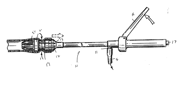
Fig. 1 is a perspective view of an applier of the
invention;
Fig. 2 is an exploded perspective view of the coupler as
seen in Fig. l;
Fig. 2A is an exploded perspective view of a coupler as in
Fig. 1, with an inverted impaling ring and a separate
latching mechanism;
Fig. 3 is a perspective view of the mating portions of the
anastomotic attachment coupler as seen in Fig. ~; - -
Fig. 4 is a cross section of a mating half of Fig. 3,
taken across lines 4-4; -
Fig. 5, 6, 7 and 8 are plan views of the attachment -
mechanism of Fig. 1 applying a fastener of Fig. 2;
Figs. 9 and 10 are perspective views of the introducer
portions of the anastomotic coupler with compression
halves placed therebetween;
Fig. lOA is a view similar to Fig. 10 of the coupler
configuration of Fig. 2A;
Fig. 11 is a particularized cross-sectional view of the --
mating halves of Fig. 10; and
: '. ~'
SEN-124
. ~: . - .: :
.... . . . .
21147~
g
Fig. llA and llB are views of the configuration of Fig. 2A
taken in an orientation similar to Fig. 11.
Detailed Description of the Invention
- There is disclosed in this invention an anastomotic
fastener applier 10, useful in combination with introducer
means 40,50 and compression means 20,30. Each of these
elements is readily apparent when viewing Figures
through 11, and will be further described herein.
As seen in Figure 2, there are provided compression
members or fasteners 20,30. These fasteners 20, 30 are
generally absorbable and are readily formed from known
biocompatible materials such as, for example,
polydioxanone, polyglycolide, polylactide, copolymers of
lactide and glycolide, copolymers of 1,4-dioxanone and
lactide or glycolide, copolymers of ~-caprolactone and
lactide or glycolide, copolymers of trimethylcarbonate and
glycolide, and various other polymers and blends of the
aforementioned copolymers and homopolymers. The polymers
listed above may also contain a filler such as calcium
carbonate, tricalcium phosphate, magnesium oxide or,
preferably, barium sulphate, at a concentration of about
5 to 35% weight or preferably f~om 13 to 15% weight.
Barium sulphate is an inert biocompatible material that
will render the fasteners and other components of the
coupler radio-opaque for visualization postoperatively if
desired.
These fasteners 20, 30 replace a standard anastomotic
staple line. Each of these ~asteners is plate shaped and
contains a central toric section 22, 32 which allow
positioning of applier 10 therein. This toric section
SEN-124
' ':
211~
- 10 - , ~.
also allows the pull-through function of the fastener
system. Also, at least one of the fasteners 20 has legs
or prongs 24 which may be sharpened so as to pierce `
tissue. Optionally, the prongs may be split along line
24s, to create a spring-like effect. The prongs may be
fabricated from the same material as listed for the
fasteners 20, 30.
Springs 8 may be added to provide a more uniform
controlled compression of the inverted bowel tissue.
Helical springs are shown in Figure 2, but other
configurations could be used including but not limited to ;~
leaf springs or compressible foam washers. The spring
material can be metallic or non-metallic. It is possible
to specify a spring constant to achieve a pressure to
ensure hemostasis. Experiments conducted on the colon of
anesthetized canines, as a model for human colon, would
~ suggest that the springs be selected to achieve a pressure
¦ distribution on the ring 39 of at least about 8 to 12
gms/mm2. Too low a pressure may result in leakage and too ~ ~-
high a pressure to excessive tissue necrosis. -
The other fastener 30 has multiple slots or receivers 34
which correspond to the prongs 24 on fastener 20. This
arrangement allows each of the prongs 24 to have little or -
no difficulty in alignment within the receivers 34 on the
other fastener 30. Each fastener 20, 30 generally has at
least six such prongs and receivers 24, 34.
~ .
The alignment aspect is very important concerning these
fasteners 20, 30. It is to be realized that with
conventional staples, and conventional staplers, the `
staples are pre-aligned with anvils so that the staples
are readily formed after piercing through tissue. In
'.
SEN-124
.~' ' ..
,.,
'
21147~
contrast, it is necessary to have these fasteners 20, 30
self-aligning so that the fasteners themselves meet with
one another. Thus, the prongs 24 are configured so that
they will readily be urged into each of the receiving
receivers 34. The receivers 34 are equal in number to the
prongs 24 and are wide enough so that the prongs 24 will
fit within each of the receivers 34. Of course, as seen
herein, the fasteners 20,30 can each be provided with
prongs 24 and receivers 34, which allows more secure
compression after emplacement of plates 20, 30. Also,
toric sections 22,32 have alignment means comprising a
slot and groove arrangement, which lines the prongs 24
with receivers 34.
As further seen in Fig. 2, each of the plates 20,30 has
attached to it on an internal section, an additional ring
25,35. Each of these rings 25,35 sits securely on a plate
20,30. Each of the plates 25,35 are provided with tissue
adhering pins 29,39. The pins 29,39 are capable of
adhering to tissue after suction applied to the coupler
mechanism 10 along the compression means 20,30. The pins
may be fabricated from metal or, preferably, from a
reasonably stiff biocompatible absorbable polymer such as,
for example, but not limited to, polyglycolide, or a
copoly~er of lactide and glycolide. The polymers may
contain fillers as described for the fasteners and prongs
of the device.
The plates 20, 30 are formed to be generally thin (about
.010" to .030" thick) so that they do not take up much
space within the housing of the applier 10 or between
tissue. Naturally, such reduced thickness is configured
so as to not inhibit holding strength of the fasteners 20,
30.
SEN-124
2 ~ ~ '1 7 ~
- 12 -
For example, for bowel anastomosis, fasteners 20,30
typically are made in sizes of 10, 18, 21, 25, 29 and 33
mm diameters. These allow for accurate placement for
anastomosis through a trocar, like the Endopath~ trocar
made by Ethicon, Inc., or to correspond to typical
anasto~otic instruments. Materials may be used that are
initially hard and then terminally soft absorbable
polymers, such that their consistency at the time of their
expulsion i5 soft and pliable. These permit usage of such
anastomotic rings in other sites, where absorption rather
than expulsion is the procedure the body uses to remove
these plates.
As seen in Figure 2, each of the legs 24 has flanges 27,
such that a number of useful gap setting distances are
provided. The user is able to choose over a relatively
infinite range, the appropriate spacing between the two
rings plates 20,30 with flanges 25 locked on detents 37
contained in each hole 34. This can be better understood
by reference to Serial No. 709,860, incorporated herein by
reference.
The introducer mechanisms 40,50 are generally seen in
Figs. 2, 3 and 4 can be found also in Fig. 2 coupled to
the compression plates 20,30. A first introducer
mechanism 40 is generally found to be on the proximal side
of the anastomosed tissue. A latch 51 attaches plate 20
to introducer portion 40 at notch 41. This introducer
portion 40 contains a generally conical shape 42 with a
generally cylindrical interior portion 44. It can be seen
that there are acceptor holes 46 which are meant to attach
to the prongs 24 o~ one of the compression plates. The
generally tubular internal portion 44 is formed with
ridges 48 which are able to mate with the opposite distal
SEN-124
211~4~ ~
- 13 -
portion 50. The tubular section 44 contains an interior
hollow cross-section into which the applier 10 is able to
be placed.
The introducer portion preferably consists of a material
that will soften and dissolve or hydrolyze in the bowel in
less than about 24 hours so it can be expelled.
Generally, this material may be formed from a water
soluble material such as, for example, gelatin, albumin,
dextran, alginates or chitosan, or preferably from
synthetic materials such as, for example, modified
celluloses (e.g., hydroxyproply-, hydroxypropylmethyl,
carboxymethyl-), the modified thermoplastic starches, -
polyacrylamides, polyacrylic acids, polyvinylpyrrolidone,
polyethylene oxides or polyvinyl alcohols. Most
preferable from a blend of polymers is a material which
provides good dimensional stability upon short term
exposure to moisture, e.g., during the intraoperative time -
to accomplish the anastomosis, and yet will soften and
dissolve within, say, 5 to 76 hours and preferably in
about 24 hours and be expelled when exposed to the fluids,
particularly water, in the bowel.
A good example of a material that will accomplish this is
a polyvinyl alcohol containing a plasticizer to allow the
blend to be melted processed, eg. injection molded. A
better example is a blend of 5 to 20% weight nylon 6,
about 15% plasticizer and the balance of polyvinyl alcohol
88 to 99+% alcoholized. An even better example is a blend
30 of 5 to 10% weight nylon 6, about 15% glycerine as
plasticizer and the balance a polyvinyl alcohol about 88%
alcoholized. These materials may contain also barium
sulphate in the amount described for the other device
components, and appropriate stabilizers. Other
SEN-124
.. ~.. ,:.. .... i . , -
211~7l~
- 14 -
combinations of water soluble, or hydrolyzable polymers
with non-soluble polymers may also come to mind to one
skilled in the art.
As further seen in Fig. 2, the distal or opposite
introducer portion 50 also contains a conical outer
portion. It furthermore contains a tubular inner portion
52. This inner portion contains detents 54 which mate
with the slot 45 contained on tubular portion 44 of the
proximal portion 40, as can be seen in Figures 3 and 4.
Thus, both these introducer portions 40,50 are formed in
mating relationship. Of course, distal portion 50 also
contains receiving holes 76 which are capable of receiving
the prongs of the legs 24 contained on an opposite
compression member 30 as seen in Fig. 2.
Alternately, as seen in Fig. 2A for instance, a separate - -
latching mechanism 200 is provided. This mechanism mates
with tubular portion 44, and its slot 45, on the proximal -- `
side of the device at mating section 210. As well,
latching mechanism 200 mates with tubular portion 52 and
detents 54 on the distal end of the device, at mating
section 220. Also, it will be seen that in Fig. 2A, plate
20 is already attached to introducer portion 40. However,
pins 29, 39 have been removed from the plates 20, 30, and
are placed on rings 120, 130. These rings 120, 130
contain inverted pins 129, 139. It has been found that in
certain procedures, latching mechanism 200 and/or inverted
pins 129, 139 may be used to facilitate tissue
manipulation, to perform the endoscopic anastomosis, and
ultimately to enhance healing.
As seen in Fiqs. 1, 5, 6, 7, and 8, there is also
described an applier for applying both the introducer
SEN-124
.~ ,.. . . . . - ... . .~ . ..
2~7~ :
- 15 -
portions 40,50 and the compression members 20,30
laparoscopically. This applier 10 contains retention pins
12 contained at the distal end of the shaft 14. These
pins 12 are actuated by operation of the handle portion ~ -
16. The button 17 on the end of handle portion 16 is
capable of causing the pins 12 to retract at the opposite
end of the shaft 14. Thus, it allows the engagement or
disengagement of either introducer member 40 or 50. of
course, each of these introducer members 40 or 50 will
have a compression plate 20 or 30 attached to it when
attached to the tissue.
As seen in Figs. 5 and 6, there is further contained a
knife blade 19. This knife blade 19 is activated by the
operation of handle portion 18, 18'. This knife blade 19
is caused to pierce the tissue captured within the lumen
described by any of the introducer portions which also
hold a compression member 20 or 30.
Optionally, there are contained in the hollow shaft 12
holes 11 which are capable of causing suction of tissue to
either introducer portion 40 or 50 as well as the
compression members 20 and 30. Suction is provided along
line S to holes 11.
In operation, in Figs. 5, 6, 7 and 8 the device of this
invention is delivered to the abdominal cavity in a two-
step process. First, a distal introducer 40 with a
compression plate 20 and ring 25 attached is loaded onto
the cylindrical applier at the pins 12 so that it is able
to be inserted stably within a trocar. After emplacement
within a trocar, the introducer and compression member is
positioned within a bowel section. Of course, the bowel
section must previously be cut. Thereafter, the
SEN-124
v ~, : ;., ~ .
;.. :.i. .,.. , .,. ~:, . - -
211'174~
- 16 -
anastomotic introducer is placed into the bowel, and
suction is applied so that the pins 29 of ring 25 are
caused to hold tissue thereon. The handle 16 of the
applier remains external of the body cavity through the
trocar cannula. The combined applier 10 and anastomotic
coupling mechanism 50,20 form a generally cylindrical
shape which allows the surgeon to position the coupler
body half 50,20 within the body colon lumen.
Once within the distal lumen of the sectioned bowel, the
surgeon holds the applier handle 16 while working the
bowel wall over the pins 29 located about the radius of
the compressive ring 25. To assist in the manipulation of
the bowel and secure it to the coupler half, the surgeon
may apply suction to the connector on the applier handle
via the hollow shaft of the applier handle 16 to holes 11
in the body circumference in the body shaft of the
introducer applier 10. The surgeon then pushes on the
handle 18 to engage a cylindrical knife 19. The
cylindrical knife 19 which travels toward the coupler half
to both push the bowel to clamp it to the prongs 24 and
then to cut the internal portion of the lumen held within
the cylinder 11 of applier 10. The next step is to
release the bowel and coupler half 50,20 from the applier
10, by pressing button 17 to disengage pins 12 on shaft
14.
Thereafter, the steps are repeated for the proximal or
opposite portion of the bowel.
Another form of this invention is that the pins 29, 39 are
separate from plates 20, 30 and are fired by the applier
to impale the tissue to the plates 20, 30 in a manner
SEN-124
~. , ~ , .............................................. .
. ~ : ... .
- 2 1 i ~7 ~ ~ -
- 17 -
similar to a staple, fired into a receiver. This is
described above, in conjunction with Fig. 2A.
The uniqueness of the instant invention is seen in Figs.
9, 10, and 11. Now the proximal and distal sections of
~the bowel are both fitted to each half of the coupler
40,50, the applier may be withdrawn from a trocar, and the
surgeon may grasp each of the halves so that they snap
into place. This is accomplished by mating latches 51
with notches 41. There may be ratchets placed on the
prongs 24, to enable the surgeon to create the desired
compression so that the tissue is properly anastomosed.
Of course, the design of Fig. 2A is attached so that
connection is made as in Figs. lOA and llA. Indeed,
without latching mechanism 200, but using rings 120, 130,
latching may be accomplished as in Fig. llB.
The desired compression could also be accomplished in a
number of other methods including the use of metallic
springs, non-metallic springs, compression foams, or even
a fixed gap selected externally and gauged according to
the tissue thickness.
Thus, this coupling device accompl~shes some very unique
features. First, there is compression at the serosa
junction in steps of between .5 mm to 2 mm to accommodate
various bowel tissue thicknesses. Second, there are pins
which hold and stabilize the lumenal tissue sections
during healing. Third, optimally, there is a two-step
biodegradation, first in about 24 hours for the introducer
portions 40,50 and then about 2 to 3 weeks for the
compression portions 20, 30. Fourth, the applier 10 is
able to bring the device into the surgical field via a
SEN-124
211~7~4
- 18 -
trocar. It operates as a stable work platform and
manipulator and contains an "integrated anvil and cutter"
for assisting in affixing the luminal walls to the pins 29
for removal of the excess inverted tissue portion. Fifth,
the instrument 10 also contains suction mechanism via the
hollow shaft 12 to aid in tissue manipulation, and device
placement.
Yet, what is clearly unique is that there is no purse
string suturing of the bowel required so that this
mechanism may easily be operated for laparoscopic
application. It allows surgeon's skill levels to be
lowered because -there is less skillful steps to be
performed in accomplishing anastomosis. The mechanism
resides fully within the bowel so that it is easily
removed, and takes the shape similar to a bowel dilator
which aids in insertion and expansion of the bowel lumen.
Because there are openings in the introducers 40,50 and
the compression plates 20,30, and the introducers 40,50
2~ are dissolved soon after the endoscopic procedure, there
is easy passage of gas and excrement during healing.
This, it is believed will improve healing time, and, of
course, patient comfort.
These and other embodiments of the invention have been
described as above. Of course, it may be possible to vary
the applier and fastener of the present invention without
deviating from the intent of this invention. For
instance, it i9 possible to create an apparatus with a
curved longitudinal shaft, or having a flexible shaft, or
where the shaft portion near the distal end contains a
trocar mechanism for piercing tissue. What is to be
realized is that it is the following claims and their
SEN-124
.. . .. .
., .. ~: . . ~ . . -: . - ...
.
, ~ .
: . :
2 1 1 '' 7 ~
-- 19 --
equivalents which are meant to cover the scope of the ::
invention.
SEN-124