Note: Descriptions are shown in the official language in which they were submitted.
WO 93/04077 2 1 1 5 4 6 1 PCI/US92/06957
ENZYMATIC ANALYSIS USING SUBSTRATES THAT YIELD FLUORESCENT PREClPITATES
This iL~re,Lou was made with U.S. Go.~ support under grant GM 38987 awarded by the U.S.
National T--';' of Health. The U.S. Go.~ has certain rights in this ;t "--
s
FIELD OF THE INVENTION
This invention relates to a class of novel lluu~ug~ic ~ ~ for d~ g enzyme activity,
particularly that of gl~cG~;d~, pl,o~ t~C~- and sulfatase C~ ~ The enzyme acts on the l~r u~ a~e
lû substrate to yield lluo,~e..t products that are sperifirslly formed, -- c, and i -coh ' '- in aqueous systems.
BACKGROUND OF INVENTION
Detection of enzyme activity is useful in the analysis of a biologic_l or chemical sample, such as
15 whole o.g~us."s, cells or cell e~tracts, biological fluids, or chemical mixtures. For e~sTnpl~ information
about ",~t~bolis"" disease state, the identity of microG~ .;;."~, the success of a genetic ~ ip~ ion~ or the
quantity of toxins, can be gained from evaluating the activity of certain enymes. Furthermore enzyme
col,ju~at~s are often used as sensitive bioanalytical tools for dPt~^~tion of analytes.
Enyme activity is often detected through the use of a synthetic svt 5~- . The endogenous substrates
of an enyme are used in dPcig~ing synthetic sub~ dt~s. Several ~l~co~;dasc enzymes are known to target
specific glycosides (R-O-Gly) to yield the co~ ~uAing call~h~d-ale and an organic alcohol or phenol (R-
OH). Pl.o~ e enzymes catalyze the conversion of certain pho~ mnG ~ O-P(O)(OH)2) to
i,.o,~ ic pb~, ~- ^ (P;) and an organic alcohol (R-OH). Similarly, organic alcohols or phenols result when
25 sulfatase enymes liberate il~GI6~ ~iC sulfate from some sulfate r~ (R-O-SO3H) or when
6~ - ' -e enymes hydrolyze aryl esters of P 6~ 1;' -~ '- acid (R~C=O)-C~H4-NH-
(C=NH)-NH2). C&l~ lC acid esters (R-O~C=O)-R') are L~d-ul~d by esterase cnzymes to alcohols and
acids. Cytocl.,u.,.c enymes o~cidize aryl alkyl ethers to give the phenol and an aldehyde or acid.
Most ph~ and sulfatase enzymes are r~ c~ for the structure of the alcohol. Two types
of i ' - . ' enzymes have been ' - ' ~ i, however, that have diffcrent optimal pH for their ~
activity (pH optirna about 10 and about 5 ,~ ). The aryl sulfatase enzyme most closely .~ the
acid 1' ~ ~ ~ in pH opt~ ~ and ~ t~ tumover. G- ' ~L is a cell surface protease
.'- ~ of several human tumor cell lines, which is not d~ in nonnal human cell strains.
35 r - - have structural ~ ;.GU.~ .~t~ that range from those that Ljllol~ oeters of the lower c&,l,u,.~lic acids
(usually ~ about 4 carbons) to the ~lipase enzymes that optimally L.~ ~ esters of the longer ca.l~a~lic
acids (usually ~ about 8 carbons). There are ~everal ~tu~Lu~ enzymes (is~ ) that differ in their
ability to if' ' -~' aryl ethers d p .. ,l;.. g on the source of the enzyme. Table 1 lists some ~ ly
WO 93/04077 PCI'/US92/06957
investigated enzymes snd their tsrget groups. 211~ 4 61
TABLE 1 REPRESENTATIVE ENZYMES
S E.C. NO. ENZYME TARGET GROUP
3.2.1.20 ~{;1. r~ A~ a-D41ucose
3.2.1.21 ~B~' ~ -D~Iucose
3.2. l.æ ~7~ -D~slrt~cP
3.2.1.23 ,B (;~ t~c"~P, ,B-Dfi~lg^tocP,
3.2.1.24 c~-M^ n.lc;AgcP a-D-M?nn.. cP
3.2.1.25 ~-MP~ns~ciAscp~ ~ D ~?~nss~P
3.2.1.30 N-Acetyl-B-~Iu~oc-.. ;.. iAsc~P ,B-D-N-Acetyl41uc~s-.. :.. F
3 .2.1 . 3 1 ,~lucuronidase ,B-D~lucuronic Acid
3.2.1.38 ~B-D-Fuc~ciAscP ~B-D-Fucose
3.2.1.51 a-L-Furn~;AscP cr-L-Fucose
3.2.1.-- ~B-L-Fu~Q~iAscP~ ,B-L-Fucose
3.2.1.76 L-Iduronidase cr-L-Iduronic Acid
3.2.1.4 Cellulase ~-D-Cellobi~se
20 3.2.1.-- ~-A.~;,.o~,~. - Asce ~-L-A. ' ~r~.. ,nose
3.2.1.37 ,B-Xylo~;dase ~B-D-Xylose
3.2.1.18 a-N-Acetyl-r.e.l,i.. ;.. ;Asc~P Ix-D-N-Acetyl-r,cu~ .. ",ic acid (Sialic acid)
3.1.1-- gllgni~ nb~ t~ce aryl esters of p-g~^ni~linQbpn7n;c acid
3.1.3.1 alkaline pho~ ~ aryl or alkyl rLQ~l.h~ n~c-Pr5
25 3.1.3.2 acid l.hh~.k~-_~ aryl or slkyl rh~ r ~n~
3.1.6.1 aryl sulfatase aryl sulfate ~ .-r_ 4
3.3.3.41 4-n;l,~.phl"yl phh~ e aryl I ' c~
The synthetic sub~lldt~s for rnany enzymes, inrbl~ling those in Table I as well as many esterases and
30 .,j~cL,u,uc enzymes, are c~- :c~ y based on the same ûrganic alcohol or phenolic p,~u.~ , differing only
by the nature of the leaving group (e.g. ~ h~ , sulfate, g~ Ai~-b---- , ~l,o.~lic acid, c~l~b~d ,
or alkyl alcohol). The synthetic substrate should not inhibit the ~"~tic reaction so that the enzyme can
produce enough product so that it can be detected (enzyme ~mrlif on of the det~tion product). Most
synthetic ~ have been designed o that the presence of the enzyme (or enzyme conjl " ) results in
35 a d~l~ q~'- pbenolic product, e.g. f~ of a soluble colored or lluo.~t product or f ~ of a
~. ,
hs~ - that yield soluble cLuluogc~uc (but noullUG~lt) products include p~ ph^~P
or sulfate ~n~ or gl~;,;d~s of o nil~u~LG~ol~ p-ml,.,~ 1 tL~ ` ' -` and ph -1l' ' -l-
40 Fl og. - ~ - derived from such phenols as various 7-L~d,u,.~ s, 3~ ' ~r ~..., 8-
h~d.u~J~.~G 1,3,6-tfl '' ~ acid, flavones or various d.~ u5 of ~- or ,B -~,' ' ~' typically yidd
soluble lluu.~ products. Although assays based on ' - ~.l products are generally p.~,f~ d because
WO 93/04077 211 a 4 61 3 PCr/US92/06957
of their greater sensitivity, they are deficient in a number of p~U~t;Oa for analytical ...~...c...~..t of enzyme
activity in vivo and in vitro.
None of the reported f' - ~cA ~ that yield soluble products are optimally detected below
5 a pH of about 6. With many ~ ~ t~ ~ it is no~, ~ to adjust the pH of the dye product to above 10 to
obtain the ~ lluu~c e ,fl..,;~. Assays that require such a change in pH or the addition of other
d.,~elcr reagents are not readily adapted for highly ~d analytical p~uccdu~co~ In addition, soluble
reaction products, whether nuc,.G~nt or colored, readily diffuse away from the site of activity, especially in
in vivo ll r 1 ' ' ~
Certain aUL~ ~ for rl~o~ e, sulfatase and some glycosidase enzymes are known to yidd colored
J~'ir l~s that are not n~O.c~c~L The best Icnown of these are 5-bromo-4-chloro-3-indolyl Flm~ p~
(BCIP) lLeary, et al., PROC. NATL. ACAD. SCI. 80, 4045 (1983)], S-bromo 4-chloro-3-indolylgalq- Ic.~
(X~al), several other ~X-glycosides~ that are similar to X-gal and the co. . Ga~,ûnding 5-b(. - 1 chloroindolyl
15 sulfate ~Wolf, et al., LAB. INVEST. 15, 1132 (1966)]. Following e..~-,~tic hydrolysis, the colorless 3-
hydroxyindole iutc.ll,edidt~ are converted to insoluble indi~oid dyes by ~.YiAq~infl with a second reagent or
more slowly by m~le Igr oxygen.
Menton, et al., PROC.SOC.EXP.BTOL.MED. 51, 82 (1944), introduced a two step terhni1~le in
20 which certain phenolic products, liherated by hydrolytic enzymes, are s l~ "~ ~ly coupled to a d;s~nni~m
salt. The terhni~uP yields ~,I"~".,opl,oric, but nonfluo,~cG.,t, diazo dye products. Bu~s~nP, ENZYME
CHEMISTRY AND ITS APPLICATIONS IN THE STUDY OF NEOPLASM, pg. 160 (Academic Press1962) i-,t,uJuced imrlifiP~ cim~ .Po~c and post-coupling azo dye t~L.~ . ~ using naphthol-AS ~L~
and sulfates, as the enzyme substrates.
A ~m~ifC ~f~n of the two step t~ Ziomek, et al., HISTOCHEM. CYTOCHEM. 38 (3), 437
(1990), ~G~Jort~lly yields a red fl.,o,~.ll azo dye ,JI~ that is useful for hi- ~
of ~ * activity. The coupling reaction of the diazo color-forming reagent must be ~ at
an alkalinc pH. While this pH nay be adequate for 1 ' ' dP~ of alkaline p~ ~ ~ activity,
30 it does not permit c~ u- dct~ of the activity of acid p~- . ' and aryl ~ulfatase enymec and is
b~p' l for ~ )A of ~B fg,dl~ , (pH opt ~.2), since these enyme_ all have c"l..; -1~, low
activity in alkaline medium. FL ;' ~ ~ c, the diazo coupling reaction is not specific for the phenols formed
by the ~ ir reaction. Therefore the presence of intrinsic phenolic s in the test solution or
the L ~Isc;~l fluid can yidd false positive sig~nals. All of the above methods suffer from weak and ~.,.~ t
35 - . ~ n-.."~.., staining of enzyme activity.
..c b rlifi~ t ' . ~s are used in I ~ t~ and c~ h~ y to localizc spccific
antigens by .,~,. Success of this i ' . ~ depends on an efficient :,it~, _pf ~ deposit of e.,L~ -
WO 93/04077 2 1 1 5 4 6 1 4 PCr/US92/06957
products that contrast well with the und~ u-g cellular ~uc~u.~s Colored precipitate formed by hydrol~3is
of known ~,Lu~ olic ~.~ciy ~ .,u~ such as X-gal can be well visualized at discrete loci in cells
or tissues using light ': uaCOIl,r, if the sample has 1, r- q~l~ q' ' - ~' of the target ~ It has been
reported thst the clul r' ~ pl~, ' ' g substrate for alkaline Fh~s"~ when coupled with a
5 digo~i6_.;.~ '-'~'~~ probe and anti-digo~igenin conj~ g ~ with alkaline ~' ~-r~ , can stain nerve growth
factor rnRNA at a higher vit~ and ,~ than a standard isotope label method lSpringer et al.,
HISTOCHEM. AND CYTOCHEM. 39, 231 (1991)]. However, the enzymatic products from the
~,b~uu.ophor;c L t~ ~ are not 5~ffi~iPn~ to form a visible precipitate that contrasts well with cellular
allu~lul~s when a single molecule of the analyte must be detected, because the cL.uuopho.;c signal is
10 ina~rr~c;e..t for dPtPrtio~ The lluul~.ll p.~;~,;tate of this invention, in contrast, provides a more easily
d~tec~q~`r signal in smaller amounts.
In recent years, nu~ .uu~ no...Odioa~ e ..~,l,.uaches have been developed and refined for in situ
hybridization ~Hopman et al., MOLECULAR NEUROANATOMY, pp 43, Elsevier Science Publishers
15 (1988)]. All of these nonradioactive techniques are generally able to detect specific mRNA i~l situ without
difficulty. In contrast, the nonradioactive methods for detecting a specific gene which exists in few or even
single copies in a cell's genome using biotinylated probes, require oligonucl~o~i~es that contain several
thousand bases in order to allow for sufficient incorporation of the biotin (or other) label. In practical terms,
any probe shorter than abou~ 2,000 bases will not result in visible si~nals sufficient to detect few or single
20 copies in the cell genome utilizing either the colored preciri~qles or fluol~.,ce microscopy. The need for
a probe of such long length severely limits the ease and flexibility of the probe design because prepq~.q~irn
involves such timeio~ ng t~hni~ues Because of their stronger a~C~lm~lq~ signal, the substrates of this
invention can be used with shorter olig~ cleoti~le probes.
The sul,sl.. t,_s of this i.. ~lion also differ ~igni~- t~y from substrates p.eviou~ly described in that
most known lluu~og~ ;c ~ yield products that are ~?p.~.ably lluGles~nt only in the solution phase,
whereas the p..,f~,.,cd L l~ - from this i.~ lion are virtually nonfluo.e~..t e~cept in the solid phase. In
addition they yield ~ l_, highly lluo,e products without requiring addition of a color-d~ ,lo~.,..g and
P~ - r;~ ' e reagent. ru,i' - G, the subject i~ ' t~ ~~ are specific for a pa,li,.,l..r C~L,~ ' ~ activity, and
30 are optimally reactive at or below l ' .~ g;~l pH. As a result of these cha,~t.,.;sl;cs, the ' 9 of this
i.~v~,.,lion can detect the activity of a wide variety of enzymes and c. L.~u.~ related analytes, in living cells, in
e~tracts of living cells, in ~ ;~L;c~l fluids, in biopsy samples, in vivo and in vi~ro, without . ~ g any
plG~ g of the samples by ~ ~ t~ ~ cc~' ', or filtration and without addition of &~ouda,
reagents.
Some of the lI..u,G~t dyes used to prepare the subject s~l~ are already known, e.g. U.S.
Patent No. 3,169,129 2~rtho-hvdro~Y-~henyl4~3H)~ l;, u~ to Rodgers, et al. (1965)
(~ ' ~ ); Hein, et al., The Use of Pol~ho~.ho~ic Acid in the Synthesis of 2-Arvl- and 2-AlkYI-
s 21154~1
substituted Benzimidazoles~ Benzoxazoles and B~..zullliazoles. J. AM. CHEM. SOC. 79, 427 (1957)
(benzimidazoles, benzoxazoles and benzothiazoles); and Naumann & Langhals, A Simple Synthesis of
Dihydroxybipyridyls. SYNTHESIS 279 (Apr. 1990) (dihydroxybipyridyls). It has been recognized that
several of the dyes have very low solubility, particularly in water, and that the compounds are fluorescent in
the solid state. The large Stokes shift characteristic of some compounds in this class of dyes has also been
described. There have been several studies of the fluorescence mechanism of this class of compounds which
has been related to a high degree of photostability. Catalan, et al., Photoinduced Intramolecular Proton
Transfer as the Mechanism of Ultraviolet Stabilizers: A Redl)p.disaL J. AM. CHEM. SOC. 112, 747 (1990);
Sinha & Dogra, Ground State and Excited State Pl olul U~J c Reactions in 2-(o-Hydroxyphenyl)benzimidazole.
CHEM. PHYSICS 102, 337 (1986); Orlando, et al., Red- and Near-infrared-luminescent Benzazole
Derivatives, CHEM. COMM. 23, 1551 (1971); and Williams & Heller, Intramolecular Proton Transfer
Reactions in Excited Fluorescent Compounds. J. PHYS. CHEM. 74, 4473 (1970). None of the references,
however, indicate the use of these dyes as fluorogenic substrates.
Orlando, et al., supra at 1552, citing Williams & Heller supra noted that replacement of an o-
hydroxyphenyl group by an o-methoxyphenyl group results in nonfluorescent benzazoles. An alkoxy group
was the only blocking group described in the reference, however, and there was no indication that blocking
groups could be selected to monitor the presence or activity of enzymes.
The intPrn~tional patents EP,A,0 433 853 (Boehringer Mannheim Gmbh., 6126191) and ET,A,0 158
225 (Miles Laboratories, Inc., 10/16/85) describe the general state of the art at the time of the invention.
DESCRIPTION OF THE DRAWINGS
Figure 1: Synthesis of Substrate. Figure 1 is a diagram of the formation pathway of some typical glycosidase
substrates. In step 1, the fluorophore is glycosylated using a modified Koenigs-Knorr methodology in which
a protected carbohydrate group is added to the hydroxyphenyl-quinazolinone. After isolation of the protected
intermediate by column ~l~ullldLu~l-y or by Inc~ iLdLiOn of the remaining starting material combined with
recrystalization or trituration, the protective groups are removed (step 2) to yield a nonfluorescent 2'-
glycosidyloxyphenylquinazolinone.
Figure 2: Characterization of the fluoro~enic precipitatin~ substrates.
A) Fluo.~c~ilce chdld~ li~Lion of a fluorogenic ~ it~ g substrate: a. emission of 2 mM 2-(4'-
methoxy-2'-phosphoryloxyphenyl)quinazolinone (3e); b. emission of the precipitate resulting from incubation
of the substrate (2 mM) in the presence of excess alkaline phos~lldl~se; c. emission following dissolution of
the pl~ iLdl~. The emission measurements were made in 0.1 M TRIS pH 10.3 containing 50 mM NaCl, 10
mM MgCI2 and 0.1 mM ZnCI2 using a Perkin-Elmer LS-50 fluorometer with excitation at 400 nm, excitation
slit 3.0 nm and emission slit 2.5 nm.
B) Coexistence of fluorescence and precipitation: 5 mM 2-(4'-methoxy-2'-
phosphoryloxyphenyl)quinazolinone (3e), in 0.1 M TRIS pH 10.3 containing 50 mM NaCI, 10 mM MgCI2
and 0.1 mM ZnCI2, yields 210 units of fluorescence by action of 10 ~lg/mL alkaline phosphatase in 20
WO 93/W077 2 1 1 ~ ~ 6 1 PCr/US92/06957
seconds. The ll..o.c~..c~ can be .l d by addition of 0.6% Triton X-100 as a result of ~ iLle
~Iicc~7~ inrl
C) Light- ~& increase as a result of pl__ ri' 2 mM 2~4' ' l. y-2'-
pLu~Jhû~11O~ JL~u~l)q ' - (3e) s g increases from 450 units to beyond the d~t~ limit of
5 1000 units in 20 seconds by action of 10 ~g/mL alkaline pl.h~ t~_7, showing rapid fo, - of a p,~ ri~
The ~u~t;c reaction was in 0.1 M TRIS pH 10.3 ~ ~ .g 50 rnM NaCI, 10 mM MgCI2 and 0.1 mM
ZnC12. The 5~ltu.;ng ...~u.~...~t was rnade in a Perkin-Elmer LS-50 lluor~ ter with 5 nm slits and a
coi.~ri~1~n~ eYri'^'ior and emission ~ .lc..gth of 420 nm.
D) Critical concentration: Various concentrations of 2-(4'-methoxy-2'-
10 phosphorylu~ hcl..yl)quinq7nlinnn~ (3e) were reacted with 50 ~LL of excess alkaline phoc~ in 200 ~Lsolution of 0.1 M TRIS 2 pH 10.3 c~ E 50 mM NaCI, 10 mM MgCI2 and 0.1 rnM ZnC12. The
resulting precipitate and fluo.~nce were .,.~.ned in a CytoFluor fluo.~cc. ce plate reader (Millipore) with
excitation at 360 nm, emission at 460 nm using sensitivity setting 3. The figure shows a critical co,.cenl.ation
of 2.5 rnM.
E) pH d~pen-l~nre of p,~i~,~t~lion: 2-(4'-Methoxy-2'-phosphoryloxyphenyl)q~inq7nlin~.n~(3e) was
reacted with 50 ~L of excess alkaline pl-o~l~h~ce in 150 ~LLL solution of 0. I M TRIS pH 10.3 c~n~ ;..;..g 50
mM NaCl, 10 mM MgCl2 and 0.1 mM ZnC12, 0.6 mM. The mixture was titrated using 50 ~LL of various
conc~.-t-dtions of HCI to obtain the desired pH, then nl~ul~d in a CytoFluor fluo-~e..cc plate reader with
excitation at 360 nm and ernission at 460 nm using sensitivity setting 3. The figure shows a pK~ of about 8.8.
Figure 3: Detection of Con A ~ tC.l~ usin~ a nuoiuFc.lic precipitatin~ substra~e.
A) Phase contrast image of NIH 3T3 cells;
B) Image of NIH 3T3 cells stained with a fluorogenic p,e~ ';--g substrate, 2~5'-chloro-2'-
phosphorylo.~ ,.,yl)-6-chlolu~u,n~l~linone(8e); exposure time 8 seconds using filters sptil ;7 ni for Hoechst
25 33258. The photos were taken in a Zeiss lluol~.ll usco~c with Fujicl..u,..c 1600 slide film.
Figure 4: Detection of EGF ReceDtors usin~ a flLIulo~ ic l~.cc;~;tatillQ ~uI,~tla'
A) Phase contrast image of A431 cells;
B) Image of A431 cells stained with a lluo~o~e~ic p,e, g ~ 2-(5'-chloro-2'-
30 ~JI.o~Jhu.~loA~h~ 1)-6-chlGlutlu~ (8e);e~posuretime15secondsusingfilters~t;~ dforHoechst
33258. The photos were taken in a Zeiss nuO,~..~ "uCIu~OpC with a Kodak Ekhch.u...e 400 slide film.
Fi_ure 5: Western Blot. Figure 5 is a pLot~J6 ~ showing d~ 'ecti of subunit II of the ' ~ protein,
c .~I~Lu~e c o~idase, using standard Western blotting ~ s
35 (A) Coo-- ~c~ r Blue staining of whole protein.
(B) Color d~,~do~cd with BCIP/NBT.
(C) Stained with p~ ;r - b~ t~ le.
WO 93/04077 2 1 1 ~ ~ 6 1 Pcr/US92~069s7
SUMMARY OF THE INVENTION AND DESCRIPTION OF PREFERRED EMBODIMENTS
This h.~ntion describes novel aub~ ta uscd to measure enzyme activity. The ~ ctPtec are
no..fl..o.es~.ll but react with enzymes to yidd fluG.e~.lt phenolic products that are specifically formed,
S nontoxic to the cells, and p~ J;lalc without inactivating the enzyme. The phenolic product may result from
h~ Lulja;s of a phenolic cster or a phenolic ~ oa;de, e.g. by F~h ~a~ , s~lf?~ocP" glycosidase and esterase
enymes. ~ ly, the phenolic product may be formed by oYi~io~inn of aryl alkyl ethers, e.g. by
cytochlvll.e enzymes.
The p~cr~ ,d au~all - of this invention are blocked fluorophores ~ .. ted by the formula:
BLOCK-O-Xn
where the fluorophore Xn contains a rnin~ lm of 2 aromatic rins, two of which are typically linked rather
than fused together. The aromatic rings include u"~t~ ted heterocyclic ring ~l~u~lul~5. Each of the two
15 linked aromatic rings may be fuscd to additional aromatic rin~s. Typically, at least one of the aromatic nngs
is fused to at least one additional aromatic rin~. In general, fusion of one of the linked aromatic rings to at
least one additional aromatic ring inc-~ses the wavelength at which the solid product can be excited and at
which the fluorescence can be detected, which is beneficial for some applications. Fusion to an additional ring
also typically results in the product becoming less soluble in water, which is favorable to precipitation.
BLOCKis a group that changes the excitation or emission properties (i .e. al,soll,_~ce or ftuol ~c~i.lce)
of the fluo,u~Jho,c and is capable of being cleaved from the remainder of the substrate molecule by action of
an enzyme. Preferably BLOCK blocks the long wavelength (greater than about 450 nm) lluGl~i;,eence of the
nuolopllolc.BLOCK is selected to be specific for the enzyme of interest. Typically, BLOCK is a monovalent
25 moiety derived by removal of a hydroxy group from ph~ , from sulfate or a biologically compatible salt
thereof; or a monovalent moiety derived by removal of a hydroxy group from an alcohol or from a carboxy
group of an alipho~ir~ aromatic or amino acid or of a peptide; or a monovalent moiety derived by removal of
the r ic hydroxy group from a mono- or pol~saccllal;de. Preferred monovalent blocking groups include
the target groups listed in Table 1, which includes some of the enzymes that will cleave such groups from the
30 ~
When BLOCKis a~Ja. ~t from the f~.~hlde. of the substrate ~' '~ by action of an enzyme, the
result is a visible p.~ I~ o A visible p,~ `~r ' '^ means it is do~ A by a light sensitive l ~' - . e.g.
a change in spectral (e~ jonl~ ~) properties, a change in light acdth.ing, or visible crystal formation.
35 Plefu.dll~ the pl~;~ t~ is Ituvl~ll. The favorable pH range for p., . on and de'ertion of the
nllol~nt products is from below about pH 2 to above about pH 11, most favorably in the range of pH 5-8,
which r..~ p~c_ c the physiological pH for in viw appl: -
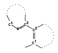
The visible pl~ tt generally has the formula H-O-X~, where X~ is a I1UOIU~JhOI~; of the formula:
WO 93/04077 2115 ~ 6 1 8 Pcr/us92/o69a
C ~N~C~C2~
s 11 .
~c1
that is covalently linked through ct to the o~ygen -O-.
The carbon atoms of -Cl=C2- are joined so as to ~ , ' ^ a first 5- or 6-.. ~ l,c.ed aromatic r,ing
that may contain at least one of the hetero atoms N, O or S. C~mmnnly the -C2=CI-O-H portion of the
n~,OI~..l p~;~;tatc defines a phenol or a rqphthf l Less commfmly this portion of the lluolesc~lt
Pl~- r contains a hetero atom.
The carbon atoms of -C4-N=C3- are likewise joined so as to ~ ^ a second 5- or 6 .. e.,ll~.~,d
aromatic ring that contains at le. st the one nitrogen h- te.. t~ that is between C3 and Cq. This second ring
may also contain at least one Bd- ti~ -1 hetero atom N, O or S, as well as oxo, thiooxo, sulfone, or amino
fi~lctif nq1itiec,
The first and second 5- or 6-.. ,e.. ,l.~.ed aromatic rings may be joined by a bridging ring between said
first and second rings. The bridging ring includes at least c2 and C3 and can contain a heteroatom N, O or
S. The bridging ring may be a 5- or 6-...e...be.~d ring and rnay be saturated or uus~tuldted~
Each of the first and second 5- or 6 .~ .cd aromatic rings may be fused to at least one ~
25 aromatic ring that may contain at least one of the hetero atoms N, O or S. Preferably, the lluo.vyho.c
contains at least three aromatic rings, two of which are fused. Typically the second aromatic ring which
contains at least one nitrogen ~ u..tu... is fused to a third aromatic ring.
Each of the aromatic rings may be further ~ifi.~ by ~ ti.~ of any hyd.v6_.~(s) on an arornatic
30 carbon with a halogen atom, lower alkyl (about 14 carbons), pc.ll~o.valkyl (about 14 carbons), alko~y (about
14 carbons), nitro, cyano or aryl, or any c~ ' t. thereof. The p.efe..ciid halogen s~b~ti ~ are F, Cl
or Br. Halogen and alko~y '; on the romatic rings appear to have a ~ .~- r..: 1 effect both on
reducing the solubility and ill~JIO~g the nuO--~.~ p~ot~lif-s of the lluv-es~lt solid.
WO 93/04077 9 2 1 1 5 ~ 6 1 Pcr/US92/06957
In one e~ ;, - u~ of the ...;~ - H~-X" has the ~l~u~lu~e;
S HO
where W is (CH3)2C (i ,o~o~"lidc,lF), -CH2-, -CH= (methine), S, O, or ~ R)- wherein R is H or lower
alkyl (1~ carbons); and Z is ~C=O)- or -CH=; and n is 1 or 0. When W is -(N-R)- and Z is -(C=O)-, the
10 products are qu ~ s (also referred to as qL - ' ). When W is ~N-R)- and Z is absent (n = 0),
the product are ~ When W is S and Z is absent (n = 0), the products are bPn~n~hiq~r~le~
When W is O and Z is absent (n = 0), the products are bPn7n~ When W and Z are each methine, the
products are q~- nolinPs When W is iso~..u~lidcnF and Z is absent (n = 0), the products are in~olin~c
When the first aromatic ring and the second aromatic ring are both 6 - - -- I-f .ed rings that together
form a 5- or 6- ."~n~ .ed bridging ring between them, the product~s are pl.~ h~idines. The bridging ring
may oe saturated or u..s~ lu~tsd. When Xn is a pl, .,~ .;dine, the precipitate H-O-Xn h_s the ~lluulu~;
In another e .1~;.~ F~,~ of the invention, the fluo~upllGI~; Xn is a quins7Oli~one~ bPn7imir1az~
b ~_ ~ 1-, t ~ ~ ' e, ~ - OI;' F, an in/~olinP" or a r I ' ' ~, and at least one of the aromatic rings
is further modified by, ~ ;on of one or more Lydlot,e.. atoms on an aromatic carbon. One or more
25 s lJ~(ifi,-~..~(s), which may be the same or different, are F, Cl, Br, lower alkyl, pe.lluu~ualkyl, alko~y, nitro,
cyano or aryl, or any c ' ~ - thereof.
In yet another ---1~1-- ~t of the i ~ lio.., the lluo,~ Xn is similar to a ~u;~ nF,
1 ' e . ~ , L ' ~ or an indoline but is further - - -n~l; fi~d in that at lesst one
30 of tbe aromatic rings is fused to at le st one ~ ' ' ~1 aromatic ~ing that may contain d least one of the betero
atoms N, O or S.
The p,~,fe.~d nuu~ube~ic ' 11 - for this invention have one or more of the following p.upc.li~.
1) generally soluble but r~f ~ ~It in water but n,l~ g a highly n-,O,~.t solid product in an
aqueous solution co~ E the -~bf ~ and tbe specific enzyme;
2) a low residual rolubility and rapid p~ A rate for tbe solid product relessed by action of the
enzyme;
3) reactive over a wide range of pH, generally below a pH of about 11;
WO 93/04077 2 1 1 5 ~ 6 1 lo PCI`/US92/06957
P.~;"a.~ on of Fl-lolu~l.o.~-, (Xn):
The p.eft,..cd fl~,o.~,lt dyes used in preparation of the lluolubenic ,ul,st, generally fall into the
;..r~oc (Tables 2 nd 3). ~ s, b~ n~r~ ' s, ~ - ~o~ b ~ (Table
5 4), ;.tlol; ~s and F'a ' ~ -- Schif& ba_es (Table 5), which are similar in structure and also form
lluol~lt ~ iri~t~, are less p.efu.-ed because they are relatively unstable in vivo.
PrPp ~ra~ n of a number of the ~..,f~,.,.,d lluo.uyhûl~-, is desc~ cd herein as a means of ill .~l~ H ~g
the breadth of the reaction. The d i~ ;I lio ~c are meant to ill~c~P, and not to limit the choice of reactants
10 and reaction c~n~iitionC that can be used to prepare the requisite lluolU~,c..ic ~ - By ~ r U~l' A choice
of, ~,5~;n ~ , in particular, the ~.u~ ;, of solubility, nuGI~.ce intensity and wavel~glhs and product
phr~ ity can be mr~jfiP~
Table 2 lists ~u~,.~ntative 4(3H)~l~inc~7n!inonp-c their spectra and the visible color of the fluo,~..t
15 crystals, according to the formula:
Ri~NH
~:~4
Ho~R3
R2
2û
Among the methods that have been s~ rully utili~d to prepare the subject q~lins~rtlinonPC dyes are the
following:
1) By heating of egUim~lsr amounts of an 9n~h-9nil '1P with an aromatic aldehyde in the presence of
2', catalytic amounts of p-trJlu Ifonir acid (TsOH), a diL~d~uy; ~7rt!inonP is formed, which is o~idized by
asuitable-l~iAi7ingagentsuchasdichlo,ud;~ oclu;~n~(DDQ)totheco.~ 7l~N~gy~R~ ol~(E~ample
1).
2) By reaction of isatoic ' ;d~ides with ~ ~ in the presence of cahlytic amounts ûf base in an inen
solvent (US Patent 3,655,664 to Pater (1972) and F- , '^ 2).
3û 3) US Patent 3,526,627 to Brooks (1970).
WO 93/W077 PCI`/US92/06957
11 2115~61
T~ble 2. 4-(3H)~in~ nPC (a)
4~3H)~ ' Rl R2 R3 R~mp lC] YieldE~Mm"~2Color
[ I
la 2~2' hjd~uA~l' yl) H H H H 297-298 64 490 b-g
2a 2~2'-hydroxy-S'- H H H OCH3290-29289 SS0 y
3a 2~2'-hydroxy-S' ~ I rl ~1)- H H H NO2 >350 74 470 b
4a 2~2'-hydroxy-4'- H H OCH3 H 284-286 35 4S0 b
-~ ~"h~
Sa 2-(2'-hydroxy-4'- NO2 H OCH3 H > 350 42 b
mcthoA~,h_nyl)~nitro-
6a 2~2'-hydroxy-S'- Cl H H OCH3342-34470 550 y
he yl)~chloro-
7a 2-t5'-chloro-2'- Cl H H Cl ~350 70 510 y-g
h~d-uA,~h~..yl)~chloro-
8a 2-(2'-h~.i-uA~"h~ 6- Cl H H H 336-338 30 500 y-g
chloro-
9a 2-(S'-chloro-2'- H H H Cl >350 60 510 y-g
lOa 2-(2'-hydroxy-4'- Cl H OCH3 H >350 64 b-g
methoxyphenyl)-6-chloro-
lla 2-(3',5'-diehloro-2'- Cl Cl H Cl >350 45 550 y
h~dru~ JL~",yl)~chloro-
12a 2'-(3',5'-dichloro- H Cl H Cl >350 75 y-g
2 l I~ 1 I UA~
13a 2-(2'-hydroxy-S'-nitro- NO2 H H NO2>350 96 525 y_g
phenyl)~nitro-
14a 2-(2'-hydroxy-S'-"it,u~,h~nyl)- Cl H H N2 >3S0 86 480 g
~chloro-
lSa 2 (2~ L~J~UA~I' yl)~nitro- NO2 H H H > 350 63 560 y
16a 2-(5'-ehloro-2'- NO2 H H Cl ~350 69 y
l,,~J,uA,~,h~..yl)~nitro-
17a 2~2-l,yd~vA~ ,~' ' yl)- 3S2-354 94 nf
18a bis-2,5-~(3H)-., ~ ' ~11- >350 8 r
l.yd.~, r~-
lColor of ^ ~ - b-g = t' g,~n, y = yellow, b = blue, y-g = yellow-green, g = grecn,
nf = non-n-,v..i t, r = red. 2 Emission max. of solid [nm].
WO 93/04077 2 1 1 5 ~ 6 1 12 PCI/US92/06957
Table 3 lists lc~,,~.t~ , benzo 1(3H)~ their spectra and the visible color of
llYo.esc4.lt crystals. The c~ u- .A' in Table 3 are prcpared by sirnilar p.ocedu-~ as used for the c~ . s ' -
in Table 2 but starting Wit'Y . . ., Iy ~ d . ' ' -' --- buA~lic acid derivatives.
S Table 3. benzo-4~3H~,t- ~ o^~s F~ .,LI~l~ to the formula:
~f2
H0~bjJ
# bcnzo-4-(3H)~u;r.~2ol;.,onc, Rl R2 mp ~-C]YicldEMm,~ of Color
[9GI solid
lb 2-(2'-hyJ.u~,hc,.~l) H H > 350 49 --510 y-g
lS 2b 2-(2'-hydroxy-S' ~ H OCH3 35~58 28 --570
3b 2-(S'~hloro-2'-l-~dlu~ c.. yl)- H Cl > 350 57 --510 y-g
4b 2-(3',5'-dichloro-2'-hy~ l,cnyl)- Cl Cl > 350 75 --550 y
IColor of nuorei~c~rlcc: y-g = yellow-grccn, y = ycllow.
Table 4 lists representative b~n7nYq7~ 1es, b~on7imi~q7r~les and ben_o~hiq~ s, their spectra and the
visible color of the nuol.,s~t crystals according to the formula:
~0 R2
~ \~
FlYU.UIJhO~,S of this type are co,l~_..;e..ll~, prepared from .~ ..up 'y ' ~ de.;;_t ~ of o-
30 Y~ l, o-z- :--otl ûp~ -l and o pl.e.. ~h ~ and the co,.~l.on~l; .g s~,l,sl;~ 1 de.i~ ~_s of a
benzoic, . ' ' -:~ or pol~ lic aromatic or I l u.,~_lic acids or aldehydes a~o..ling to p,.,: ~ known
in the art, jn. lu~lh~p
1) BY C~ndnn-~t;nn of a) o ,' ylr--~;~ b) r - ..~pl f ~olC, c) ~hioph~nolc with salicylaldehydes
followed by o~ ;on with Pb(OAc)4 (Stephens et al., J. CHEM. SOC. 2971 (1949)].
2) By heating of o -t' ~ c with ~li~ ~ in DMSO (Dcleg;o.~ , DYES AND
PIGMENTS 12, 243 (1990))
WO 93/04077 13 Z 1 1 5 4 6 1 Pcr/us92/o6957
3) By pol~l.G ",ho~ ic acid catalyzed ~nAd .. ~ of carboxylic acid derivatives with o-amino, o-hydroxy
or o .-.c.~ oyl arnines (Hein et al., JACS 79, 427 (1957))
Table 4. ~ .. 9 1 ~ and bu~.
C~ R~ R2 X mp t-Cl Emi~sion wavc Color
Icngth ~ tnm],
S lc 2 (2' L~ C~ I) H H O 126 b
t
2c 2-(2~ UA~ ) H H NH 242 b
3c 2~2'-lly~l.ut.,~ ) H H S 127-28 52û y-g
4c 2~2'-1.~1.u~ l.th~l) H H S 11~12 520 y-g
~C~
Sc 2~5'-amino-2'- NH2 H S - 660 r
hydroxyphcnyl)
b~n~nlhi~ 7~nl ~
6c 2-(2~-hydroxy-5~- NO, H S 2~0-12 520 y-g
nitrophcnyl)
b~n7n~h~ nlc-
7c 2-(~',5'-dichloro-2'- Cl Cl S 186-88 5S0 y
hydroxyphcnyl)
bcn7nlhi~7nl~
8c 2-(2'-hydroxy-5'-mc~hoxy) OCH3 H S 74-76 600 o
brn7l)lhi ~ ~n¦e
9c 2-(2',5-d;by~.ux~,l,c,,~l) OH H S 197-94 550 y
tj~A7.~ 7Ale
IColor of tluo~c ~ b = bluc, y-g = ycllow-grcen, r = red, y = ycllow, o = orangc.
Table S lists some Schiffs bases and the color of their fluorescent precipitates. Schiffs bases are
prepared by heating of an aromatic aldehyde with a s- b~ d aniline in a suitable solvent such as EtOH or
20 toluene (Kresze et al., Z. NATURFORSCHUNG 10B, 370 (1955) and Example 3).
Table 5. Schiffs Bases according to the formula:
R2~
R3 OH ~3R1
WO 93/04077 2 1 1 5 4 6 1 14 PCI'/US92/06957
Schifîs basc R~ R2 R3mp ~-ClYicld [9G]Color~
ld 2~ l.oa~.L~ ~ NMc2 H H13~38 89 o
" ' ~ . '.~1 uninc
2d 3,S~ichloro 2~ 'r ~- NMcz Cl Cl-- 72 r
~,~ q~~
~ r ~1 iminc
3d 2-hydroxy-5 - ''J''--'L/'-'- - P~- NMc2 NO2 H212-14 99 r
J r .~ ninc
S 4d 5~hloro-2-hydroxy t~L~ - NMc2 Cl H188-90 84 o
4'~1u,,dh~ l irninc
Sd 3,5~ichloro-2-hyd~xy- OMe Cl Cl11~16 96 o
r ~ ,~1 i ninc
IColor of lluo~cc..~ o = orangc, r = rcd
10 PreParation of FllJu,u~.,ic Substrates
In certain i~ c, especially where BLOCK ish.co.~,o,~t~dto yield a simple aliphatic ether
substrate for a cytochrome enzyme, it is y~ef~ b!F to h.co,yol~teBLocK before formation of Xn (for instance
see Example 10) Generally, however, the substrates of this invention are prepared by the following steps:
I) preparation of a suitable lluo,u~,ho,e such as those already mPn~io~d above; and
15 2) reaction of the fluorophore with an ap~..u~ ate form of a blockin~ reagent to form the substrate.
,
BLOCK is typically bonded to Xn by reaction of a reactive form of BLOCK with the hydroxyl group
present in the unbound form of Xn through the inte.~ ,a~y of a reactive derivative of BLOCK that can
s~h~ tly be converted to BLOCK. ~ûr instance, pl~o~J~h~ is inco,yolst~ using a reactive form of
20 pl~ such as r~ l ho,uu~ oxychloride in Example 7 or such as via pl~o~ }~"~ itP .,Le.l,.sl,y in E~ample
7. Sulfate is typically i.,~,o.~ ' by reaction with chloros~lf~nir acid as ~ l in Example 6.
C...lu.~ esters are typically i...."G,~ted by reaction with an act,~-_ d form of the acid (for instance
anhydride, rnixed anhydride, acid halide) as shown in F-- ,1~ 8 and 9.
Gl~_usid~c are typically prepared by a ~ifiP~ Koenigs-Knorr ~ involving l~ of
the unbound form of Xn with a soft acid catalyst (for instance silver c~.L- ), an - t;i_ ~ p,. I
c L-~ ' (APC) d~ , and a -~ ~ ` base (for instance sym-collidine), under ' ~.I,u~s
C,..~ ;o,!c (Figure 1). The APC will contain one or more sugars with an L,'-~_'- g group at the
position of the sugar to be attached to Xfl. Typically the APC is a l ' ~ d sugar, where a halogen is the
30 - ~ g group at the ~ - ;c position. De~ndi..g on the reaction c4 l;l;o~, the sugar(s) involved, or the
-- r isomer required, other activation groups at the f~ ,-;C position of the APC can be used, most
c - ly trichlo.~ ' , I' ~, ' ~1 or acetate. This will result in the p,. ' ~ of a -- " - ~t
;de i -' 1~ After isolation of the p,~ ;dc o' . the p~o~ C groups are
removed from the p,ot.~t~ gl~_os;de, using p.u~s ll r upr to the p.ot~ti.,g group(s) present.
-
WO 93/04077 2 1 1 5 4 6 1
PCr/US92/069~i7
_
Synthesis of ~ ~n~tive ~r~ of the s-ubject substrates that contain glycosides are given in r~
4 and 5.
The following Tables 6-8 contain ~c~ /e pl-h~ DulJallot~s. Althou~h all of the S~all~t~5
in Tables 6-8 are ~.I.h~ -8-- -q, any s-uitable blacking gr~3up p~ iOllaly d~;~ could be ~ ~1 to prepare
S the same mnge of a~all - for detecting or analyzing a p L~,ular enzyme. For ~ , Table 9 ill-- -
some of the same a~ allot~ that can be rnade as ~Iy~s;d~. The number of phocpb~t~ !, b~t~ - described
herein are merely ~ .,~entOIive of some of the choicea available for de~rtihrl of pl~ ~, enymoe. The
range of choiccs are meant to illllctr?'e, and not to limit the range of possible n~O.ub~ c â~sll ~ with a
variety of properties. Any of the doecribed II~GIu~ho~ can be used to prepare a substrate for a wide range
10 of enzymoe. By Opl~lupriOte choice of fluG~uj)hûl~a~ blocking groups, and ~ t~, in polli~ the
substrate can be tailored to give desired properties of reactivity, solubility, nuû.e~,.ce intensity and
wo~ cgtils, and product photos's~ility. Table 6 gives a Dullul~aly of the a~llth~is of some .Iu;
phn~,h~t c prepared as in Example ?. Table ? gives a DI~I~Ullol,~ of the synthesis of some l~ ~i~u;~ ..lin~n~
phnCrhs~-s prepared as in Example 7. Table 8 gives a summary of the synthesis of some bPn7nthis~nle
15 phocrhs~c,
Table 6. 2-phosphoryloxyphenyl4-(3H)~uinazolinones(disodium salts) according to the formula:
o
R3 ~H
~
0 2
O=P--O -No+
O~Na+
~ 4 (3H) ~ -' 2' R~ R2 R3 Yicld Yield of R~
pho~ph~-s (disodium salts) ~%] in~ .,didt~ PrOH/NH3/H20
di-t-butyl 70/10/20
eskr [9ol
lc 2-phenyl H H H 76 99 0.38
2c 2-(5' -' .~/h~ l) H H OCH3 86 90 0.36
3e 2~4'. ' ~,h._.~l) H OCH3 H 89 99 0.36
4c 2~5' -r' ~l~nitro NO2 H N2 84 95 0.S2
5c 2-(5' . ' ~ I.c.. ~l)~chloro Cl H OCH3 90 92 0.37
6c 2-phcnyl~nitra NO2 H H 92 96
7c 2~5'~1,1O.~",hcn~l)~nitro NO2 H Cl 83 89
8c 2~5'~-' "-r' ~I)~chlor~ Cl H Cl 82 86 0.39
9c 2~5'~hlo~yh.~ 1) H H Cl 95 96 0.38
WO 93/04077 ~ 4 6 1 16 PCI`/US92/06957
Table 7. Benzo 4~3H)~ 2'~hn~ alt) ~ .I;.. g to the formula:
o
S ~)~
o
O=P--O ~Na+
O-N~+
# Ben~o4~3H)- R Yield Yield of ~ ' Rf (rl~
4~ ol; oa~2'-p!~ [%1 di-t-butylcstcr[%] i-PrOH/NH3/H20
(disodiu,.l salt) 70/10/20
lf 2-phenyl H 87 91 0.36
2f 2~5'-chlo.o~,h~yl) Cl 90 92 0.37
Table 8. Benzothiazole rhr.crhq~es (disodium saltc) accordirlg to the formula:
Na203P-O R
~\~
p.. ,.J~h;->-~1~-2'- R~ Rz Yield Yield of inter- Rf (rllo~
Fh''Crhq'e (~ ... salt) [%] mediate di-t- i-proHtNH3lH2o
butyl ester [ %] 70/10/20
lg 2-phenyl H H 96 98 0.37
2g 2~5~.......... ~LuA~Jh~l) H OCH3 79 -- 0.39
3g 2~3',5'-dichlG.u~ l~w~l) Cl Cl 71 -- 0.38
35 Table 9 gives a ~u.. ~. r of the s~ ' of some qu ~ gl~ ;des l1 ~ d;.. g to the formula:
o
R1 ~
Gly~
R2
WO 93/04077 2 1 1 5 4 6 1 PCr/USg2/06957
-
Table 9. 4-(3H)-Qu~ .Ar Glyuos;d~s
# 4~3H)-Qu ~ F Gl~a;d~s -Rl R2 R3 R~
lh 2~2'-gala,lop~ ~A~ n~I~- H H H H
2h 2-(5'-chloro-2'- Cl H H Cl
gal..ctu~ loA~/}; ,yl)-
6-chloro-
3h 2-(2'-gal.. ~lo~ loxy-5'- H H H OCH3
methoAy~,h~,..yl)-
4h 2-(2' LlUCrJ~.Y. ~ ~ ano- H H H H
syloxyphenyl)-
Sh 2-(2'-cellobiosyloAyl.hc.. yl)- H H H H
6h 2-(2'-~lu~ly~dllOa~l~Ay~Jh~,,lyl)- H H H H
lo 7h 2-(2'-1l~u,O~yl ulOayl-o~y~JLc~yl)- H H H H
8h 2-(2'-Fuco~yl~losyl-oxyphenyl)- H H H H
Pror~erties of Preferred Suhs~rates
As co".l)~..~ with other synthetic substrates, the fluorogenic precipitating substrates described in this
invention normally have high enzymatic turnover rates and moderate affinilies for the enzymes. Turnover rates
20 of the substrates can be de~ lu~t,d as in Example 10 and expressed as micromoles of product per minute per
rnilligram protein (k2, in units of /lmol-min'~-mg'l). The affinity of the substrate is deleluu~l~ by its
dissociation constant, KM, in millirn~lsr units. The enzymes can bind to and catalyze conversion of the soluble
:~uba~ eS into det~sblA reaction products that are a~p..l~.ltly less soluble and will p.~;,o.~le in aqueous
solutions. The p~efu.~,d dc~c~ ' le reaction products are lluol~nt p.~ s The p.e~ ti~ n however,
25 andthuslluol~ce, dependsonthereactionproductc ~ntl t; ortheinitial auball ' c Du~Clt~_~iOrA used,
as well as the i~ t~on state of the product's phenol group. There are two p..._ a dctu~l~u~... the
p.~ ion. i.e. critical co~c-.~t-~ on (M and pH d~ ,r~ (pK,). A method for detu,llull~tion of tbese
palal~,t~,~a is given in EAample 11. Table 10 gives the relevant p~ualll~,tu~a for the e.~1l.~tic reaction and
P~- r - ~~~ of q--~ based alkaline p!rn~, ' ~ ~ ~bL~ -
Table 10. Ch.. ~t~,.i~tion of ~;.-- .~1;. ~.ne ba~d S-~a(~ -S for pl~o~ '-~ enzymes
WO 93/04077 PCr/US92/06957
2115461 18
Q~ g71~ Ç PL~ k2 KM MC(I) PK,(2)
(~mol/min.mg) (mM) (mM)
2-phenyl 188.81 5.00 1.5 13.5
2-(5'-metho~ h~ l) N/D N/D 0.8 8.8
2-(4' ' ~A,_.~I) N/D N/D 2.5 8.8
2~5' ~.~.tho,~ .. yl)-6-chloro N/D N/D 1.2 10.5
2-phenyl-6-nitro 150.66 9.1 1.2 8.8
2~5'-cl.lo.opl~ 1) N/D N/D 0.4 8.5
2-(5'-chlorophenyl)-6-chloro 618.60 75.0 0.1 10.5
2-(3',5'~1im~h~".~,he.. ,~1) 215.60 10.8 0.3 11.0
The assays which use the ~uLallates of this invention are rapid and highly sensitive. Due to the high
pK,-values of the fluo.opho.es (pK, ~ 8.5) and the fact that the protonated neutral form of the dye is the
fluorescent species, these assays can be carried out within a relatively wide pH-range that is near or below the
15 pK, of the phenolic group. Formation of the nuo~ t precipitate does not require addition of any particular
additives beyond the enzyme, substrate and appropriate buffered medium to facilitate the enzymatic reaction.
Abso~b.u.ce and fluo,~sce..cc of the precipitate is pH insensitive and exhibits a rnaximal intensity that can be
detected at a wavelength ~hat is greater than about 100 nm longer than the lon~est wavelength for maximal
~citP~i~.n of the precipitate. This a, r,~.able Stokes shift has the significant advanta~e of reducing background
20 fluol. scence in the sample.
Detection of EnzYmatic ActivitY Usin the P,~il,;t~th~ Substrates
The present invention can be used to qualitatively or 'I' vely detect the activity of any enyme
25 that is capable of cleaving the blocking group from the remainder of the ole Ic to yield 8 lluol~.lt
phenolic detecti- n product. The enyme may act by L~d~ul~;~;s or by a nonl.ydn,lylic ~ ~ ' . either
~ ' resulting in formation of the same phenolic d~t~ction product. The enyme may be sctive in a
living or nonliving system.
The method for dt't"r~ing the activity of an enyme includes the following steps:
A) c4 ~ g a sample ~ .e~t d of c~ t~; ;ng the enzyme with a a~lbalrat~ of the type described above, under
C~ ion~ suitable for the r~ .ll~tion of a visible pl~:~ e; and
B) qualitatively or ~ Iy evaluating the pl~ ,
The substrate rnay be cc ~ witb the sample by any means that f~il ~ contact between the
WO 93/04077 2 1 1 5 4 ~i 1 PCT/US92/06957
19
enzyme and the substrate. The contact can occur through simple mi~ing, as in the case where the sample is
a solution. The solution can vary from one of purified enzymes to cell e~tracts to ~ ,d ~, ~IOo;cal fluids
such as urine, cerebral spinal fiuid, blood, Iymph fluids, tissue ~~ ge ~, mucous, saliva, stool,
ph~ r'cO;cal ~.el;ons, etc. In some cases it is d '~ to separate the enzyme from a mi~ture of
S 1, le ' ~or fluidsin the solutionprior to ~ n' l withthc~ ~ tl Nu vus l ,~ e~ist for
~ t and ,~.~.;f, ~; of proteins, ;- ~lu 1;-,g cnzymes, from generally crude mi~tures with other proteins
or other biological ~ ~lv~ ~- These include such means as el~tnv~Lo..,tic t~ and liquid, size-
e~ h~ion ion-e~change, affinity and adsorption cl,lv...dtvo.a~,hy. Theje share the common feature that the
products are coll~rtPd in fractions tbat are cha~,t~ lic of the given protein.
~ ollowing the separation or purification technique, the ~uI,~ may be atded to the solution directly
or may contact the solution on an inert matrix such as a blot or gel, a testing strip, or any other solid or semi-
solid surface, for example where only a simple and visible dc.. ~ dtiOl. of the cr.L~..~tic activity is desired.
Example 12 provides a typical p.oceJu.~ for delecting and .~ g the e.lL~ "- activity in solution and
15 after adsorption onto a synthetic ...e.ub,àn~. Example 13 provides a means for d.,t~ting this enyme activity
following separation by a chromatographic tPrh.~ uP E~ample 14 provides a means for dP~ec~in~ this
enzymatic activity following separation of a mixture of proteins by an el~t(ol)hol~tic technique. Any inert
matrix used to separate the sample can be used to detect enzyme activity by observing the Sluv~c.lt deposit
on the inert matrix. The enzyme facilitates precipitation of high local concelltr~.tions of the enzymatic products
20 where it is immobiliz_d on the inert matrix.
The immobilizing matrix on which substrate and sample come in contact may be a ",~.,.b, ~e.
Enzymes from various hicl~g;c~l sources can be imm~bjli~ on nylon, nitroc~ osP, or other ~ hIAU~CS
without al",.~;able loss of c..lL~lllal;c activity. A solution of a suitable lluvlvg_.~;c P~ g substrate is
25 then added to the ,..~n,l,.A~e supports. Using suitable ill- ~ ~ 1, such as provided by an ultraviolet lamp,
the immobilized enymes can be v;sualiL~d in a ~dot blot~ as lluv~s~nt spots on the 1 ,, r (see E~ample
12). This ~l~t~l;oll ~. .,tha~cls6~ is co..~,_..;e.,t, ;~ , and very sensitive. A mass of 0.5 ng alkaline
p~ C~Ac" can produce a dense and bright nuo,~ -t spot on the ' -- that is clearly visible by eye
when illu~ by a ~..;. - -' UV lamp. Such d 'b~ t ' l -~ little or no ;' ' t~
30 u.sb~ - are pa.li-,..la.ly ~ in clinical d:aL~ For c- . 'e. det-,. - g the serum level of
alkaline pl"~ La~, activity on the . ..~ P supports as d~,il~cd above could be of help in d a" ~ g
Paget's disease ~Farley, et al. J. BIOL. CHEM. 225, 4680 (1980)~.
Another use of the lluo,vO_..ic pl~ - r ' ' g I b~ ~ with a solid matri~t is in analyzing is~L~".e s
35 of a pa. Lculaf enzyme. This . "1- ~ may be }. I;~.UIa~ useful in clinical ~' ~ ~ where it is known,
for ~ , that the hepatic ;~u.c ~b~ of alkaline y' , ' changes in response to liver disease
-. et al., AM. J. CLIN. PATHOL. 57, 625, (1972)~. The i~ L-J - ~4U~U can be ~O~ -cly
obtained by; ' g the ele~l.v~l o.etic gel of a human hepatic sample run under n ' g c~.~...l;l;n~c
W O 93/0~077 PC~r/US92/06957
2115~61 20
(as in Example 14) with a nu~O-Og~ c P~'r~ ~ g substrate for F~ ,~ , since the small substrate
-'- le can readily p~,..etl ~e into the gel medium to react and form a highly lluo~ t p.~ :y~ S- It is
~t. od that the subject pho~ e ,ul,alldt~,s will also be useful for analysis of acid p~ or total
~ r ,~~ isoe..~..,cs, and are not limited to de~~ing aLlcaline ~h~ is~.,L;.,.es. Other is~.~,...
S spectra, e.g. for c~luul,...".c enzymes, may be similarly e~ using samples from different .1 ~ or
different tissues from the same l1,
The subject s.ll,ah~tes may also be c~.. h:nrd with samples that are or contain whole cells. The
n~O,og~..;c pl~;~Jilatillg ~.~IJall..t~,s readily enter live cells and react with cl.dc&c"Oua activities of p..,ti.,.,l~r
10 enzymes such as ~-gqlq~tosi~lqce and alkaline Fll-ocl.k ~ under normal physiological c~n~litisrC The
substrates can also be used for staining the ,ndo6_..ou~ activities of alkaline phosl.~ e in a cell that is fixed
and treated with routine hiclof l.P .. :c~l or cyto.~b~ I p~Jcf~ulus. Although most of the aul~trdlf s have been
found to enter the cells by passive diffusion, the substrates may enter the cells by any t.~h.~i-lu~ that is suitable
for tIa"alJolthlg the substrate across cell n~ b~.u,fs with minimal disruption of the viability of the cell and
15 integrity of cell n~c"~b~ cs. Examples of suitable p.o~e,aes include action of chemical agents such as
dctctge.~ts, enzymes or ~d- ..Oc;-lf tl;rho~ e receptor- or transport protein-m~Ai~'e~' uptake; pore-forming
proteins; microinjection; elcctr(,l)o.~tion; hypo-osmotic shock; or minimal physical disruption such as scrape
loading or bG...ba..l...~..t with solid particles coated with or in the presence of the substrate.
The enzyme being evaluated may be present in the cell either as the result of expression of an
endogenous gene or of a foreign gene i"tr(,.lu~ed by means of viral trqncf~xtion or genetic m~r~irulq~ion (see
Example 15). For example, the gene that encodes ,B-gPl~rtocirlqce is often fused with other genes or with
genomic regulatory f~lfmPn-c The resulting DNA constructs are then i~t~uduoed into the cell of interest, and
,B-gql~ tO~;ft~f expression is assayed to ensure proper gene GAIJlf~ioll. Using this tPrhniflue~ one can
2'i investigate expression ef~l.,ic..c~ of the ~ o~ g gene, which may be affected by promoter and/or l.;~,lf~or
m-~nirl~ ;fmc The nontoxic and sensitive dP'P~ti.~n of the enzyme activities in live cells is very useful in
testing the success of gene fusion, p..,tiuul~ when it is desirable to reuse the tested cells. For P~ B-
gal~oc;~lscP activity resulting from lacZ gene e.~l,.~aion has been used to detect the ~ ti ~ r of lacZ
gene fusion ~r.~t~u~ta in oells that lack e..dogc,.Jua ,B-gal-rl~ ~ activity. The lluu~u~ic pl~ r g
30 sub st~ for ~B-gPl ~ tQ~ release 8 well-retained 11UUI esoe.,t p,~;pit~t., in lacZ positive oells and allow easy
id~ulirl~liun and further sorting of the positive oells. The aub~ can also be used to probe cell p~_' t;
or inert samples for cells eA~ 7 the enzyme, such as in the d.,t,_. of bacterial c- - - on of
biological samples. Also the e~ of e..dGg~.Ju~ enzyme activity in tissue or cells by the
COI~ lluu,~ ~b~a ~isofci 7 ~ ingaining h~ful ~n aboutthehi ISC;CBIdist~;bulion
35 of the enzyme, du._loy-- D ~ L~_-SpC;rlC tA~JI~a;Oll of the enzyme, or cancer related eA~.,~;on of the
enzyme. In either live cells or fi-Aed cells, the enzyme activities are reflected by the " ~ t pl~_ r
at the activity sites.
WO 93/04077 2 1 1 5 ~ 6 1 Pcl/uS92/o6957
_ 21
The substrate is c~ with the sample under con~ nC suitable for the fu. " of the
p~ Jitale. P~,fe.dlJly the sample is in an aqueous buffer at a pH greater than about 2 and less than about 11,
more p.efe.~ly at a pH between about S-8. The Co~ t~ t;~m of the substr~P must be s~ffiriPn~ to give a
~P~Pr~r1-lr reaction product. The conc~ n sufficient to give a dPt~ reaction is related to pH, with
5 a lower c ~ - ~ required at a lower pH. A co..cent~ _ or of substrate between about 0. l mM and 1 mM
iS ~- rr.~ t for formation of p.~,;,;~ at a pH of about 8.5 or lower. A co-~~- of substrate greater
than about S mM is :wf~..,;~,..t for forr~ ti of a p.eci~: tc even above pH 11. At pH greater than about 8.5,
a conc~,nt.alion of substrate greater than about 2.0 mM is ne- y to forrn a ,Jl~iJJ;h te in solution. Where
the enzyme is at a fixed location, a lower c~ t. ~n of the substrate may result in rul ''on of a visible
10 pr~ciy;tate. Typically, the pl~c;p;tate forms within several minutes after interaction of the substrate with the
enzyme. Usually, optimal p.ecipitation is obtained within about lS minutes to about one hour.
To facilitate the detection of the visible p.~ci~,;tate, the ~ci~o~ion or emission properties of the
precipitate are utilized. For example, the precipitate (H-O-Xn) is excited by a light source capable of
15 producing light at or near the wavelength of maximum absorption of the fluorescent product, such as an
ultraviolet or visible lamp, an arc lamp, a laser, or even sunlight. Preferably the fluorescent precipitate is
excited at a wavelength equal to or greater than about 300 nm, more preferably equal to or greater than about
340 nm. The fluo,~e.,ce of the precipitate is detected qualitatively or qn~n~it~ively by de~P~ti~n of the
resultant light emission at a wavelen~th of greater than about 400 nm, preferably greater than about 450 nm.
20 The emission is detected by means that include visible inCperti~n, photographic film, or use of instl -. u~ion
such ac fluorometers, quantum counters, plate readers, micluscopes and flow cytometers, or by means for
amplifying the signal such as a photomultiplier.
Identification and quantitation of the activity of enzymes from various sources and in various
25 arpli~ iorlc can be sensitively, specifically and yet versatilely pe~rullu d with the use of the fluorogenic
p.,~ ;,.p substrates (see for instance FY---rl~- 12, 13 and 14). This sensitivity and specificity is based
on the high tumover rate of the sub~ tes~ dense fluol~..ce and high pbot c~-' lity Of P~ r ~ ' ' products
and vast increase in the turbidity of the assay systerns. For example, in 10 minutes, an activity eq.li~al~nt to
10 ng of purified alkaline ph~c~ c~ can be easily detected by the lluu~ ce resulting from the hyJ~ul,~ s
30 of a qllin~7nlin~ne pho~ that is ..,~La.~d in a cuvette and a nuu.u.n~,tcr. The ! l~t~a~ llyd,uly~;s also
causes a sharp increase of the sample's turbidity (Figure 2). Thus the &4U~,~iug U~SUI~ ' in a nuu.u.u~,ter
or a s~oct.~l~' can give ad l.t;~ and afrllll~atiie iurc.l ;o. about the specific presence of the
enzyme and yet provide an even more sensitive means for tracing alkaline ~' ~, ' in ~ t ~ less than
1 ng. High C~ C activities may be directly observed by eye as a turbid p. r-' tl~ al r il~g in the
35 C..L~u~tic reaction. The co .~ t of nuoles~ce and turbidity can help ensure a double i~1Pn~ifir~inn
of the enzymes. A lluo.~..ce plate reader utilizing a front-face ~uC8~ul~ul~ t g~u~tl ~ is found to be very
suitable for u,~,,;-.g a sample of high turbidity that results from either the sample itself or from p.v ;l.;- ~;fm
during the Crl~ t;C reaction (E~ample 12). The lluu~ug~l;c ~.~ ri~ b~ in this invention can
WO 93/04077 PCl`/US92/069~7
2115461 22
t}._.~,fu,c be used for fast and !~m~ir detection or s~.~..;,.g of target enzymes isolated from many sources.
Detectin~ ActivitY of Enzymes as Co.,iu,cates
S The s~L~t-,s may be used in cv.. ju.. ~,t;o,. with enzyme COnJ-Odt~ to localize cellular .~cepto.~, to
probe gels and blots; to localize hybridization probes; or to probe cells and tissues that do not el~press the
enzyme, for example, by enzyme-linked -~ Ih,"t assay (ELISA), or enL~,,.c ~ hi~
or c.~ ,y, or other e, yl,.e .l~idt~ techniques. ECILYI~.e . ~ ~ d tPrhn q~.Pc take advantage of the
-ti betweeo specific binding pairs to detect a variety of analytes. FY' ,1-- of specific binding pairs
10 are listed in Table 11.
TABLE 11 REPRESENTATIVE SPECIFIC BINDING PAIRS
antigen ....................... .................antibody
biotin .......... ........................ avidin (or streptavidin)
IgG~ .................................. protein A or protein G
drug receptor ......................... drug
toxin receptor ........................ toxin
carbohydrate .... ....................... Iectin
peptide receptor ...................... peptide
protein receptor ...................... protein
carbohydrate receptor ................. carbohydrate
DNA (RNA) ............................. aDNA (aRNA)*
25 ~IgG is an immunoglobulin.
*aDNA and aRNA are the antisense (comrle~ .y) strands used for hybridization
In general, an enLyllle~edi~ted technique uses an enzyme attached to one member of a specific
binding pair or series of specific binding pairs as a reagent to detect the c4 , I y member of the pair
or series of pairs. In the simplest case, only the ....,n~ of one specific binding pair are used. One member
of the specific binding pair is the analyte, i.e. the ~u~ re of analytical interest. An enyme is attached to
35 the other (c~ . ' n~ .~) member of the pair, forming a ~ conj~.Oate. The c~
c~ ;_0 attaches to its .~ . ' ,~ analyte to form a c~. . !( ~ ~ binding c~ "' Alt~,. v_ly,
multiple ~;pecific binding pairs may be ~JU~ ly linked to the analyte, the c .1 .~ cvlli~ c ~, or to
both, tesulting in a series of specific binding pairs ~,os~d between the analyte and the ~'-'--' ' IF enzyme
of the c . '- y co..j. g i.,cv.~ in the specific binding cl . ' Table 12 shows the
, ç.~ . :e~ of specific binding cv. . ' ~s with and without ^~ jonsl specific binding pairs
~ between the c ~. "' .~ CO.. j1 o ' and the analyte.
WO 93/04077 2 1 1 5 4 5 1 Pcr/usg2/o6gs7
-
TABLE 12 R~r~TATIVE SPECIFIC BINDING COMPLEXES
ANALYTE .... ADDlTlONAL PAI~S .......................... COMPLEMENTARY
S CONIUGATE
DNA......... aDNA t-: .......... avidin ................. ti- ~L~e
DNA ........ aDNA--antigen antibody--biotin .. avidin ..... b-~' enL.~ C
DNA......... aDNA c.r.2~.. c
DNA......... aDNA - biotin ............................. aviJi,- enL~lle
10 DNA ....... aDNA - hapten~ ............................ anti ka~t~-, e.,L~".e
RNA ........ aRNA--hapten*............................... anti-bapten e.lLylllc
RNA ........ aDNA - biotin ............................. avidin - enzyme
antigen mouse antibody anti-mouse~biotin .............. avidin CnL~ C
antigen mouse antibody anti-mouse ..................... mouse anti I~I~IIIC. enzyme
15 antigen............................................... antibody cn~",c
antigen antibody--hapten~.............................. anti-hapte~ "c
c~.~hyd,de . Iectin ~;G" ............................... avidin--enzyme
,c pt~ ~ ... Iigand--biotin ............................ anti-biotin enzyme
IgG ........ protein A--hapten~ ........................ anti-hapten--enzyme
20 ~a hapten is any group for which there is an antibody, typically low mOI~`I~ wei~ht m~le~les such as drugs,
dyes, and aromatic m.~ P5
~for instance a drug receptor, a to~in receptor, peptide receptor, protein receptor or carbohydrate receptor
2S -- is a covalent bond between two reagents; all other bonds are noncovalent
At one end of the specific binding complex is an analyte. The analyte is any n~^,le lar species for
which there e~ists a ~ , `~ agent that forms a specific binding pair. Typically, the analyte is a
30 cn ~ of a biological cell or has been isolated from a l.ologi.^sl cell. The analyte may be any of the
agents listed in Table 11 above. If the analyte is part of or derived from a ' ~1og ' cell, the cell rnay be of
anirnal, plant, bacteria or yeast origin. The crclls may be living, or they rnay be dead. The cells may be
isolated, in tissue, in viw or in vitro. The analyte rnay be derivod from a bi~lo~, 1 cell by any process that
permits ~p~u_ t n from the cell such as by disruption, e - ~,..rlu~ , - adsorption, or eL~ o.~Lc
35 or el~l--, ' ~ etic &,tJ, -
At the other end of the specific binding comple~c is the ~~ , '- ~ con;. ~ ,v~ g the
enzyme. A~ ' of the enzyme to the r . '~ .~ ~ouj~ g ^ is typically by a covalent bond.
A' ~ , the high affinity of ~ ~ ~- ~ may be e . ' ~i- d, using an e.,~.~llle e.~...e t; to hold
40 the enzyme to the specific binding c ~ p' N~ v~ rnethods and reagents e~cist for making the covalent
bond such as ~ ~ or ~ 12 ~Jy~;~ly'l ' r ~ r-~ (SPDP)[Biochem J 173, 723 (1976)].
,ly it is cv~ e to couple l ~ ~ d gmes to ~ ~' t~d analytes (or to b t- ylat~,d
WO 93/04077 24 PCI`/US92/06957
,nte..l.~l;~t~ that can fomm a second specific binding pair with the analyte) via the inte...ledia~.y of avidlh or
:It~GIJta~;d;" since the latter reagents have four biotin binding sites each (see Example 19). Many ligands can
be C~..j~dt~ with biotin without loss of their affinity for the CG , I ' ,~ e~bc~ of their specific binding
pairs. Cl~_os;d~e and "h-~, ' enzymes are f ~ ntly chosen as label enzymes because of their high
S tumover rate, low cost and unique capacity to detect with high sc.,;.it;v;ty ~ olog;cal analytes in samples that
have no ~-L~ activity. The enzyme ~ ' in the specific binding complex interacts with the subject
substrate to remove BLOCK and form a visible p.~ Detection of the interaction of the substrate with
the C.lL~ unjugated specific binding complex, and thus the presence of the analyte, is by ~ G~G~CC~ light
sodtt.,.;llg, or visible a~",ca.~ce. Unlike virtually all e~isting reagents for dPtecfion of this ~n
10 removal of BLOCK results in formation of a d~ t lr fluorescent p.~ te precisely at the site of the
h~t~,.a~li(JI.. A sample thouht to contain a specific binding complex in a~ i- ion with a particular analyte
can be c~n~tr~P~i with the ~ ,.op~iate substrate in any of the ways previously described. Similarly, following
the formation of the p,~;~,;late, the desired qualitative and y~ it ~i~e .ueasu.c...~,nts are likewise obhined
using p~uc~lu~ci~ co...l,a~able to those previously described.
lS
These unique substrates are useful for enzyme-mediated methods used in standard blotting tP~hniqups
for identifying and semi-yua~litaling specific species of proteins, RNAs or DNAs. For example, the dot blot
c. i...~.,t~ include immobilization of proteins or nucleic acids on ~ ub~nes followed by specific detection
by antibody-enzyme or avidin-enzyme conjugates along with the fluorogenic p-~ipilâting substrates (FY9mrlPC
20 12 and 16). For the nucleic acid dot blot, the immobilized nucleic acid is allowed to hybridize with biotin-
labeled co...~,le.u~nlary DNA or RNA probes before applying the c.~zy,..e-avidin or -streptavidin conjugates.
The detection sensitivity of the dot blot using the subject substrates is equal to or even greater than those using
the colored p,~;~;t~ti--g substrates, i.e. 5-bromo4-chloro-3-indolyl phocphstP for pho~ s~, 5-bromo4-
chloro-3-indolyl gslsrt.~cid~p (X-gal) for galsrtoc;~loce and 5-bromo4-chloro-3-indolyl sulfate for sulf9~9cP
Westem, Northem, and Southem blots, however, are designed to specifically l~..i~ the proteins
and nucleic acids following ele~l-u~l.o.~t;c scpa-_ ~ The :~Ja~ ' ~ bands are then typically t. -f~ ~,d to
- supports that are suitable for ~ub~ n' binding of protein-specific -o-~ibo~lips or DNA or RNA
._~u_..c~s, as well as for reaction with the nuO-.gcl~;c ~ r - - g ~ !~t~ ' The recoh~inn ds~ d
30 on the I r ~;xl Illc~lb~ cs by use of the ~ ;ng ~ - is c , - _~le to that obtained by use of
ch,l )j' ;c ~ .;; g sub a~ s, ~ s~ and r~ioiC~tope labeling.
The tluul~.~t ~ in this ..,v~tion provide a unique , r or ~; to improving hi~ ` or
~ - -1 d- ~ rljo ~C As stated l,..;viou~l~, these t ' , ~ ~ can be used to probe for an infinite number
35 of antigens and DNA or RNA s,l ,~ Sinoe most oells or tissuPs have little or no a~'oll~ ~nce, the
signal, i.e. the nuG.~.,t ~ i, - resulting from an alzyme reaction ~ Yi~t~ with the analytes being
detected, has an o.~,. ..'n~' g contrast over the dark background, thus allowing very sensitive detection of
a .~,la~ ,ly small number of analyte ~Ir ' FUI~ U~GI~ the unusually large Stokes shi~ft found in most
WO 93/04077 2 1 1 5 4 6 1 PCI`/US92/06957
of the subject dyes (r,~u~,ntly over lûû nm, often over 150 nm) further enh ~reS re~l~ ln of the signal
fluo.~..ce over the background.
Fluo,~ labeled ' ~ ' or ligands have frequently been used to stain cell^surface l~p'l D.
5 The lluo.~.~t antibody usually has higher de~tion sensitivity than the nuO.~..tly labeled
ligand, since an antibody can be CO~ i with relatively more lluo~uphores without loss of bi~ Bi~ I
activity. Moreover,thee,~ depositinnofl.u...~,.u~lluo.~.ial rle '-~furlherc I - the
sign2h This is shown in c~ - vJin A (Con A) receptor vi~ ion in NIH 3T3 cells (Example 17, Figure
3). Using biotinylated Con A, a sll~L vidh~-alkaline pllo~.h - cP conjugate and a nuolu6_.lic p.~ ;l';~ g
10 substrate for alkaline Fh~ ,' , the Con A receptors can be observed under a conventional ll-lo.~,.ce
uscuyy as much brighter and more dense nuu.~cent spots than can be observed using common lluol~"t
Con A staining ~Prhnj~luPc As another de...u..~ tion of the advanta~e of the p~y;hling ' -s,
~;de,,.~l growth factor (EGF) receptors present in human epidermoid carcinoma, A431 cells (E%ample 18,
Figure 4), are difficult to visibly detect using EGF labeled with a single fluorophore, even though binding
15 G~pClill--,.-ts indicate that these lluGlcisce.lt EGFs have a high affinity for lhe receptors of A431 cells (the
.l;,~ i ~ion constant is sbout 2.5 nM). Raising the EGF concentration generally results in nr~ncre~ific staining
that cannot be blocked by lml~beled EGF. Certain cell receptors are present in such low quantities that
detection using even the most efficient fluo,uphor~s such as the phycûbiliproteins is not possible. However,
the EGF receptors in A431 cells can be visualized as dense, bright and punctate fluorescent stains by use of
20 biotin EGF, streptavidin-alkaline pho~l.h. ~ , a fluorogenic precipihting substrate for alkaline phocrh~co,
with detection by conventional fluorescent rnicroscopy. This staining me~hod is specific for the EGF receptor
since staining can be totally blocked by unl~ d EGF.
It is obvious that, for hi ~lo~ ;r~l and cytochemical applications, a fluorogenic precipitating substrate
25 is superior to a ch.o,..olJholic plc;iy;tatil~g substrate in terrns of signal over noise, and is superior to labeling
with radioactive isotopes in terms of both detection sensitivity and spatial resolution. This makes the
O~Uge~iC pl~;~U-~A~ g s~ palliC~IL~ useful for in sit~ hybridization for d~ ing the amount and
d;Dl.;l ulion of a specific Y~. - e of RNA or DNA in 8 single cell, either from the cell genome or from an
invasion of a foreign gene such as a virus, ~ ;u... or fungus (Example 20). Modern DNA D.~..lhes;s has
30 ~ultud an ~ and routine p.~pa, ti~ and labeling of an oligr Ic4ti(le with lengths up to about 100
bases. The nuGl~Jg~ ic P.. . E ' t~ in this invention, can be used to detect ~...e co,.jl 1 s
bound to these short, sparsely labeled olig~ ~!~t '- probes. The nuu~ t p-~ resulting from few
or even single enzyme conjl O l~ that are l~;'tr~i with a probe as short as 20 bases may be visible by
co..~rc.~t;u..al tluol~ce us,_u~,y. The il..~,.u.~d ~t~tsl~ility of the p~ r;~ ~ products and their
35 ~ high pl oll,c~l-ility invariably enhance the signal well above that ob ' 1~ with direct ~ u~hole
c,o..j~ atus of avidins or bo~ s The sLl,~l. in this invention represent an important advance in in situ
l..~.;dia_ti- for mRNAs, viruses as well as genomic DNA.
WO 93/04077 PCI /US92/06957
21154~1 26
Modern flow ~lu,.,~ tl.~ has been a powerful tool for idc..~irri..~ and sorting cells (see for instance
the book Flow CYt~,",.,tly and Sortin~, Melamed, Lindmo and MW. A~lc~h~ Wiley-Liss (l990) for
ba~g,ou..d and oppl ~ ~ of flow c.~lu...~,t.y). The diverse qprlirqti~nC of flow cytometry in cell biology
and clinical d:o~.-r~:c greatly rely on de.- lo"--- ~ of lluu~nt dyes and d.~ g prhniqu~s In most
S cases, nuo.~ labeled r ' ~ ~, ' I....Cie,~ ~;1 g cellular analytes, }~a.li~ la,ly cell-surface antigens or
tOl~, are su~rully used to analyze and sort cell p~pu~ nc on a single-cell basis. The enzyme-
~mrl-r- Iti~ t~- l S~-~ using the ' ~t~Ole~ d~ri~l in this invention provides a higher ~:~_ "e signal,
and therefore pe-mits more sensitive cell analysis and cell sorting by a flow cytometer. For exam~'e, Con A
or EGF .ccc~to,~ on a cell l~ "-I"~.c can be ~ t~ and sorted by use of a biotin-labeled Con A or biotin
10 EGF, a streptavidin e..~r,..c cu~ 6dte and a co. I~l~"A;..g fluorogenic p.uc;~,;l g substrate. His~
studies suggest that the lluo.~..l p.ccip;Ltcs may irreversibly deposit on cell l..c...~ ouS or cjt~ c
~tlu~;lulGs. The nuul.,s~,.,.lt pr~c;~J;ldtc will permit a lower detP~tion limit of cell-surface receptors, thus
allowing more precise cell analysis and sorting based on the numbers of receptors present on any given cell.
The p.~,c;p;tat~ also provides a detectable scatterin parameter in addition to the fluorescence. The flow
15 ~ t~l's facile use of multiple ~ ".~t..~ allow the characterization of an analyte from diverse aspects.
Therefore the double ex~min~ion of the nuo~isc~i~)ce and the scattering rendered by the fluorogenic
p.~c;~,;tatin substrates may facilitate the collection of more complete and useful information about cellular
analytes using a flow cytometer. Similarly, cells in a population can be distinguished and sorted by the
endogenousactivitiesofglycosidase, phosph~ce, sulfatase, guanidinobPn7--a~ce, esterase, cytochromeoxidase
20 and other enzymes that liberate a fluo~esce.~t precipitate from one of the subject substrates in this invention
as analy_ed in the flow cytometer.
The following P~r~mpl-- are included by way of illustration and not by way of limi~ ion
25 EXAMPLE 1: SYNTHESIS OF A QUINAZOLINONE DYE:
Synthesis of 2-(2'-hydro~,he..Yl)-4-(3H)-quinawlinone(1a). Equimolar amounts of anthr ~ e (1.3 g,
10 mmole) and sali~,y'~ dP (1.2 g, 10 rnmole) are s ~ drd in 15 mL MeOH and refluxed for 30 minutes.
After cooling of the reaction mi~ture to 20C the orange product is isolated and washed witb MeOH. Yidd:
30 2.2 g (94%). This product is s ,l~ "~led in EtOH and reflw~ed in the presence of catalytic amounts of p-
tr~ nPcl~lfonic acid (TsOH) for l hour and the formed colorless dihydroquin~ inonP c~ , -L ~ iS suction
filtered. Yield: 1.8 g (839G). The Jih~ l; on~ is ~L~ d in MeOH and 1 mole equivalent of
DDQ dissolved in MeOH is added. The c..~ is reflw~ed for about 0.5 hour until the thin layer
c,l,le o~ ll ('I'LC) shows the ~ ~ ~.ce of the blue lluu~nt dihydro~...~ 1 Yield: 1.56 g
35 (85%); mp: 297-298C.
The 4-(3H)~u ~7-~l;n.~--r de,i~dti~ such as those in Table I can be s.~ -,d by using v~, onc of the
p,oc~h"e described in this example.
WO 93/04077 2 1 ~ ~ 4 6 1 PCT/US92/06957
_
EXAMPLE 2: SYNTHESIS OF A HETEROCYCLIC-CONTAINING QUINAZOLINONE DYE
Synthesis of 2~2~-h~dlux~v~lid~1)4-(3~)~ o~ ~ (17a). Tû a stirred solution of 20 mmole of 3-
IIYdI(JA~;C~ C (2.6 g) in 10 mL d;l.._tl,~l~l ~~ is added 3.2 g (20 rnmole) of isatoic u~llyL;de.
S The miAture is heated to 80-100C and then S mg of p~.dc,cd po~ - hyd,oAide is added. The nuxture
is kept at this t~ Je~tule fûr 4 hours then cooled and the l,.~;p;t~te is isolated, washed with cold
d;,.,ell,~lfo,,.~-.udc and m~hqnol. Yield: 2.2 g (46%); mp: 190-192C. Color of fluo,~nce: blue.
The 4-(3H)~I~ins7Olinon~c and bis4-(3H)-quins7r~linon~c such as those in Table 111 can be prepared under
10 c~>nr~i~ionc sirnilar to those described in this example.
EXAMPLE 3: SYNTHESIS OF SCHIFF BASE DYES
Synthesis of 3,5-dichloro-2-hydroxybenzylidene-r~-dimethylaminor~henvl imine (2d). 0.95 g of 3,5-
15 dichlorosalicylaldehyde and 1.05 g (10 mmole) of N,N-dimethyl-p-phenyl~neli~min~, hydrochloride are
dissolved in 15 mL of MeOH and the solution is refluxed for I hr. The resultins precipitate is filtered and
washed with MeOH. Yield: I . I g (72~o).
Under similar conditions Schiffs bases such as those in Table ]V can be prepared by reaction of the
20 appropriately substituted aromatic amine d~rivative with the appropriate aromatic aldehyde.
EXAMPLE 4: PREPARATION OF A SUBSTRATE CONTAINING A B-D-GALACTOPYRANOSIDE
BLOCK AT THE HYDROXYL GROUP OF 2-HYDROXYPHENYL-4-(3H)-QUINAZOLONE.
2~ The following co,..po~ d was prepared:
OH
NH o
N~10
SYnthesis of 2-(2-(11-D-~alacto~ ..o~lh)x~ 1)4-(3~)~l ' e (Ih). Under ~ hyd~u~ tor~ ;nnC a
mixture of 2-(2-l,jd.oA~ ..yl)4~3H)~uins~n' ~(la)(10.0 g, 42 mmoles), acti~ated 4 A m~u'e _'~r sieves
(2.0 g), and anhydrous methylene chloride (130 rnL) is allowed to stir under dry N2 for 1 hour at room
35 t~ ~.ah.c. Sym-collidine (6.6 mL, 50.4 mmoles) and silver c~.bûnatc (13.89 g, 50.4 rnmoles) are then
added and the mixture is stirred in the dark at room t~ tu.~ for 30 minutes. 2,3,4,6-Tetra-O-acetyl -D-
galaclo~l bromide (20.71 g, 50.4 mmoles) is added slowly with stirring and the mixture is stirred under dry
N2 in the dark at room le.. l.c.dt~ i for 6 days. The rnixture is filtered throu~h a pad of ~ e~ earth
WO 93/04077 28 PCI'/US92/06g57
and the residue is washed with chloroform (5 x 50 mL). The c.J, l.j~A filtrates are cA~ .tcd with I n
aqueous HCI solution (1 x 250 mL), saturated aqueous NaHCO3 (1 x 250 mL), 0.1 M aqueous Na2S2O3 (1
x 250 mL), and water (1 x 250 mL). The organic layer is dried over anhydrous MgSO4, e~ . ~t~, and
dried in-vacuo to a light yellow solid (27.69 g, 116%). This crude ~ ,..e,d;ate is cl..o,.~t~,O. ,' - lly
5 ~,a...tcd on a column (8 cm x 32 cm, 700 g) of silica gel (35-70 ~1) and eluted by ~t~,p w .se elution using 25 %
ethyl acetate 75 9~ heAanes (2.0 L), followed by 33 % ethyl acetate 67 % hexanes (7.0 L). A total of 120 x 75
mL fractions are collPr~P~i Fractions 57-71 are shown by TLC to contain the desired product (Rf = 0.14
when eluted with 33% ethyl acetate 61% hexanes). The protected glycoside is isolated by e~al.G...tion. The
product is dried in vacuo (13.4 g, 56.1 %). IH NMR (CDCI3) ~: 8.45-8.42 (d, lH); 8.18-8.15 (d, lH); 7.8-
10 8.0 (m, 2H); 7.56-7.61 (t, lH); 7.39-7.44 (t, lH); 7.05-7.08 (d, lH); 6.97-7.00 (t, lH); 6.40-6.43 (d,lH);
5.71-5.77 (t, lH); 5.56-5.70 (t, lH); 5.29-5.33 (t, lH); 4.34-4.38 (m, IH); 4.17-4.26 (m, 2H); 2.23 (s, 3H);
2.06 (s, 3H); 1.99 (s, 3H); 1.90 (s, 3H).
A suspension of the above protected galq~ tocide (568 mg, 1.00 mmoles) is prepared in 100 mL methanol and
15 30 mL methylene chloride. 250 ~LL of 1 M K2CO3 (0.25 mmoles) is added and the mixture is stirred at room
t~ c.~tulc for I hour. The reaction is dete....i,.e~ by TLC to be complete (desired product at origin, starting
material at Rf = 0.55, deco",yosition product at R~ = 0.84, 50% hexane, 50% ethyl acetate). The reaction
is quenched by adding a mixture of I g IRC-50 strong acid and I g IRA-93 wea~ base Amberliten' ion
exchange resins. After 10 minutes the resins are removed by vacuum filtration and washed with methanol.
20 The filtrate is evaporated and dried in-vacuo (250 mg, 62 %). This material is proven to be the desired product
Ih by IH NMR (DMSO-d6) ~ 8.52 (d, lH); 8.28 (m, 2H); 8.25 (d, IH); 7.74 (t, IH); 7.45 (m, IH); 7.0 (m,
2H); 6.20 (d, IH); 5.25 (m, IH); 4.55 (m, 2H); 3.80 (m, 3H); 3.55 (m, 4H); 3.22 (m, IH). Infrared minima
at cM-I 751 (s); 766.94 (s); 1052.1 (s); 1080.0 (m); 1091.0 (m); 1490.5 (s); 1580.0 (s); 1583.1 (m); 3381.9
(s); 3389.7 (s); 3396.0 (s); 3402.2 (s); 3409.7 (s); 3416.7 (s); 3419 4 (s). Melting point/de~o",pos;tion 140-
25 165C. With the exception of products such as the glucuronide (Example 5) that require additional steps toremove protecting groups, glycosides derived from other carbohydrates are prepared s
imilarly. Phenolic
p~UI~Ol~ other than la react similarly.
EXAMPLE 5: PREPARATION OF A SUBSTRATE HAVING A 13-D-GLUCURONIC ACID BLOCK AT
30 THE HYDROXYL GROUP OF 2-HYDROXYPHENYL~3H)-QUINAZOLONE.
The following co .l.u~ .rl was prepared:
WO 93/04077 2 1 1 :~ g 6 1 Pcr/US92/06957
29
Synthesis of 2-(2-methyl-(2,3,4-tri-0-acetyl n-D-~lucG~ au~;du~....~l)o~-~lJl.e..yl)4-(3H)-4u;~ oof.
Under anl.yd.ous con~litionc a mixture of 2~2-hydlu~ he..~ 3H)~,:nlz~l^n~ ( la)(1.95 g, 8.2 rslmole), dry
sym-collidine (1.31 mL, 9.9 mmole), silver ca.~. - (2.71 g, 9.8 mmole) and activated 3A -~D I sieve
is allowed to stir in the dark, under an ~ c of dry nitrogen gas, at room t~u,pc.~tu..i for 1 hour. 2,3,4-
S Tri-O-acetyl-l~bromo ct-_ gl~p;. - ' ol,;cacid, methyl ester (3.90 g, 9.8 mmole) is added slowly, and
this mi~cture is allowed to continue stirring as above, p.ut~t~ from light, for 190 hours. The reaction mi~cture
is filtered through a pad of ' - earth, the p.. i. is washed with chloroform (5 x 15 mL) and
the co"l~;..cd filtrates are extracteld with 1 M aqueous HCI (1 x 100 mL), satu. Ied aqueous NaHCO3 solution
(1 x 100 mL), saturated Na2CO3 solution (1 ~ 100 mL), 0.1 M Na2S2O3 solution (1 x 100 mL) and water (1
10 x 100 mL). The CGI~ 'ed organic layers are dried over anhydrous Na2SO4, filtered, evaporated, and dried
in vacuo to a tan foam (5.11 g). This sample is applied to a column of silica gel (300 g) and eluted by
gradient elution using 3: 1, 2: 1 and finally 1: 1 hexanes in ethyl acetate as eluent. Fractions c~ n~ np the first
UV absorbing product to elute from the column are csmhinP~ and evaporated to a colorless foam (940 mg,
21~). TLC (SiO2) (2:1 hexanes:ethyl acetate) Rf = 0.24. ~H-NMR (CDCI3) ~: 8.4(d,1H); 8.1(d,1H); 7.95-
15 7.87(m,2H); 7.6(dd,1H); 7.43(dd,1H); 7.08(d,1H); 6.97(t,1H); 6.61(d,1H,H-1); 5.60-5.52(m,2H);
5.45(m,1H); 4.45(d,1H); 3.7(s,3H); 2.10(s,3H); 2.04(s,3H); 2.00(s,3H).
2-(2-O-n-D~ c.">~,..i)os;duronate. methyl ecter)-4-(3H)~uinazolone. A s~.cp~Pncir.n of 2-(2-0-(2,3,4-tri-O-
acetyl 13-D-~lu-~o~,y~u.osiduronate, methyl ester)-4-(3H)-quinazolone (700 mg, 1.26 mmole) in anhydrous
20 rnP ha~lol (70 rnL) is cooled to 0C in an ice-bath while under an ~ c of dry nitrogen gas. A solution
of freshly prepared sodium methoxide is added (1.4 mL 0.90 M solution) and this mixture is stirred as ahove
for 4.5 hours then at room tc,..p~,.atu.c for 2 hours. The reaction is neutralized with washed, dry IRC 50
(H + ) resin (pH 4), filtered, and evaporated to a tan powder which is dried in vacuo overnight (530 mg, 989~).
2~2-O-n-l)- l~luop~.. os;duronic acid)4-(3H)~uinazolone (4h). A solution of 2-(2-O-~-D-glu~py~ o-
s;du,onate, methyl ester)-4-(3H)~ (100 mg, 0.23 mmole) in water (25 mL) is added to an ice-cold
solution of 0.08 M LiOH (4.36 mL, 1.5 equivalents) con~ g ac~tonil-ile (10 rnL) and stirred at 0 C for
3 hours. Following ~P~Jtr~ n with IRC 50 (H+) resin, the mixture is filtered, the ~ l is e~
under rceduced pressure, and the aqueous solution is lyophili7~ to a tan powder (69 mg, 71 %). An analytical
30 sample can he purified by SPpt~a~px LH 20 column eh; gla,Jh~ (28 ~ 250 mm) and eluted with water.
r; _t-- - c~ Ai~.E the second c~ ~A~ to elute from the column are l~r~ PA to a colorless foam (27
mg from 50 mg applied to the column). TLC (SiO2)(7:1:1:1 ethyl a ~ . r~1 P .c acid) Rf =
0.57. The IH NMR in (d6-DMSO) is co~ t~, ' with the p.u~,owd structure.
WO 93/04077 PCI`/US92/06957
2115~61 30
EXAMPLE 6: PREPARATION OF A SUBSTRATE CONTAINING A SULFATE BLOCK AT T~
HYDROXYL GROUP OF 2-HYDROXYPHENYL 1~3H)4UINAZOLONE.
The following cc. .~ was prepared:
S o
~H
o=s=o
o Na
S-rnthesis of 2-(2-ll~fuA~ lfonvloAY)-4-(3~1h~ li .o"~ sodium salt. Chlorosulfonic acid (0.33 g, 2.5
mrnoles) is added to 188 ~L pyridine at 0C followed by 2{2-hydlu~yk~ l)4~3H)y ;~ l; .nnP(la) (0.6
g, 2.5 mmoles). The mixture is heated at 60C for 24 hours. The pyridine is removed in-vacuo and the
15 residue is redissolved in water. The solution is neutrali_ed to pH 7.0 with NaOH and the product is purified
by chromatography on a 3 cm x 30 cm column of lipophilic Sephs~ LH 20 using water for elution. The
product~on~sining fractions are combined and Iyophiti7~d to a colorless solid. TLC: (10:5:1 ethyl
acetate:~n~ths-lol:water) R~ = 0.3.
20 EXAMPLE 7: PREPARATION OF A SUBSTRATE CONTAINING A PHOSPHATE BLOCK AT THE
HYDROXYL GROUP OF 2-HYDROXY-5'-CHLORO-PHENYL4-(3H)~-CHLORO-QUINAZOLONE.
The following co""~)ou,.d was prepared by two different routes:
Cl~H
(NH4 )2 b;~l
30 Svnthesis of ~ ;.... 2~5'-chloro-2' Pl-os~ l.o",luA~-l.e..yl)-6-chloro4~3H)~u: - vl;Ao~r (8e). 2~5'-
Chloro-2'-b~ld.~.A.~,heu~1)-6-chloro4-(3H)yu ~ .r,1.53g(5.0mmoles)issddedtolOrnLdrypyridine
at 0C followed by 0.767 g (0.466 mL, 5 mmoloe) ph~c~ n~ o-Aychloride, d -I-~i in 5.0 mL dry pyridine
under N2 (g) st 0C. The reaction is c "' ' within 2 minutes (silics gel TLC; ethyl a - ' -' water
7:1:1). Thesolutionis ' 'topH?.0bytheadditionof0.68rnl(10mrnoles)ccr~ t ~
35 ~'dl-J~.idC in 20 ml H20. The product is purified by cL. 'c,, . ' ~ on a 5 cm -A 17 cm column of (35-70
~) silica gel. Elution of the column is carried out with a stepwise gradient, starting with ethyl acctate (1000
rnL) followed by cthyl ~c " ~' ~' ~._ 7:1:1 (1750 mL). Fractions r- ' ~ the product are
C~.. 1-:. ~1 The solvent is removed by rotary c~ snd the product is dissolved in wster and Iyophili7Pd
WO 93/W077 2 1 1 5 ~ 6 1 Pcr/Usg2/06957
(S63 mg, 29% yield). TLC: Rr = 0.17 (7:1:1 ethyl p~- Inr ' .l water). IH NMR (DMSO d6) : 7 39
(d, lH); 7.57 (d, lH); 7.70-7.90 (m, 3H); 8.07 (s, IH). 3~P NMR (DMSO d6) 0: 1.2 (s).
An.l,lor.;u". 2~5'-chloro-2'-pho~"ho. ylo~ le~ chloro4-(3H)~l~in~7nlinon e 2-(5'~hloro-2'-
S hyd.-~x~Jhe. yl)~-chloro4~3H)~ >~ ,122 mg (0.4 r~nole) is added to (25 mL) .~ hyle~lc chloride
at room te.l.pc.~lu.e followed by lH-tetrazole (84 mg) and di-t-butyl-N-N di~ yl~ o~ nt~ Aj~P (160 mg).
This rnixture is allowed to stir for 1 hour, aRer which time the phnsFhite product is oxidized to the F~hn~
using m-chlolope.lJcn~oic acid (160 mg). The product is isolated by vacuum filtration and is purified by
chromatography (5 x 17 cm column, 35-70 11 silica gel) using chloroform for elution. The product^con~ining
10 fractions are combined and the solvent is removed in-vac~ o. The residue is dissolved in P~ le (25 rnL)
cn~.l,.;n;l~g trimethylsilylimidazole (10 Eq). The reaction is quenched by the addition of H20 con~ining two
equivalents of arnmonium hydroxide. The product mixture is separated on a colurnn 3 x 30 cm lipophilic
Sephq~ LH 20. TLC: (7:1:1 ethyl acetate~ ol:water) Rr = 0-17-
15 Phosph~tPc of other phenolic dyes are prepared by similar chemistry, of which the phnspllorous oxychloridepl~celule is usually the preferred route.
EXAMPLE 8: PREPARATION OF A SUBSTRATE HAVING A GUANIDINOBENZOATEESTER BLOCK
AT THE 2-POSITION OF 2-HYDROXYPHENYL4-(3H)-QUINAZOEONE.
The followin~ compound was prepared:
o
~o~
NH
H2N NH2
SYnthesis of 2-(2-P ~ A;~ote.~lox~)-4-(3H)-al~in-~7rl~ Under '~yd,ous c.. ~l~-1ilinl~C a mixture of 2-(2-
hyd,(".~ 3H)~lonP(la)(1.00 g, 4.2 llunole), dicyclohexylca.l~ ' '~ (2.17 g, 10.52 mmole),
and p ~acid (1.50 g, 9.69 rmnole) in ~ ~d.ous dimethylru~ P (25 mL) and dry pyridine
35 (25 mL) are allowed to stir at room l~.-,~.a~u-~ for 18 hours. After this time the reaction mixture is filtered,
to a clear yellow oil and cryshllized by trituration with chlorofor~n (70 rnL). The resulting
colorless nonfluo,~at solid (2.24 g) is purified by reversed phase MPLC cl..~...~tography to yield the pure
product which is ~I.a...~l~,.i2~ by 'H NMR.
WO 93/04077 PCI /US92/06957
~11S4~1 32
EXAMPLE 9: PREPARATION OF A SUBSTR~TE HAVING AN ESTER BLOCK AT THE HYDROXYL
GROUP OF 2-HYDROXYPHENYL4~3H)-QUINAZOLONE.
The following ~~ . u J was prepared:
o
~NH
o~3
CH3
Synthesis of 2-(2-aceto%vphenyl)4~3H)~uinazolone. A s~crencion of 2-(2-hy:l~u~ Jheuyl)4~3H)-
quinazolone ( la)(25 mg, û. I mrnole) in acetic J.yd, ;dc (2 mL) is heated to reflux for 2 hours, cooled to room
t~ Jeldt~e, and the excess acetic anhydride is removed by vacuum distillation below 40 C). The resulting
15 solid is dissolved in chloroform and purified by silica gel chroma~ography using elution with chloroforrn to
yield an off-whitepowder. TLC (SiO2) (eluent = chloroform) R~ = 0.14. IH-NMR (CDCI3) ~: 8.31(d,1H);
8.07(d,1H); 7.81(m,2H); 7.62-7.50(m,2H); 7.44(dd,1H); 7.27(dd,1H); 2.32(s,3H,-OAc). The octanoate is
prepared similarly.
20 EXAMPLE 10: PREPARATION OF A SUBSTRATE HAVING AN ETHYL ETHER BLOCK AT THE
HYDROXYL GROUP OF 2-HYDROXYPHENYL4-(3H)-QUINAZOLONE.
The following compound was prepared:
û
.~f~
30 SYnthesis of 2-(2-ethoxYphenYI)4~3H)-~u Jl~ e An eql~imnlq~ mixture of _nthrnil- ~- (136 mg, 1.0
mmole) and 2 ethu~ Phyde (166 mg, 1.0 mrnole) is ~ dcd in ' ,~l (30 mL) and heated to
reflux for 3 hours. After cooling, the Schiff's base is isolated by vacuum filtration, ~ lr~ in ethanol (50
rnL) c, ~;; g p~ fonir acid (33 mg, 0.17 rnrnole), and heated to reflux for 1 hour. The resulting
d;hyd~u~ n~ is treated with 2,3-dichloro-5,6-dicyano-1,4-~ oll~ (DDQ, 227 mg, 1.0 mmole)
35 and heating is ~ as above for 1 hour. After cooling to room te...~.,.lu,~, the p.. , - ~ solid is
filtered and washed with ' -' The product is recrystallized from . -I to yield a colorless solid.
IH-NMR (CDCl3) ô: 8.54 (d,lH); 8.30 (d,lH); 7.77 (m,2H); 7.51-7.43 (m,2H); 7.15 (t,lH); 7.05 (d,lEI);
4.30 (q,2H,-C~CH3); 1.60 (t,3H,-CH2CH3).
WO 93/04077 2 1 1 5 ~ ~ 1 PCI/US92/06957
33
EXAMPLE 11: CHARAt 1 kRIZATION OF THE FLUOROGENIC SUBSTRATES AND THE
FLUORESCENT PRODUCTS IN SOLUTION AND IN SUSPENSION.
1). Solubility. All of the subject b~ ~ll..t~ for I ' ~ p~ f~.~ce, glu~ - and g. ' -b ~
5 are highly soluble and &o~Jluol~lt in water. Other gl~_oa;dd~ a~al~t~ show variable water solubility
and are pftr~,~ably prepared as stock solutions in an organic solvent such as J;,..ell.y6ulroxide (DMSO) which
can then be added to the enyme~'q~ g sample in an applutJl e buffer. S~' t~ for esterase and
oxidase enyme,s are nonlluo,~.,l and tend to have low water solubility. They are preferably d;saùl-~ in
DMSO before addition to the enyme-con'~ g sample.
2). Fluo,c;,cc..ce spectral cl.ala~te.;zalion of the solids. Since the nuo,~.lt dyes that are formed on removal
of BLOCK have very low aqueous solubility, it is difficult to directly disperse the solids in buffer for spectral
determination. An altemative p~uceJu~e is nece;~,y for 6~ .dth.g this information. When substrates
CO~I~d;l~;ng the polar BLOCK ~roup, phosphq~e, âre hydrolyzed by the enzyme, the resulting products are much
15 less soluble and are present in water as a fine dispersion of fluorescent precipitates. Since a turbid sample is
normally u ~-rcl~lPh~le for optical ll.cdsu,u...~nt, fluo.~c~..ce spectra of a nuo,c6cent p~ dt~ should be
l..cdsul.,d at a concent~alion as low as possible. Usually a conc~,..t~tion around the critical concc,.lt,ation
required to co.,.,....cc p.~i~);tation (with an optical density less than 1.0) is used. The required conc~ tr~tion
can easily be obtained by reacting the same co"ce--h~tion of substrate with an excess of alkaline r~ D ~PI ' ~ ~
20 (Calzyme LabGldt()fies, Inc., San Luis Obispo, CA) in a reaction buffer (0.1 M TRIS pH 10.3 c g 50
mM NaCl, 10 mM MgCI2 and 0.1 rnM ZnCl2). Fluo.~sce..~e eXciJ^tinn and emission spectra of the reacted
substrate sample are ~,,easulud in a l-cm cuvette and in a Perkin-Elmer LS-50 fluorometer using normal
lluo,es~.,ce s~,e~lrul~ acqui~ition p,uceJu,~s. Spectra for some of these cGll")ou"ds thus obtained are
~qbul ~ d in Tables above.
3). Det~, q~ on of the critical Cull~ltl_ on and DH d~.~f..,d~ ~ce for the V~ ;L ~ n The critical
concenl,al;on (Mc~ in units of mmoles) and the pH d~r-~ e (pK,) for ple~ are both iul~-ûltant
pala~etu~a for judging the p.e r ' ''fln p.~"Jc.Les of the enz.~ ~jr products obtained from the subject
~ ': Below the critical c - n~ the product will not be detected as a nuvl~lt pl~_ ri~ '
30 Ob;iuualy these two pal~U~t~a eo..tr;~ to the p~r~ ;~;J4~;0 in a co,.~,l t~ way. As a simple ~p,l ~, one
rnay define the criticdl c ~~ t; as the c~n~ for the p,~ . - at a high pH where
all of the phenol group is ionized. Similarly, ~ by lowering the pH results in p,~ , -- at a
c ~_c nb below the criticdl con~tl - that is obtained at high pH to an e~tent that is related to the pK~
of the phenolic group. It is a .,l.a...ct.,.;~li of the plef.,.l~l ~LI ~ that only the product Pl~, ! is
35 l1~.Vl~l. In this case the p.~ , - is 4 ' by its ilUGl~Ce which results in a COIIG'--- of the
" ~ cc with the degree of p,~ r;~ :
To obtain the critical c~ c~ t~ n for one product the Fh~ allat~, (le) (200 ,LL) in a 96-well plate
WO 93/04077 PCI /US92/06957
~llS~61 34
is added to 0.1 M TRIS, pH 10.3, c~ g 50 mM NaCI, 10 rnM MgCI2 and 0.1 mM ZnCl2 to yield a final
Cu .CeJt~ t;cm of 0.1 to 10 mM. 50 ~L of a 1 mg/mL solution of alkaline ph~s~ , is then added. The
lluv~c* d~i_lo~.... - is complete after S minutes as el in using a CytoFluorTM 2300 lluv,~ce
plate reader (Millipore, Bedford, MA). A ~ t;t showing ~ r-~ ' le nuO~ -e is det4.~,ed as
5 the critical conc~.ltl ~
In a 96-well plate, 150 ~L of the substrate solution at a final ~ i below the critical cn~ t.,.ti~m as
dete.uu ed above is cornhinPul with 50 ~LL 1 mg/mL alkaline phoc~.h ~ _~ for sufficient time to allow c~ , l P
substrate hydrolysis. The solutions are acidified with 50 ~lL HCI at c ~. tl ~tiuns lj r uy~ for - ~u,li..g
10 the pH of the reaction mi~ture from 10.3 to 2Ø The fluo,~..ce is read in the CytoFluor apya~tu~. T_e
pH showing half maximal lluol~;,ce.~c~ is the observed pK. of the ~ub~lrat~.
Alternatively the critical couc~..l~ on and the pK~ can be d~t~...u..e~ in a cuvette by measuring light scattering
of the product plcui~itale using a ~ u~1~otclmP~pr or a lluor~,...- t~r.
4). Kinetic assaY of some of the substrates. The specific activity (k2, in a unit of micromole per minute per
milligram protein) and Mirhqplic constant (KM- in a unit of mill '~) for hydrolysis of the substrates by the
enzyme are listed in Table 10. All of the specific enymatic reactions are made in the following buffers:
For ,B-galvqct~cir~qcp 0.1 M phocph~e, pH î.0 c/~n~inir~g 0.11 M 2-merc~ptoethqnol and 1 rnM MgCI2.
20 For alkaline pl.os~ 0 1 M TRIS pH 10.3 cO.~ ; g 50 mM NaCl, 10 mM MgCI2 and 0.1 rnM ZnCI2.
For acid pho~ qce 0.1 M acetate pH 5Ø
For c- lf -qcP 0.1 M acetate pH 5Ø
Because the kinetic assay involves variable amounts of p.~ilJ;tate with variable light scattering"..e~s~ nt
25 in the nuolu~ ter as d~.;l,ed above cannot be used here. Instead, a lluG~ ce plate reader with front-face
-- G ~,.lt 8~...~ such as the CytoFluor~M 2300 (Millipore, Bedford, MA) is p,~if~"~d for ,_ ~ g
the ~ , - The following protocol i11 ~f the kinetic aCsay.
4.1). In a 96-well plate, pipette 200 I~L of a ~ in tbe aL~taline ~ reaction buffer
30 with a final conce.~ of 2 to 6 mM, then add 50 ~L 1 mg/rnL aL~aline I ' ~ ~ The total l-yd,ul~
of the ' ~' is usually ~ p' within S minutes. Fluo~ce of the t~ . is ~d in the
CytoFluor with ~,.o~ ;t~ and ~ : ~ and ernission settings as have been d~ - ' with the
fluo~u~ tu~. The ll~.v.~..~ signal versus the amount of the p~ ;r- is then . ' 1 ~' -' for each
b~
4.2). In a 96-well plate, pipette 200 ILL of a ~ t solution in the reaction buffer with final
e n tl - - from 2 to 30 mM. Add 50 ~LL 10 ~g/ nL alkaline i ' ~ . ' - to initiate the c L/ ' - ~ reaction.
Read the ll-,u.~e of the resulting P.~ `;r ' ' aRer 10 minutes reaction time and calculate the P~ r
WO 93/04077 2 1 1 ~ ~ 6 1 Pcr/US92,06957
fnv~ r, te per minute by use of the d~ lluu~nce signal versus the amount of the p,,
4.3).Perforrn~'w''~ plotsofthe _~ t ' C~--r~q~ ;Qn~ andthep-~ ,;t,~ter~ ratetoobt-n
the ~pecific activity and ~':~'-~" const. nt (Stryer, L., R;, _L~ - .y, pp. 189, W. H. Freeman and C. . ~,
5 New York, 1988).
M .,..lCllt of the kinetic pa.r".~.a of t_e ~ul ~t- ' for other enzymes is done similarly.
Altc. ~,ly, in ~)ri,.c rle, turbidity or light scattering Ill~Ul~,ul~,~lta in a al~l.o?l~ntor.-~P~er or a nuo,~,."~tu.
10 can be used to .~ltiL~te the amount of p.~;l~ t~ in the cuvette. If a lluo~uetcr is used, the PYCi~ t; and
emission should be set at a same v.~ ..glll.
EXAMPLE 12: lN WTRO ASSAYS OF BIOLOGICAL SAMPLES SUCH AS CELL EXTRACTS, SERA,
TISSUE PREPARATIONS OR BIOPSY SAMPLES FOR ENZYMATIC ACTIVITY USING THE
15 FLUORESCENT PRECIPITATING SUBSTRATES.
1). Solution assay of enzvmatic activity. In this case the substrates react with the enzyme s,-mples and the
resulting lluo~ .ce and/or turbidity of the hydrolytic products is measured in a fluo,û...cter or a ll,_o,~..ce
plate reader for quantitating the enzymatic activity. For instance, acid or alkaline phosphqtsce activity can
20 be ~ - ' in solution by use of a high concentration of one of the subject phocph^~scP suball~t~ such the
a quinazolone phosphs~c (le-9e). For det~...u..alion in the CytoFluor fluo.e~e..ce plate reader, 200 ~L of
a q~ins~nlcn~- rhosphs~ with con_e.,lratiûns from S to 10 mM in the reaction buffer best suited for the type
of phna~ v activity to be "~ ,d is pipetted into a 96-well plate. Sul.s~uc..tly 50 ILL of standard
(purified) acid or alkaline pho,l~h ~ or the sample to be tested is added to the substrate solution. At 10 to
2S 30 minutes reaction time, nuû~ lCe of the hydrolysis Pl~;~;r is read in the CytoFluor ~ p ~ There
is good linearity between the lluu~escence resulting from the hydrolysis product 6e~_ ~ I by alkaline
ph - . ' activity when l ng to S ~g of purified calf intestine alkaline pL~ ~~ ~ ' iS used. Since the
pl~ r'- ' fiul ~^ iS favored by acid, similar results are ûbtained with various acid, ' ~, ' en_ymes
using the identical ~ ~ This linearity is then used as a standard curve for d~tu....u.;,.g the rt , -
30 activity in a sample from other 1 ~ l~g;cal sources. A similar d~ nt c-n be ~rulllled using a cuvette
by ~ g the turbidiq of the p,~ , 1 hydrolysis product in a spectronuG,~",et....
2). Solid-phase detection of ~ L~ activitY (~dot blotst' and related t-~hnirlue~ Solid-pha~e de~-tinn
t~ s are pe.ru""~,d by -~ 1i7i-~g t_e enzymes being detected on a suitable . - ~nl"~ r. wch as a
35 rlitroc-elh~lo~P ~ The ~ 1i7Pd en_ymes react with a Dolution of the fluul~ p.. :, g
D~;-h l~ to yield distinct f ~ Dpots on the h.
Enymes from various biological sources are readily imm~ili7P~ on an Tm nobi1On-P h. - (Millipore,
WO 93/04077 PCI`/US92/06957
36 211~461
Bedford, MA) with retention of activity. After spotting the samples at a range of ~ using a
u~;~l the spots are allowed to air dry for at least l0 minutes and the resulting . - ~ f is i~ in
the substrate solution. Detection is very sensitive. For esample, the activity of 0.S ng of purified calf
intestine aLkaline t hc, ' e can be easily v.~li2~ using a c~ UV-lamp (EX 365 nm) with even
S lower levels d -e ' 'e over bacl;~.uu..d using - ~n e~ .
EXAMPLE 13: DETECllON OF ENZYMATIC ACTIVITY IN SAMPLES SEPARATED BY A
CHROMATOGRAPHIC TECHNIQUE
10 ~-gal~o-:~-c~ from E. coli (MW 540,000, appr~si ~ 1 mg) and ,~-glucuronidase from E. coli (MW
280,000, a, rJ~u~h~lely 1 mg) are dissolved in 25 ~L of 0.1 M phosrhqte, pH 7.0 c~ g 0.ll M 2-
..lc..-l.t~h~-.ol and l mM MgCI2 and ~ phed on a l x 35 cm column of BioGel A 1.5 M
equilibrated with the same buffer. Detection of the protein absorption at 280 nm is used to detect isolated
protein fractions. This results in two well-separated peaks. To 25 ~LL aliquots of each fraction, is separately
15 added 5 ~lL of a S m~/mL aqueous solution of 2-(s~-methoxy-2~-~alactopyranosyl)phenyl~-(3H)~llinq7nlrr
(3h) or 2-(2-0-13-D-yluco~Jy~nosiduronicacid) 4 -(3H)-qllinq7l~lone(4h)~ Visibleyellow nUOI~out p.~ipit~te
for nation occurs when the g~lq~oci~e substrate is added to fractions that contain the first peak whereas visible
formation of a green fluorescent p,~".~tate occurs when the glu~ unide is added to the fractions that contain
the second peak. No cross reactivity is observed.
EXAMPLE 14: DETECTION OF ENZYMATIC ACTIVITY IN NON-DENATURING GELS FOLLOWING
ELECTROPHORETIC SEPARATION.
1). Detectin~ enzvmes on native ~els usin~ ~,~;,,;l.,t;a~ substrates. Gel eleello~,ho,~;s is a common method
25 foride.ltir~i..gproteinsinfluidsandtissueho...og- Innativegelel~lrupho,~;s,theproteinsretaintheir
activity during the process of separation and thus can h~,~,o~ lic?lly be identified by activity assays. By
~ the native gel with various p.c. ;ti' '; ~ .~ tl ~t specific for the analyte enzyme, one can identify
whether the enzyme is present in the loaded sample. These methods are used for in~ ;Ld~;-. the OA~ C -
of both . ~L_..ou, enzymes and O L~ - fu6ion proteins and for the d ~ of the enzyme in 6amples
30 ~ .c~ ~ of c~ ; .e the enyme. A protocol corm~mly used in these OA~rill~cnt~ is as follows:
1. The enyme~ .g sample is miAed with the sample buffer (0.075 M TRIS-HCI in 50% glycerol, pH
6.8) to a cu~ce~ltl l~ -r that is ~I r u~.lihte for loading the enzyme. The sample is loaded onto a 4-20% gradient
a~ 8 gel. The gel is run at 30 mA for ..~ ly 30 minutes.
35 2. The gd is i ' ' in a buffer that is optimal for the cnzyme being detected and that contains 1 mM of
the ~a For aL~caline pk- . ' the buffer used is l00 mM MOPS, 50 mM NaCI, 1 mM MgCI2, 0.I
mM ZnC12, pH 7.5. The . r~~ e of l1UU~O~t bands is ed using a I ~ ie~ lamp.
WO 93/04077 2 1 1 5 ~ 6 1 Pcr/US92,0695,
; ~ . i. , .
2). Co...l"";n~ the sensitivity of enzyme d~t~tio.. usin~ y.~:;lJ;t~ti"~ substrates versus standard C~ ~
Blue detection. Dec~;..g amounts of alkaline Fh , ' are loaded into the lanes of two gels. The amount
of alkaline l.ho~.ho-~;e loaded into each lane varies from S llg to 10 pg. After clf~lloyhol~;s~ one gel is
d~ lopcd for v ' - using standard r~- - p~ g p~Ul~ the other by ' ti~- with
5 1 mM 8e as d~,;l~ above. The d~t~ ' '- quantity of alkaline phc ,~ - using standard
Con n^~c;e Blue-staining methods is 1 llg. ARer: ' . with 8e for 30 minutes, 1 ng of dkaline
p~ ~ is clearly visible using a I " -'~ UV-lamp. ARer apprnY - '~ 20 hours jn~ q~inn. 0.25 Dg
of alkaline pho~l~h~ce can be visibly detected without any signal f~nh~ e.... ~.~ These experiments indicate
that incubation with the yl~ ;t~ g substrate provides a method for enzyme de~ction that is 4000 times more
10 sensitive than standard Coomqccie Blue-staining mf tho~lC As a negative control, it can be de...o.,al"~ted that
the alkaline pho~ 95f substrate does not stain lanes in an elf~l~ ol)horese~l gel that are loaded with either 2-~lg
bovine serum albumin or 2-~g ,B-galP~lc~ ce whereas the subject g~ t~ c~ substrates stain the
g~ nSirl~cf~on~aining lane but not the alkaline phocrh~ cf~- band. Multiple proteins or extracts can be run
in a single lane wjth only the enzyme specific for the synthetic substrate being detected by the staining.
EXAMPLE 15: LABELING OF AN ENZYME IN A LIVING CELL EXEMPLIFIED BY LABELING OF
LACZ POSITIVE CELLS IN TWO DIFFERENT TYPES OF CELLS WITH A ,~-GALACIOSIDASE
SUBSTRATE THAT YIELDS A FLUORESCENT PRECIPITATE.
20 1. Fil,.ubla ;t Cells
1.1 Cell Lines: NIH13T3 cells (lacZ negative) and CRE BAG 2 cells (3T3 cells transformed with a retrovirus
CO~ g the l~cZ gene) are c.,.~JlG~_d for cellular assays. Both cell lines can be obtained from American
Type Culture Collection Co., Rockville, MD. The cells are grown in a humidified ~ o~l)hf- e of 5 ~ CO2
25 in Dulbecco's modified Eagle's medium au},pl n' ~ ~ with 10 % calf serum, 50 ~lg/mL g ~ 9 -'- in . 300 ~Ig/mL
L-glu~-- i..e and 10 mM HEPES pH 7.4.
1.2 Stock Solution of lhe Labelin~ Rea~ent: The ,B-gql~t~ t~C~ substrate Ih is d;saolv~,d in DMSO to get
a 10 mM stock solution.
1.3 Working Medium: 100 ~L of the dye stock solution is added to 10 mL of fresh culture medium to prepare
a 'wu~l~h~g medium~ cou~i- ;..g 100 ~M of the a..batl tc lh. This medium is then filter-sterilized by passing
through an Acrodisc~ filter (0.45 ~ pore size).
35 1.4 Staininr and Ft~ ;o" of Cells: Cells growD on coverslips are l~aF~ ,d to the working medium and
. ' d at 37C under normal culture c ~ ~ Cells are ~ ~' at the desired time for their
lluo.~.cc under a Zeiss uSCOpC e~ ~d with a Hoechst filter set (typically ~ on at about 360 nrn
and emission past 480 nm). After 60 minutes of ~ nn, nUO,~.~t spots can be observed in tbe
WO 93/04077 PCI'/US92/069~;7
~115~61 38
c ~'nl-la~ area in the lacZ positive CRE BAG 2 cells but not in the 3T3 cells. Aher 6 hours, the
lluo,~,.cc. intensity of stained CRE BAG 2 cells reaches its highest level.
1.5 Cvtutu>.icitY and Cellular l~P~n~ion This substrate 6hows no cytotoxicity. Cells I i~ ' in a 100 ~M
S working medium of the galP ~ , substrate lh for 24 hours look morphologically normal and have the same
popul~'inn doubling time as the control. Cells p.e; ' ~t~d in the working medium for 6 hours can be
s,Jl~ iln -~d and i ' ~ in fresh medium resulting in the formation of a second genf ._ nn of cells that is
normal and which does not contain the fluolw~.nt p,~ ip~ P
10 2. Yeast Cells
2.1 Yeast Strain: Yeast strain EG123 is transformed with plasmid pLG~-312S, which carriw the yeast CYCI
promotor re~ion and ini~iation codon fused in frame with the lacZ gene. The cells are grown in a synthetic
medium selecting for plasmid ~"';-'~f ~ " e to a density of about 107 cells per mL. Cells are collected by
15 centrifugation and r~cllcrçr)rl~d in Z buffer to obtain a cell suspension. Z buffer contains 0.2% ~B-
mercaptoethanol which improves the permeability of both the yeasl cell wall and the plasma l~ lbl-ulC.
Nonl,a.,sro,."cd yeast cells are used as the control.
2.2 StaininQ Solution: The gqlqc~oC;~sse substrate (Ih) is first dissolved in DMSO to ~et 10 mM stock
20 solution. This solution is diluted 1:50 with distilled water to obtain a 200 ~M staining solution.
2.3 Stainin~ and Examination of Cells: To inhibit endogenous vacuolar gqlqr toCi~lqcp activity, yeast cells are
first p.~: ' 1 with 300 ~M chloroquine for 20 minutes at room te."~,c.~tule, then mixed with an equal
volume of the substrate~ont ~ g staining solution. Most of the laæ positive cells become fluorescent within
25 15 minutes. A crystalline l,.~;~,;late can be observed in the cells. while the laæ negative cells remain
u ~ ~; ed for at least 2 hours.
EXAMPLE 16: WESTERN BLOT ANALYSIS USING A FLUOROGENIC SUBSTRATE FOR
PHOSPHATASE THAT YIELDS A FLUORESCENT PRECIPITATE.
1). SDS-Gel Elc~l~uuhû~e~;s of Bovine Heart C~tuch~u..lc c Oxidase:
1 .1 . Cytochrome c o~idase from the bovine heart ~ A- ;al inner ..._.,lb, ~ is highly purified L`U' ding
to the method of Capaldi and Hayashi, FEBS LETT 26, 4æg~238 (1972).
1.2. 50 ~11 of pure .,~tu h.u...~. c o~idase (20 mg/ml) is dissolved in a ~ ti~n buffer to a final
co..centl, ~'nn of 1 mg/ml for e.l~l,u?hG,~s;s.
WO 93/04077 2 1 1 ~ g 6 1 Pcr/US92/06957
39
1.3. An 18% a~ gel co, t~: - .e 6 M urea, 0.1% SDS is used for gel ele~l~u~ho~;s - CD..lh.g to a
~.vc~l-,.e p~l;r~h~i by Zhang, Lindorfer and Capaldi, BIOCHEMISTRY, 27, 1389-1394 (1988). 15 llg of
protein is loaded onto each lane. The subunits of ~l~Ln,ll.e c o~idase are separated during ele~llu;)hol~;s
(there are 13 different subunits in bovine c.~hl~ - c o~idase).
1.4. The samples are prepared in three identical groups (A, B and C) so that the gel can be cut into three
parts. Part A can be stained for Coc ~ c Blue viC~sli~qtion of the protein bands and parts B and C are used
for the blot anal)~ is
~0 2. Transfer of the Proteins from the Gel to a Nitrocellulose Me.,lb...ae;
2.1. l. ~ ely after cleclrupho,csis, Part A of the gel is cut for Coomqccie Blue staining.
2.2. Parts B and C of the gel are washed in a transfer buffer contqining 20% m~hqnol for 20 minutes at room
5 tC,ll~Jc,~lul~ to remove the SDS from the gel The nitrocellulose ...c...~.~.ne is also thoroughly soaked in the
transfer buffer prior to assembly of the ~transfer sandwich~
2.3. Protein samples in both B and C, resolved in SDS-gel electrophoresis, are semi-dry transferred onto a
nitrocellulose ~ e following a standard ele~tIol~horetic elution procedure published by Harlow and Lane,
20 ANTIBODIES, A LABORATORY MANUAL, Cold Spring Harbor Lab (1988).
2.4. The llle.lllJlal)e is then washed with TRlS-buffered saline (TBS) and the additional protein binding sites
on the IIIC,.IIIJI~IC are saturated with 5% nonfat dry milk (blocking solution).
25 3. J.. ~ od~te~lion of specific pol~rpeptides with a ,~l~ci(J~t~ti,,~ rhc ' ~ substrate (le) on nitrocellulose
..,~,..,1"2.,.~.
3.1. The nitrocellulose ~ .nl~ ~ is; _~ ~ with a polyclonal antibody that is specific for Suburlit II of
Cyt~h.~ ~ C o~idase (final antibody c~ t;- 20 ~g/ml) at room I , I' e for 12 hours with
30 ~
The ~ lB~ e is then washed with TRIS-buffered saline c~ ; ; g 0.1% Tween 20 (lTBS), to remove any
unbound n~ ';l~l;rc
35 3.2. The ~ ~ is i _' t~d with the e~.c ' ' -'-~ s~ud~,~ antibody at room tc r~ ~ for 1 hour
with a~i' Alkaline ~l r~ ' co.,j. ~"~ goat ~ l~ b'~ are used with 1:3000 dilution in
TTBS buffer ~ ~,~ 1% nonfat milk.
WO 93/04077 PCI`/US92/06957
21154~1 40
3.3. The . r .~ is washed with TTBS buffer to remove unbound ~conda,y on~ibDd~ This is then cut
into two identical parts: B and C.
3.4. ~emhra~P B is ~ in the ~k~l.' substrate BCIP/NBT (BioRad), while C is i~ i with
S the lluo.uge.lic, rhc ,' substrate le.
4. Result (FiQure 5):
4.1. After i..- ul ~;ol) for 15 minutes, the il~ullu~o~ e peptide band in ~ bl~u~e B is visualized by its
10 blue color using BCIP/NBT staining. The staining on a .u~..lb..u~e kept in a dry Petri dish fades within 3
days.
4.2. After 30 minutes, a briBht fluorescent band of the i~ uno~c~ e subunit II is visuali ed under UV light
in ...~ I,.a.~c C. The band, kept in a dry Petri dish, is still fluorescent after 3 days. No other transferred
15 bands are stained on the ll.e...l,.ane.
EXAMPLE 17: DETECTION OF A CELL SURFACE RECEPTOR FOR LECTINS.
1). Cells. NIH 3T3 cells (American Type Culture Colle~ti~n, Rockville, MD) are dish cultured then digested
20 with trypsin and tlau~fu~lGd onto glass coverslips. The cells are suk~lPI~ed and s~sl.ili7~d on the slips for 12
to 24 hours before use.
2). StaininQ. The slips are ll ' l~d into a staining dish, washed with PBS, then rinsed in 3.7%
formaldehyde PBS solution for 15 minutes at room t~.l-p~ tlJIe. The slips are washed with PBS, then
L d with O. l llg/rnL ~ ' ' co..c~.~ lin A (Molecular Probes, Inc., Eugene, OR) in PBS solution
for 30 minutes at room tc,,.~ G. The slips are washed with PBS buffer, then: ,n.' ~ ~ with 1 ~g/mL
streptavidin alkaline ~h- ,~ (Molecular Probes, Inc.) in a reaction buffer (0.1 M TRIS pH 7.8 c~ p
0.15 M NaCI, 50 mM MgCI2 and 0.1 mM ZnC12) for 30 minutes at room l~ - 9-~ ~r~ C. The slips are washed
with the rcaction buffer, then i ~ ' with 0.1 mM of a ~ ..ir - g alkaline ~ ..tL_, substrate for 15
30 minutes at room t~ r ~. The substrate is filtered through a 0.2 ~LM filter (Millipore, Bcdford, MA)
before use.
3). Detection. The slips are washed with PBS then essmir~ under a Zeiss " ~ ~..ce u~ope e~uippe~
with suitable filters (typically Psri~?.inn at about 360 nm and emission past 480 nm). The product appears as
35 b " ~, lluoles~t spots that coincide with the cell with Pee~n~islly no e~tr~^P~ sr ba~g,. ~ For
c~ ol. with a direct nu~ B, fised cells are ' d with 0.1 ~g/mL nuOI~i
i~tL~ con;, ~ r~n( ~al;ll A (FITC-Con A) in PBS solution and ' ~ washed with PBS.
The FlTC-Con A stained cells are much less bright than the cells stained with the pl-- r: g ;~
WO 93/04077 2 1 1 5 4 6 1 PCI/US92/06957
41
Altu.l.dti~el~ the lluG~G~e.,t labeled cells can be detected and the lluolGs~.~ u ~ ~ by flow cytometry.
EXAMPLE 18: Dkl I~CTION OF A GROWTH FACTOR RECEPTOR.
5 1). Cells. Dish-cultured A431 cells (American Type Culture Collp~tion~ Rockville, MD) are digested with
trypsin and tl~r~.lcd to glass cover-slips. The cells are E hc~ltl~ed and s~ hili7~d for 24 to 48 hours before
use.
2). Stainin~. The slips are tl~.ar~.lcd into a staining dish, washed with PBS, rinsed in a 0.5% formaldehyde
10 PBS solution for 15 minutes at room tc~pc~atulc, washed with PBS, then i ' ~ in 50 ng/mL biotinylated
epiderrnal growth factor (biotin-EGF; Molecular Probes, Inc., Eugene, OR) in PBS solution for 30 minutes
at room t.,.l~ ltu~e. The slips are washed with PBS buffer, then inrubq~Pd with I ~g/mL streptavidin alkaline
pho~ c~ (Molecular Probes, Inc.) in a reaction buffer (0.1 M TRIS pH 7.8 cgn~qinin, 0.15 M NaCI, 50
rnM MgCI2 and 0.1 mM ZnCI2) for 30 minutes at room te...~ tu~c. The slips are washed with the reaction
15 buffer, then incrlb~q~pd with 0.1 mM of a precipitating alkaline phosphq~qce substrate in the reaction buffer for
15 minutes at room t~ pC~tulc. The substrate solution is filtered through a 0.2 ~LM filter (Millipore, Bedford,
MA) before use.
3). Detection. The slips are washed with PBS then PYq-minP~d under a Zeiss fluorescence rnicroscope equipped
20 with suitable filters (typically Pl~ci~'ion at about 360 nm and emission past 480 nm). The product appears _s
brilliantly fluol~.lt spots that are obscrved only in areas of the slide where the cell is observed with
f-ccPn~iqlly no extracellular background. For coll,l)drison with a direct fluorescent conjugate, the fixed cells
are ;. - vl ~d with 50 ng/mL solution of fluorescein EGF snd s~ u~ lly washed with PBS. The fluol~ei"
EGFstainedcellshaveGAIle..lclylowvisibilityandverylowrhotos~ql~ilitywhichprecludestheirvicl~qli7qtir~n
25 Alternatively the lluolG~nt labeled cells can bc detected and the intensity q~ le~d by flow cytometry.
EXAMPLE 19: ENZYME-MEDIATED IMMUNOHISTOCHEMICAL DETECTION USING THE
PRECIPITATING SUBSTRATES.
30 1). Cells snd Detection Kit. A cbll,..,c.c;al ~ .,I;c kit which is normally used to diagnose systemic lupus
cryth~ (SLE) and related ~1 - diseases is adapted to d ..~ -1. the utility of the pl~ g
for c .~ ue ~ ' - 1 d~ t~. Iio~ The kit includes slides c- ~ e wells of fi~ed HEp-
2 cclls (hurnan epithelial cclls) and a positive control c~ ;..g of anti-DNA - hoAiP-~ isolated from hurnans
v~ith the disease. Normally, the slides are, ' ' with scra from patients and then probed with lluol~ill-
35 co..j~ O ~ anti-human 'i~ ~ ' Using this method, the nuclei of thosc c,ells that have bcen h ' l~ with
SLE po ,;l;~., scra appear to have typical ~ C&~ that is iAPntifiPd by their grecn lluo~ ,e using
standard nuol~.c~ I o~co~,; whereas the negative controls have no lluol~ce.
WO 93/04077 ~ l 1 5 4 f~ 1 PCI`/US92/06957
42
2). Sample P~,v~ution. A four-step ' - protocol is used to prepare the slides. All reagents are diluted
in 1% bovine serum albumin (BSA) in TRlS-buffered saline, pH 7.5 tTBS; 100 mM NaCI, 100 mM TRIS).
Each: ' is '~2 hour. Slides are washed three times with TBS between: ~l ' t. ~ A typical p~U~UlC
consists of the following steps:
S
1. The wells are ;... ,Jl,~t A with either SLE po~;ti~e or -negative human sera (provided in the kit).
2. The wells are: _L ~ ~ with bioti~ d goat anti-hurnan (20 ILg/ml).
3. The wdls are i ~ ~b t. d with streptavidin (50 ~g/mL), a reagent that has f.,ur binding sites for biotin.
E~cess binding sites are available for binding the next reagent.
10 4. The wdls are i-l ub~t~A with a 1~ t~ enzyme that will remove BLOCK from the subject ~ul~sll- e
Typical enymes are biotinylated alkaline phn~ tA~P or biotinylated ,l~-gals~tn~ scP both used at about 20
g/ml.
Alternatively, llu~ uil5 other co...l,;nations are possible including the single reagent of an enzyme-
15 coupled anti-human antibody, in which steps 2, 3 and 4 are corr~hi~PA
3). Stainin~ with the r)recir~itatin~ substrate. In the preceding steps, a series of molecules is effectively bound
to the nuclei of those cells that are initially inrnb3tP~t with SLE-positive sera. The final molecule in the series
is an enzyme that will cleave its substrate to fotm a fluorescent precipitate thal is deposited directly over the
2û nuclei of the HEp-2 cells. The protocol in part 2 of this example is follûwed by a 30 minute inrnt~inn in a
suitable substrate typically at 0.1 to 3 mM but prefetably about 1 mM in a solution that is op~ -i for the
given enzyme and for the substrate. For alkaline phocph~t~cP and the substrate 8e this is about 100 mM
MOPS, 50 mM NaCI, 10 mM MgCI2 and 0.1 mM ZnCI2, pH 7.5. Those cells that are initially ;," "l,.t,~ with
SLE-positive sera have brightly fluo.e~.,t nuclei using a standard fluo.es;c..ce l.uc.oscope equipped with
25 filtets ..~ Iu~r;dte to the dye. No signal is detected in those wells initially in~nt~tPd with the negative sera.
EXAMPLE 20: DETECTION OF IN SITU HYBRIDIZATION.
I. Tstret and ptobes. The following ~uc~u~cs illustrate the ~le~ction of human actin m-RNA and genomic
3û genes. The cells used are hurnan s~ d i~ ca.~;nv..~ (A431, American Type Culture C~llectinn) and NIH
3T3 cells. The actin probe is 5'-biotin dX CAC GGA GTA CTT GCG CTC AGG AGG AGC prepared on
an Applied Bic, ,J;.l.,...s DNA S~.lthe~;~, .
II. Rea~ents. The following reagents are required:
Buffer Cr ~
A) 1 L of 20 ~ SSC buffer CDn~. t~ t~ (3 M NaCI and 0.3 M Na3 citrate, pH 7.0): 175.2 g NaCI, 88.2 g Na3
citrate and 1 liter water, brought to pH 7.0 using HCI.
WO 93/04077 2 1 I 5 61 Pcr/us92/o6957
43
B) 1 L of I M TBS (I M TRIS and 1.5 M NaCI, pH 7.8): 121 g TRIS base, 87.6 g NaCl, brought to pH 7.8
with HCI.
Wor~ing Buffers:
5 1) 100 mL Buffer I (fi%ation buffer, 3.7% formaldehyde in PBS): 10 mL 37% formaldehyde mixed with 90
mL PBS.
2) 100 mL Buffer 2 (per~Po~ 7~ion buffer, 0.1% Triton X-100 in PBS): 0.1 mL concent, ~ Triton X-100
dissolved in 100 mL PBS.
3) 100 mL Buffer 3 (RNA digestion buffer, 2 ~t SSC): 10 mL 20 x SSC mixed with 90 mL water.
10 4) 100 mL Buffer 4 (hyl";d,~tion buffer, 25 mM NaH2PO4-H2O, pH 6.5, S0% fv. - -'., 2 x SSC, 2 x
Denhardt, 0.1 mg/mL calf thymus DNA, 0.1 mg/mL E. coli t-RNA and 15 % dextran sulfate). Following is
a preparation ~J~vceJu~e for Buffer 4:
Dissolve 10 mg calf thymus DNA (Sigma #1501) in 2 mL water by sonication until a clear hc""vg~nous
solution is obtained (this requires about 20 minutes in a water sonic~ n bath).
15 Dissolve 345 mg NaH2PO4-H2O in about 35 mL water and bring the pH to 6.5 with NaOH.
In a ,.,e~ .,t cylinder, add the phosph~P buffer (about 35 mL), the sonicated DNA, 10 mg t-RNA (Sigma
#1753),10 mL 20 x SSC, 2 mL 100 x Denhardt, 50 mL formamide (Sigma #7503) and water co..~ P~
to 100 mL volume. This step yields a pre-hybridization solution.
Dissolve 15 g dextran sulfate (Sigma #8906) in the pre-hybridization solution. This is the final hybridizatioD
20 buffer.
5) 100 mL Buffer 5 (post-hybridization buffer, 7 x SSC and 65~Yo formamide): 35 mL 20 x SSC and 65 mL
formamide (Sigma 7503).
6) 1 L Buffer 6 (detection buffer, 1/10 TBS, l ~o BSA and 0.1% Tween 20): 100 mL TBS, 10 g BSA and 1
mL Tween 20 dissolved in 900 mL water.
25 7) 100 mL Buffer 7 (non-BSA wash buffer, 1/10 TBS and 0.1% Tween 20): 10 mL TBS and 0.1 rnL Tween
20 dissolved in 90 mL water.
8) 100 mL Buffer 8 (reaction buffer, 1/10 TBS, 50 mM MgCI2 and 0.1 mM ZnCI2): 10 mL TBS, 1 g
MgCI2-6H2O and 0.1 mL 15 mg/mL ZnC12 solution dissolved in 90 mL water.
9) 1 L phG~ buffered saline (PBS).
III. Stainin~ P,v~lu,~.
1) Two slips of 3T3 cells and 4 slips of A431 cells are placed in the staining dishes with a cover glass then
the cells are washed with PBS.
35 2) The cells are fi~ed by adding Buffer 1 at room h~lr .; for 15 minutes then are washed with PBS.
3) The cells are p~"m~ li7~d by adding Buffer 2 at room t.,..~l.e. ci for 15 minutes, washed with PBS and
then washed with buffer 3.
4) Two of the A431 slips are ;- - ~ ed with 0.1 mg/mL RNase A Buffer 3 solution (prepared from 10 mg
WO 93/04077 2 ~ 1 5 ~ ~ ~ PCI/US92/06957
44
Sigma #5500 RNase A in 10 mL Buffer 3); the rest of the slips are ;~ I,o~d with Buffer 3. The inr~J~q~ion
is done at 37C for 60 minutes. All of the slips are then washed with Buffer 3.
S) The bioti,. ! ' ~ actin probe solution is prepared at 0.25 ~g/mL in 24 mL Buffer 4 (60 ~L of 100 l~glmL
actin probe stock solution is d;s~l.,_d in 24 mL buffer 4). The cell slips are rinsed in 8 mL of the probe
5 solution at 37C for 10 minutes for p,~: vb~t;nl~ The cell slips are placed in a 100C oven for 20 minutes,
then placed back on the 37C: _' 1' to proceed wit_ the hybridization for 10 to 20 hours.
6) The hybridization is stopped by removing the probe solution and then the cell slips are post-washed 4 times
with Buffer S at 37C for 10 minutes.
7) A streptavidin alkaline ph~p' cor,j, ~, is prepared at 0.6 ~g/mL in 24 mL Buffer 6 (24 ~L 0.6
10 mg/mL stock solution dissolved in 24 rnL Buffer 6). The cell slips are rinsed in the conjugate solution at 37C
for 30 minutes.
8) The cell slips are thorouhly washed 3 times with Buffer 6, 3 times with Buffer 7 and 3 times with Buffer
8.
9) A 1.5 mM solution of the lluolcsc~ ilatil~ substrate for alkaline phocrh~9ce is prepared in 24 mL
15 Buffer 8 (1.2 mL of the substrate 30 mM in DMSO stock solution is dissolved in 24 mL Buffer 8 that is
further filtered with 0.2 ~m filter rnade by Millipore). The cell slips are rinsed in the substrate solution, and
left at room tu...~,e.~tu-~ for 45 rninutes to complete the enzymatic reaction.
10) The reaction is stopped and the cell slips are washed with PBS.
11) The cell slips are mounted for eYqminqtion by fluorescence n~icroscopy.
Modifications familiar to one skilled in the art permit use of det~rfion reagents such as alkaline ~l~o~l.h~_c~-
conjugated antibodies to digoxienin to detect dioxienin labeled probes, ,B-alactoside-conjuated avidins to
detect biotinylated probes or alkaline pho~l~h ~ ~ sul~ ates to detect direct alkaline phocphq'qc~-labeled
probes.
EXAMPLE 21: USE OF THE FLUORESCENT PRECIPITATING SUBSTRATES IN CELL ANALYSIS
AND SORTING BY FLOW CYTOMETRY.
Basically, c~tu...ct,~ e,.pe,;.,,cr.ts cûnsist of two rnajor steps: 1) cell p.G~,a. and labeling; 2) cell analysis
30 (and sorting if desired) in a flow ~lû..-~.t~,,. The first step includes preparing a h~ .,.o~. e cell, ~l' .-c-O -
and . r~ tJl- cell staining with the p.~il-;'~'i .g s ~` ;t The staining cr)nrlifion~ typified by Con A and
EGF .~tu.~ in F- . Ies 17 and 18 may be ef~.~".~. However one must use a centrifuge to wash the
cell ~ f'-c on being evaluated. In addition, if an intracellular C4. ~ is analyzed in a living cell such as
the lac~ e.~ .~;o.. in E~ample 16, the substrate concent.dlion for final staining should be raised to 1 to 1.5
35 mM to ~- F- e for the substrate's slower diffusion into cells.
The second step includes selection of a .,~ lo....,t~r, sorter and laser source that should be able to e~cite the dye
oear its absorptiûn .-.~ t~ for cell ~ fluo~G.. ce and dete.l.. ,nc cell velocity and other
WO 93/04077 2 1 1 5 q 6 1 PCr/US92/06957
in-tlu--~ tal c~-n-litionc rhPp~Pnfling on the specific research purpose. Several of the subject dyes are excited
in the ultraviolet near 360 nm and the high Stokes shiR makes the autofluor~c~ince relatively low. The
di~-- -- on against background is further improved by the c..h~l~r~ signal from the enzymatic
9mrlific~tir~n These p~u~ties perrnit dP~PC~ n of rare binding events and detpctinn in the presence of a
5 Sig,;r~c~ .o-~..ce background.
P~c~ ion of the fluol~inl product from solution permits detP~tion of the fluorescent product adsorbed to
the surface of the cell, even if the cell is living or in a flowing solution. Any p.~;l,;tatc~ particles that are
not ~csoci~-Pd with the cell can be separately detc..l ined and disregarded by their different light scattering
10 properties. These reaents and techniques permit amplification of the signal over that obtained using direct
fluorescent conjugates.
It is to be understood that, while the foregoing invention has been described in detail by way of
illustration and example, numerous modifications, substitutions, and alterations are possible without departin
15 from the spirit and scope of the invention as described in the followin claims.