Note: Descriptions are shown in the official language in which they were submitted.
W093J~3670 P~T/US92/067X9 l'",'''''~
2 1 1 S ~
DETERMINING BRAIN ACTIVITY INCLUDING
THE NATURE OF BRAIN LESIONS
: BY ELECTROENCEPHALOGRAPHY ,: :
Portions~of the work leading to this ,. -,.-,
application were developed:under a grant of the National ,, ',''~
Institute of Mental Health (NIMH~ under Grant No. ~ ,
MH 40705. The ~IMH may have rights in this application. .,'
~; 5 : ~ : :
:~ BAC~GROUND~
: Diagnoslng disorders and affli'ctions in the ,:
human~brain~with non-inva~sive procedures~is important ',::
10~ medlcally~:~and scienti,fically. Determining~activation '.
during tasks, cogn~itive processing, or in other
: condit~lons as evidenced by brain activlty and through : .,.',
non-lnva~sive~procedures lS also most valuable.~
Assessing~ non-lnvasi~vel;yl~when the brain ~is~,experiencing
5~ ,normal~à~ctivi:ty provides additional valuab:le data. ;,,
; This invention~relates to:determining the
na~ure~of brain leslons using quantitative electro~
phys~iol~oqy.~:In particulàr, the invention~relates to
~20 ~analyzing~e;1S,ectroenc;ephalographic information~ a manner 4.'!'1 ' ' ~,
t~o~pe~rmit~assessment~:of the nature of ~rain lesions.~ The
nvention;~ls~further~dlrected to give a characterization :'~,',',
of~::aff~lictions such~as~ dementia, being~selective for ~ : ~ .'"
mult;i-infar:ct dementia ~r:Alzheimer's;:d'isease,~ Pick:'s ;~ :
5~ ;disease~and demyelinating`diseases such~a:s~ multiple
s c l er o s i s
The invention is also directed;to determining : ^,~
activation tasks~by using~quantitatlve~eIectrophysiology, ,~
30~ particularly the actlvation of specific brain~regions
rendered in mental:processing such as motor and memory .;
activity, cognitive processing or other conditions. ::~
; Brain-imaging used by physicians in clinical
: 35 ~practice incIudes structural imaging and functional
:
W O 93/03670 PCT/US92/06789 : `.. :
2 ~ :L 5 ~ 5 ` ` : `
2 ~
imaging. Structural imaging i5 effected by computed ~-
axial tomography (CAT) scanning or magnetic resonance -
imaging (MRI) scanning. Functional imaging is effected
by positron emission tomography (PET), single photon
emission computed tomography (SPECT) or electroencephalo-
graphy (EEG)~ t ~ ~;
Structural imaging is performed for determining
the location of a brain tumor or other kind~ of gross
structural alteration of the brain. Functional imaging
ests are performed~to determine functional alteration in
the~brain where there may not be significant structural
alteration. ~These broad categories of tests are comple-
mentary. A physician evaluating a neurological or
15 psychlatric illness~could perform a test from both cate- :
;gorles to~assess and/or~diagnose a patient's~condition.
The-present invention particularly concerns~functional
imag_ng.~
2~0~ PET scanning measures brain metabolism and can ~i
identify areas that are hypoactive. SPECT scanniny !-
measures~oerebral blood flow, which is an~indirect ~ ~
measure of metabolism~a~nd therefore-~rain function.` Both '"--of~these technologles~yield useful physlologlcal informa~
25~ tion.~ For~eXamp1e,~Alzheimer's disease presents with
hypometabolism or~bypoperfusion of the parietal lobes
bilaterally and multi-infarct dementi~a presents with~ ~
multiple foci of hypometabolism and hypoperfusion. ~PET ; -`
and SPECT scanning are expensive, reguiring investments ~,;
of miliions of dollars initially. A1so required are many
hours of technician time per test and the produ~tion~and
injection of radionuclides into a patient. .
EEG brain mapping is relatively~less expensive
and can be performed without the need for radionuclides.
Technician time for performing the scan also is less ~-~
costly. A disadvantage of EEG mapping, however,~is that
.:
- ~ .~.. ..
:, -
W093/03670 PCT/US92/06789 ~ '~
2~65~3
it has not been possible to analyze the informa-tion '.'.
obtained by the electroencephalogram to diagnose and
assess effectively different condltions of the brain, and `
thus diseases and:disorders of the brain. : ?
nformation;which:is obtainable ~rom~an EEG ,
includes conventiona~l EEG data representative of ,;',
electrical~activity~in~different brain regions, When ..
this~data is digitlzed and~processed as in quantitative ,-,
EEG;(:'iqEEG"), :it~:is~possible to obtain topographical : '.`.~.. ,.'
bra~in~mapp~ing; of electrical activity in diff~erent brain .,.,.',
regi`ons.~From a~qEEG;~-un:it, it is also posslble to obtai~n :.:
meas:urements~of absolute:~power and relative power, and
, - ~ . .
evoked potentlals. Quantltatlve EEG techniques represent ..
15~ an advance over traditional EEG methods because they ' '~
;permit~the~detectionj~o~ trends which are dif~ficult or
mposslbl:e~;to:~discern~by~d~irect visual inspection of the
EEG~voltage:tracings.~ Previous efforts to generate : : :,,~'
images~d~epictlng:quantitative EEG data h~ve:~had limited.
20~ cllnica~l;àpplicab:ili'ty because they ha~e~ not~been:shown
Gon~v}nclngly to~ be~a~ssociated with speciflc;cllnical
syndrome,s:or diagnoses~ for example, the presence of a
'qEE~ brain~map ~f~re~i;ons:with large~amounts~of power~in
e~de;lta~:band~may;:~reflect~an~electrophysl~logic encepha~
`2js~ lo`pathy~from~many~dis'eases, without distlnguishing. : ~ ~ ~",
;betw~een them~
A~short~all~of~all these EEG ~nd gEEG data and
information which are analyzed independently is the ~ :
i: 30 ~: lnability to provide information regar~ing brain physlo-
logy that is.substantially equivalent~to~information from
PET~or SPECT scans.
: 'SUMM~RY
By the present'invention, there is:provided a
method and means of minimizing the~disadvantagès of EEGs
W093t03670 PCT/US92/06789 ".~: '
2 ~
and providing for enh.anced techniques of quantitative EEG . ,'-,
, analysis.: The invention provides for information about !j,,,,'
~': : brain electrical:function that can be associated with
specific diseases and syndromes and thus can assist in : ~.. '
5 es:tablishing different dia~gnoses. ...
According to~the invention, the determination '~
of the~electrical output~of-~a brain:region comprises ...
obtaining::first data~representative of energy in the :'
lO~ :,brain.region~in a primary~frequency domain. Second data ,.
repr~esentative of~energy:in the primary frequency domain , ~ .''.. '
:relative~to~the;ener~gy ~i~n a~secondary frequency domaln ~ . ~ ' ,.
are~determlned. ~
15~: The first~data and the secondidata are then
related~ thereby~obtain:ing a~representative~.:value of the ~ ;~'
electr~ical.o~utput~ln~the~:brain region.- This r~elationship
is,.~és'tabllshed on the~combination of:the first data and~ i,'.''. ;th:e~second,~da~a.~
The:~repreienta;tlve value;obtalned~by thls
omb1natlon~o;f flrst-data and~second~data~is;~a~concor~
''dance value~or~a~discordance value.~: S.uch~values are ~
quantlf~ied~re~lat~lve~tg the~de.~arture of~:the~:-first data ~ , ,'.. ',
25,~ and~sec:ond~data~:f:rom~a~selected:base:value:.~ The
concordance~value'is~ind:lcated by:departur~o~ both the~
f,irst~data~and~the~second data:in a~:~f~irst;~direction from
a~selected~base value.:.~A discordance value~:is~;indicà;ted : ~ : : ,,,',.
:by departure of the~first data and the~:~se~ond,:~data in:~ ',
! 30: opposite directions from a selected base value. In
addition to the concordance and discordance states,:there
is~a~state~of~ "no concordance" and "no:discordance". ; . :
Th~is~is re~ferred~to s~ "no cordance". This condition~ "`
also provides information about brain activity.
, Preferably, the concordance value:and discor
dance~value are quantif~ied and mapped topographlcally
W O 93/03670 PC~r/US92/06789
rj O
~,,.
- relative to the brain region. The mapplng is cffected in
selected frequency domains and is employed to assess and .~
assist diagnosing disord:ers and afflictions characterize~
;: by lesions in thè brain. This mapping is referred to as - :'.'.'
, . ~
cordance brain mapping.
he first data and second data are selectively ~
absolute power and relative power, respectively. : .. ~'"
Absolute power: is a determination of the intensity of :~.
lO .~electric activity in a given frequency domain in a brain .~
r~egi~on.~ Relativé~power~is a measure of the proportion of ~.
e:lectrical~activity~ in~a~given frequency domain; in a ''.-
brai~n~:;regi:on.~C:ordanc~e~mapping represents an enhancement : ''.
of quantified~EEG methods that adds significant
15 ~:sensitivity for at least the detection of deep or ;:
;c~ort~ica~l brain~l~esions`.~
The invent~1on~provides for information about ''.
bra:i:n~electri~al:function that can be associated with
20~ speclf;ic~diseases~and~:thus~can distinguish~between
di~fferent~;diagnoses~
: Furt~er, the: lnvention provldes~for information `~
about~:~bra'in:~fur.~tlon:associated with activation tasks,
25~ 6uch`tasks being:~selectively~a: cogniti~e, perceptual~
emb~ional,~ specific~:~memory task, a motor:task, or :: ' :'
cognitive~processing.:~Preferably, ths information is ~
obta;ined~:from a concurdance or discordance value. This
information is selectively the activation:,~ deactivation,
~ or absence of activation effect during:a task.
: In a further preferred form of the invention, '''~
concordance in a:selected frequency ~omain is~associated
with~normal perfusion in the brain.: Such a conrordance '~
35 :~:value correlates with both the mean perfusion of tissue : ~ ....
and~the valume of tissue with specified perfusion~
Gharacteristics.
" ~
, ,
W093/0367~ PCT/US92/06789 '-'`-~
~ r c 5 ~ 6
q ~'3 ~ The invention covers the method of determining
the electrical output in regions of the brain, apparatus , ','
for providing the determination,-and the use of such , ;',
methodology and apparatus to perform assessments and '''~
5 characterization of the human brain. ~ '-
The invention is now further described with ,',
; reference to the accompanying drawings.~ ',
IO~ DRAWINGS
Figure;l lncludes three views of scans of a ~ -~
patient with~multi-infarct dementia. Figure lA is a ,'"
braln map lllustrating discordance in the two frequency
15 bands in a linked ear montage; Figure lB is an MRI scan '`
lustrating the~same brain region; and Figure lC is a
SPECT~scan illustrating the same brain region as the
bral;n~map,~
20~ Figure 2 1ncludes three views of~ scans of the . ',~
;same patient as in Figure 1. Fi,gure 2A~is a brain map 1,
illustrating concordance in the one frequency band in a
linke~d~ear~montage;~ Figure 2B is^an ~RI~scan i~ilustrating
the~same~brain region; and;Figure 2C is a~S~ECT scan ~ ,~
;25~ 'illustrating the same~ brain region.
Figure 3 includes three ~iews of scans~ of~a ~
patient with dementia~of unknown etiology.~ Figure 3A is ,`,
a brain map illustrating discordance in the one frequency ,s,"
'30 band in'a linked~ear montage; Figure 3BIls an ~RI scan
illustrating the same brain region; and~Figure 3C is an ~ :
MRI scan illustrating the same brain region. ~ ''
, ; .
Figure 4 includes two views of scans of a ~ ,
~'~` 35 ~ patient with Alzheimer's disease. Figure 4B~is a brain
map illustrating discordance in the one frequency band in
~ a reformatted bipolar montage, herein termed bipolar
-,, ~ :
W093/03670 PCT/US92/~67~9 i"`'.' '.-'-.
2 ~ 5 ~ - -
. montage; Figure 4A. is a PET scan illustrating the same ,
brain region. , .-
; Figure:5 i~ncludes two views of scans of a ,-,
5 ~ patlent with Pick's disease. Figure 5B is a brain map . ;,
illustrat~ng.discordance in.the one frequency band in a . .,''-
bipolar montage; Figure 5A i5 a SPECT scan illustrating
the~same~brain:region.
O~ F~igure~6~are further brain scans of the patient ; '
illustrated:in Figure;5.~In Figure 6A, there is illus- ~,
ratéd~ab:solute power in~ our frequency bandsi~Figure 6B : '.
lustrates~relatlve~power in four frequency bands; ;~
Figure 6C illustrates discordance maps in four frequency. :':
15 ~band~s; and Figure 6D illustrates concordance maps in four,.
equency~bands.~Fi~gures 6A and 6B are~obta~ined in a
inked~:ear~montage;:~F~igures 6C and 6D are obtained in a
bipQl;a~montage.`~
20~ Figure 7~ nc~1udqs two views~ of sca~ns~of a
patient~-with multipl~e~sclèrosis. Figure:7B is~a~brain~
map~ lustratlng dis¢ordance in the~one fre~uency: band in:. ,"~:
a'~ ~ lar montage;~Figure~',7A is an~MRI s'can~illustrating :;~
the'same~b'rain regl~on~
Figure~8~includes~two ~iews~of scans~of a
;c~o;n,trol~subject with~white-matter disease. Figure 8B is~
a~r~aln~map illustrating~.discordance in~the one frequency~
band in a bipolar montagé; Figure 8A is a SPECT s~an
30~ illustrating the sam~:brain region.
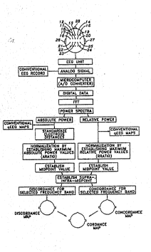
Figure~9~:is~a schematic,of~major components
illustrating:the~data~processing and~flow to obtaln:the ~~ .`.~,',
cordance map from:the:electrical output:ln~a;brain ~ :,,.",i
35~ egion.~
W093/0367n PCT/US92/06789
" .-, . . . ".:
2~ 8
Figure ~0 is a blo~k schematic illustrating the
relationship of a patient relative to appa,atus for
obtalning cordance mapping.
: ..,
Figure 11 are brain scans of the patient
illustrated in Figure l and 2. In Figure llA, 'here i~
1lustrated absolute power in four frequency bands;
Flgure llB illustrates relative power in four frequency
bands; Figure IlC illustrates discordance maps in four ~-^
lO ~frequency;bands; and Figure llD illustrates conaordance
maps in four frequency bands. Figures llA and llB are
obtained in a l1nked ear montage; Figures llC and llD are
obtalned in a bipolar montage.
:;
Flgure 12 are brain scans of the patient
illustrated in Figure l and 2, the relationship being in
~ . - .
a linked ear montage. In Figure 12A, there is illus-
trated~absolute power in four frequency bands; Figure 12B
llustrates relative power of four frequency bands; 13 ;~
20~ F~igure 12C illustrates discordance maps in four frequency~
bands~; and Figure 12D illustrates concordance maps in
four~frequency bands.
Figure 13 is~an alternative preferred version '~
~of~Figur~e 1 set up~with an improved computer program and
with~data obtained in the bipolar montage. Delta and
theta maps of Figure l have been replaced with beta~and ~,~
theta maps as indicated.
~30 ~ Figure 14 is an alternative preferred version
of Figure 2 set up with an improved computer~program and
with data obtained in the bipolar montage. A delta map
of Figure 2 has been replaced with a theta map as
ndicated.
Figure 15 is an alternative preferred version
of Figure 3 set-up with an improved computer program and -
.
~ . .
,:
r ~ J I ~ I ~J U .1 M~ j ~
T/ U S 92 / 0 6 7 8 9
2 1 1 J 6 5 0
with data obtained in the bipolar montage. A delta map
of Figure 3 has been replaced with a beta map as
indicated.
;5 ; ~ ~Flgure 16 is an alternative preferred version
; of Figur~ 4 set up~ with~ an improved computer program and
,wi~th data~obtained in the bipolar montage. A delta map
of~Fl~gure~4 has been replaced with a beta map as
ndicated.~
Figure 17 is~an alternative preferred verslon
of~Figur~e~5~ set up with an improved computer ~rogram and
wlth~d~ata~ obta1ned~1n~th~e bipolar montage. A new theta ;
map is indicated.
Figure~18~is~an~`alternative preferred version
of~Fl~gure~ 6~set~up with;~an improved comput~er program and~ ~-
1th~;data~obta~lnéd;1n the bipolar~montage.~ New C
discordànce)~and~D~ concordance) maps are indicated.
Figure i5~is~an alterna.ive preferred version~
o~ ~igure~7 s~et~up with~an improved~computer program and
w~i$h~ data~obtaLned~in the~bipolar~ monta e~.~ A delta map ; -
oI~Figure~7~s~=e-1d-ed~with;a th~e-~ma=~a=~;indlcated~
;Figure~20~is~an alternative~preferred version
Figure 8 set~ùp~with~an improved~computer prog~am and ; ?
with data;obtained~1n the bipolar montage.~ A~new theta
map is~indicated~
Figure 21 is an alternative p~eferred version
of~Figure~ll set~;~up~with an~improved~computer program and
with~data obtained in~the bipolar montage.~ A new C
(discordance)~and~D;~(concordance)~are indicated.
Figures 22A, 22B and 22C are alpha concordance--
and discordance~maps for the three CON;subj~ects wlth the ;~
SU0STITUTE SHEET
.. ,,,, . , .,, ., . . .... ,, , .. ,, ... ,, .. .. . . . .. , ; ,, . ., .. . ; . - . -. - . - . - . .; . . , - ~
16~c'dPCT/PTO OI MA~
P~T~ U S 92 ~ O 67 89
, best performance on the reminiscence and hypermnesia
paradigm (EH, LD, and LG). Subjects are identlfied on
the left side, along with the ratic of correctly ~;",
recalled/not recalled items on the reminiscence paradigm
5~ (the CC/NN ratio). The higher the ratio, the better the
performance., ~The maps,from,the CC and NN,"co,nditi,on for ' ,~
,each subject are dlsplayed separately, with concordance
ma~s in the~left column and discordance maps in the right
: .
column. Each map represents the`head as viewed _rom ~ `
10~ above, with frontal regions at~the top. Concordance and ,~
discordance are mapp~d separately but on the same
colorgraphic scale,~,~where~black is intense cordance ~",'
(either concordànce or discordance), there are ,~
intermediate levels of~cordance, and'white is a value of '"
15 zero'(neither concordance or discordance, but a no ,; ,,,
ord~nce stat
Flgur~es 23A~and 23B are alpha concordance and
dlscordance maps~ror the~two CON subjects with the ,','
20~ poorest performance on the ~eminiscence~and~hypermnesia '',
parà~dlgm (MG and AS);. S~ub~ects are identlfled on the
left~s~ide,~along`with~the ratlo of corre~ctly recalled/not
r~c,alled~ite~S on,`the~reminiscence paradigm (the CC/NN , ''
ra~i;o)~ The~hlgher~the-ratio, the better the
Z5~ per~ormance.~ The~maps from~the~CC and NN~condltion for
e~achi~su~j~ect are~displayed~separately, with~concordance
maps~in the lef~ column~and discordance maps~ in the right
column~. ~ Each map~r~epresents the head as viewed from ~
above, with frontal~regions at the top. Concordance and ~ ,,,;
30~ ~dis`cordance are mapped-sPparately but,on the same ~,.
colorgraphic scale,~where black is intense cordance
e1ther concordance~ or~discordance)`,~and there are
intermediate levels of cordance, and~white is a value of '~
zero (neither concordance or discordance, but a no , "~
3~5~; cordance state). ; ; ~, .
~': ~ ' . ' ' ~ ' .. '''
UBSTITUTE SI~E~T ` ~ `
p~
; 11 2~1~65~ -
~igures 2~A and 24B are alpha concordance and
discordance maps for~two MDE subjects on the reminiscence '. - '
. and hypermnesia~paradigm (CM and AM). Subjects are
- identified on the left:side, aIong with the ratio of '
correctly recalled/not recalled items on the reminiscence
~;' : -paradigm (the ~C~/NN ratio). The higher the ratio, ~;he
; ~ better the~performance~. The maps from the CC and NN
condition for-each subject~are disDlayed separately, with ~-
concordance maps in the left column and dlscordance maps
~in the right column. Eac~ map represents the head as
e~wé~d from above,~with frontal regions at~the ~op.
Concordance~and:discordance are mapped separately but on
the~same co~lorgraphic~sca~le, where black is;l~tense '-.
cordance (either concordance or discordance), there are':'
15~ intermediate levels;:of cordance, and white is a value of ~ ."`.
zer~o'(nelther~concordance or dlscordance, but a ns
: ~ Gordance state)
Fi~ures 25A~and 25B are alpha concorda~ce and ~';''
20~ di~s~corda:nce maps Cor~two MDE subjects on ~he reminlscence
and:hypermnesla~paradigm~ SC and LM). Subj~ects are~
dentified on th~e~left side, alo:ng with~the~ratio of ~'
correctly~recalled/not recalled items on~:ths remlni-s¢ence -~ L'~'''~'"
paradl~gm~(the~CC/~NN~ratio). The higher the~ ratio,:the
2~5~ bet`ter the~performance.~The maps f~rom~the C;C and NN
condition ;for each~sub~ject are displayed~separateIy,~:with
conc~ordance:~maps ln~the~left column~and discordance maps
in the:right~ column-.~ Each map represents ~he head as ~ ..
v-iewed from ab~ve, with frontal reglons at the`;~top.~
30~ Concordance and discordance are mapped separately but:on ''
n ~ the same colorgraphic scale, where:~black lS lntense ~
cordance~(:either~concordance or discord:ance); there~are
: in~ermediate levels of :cordance, and~white is a value of i.
zéro (neither concordance or discordance,~but a:~no
35: :'cordance state).
SUBSTITUTE SHEET ~
IPEA/US : ~`
~: .
16 R~'d P~TIP10 ~ ~ MAt~ 5 ~ :~-
Q~T/US 92/~6~89 ~-
12 211 5~ 5 0 ~ ;~
Figu-es 26A and 26B are alpha concordance and
discordance maps for the two DAT subjects on the
reminiscence and hypermnesia paradigm (RK and DL). j
Subjects are identified on the left side, along with the
5~ ratio of correctIy recalled/not recalled items on the
eminiscence paradigm (the CC/~i ratio). The higher the
ratio, the better the performance. The maps from the CC
and NN co~ndltion for each subject are displayed ~ i
separately, with concordance maps in the left column and
discordance~maps in the right column. Each map
represents the head as~viewed from above, with frontal
regions at~the top. Concordance and discordance are
mapped~separately but on the same colorgraphic scale,
where black is intense cordance (either concordance or'
discordance), there are intermediate levels of cordance,
and~white is~a value~of~zero (neither concordance or
dlscordance, but a no cordance state).
Flgures~Z7A,~27B and 27C are a serles of maps
20 :~LOr a~subject in the resting state tA), during 20 seconàs i~
of~ co~ntinuous right-h~and~movemen~ (B), and during 20
s~econds~of~ continuous left-hand movement (C). The~
variable~ mapped~is theta concordance, with dar~er color~s -
show}n~g~more intense concordance. The~rest:ing state
25~ shows~ a~slight hypof~ontal pattern, while both hand
moYement conditlons;show frontal activation. Right-hand
movement shows~preferential~activation~over the left ~ ;
hemisphere, whilé left hand movement shows preferential
activation over the right hemisphere.~ All~ ma~s show the ~ -
' 30 head as viewed from above. ~ ~~
Figure~28 shows the agreement between alpha
concordance and SPE~CT in assessing~normal~regional
perfusion. Twenty-seven subjects were studied with both
SPECT and cordance. Each subject had a brain disease
known to affect regional perfusion. The bra~in was ~-~
divided into SlX reglons (frontal, temporal, and
, ,
~: s~ rlTuTE ~H~E-~ .
::: IP~
~C~ S 92/06~89 i~
12/A 2 ~ l 5 ~, 5 ,~
.~
occipital, bilaterally) and the proportion of su~Jects iIl
each brain region who had normal perfusion and alpha :~
:; 5 concordance were counted. In all but two regions, there
. .
- .
,: , ' , ..
SUBSTITUTE SHE~T :
IPEA~US-. : : : "
,-, ~ . ~ .
,~ ~, ....
W093/03670 PCT/US92/06789
' 13
was a high level of agreement between the tw~ measures in
'~defining normal perfusion.
DESCRIPTION
e~ermini~g~*he electrical o~tput of-th~ brain -
~region of a subject and hence the assessment or diagnosis
of a disorder or affliction of the brain as characterized
by a lesion comprises obtaining first data representative
O~ of the energy intensity ln the brain region in a primary
frequency domain.; These data are represented by the
abso~lute power ln the pr~lmary frequency domain which is
ef~ined by specific frequency bands. These are the
conventional four frequency bands, namely, delta, theta,
15~ alpha,' and beta frequency~bands of electrical activity.
From~these f~irst data, there are determined
'~ second~data, namely, ~the relative power,~which is repre-
sentat;ive of energy in~a selected primary~frequency
2~0~ domain~relative to'the~energy in a secondary frequency
doma.n~
Whi:~e the pr~imary frequency domain is~any one ~-~
` ~ of~t~he frequency~bands~ delta, theta,'alpha and~beta,~ the
3~ ~ 25~ secondary~re~uency~d'omain can comprise~one or more ~han
orle ~frequency band.
In some cases, the primary~frequency domain is
several of the frequency bands and the secondary fre-
quency domain is a~different frequency band or set ofbands which should preferably incorporate at least~part
of the frequency band or bands of the primary frequency
do~ain.
35~The absolute power and relative power are
related to obtain a representative value of the
.......
~ electrical output in the brain region. Relating is
.~,.~ :: :
, ",
".
. W093/03670 PCT/US92/06789 ,~
:, ,: , .
:
14 211J~JQ
, ~ effected by determining the absolute po~er and relatl=ve
.~ power compared to a selected base value. When the first .'~
.. . . .
data and the second data both increase or decrease rela- '.
?~ tlve to a selected~bas~e value, a concordance condition is '~
5~ indicated.~ Whe'n:one of the first data and the second
.data-r~espectively increase.or declease relative to the ~
selected base while the other of the first data or second
data r spectlvely;is oppositely directed relative to the
selected~ base,:~a discordance condition is indicated.
.'~ The~relati~onship~of the absolute power and
re~lative~power~is;~hen':established. When the absolute ~ :
power~ and~rel~ative p~wer~are both greater:than a selected:
base value, then a quant:ified concordance value is
15 ~ ca~lcu;lated, indicated and displayed. Similarly, when one
of~the~ abso:lute:~powers:and~relative-powers are oppositely
directed~re~lative~to:the~::selected base va~lue, then a
quantifled~discordance~value is calculated, indicated and :
`~ .dlsplayed~
The indi~cated and displayed values provide ~he
' ~ indices~of co~cordance~and~discordance that are related '.:
' .to'~:~the presence~of~;~brain leslons. The distribution of :
oncordance~and;-dlscordance values ln the brain reglon is :
25 ";~ disp1:ayed;~topograph.~cally~through cordanc~e~ mapping.~
Theré ~ ,~`there ~is~obtaiDed~a spatial distribution and
.' ' ~ information~relating~:~to~the pathophysiological nature of :
~3 `~ the~brai~n lesions~ Through this technique,:the~evalua~
f~ tion of disorders and afflictions characterizad by:: ~ :
: 30 :~ lesions~can~be a~s;sessed to assist in a~:diagnosis.
Typical of the:diseases~ and disorders that can~be deter~
mined~are~dementing:illnesses such as multi-infarct
deme~ntia:, Alzheimer's disease and Pick's disease,
~ demyelinating:diseases such as multiple~ sclerosis, as :
,~q~ :35:~ well as:1esions amcng otherwise healthy control subjects.
~rf~
j W093/03670 . PCT/US92~S789 ;.
lS
~7,~A~ The de'ta frequency band is conventionally the
slowest frequency, being from about Q Hz to 4 Hz; theta
is from about 4 Hz to 8 Hz; alpha is from about 8 Hz to
12 Hz; and~beta is from~about 12 Hz and higher, namely,
S :to about 20 Hz:or 3:0~Hz in frequency.
; In the exemplified version of the invention,
th~e~primary fr~equency~domain incorporates any one of
the~se~bands.~ The;~selected sec~ondary frequency domaln
O~ includes~a~ll of the delt~a,~theta, alpha and beta
; fr:equency :band:s..~
The~flrst data~are the absoIute power. It is
indicated in microvolts~squared and indicates the energy
intens:ity in a selected single frequency band ('~primary
. ~ frequenc~y~doma~in")~ The~ second data~are relative~power.
It~i~a~repre~s~entatlve;of:the energy in:a selected single ` :;~
;~ frequency:~band~relative~to all the frequenc~y bands
":secondary:::fre:quency~`domain"). The relative power
.`~ 20~ répresents~;a~fr~action~;of, or the percentage~of, power in
~ the~s~élected:~single~frequency band relative to the ~ : :
,`~ abs.oluté~power:in all frequency bands. ~
In~the~determination of the~electrical output
:25~ of~a:bra~in~reglon~.of~the head.of the s~ubject,~an:
obj~Qctive~:base:~alue~is first established;fo~ each :~
su~3e~t.~:This obj;ective base is conveniently;a selected
base~be~ing a~midpoint~:for the absolute~power~and~a
:: selected:base being a midpoint for ~he~relative power for-
: 30 ~each subject. It is:a midpoint of a normaIized base
; value~of 1 which is representative of~:the respective
maximum~;absolute~power:~and the maximum relatiYe power .
The maximum absolute power and maximum relative power are
se~lected ~alues: of the first data and.the seCond data,
3:5~ respectively. Values other than the maximum can be
selected as necessary. : ~ :
s'~l W093/03670 PCT/US92/~6789 ',
~' l6 ,~~ J a
s indicated in Figure 9, the energy distribu-
tion is sensed and measured by electrodes located on the
head of a patient to obtain analog signals in each
electrode for an EEG unit. Each pair of recording
' 5 electrodes;estab1ishes a channel. The analog signal
~P~ ;provides a~conventional~EEG wavef~orm record as indicated.
he analog signal~ 1S digitized by the A/D converter in a
microcomputer to~become digital data. A Fast Fourier
Transform~tFFT) process is applied to ~he digital data to
0 ~yield absolute power values for respective EEG channels.
,~ The relative power~also~is calculated. These channels
represent ;each of~ ahout 20 electrodes located strate-
'~ gically about the~;head of a patient. The absolute power
values and the relative power values each sqrve~inde- ~ ,
~pendently to provide conventional EEG brain maps as
indicated.~ Such;-brain~maps would otherwise be termed as
quantitative EEGs~ ~
' With reference to the objective base value
20 ;~which 1~s established~for~each.subject, the maximum ~
abs~olute power and the maximum relative~power is set up
fo~r~ the~values across all channels for each frequency - ,
;2~5~ The absolut~ power data and~the relative power
data~are used in'~Gombination in accordance~with the
nvention to esta~lish representatiVe~valUes to permit
;cordance~mapping. ~The absoIute power value-~serves as~a ,,~
basis for determining concordance and discordance calcu-
lations which c~aracterize the quantity and quality of
the electrical output of~the brain region. ;This is taken ~;
in the~context of energy from recording locations of all '"
electrode channels and in all the frequency bands~
35~ ; The absolute power values are processe~ by
compUter means into relative power values~by dividing,
for each charlnel, the amount of power present in a'gi~en
1 W 0 93/03670 PC~r/US92/06789
~ ~J ~rjO
U 17
- frequency band by~the total power for each channel.
Relative power~thus reflects the distribution of the
energy for a channel among the different frequency bands.
There can thus be an absolute power value and a relative
5~ power value for: each frequency band for each of the
- ele:ctrodes loca~ed about the-head.
The~absolute~power and relative power values :
,~ are~normalized by~d~ivision by the maximum absolute and
lO~ maximum~relatlve;~power.~:valUes,:respectively, across all
;;2~0~channels~and each~;of the:four frequency~bands. The
,maximum~absolute~power;value and the maximum relative
power value~s~are determlned by examining the absolute and
relative~power values for each channel, and selecting the
15:~ greatest absolute power value and greatest relative power
value,;~The~s~e normal~ized~ ratios or~values are called
;resp:ectively~the "aratio":and "rratio" and~are compared
. ~ wlth~;~the~maximum values~normalized to l.O ("normalized
, ~ base")~.~ This, compari~son:~yields the concordance and
.20~ disco~rdance~quantlfication. These procedures are
e~ffected~:by~appropriate~computing and~microprocessing
means,programmed to~ef~fect the requisite dat~a calcula~
tio~,s~and~prcc~es~,~ing.~
;25~ A~channel~:~exhibits a disccrdant:~pattern and~is ~ :
quant:ified~w:ith:a~dis:cordance value when thei absolute:
,power~is~diminished relative to its selected base value : .
while~the:rPl:ati~e power-is increas:ed in~relation~to its
selected base value.: A sel~cted~base value is specifi-
30 ~cally defined as~a percentage, fraction, or proportion of
,the~normalized value~l ("normallzed basei'). In a~dis-
cordant condition,:the aratio is less than "1/2 of the
maximum absolute power" ("selected base") and the rratio
is:-greater than "l/2 of~the maximum relatlve: power" :
35: ~("selected base"). ~ ~
' W093/03670 PCT/US92/06789 ~
.
18 2~ J o
:; :
}~ In this sense, the normalize~ value is a
"normalized" base, and the midpoint or half point of the
base is a "selected base" or proportionate value repre-
sentative of that~normalized base value. The quantified
discordance value or score is determined by the sum of
the~devia~lon of the absolute power from "1/2 of its base
value" ("selected base") and the deviation of the
relative value from-"1/2 of its base value" ("selected
base"), as can be expressed by the form:
10~ discordance score = (rratio - 0.5) + (0.5 - aratio)
; A large discordance-score de.scribes the condition of a
channel with a~ low power signal that is confined mostly
to~;a given frequency band.
J~
~ Should the absolute power and relative power
both b~ increased, the aratio and rratio are both greater
than ~1/2 ma~ximum, namely, "1/2 of ltS base value"
("selected~base").~ Such a channel is considered to show
a~concordant pattern.~ The concordance quantification
20~ score~is then equ~al to the cumulatlve~elevation above the
1/2 power level for the two normalized values, as can be
expressed by the~form: ;
concordance~score = (rra~i~o - 0.5j + (a~atio - 0.5).
A~large~concordance score describes the condition~of a
25~ ;~;channel~wlth a high~power signaI that is confined mostly
to~a~given frequency band.
The concordance and discordance values can be
expressed in terms of a mathematical erivation. This
,, ~ : ,
30~ derivation is ~et out as follows:
Let ahf = -absolute power iD channel ch at frequency
c,
band f. Typically, ch is in ~he range 1 -
j~ to 20 in 20 channels of EEG data, and f
represents the frequency bands delta,
theta, alpha, beta
, ~
~ ~ ' ,.... ,,.. , .-
:
.`j ~
W093/03670 PCT/US92/06789 ~-.
c~ 19
Then rchf relative power in channel ch at frequency
: ~band f
h, f ''
~all bands
ch,i
10 Define a~x f = maximum absolute power in frequency
band f, of all channels
r~xf - maximum relative power in frequency
: band f, of all channels.
lS ~ Normalized values of "aratio" and "rratio" are
formulatPd: ~
aratloch,~ f
rratioch f =
25:~ ' rmax f
These~normalized values are then~compared with a
::threshold level,:~g~, half-maximal values, i.e., the ~ :
seleated base valuP
30~. : If (aratlo~h f < 0.5) and (rratloch~ ~ 0.5) then
:: : channel ch is termed "discordant" in frequency
: : band f; ~ ~
If (aratioch f >:0.5) and (rratioch f ~ 0.5) then
~:;:;: channel ch is termed "concorqant" in frequency
3;5~ ~ band f.
"~
.,~
i`
W093/0367~PCTIUS92~067~9 -
r n
~1 L ~ r~ 3 U
For concordance or discQrdanse, the magnitude
: o~ the quantification score can be calculated by the
formula:
score = I rratio~-: 0.5 1 + I aratio - 0.5 1
5 whére~ denotes the:absolute value, and 0.5 represents --
he normali:zed l~:2;maximum value.
A typical~ ca~lculation of quantified values is
;set out~
Dlscordan~::Site~
arat~io =~0:.3
rratio~ =;0.7:
aratio <~ 0.5 and rratio ~ 0.5 ~: . -
15:~ so:discordance value =:0~2 + 0.2 = 0.4
Concordant~Site
aratio =~0:.~7
. ~ rratio =~0:~.8 ~ : :
i ,~0 ~ arati~o~> 0.~5:and rratio > 0.5
:, ~ so~ oncordance value~=~0.2 + 0.3 =~0.5:
3~ In some~situationsl it i-s productive t~o :~
c~onsider~a~"selected~base" level other ~han 1/:2 the
`~ 25~ maximum~power~va~lues.~: ~0r example, if a recording~is
notable~ or~a singl:e~channel with much higher~power~than
the~others,:~this~atypically high value:sk ws~ the basis of :: :
a~comparison sca:l~e.~ Such a value would be discarded~as:~
~ an:atypical value~or outlier.
,,, .~ 30~
A ~hreshold of 40% or 30% of the normalized
maximum of 1 could y:ield more useful sets of~discordance :~
:and~concordance comparisons in:differ~nt situations.
SimlIarly, situations could arise where the threshold
`~ 35 ; level~ is set at 60% or 70~ of the normallzed~maximum.
Su~ch 40%, 30~, ~60%:or 70~ values would be the;l'selected
base."
',`' ~
`'~
,,~
~W093/03670 PCT/US9~/06789
,~ 6 j~ 21
~7ith the quantitati-~e EEG rPsults, the cordance - -
mapping is topographically illustrated in a primary
frequency domain in Figures lA, 2A, 3A, 4B, 5B, 6C, 6D,
7B and 8B, respectively, and also Figures 13A, 14A, 15A,
16B, 17B, 18C, 18D, l9B and 20B. Each respective domain
is illustrated as the de~ta, thetaj alpha Gr beta ran~e~ --
in each ol the respective Figures lA, 2A, 3A, 4B, 5B, 6C,
6D, 7B and 8B~ and Figures 13A, 14A, 15A, 16B, 17B, 18C,
18D, l9B an~ 20B,~ respectively, as indicated.
The most informative cordance map or detecting
lesions is usually in the theta or beta frequency bands.
' Such mapping'is illustrated in Figures 13A, 14A, 15A,
~. -: .
16B, 17B, 18C, 18D, ~9B and 20B. The data are obtained
from ~he 20 elèctrodes connected to the EEG unit which
measure~;the electrical activity in the~;head.
Information~is obtained that may indicate the
disconnection of cerebral cortex from the fibers that
connect brain~regions one to another. This may be the
common denomlnator in Alzheimer's disease, Pick's
dis~ease~,~multi-infarct dementia and multiple sclerosis.~ '
In~these diseases, gr~adual-severing of~the connections~
'~ that~ nk~different brain areas eventually may cause the
25~ symptoms of mental~nd neurological disability. The
representative values~as given by the dis ordance and
concordance representative values in the cordance maps of
;~ Figures l~to 8 and Figures 13 to 20 as a determination of
the electrical output of these region5 of the brain
3'0 ; prcvides useful~interpretive data to enable the
evaluation of the diseases.
The results for the brain rPgion depicted in
Figures 1 through 8 and Figures 13 to 20 were obtained
35 from measuring EEG data on subjects in a supine position
with eyes closedO Electrodes were applied in the
standard 20 locations on the head. At least 30 seconds
~ W~93/Q3670 P~T/US92/0~789 i-
2 1 i .~ O
i~ 22
;~ of relatively artifact free EEG measurements of
distribution were effected. The electrodes were applied
usin~ standard clinical procedures and the data obtained
~jJ'` were stored on an EEG unit.
~` 5
In this exa~ple as-illustratedj the EEG uni~
employed was a system known as QSI 900O produced by
Quantified Signal Imaging, Inc. of Toronto, Ontario,
Canada. This system provides data relating to conven-
tional qEEG information, topographical mapping of fourfrequency~bands in the central, frontal, temporal,
parietal, and occipital brain regions. Absolute power
P~ and relative power data for the different frequency
domains are obtained from the EEG measurements.
~ ~
The avoidance of inaccurate data readings from
electrodes~about the~head can be avoided by using
~ diff~erent~relationships between any number of selected
;~ electrode channels.~ ~Computing vector relationships
ZO;~ bet~een selected electrodes avoids the effect of
referentia1 monopolar values relative to reference
elèctrodes set up in adjacency with the ears of a
`~ subject. ~As such, it has been common in EEG~determi-
nations;to use~monopolar referencing by having a 1inked
25~ ears reference electrode: this means~by having elec-
;~ trodes;in adjacency to each ear relatlvely linked. Use
of~re~erential monopolar data for the purpose~of cal-
`~ culating concordance and discordance creates inaccuracies
;~ ~ ;; in cordance calculations having to~do with intere1ectrode
distances. While rèlative power calculations are un-
;~ affected by the mon~ages selected, absolute power changes ~ ;~
are in proportion to the square of the interelectrode
distance. Thus, the fronta1 and occipital power
estimates are inflated, since these are the furthest
points from the reference electrodes. Temporal power isunderestimated since this region is closes~ to the
reference electrodes.
W093/03670 PCT/US~2/06789 --,
- . .
23
A c~nfiguration is de~cribed with refPrence to-
Figure 9 to eliminate this problem. Absolute power data --
collected from the linked ears reference montages are~
~ first reformatted. This is effected using vector
`~; 5 ~calculations set up in~ a grid of bipolar electrode data,
comprising equally-spa~ed pa-irs of longitudina~l and ~- --
t~ansverse electrode chains. Power for each individual
,'!: ::
electrode is then~recalculated by averaging power for all
Yl ~ : respectlve pairs of electrodes in the chain longitu-
dinally and transversely. Each pair of electrodes in the
hain is regarded as a bipolar pair. The concordance and
discordance for each lhdividual electrode may be
calculated from the data from the bipolar pairs either
before averaging, or from the individual electrodes after
: . i ~ :
averaging. Thè data are thén employed to establish the
maximum, mldpoint and other values as necessary.
As an example with reference to Figure 9, the
electrodes lI,~12,~13, 14 and 15 are set out in the first
2~0~ 1ine, 1~6-, 17, 18, 19 and 20 in a second linej and 21, 22,
`~ 23~,~Z4 and 25 in a~third line. The ears 26 and 27 are
indicated~relative to nose 28. In a linked ear montage,
ea~h~of the electrodes~ll to 25 is referenced to the ears
26~and~ 27 which ~re "electrically" linked as a reference.
25~ The~grid of blpolar~electrodes is establlshed along the
ne~defined~by~electrodes 11 to 15, 16 to 20, and 21 to
2~5. ~The vertical grid~is 11, 16 and 21; 12, 17 and 22,
~i~t ~
for example.~ The bipolar data are set up by measuring
the data from each electrode in relation to the adjacent
electrodes. As such, for example, the power is measured
for~electrode 18 relative to electrodes 17, 13, 19 and
23. This is repeated for each electrode~relative to its
adjacent vertical and transverse electrodes. By computer
calculation, the calculations are effe ted to obtain a
.
measure of electrical activity at each electrode and have
a power estimate in-the region of the brain. This -
,~ .
, s~:~
., ~
WO9}/03670 PCT/US92/06789
24 ~ ' r
6 ~ ~
bipolar electrode montage avoids artifacts caused by the
linked ear montage. ~ ',-;'
i, ~,. .
This reformatting method effectively stan-
~dardizes electrode~distances and may yield information
' about'-l-ongitudinally-oriènted and transverse1-~-oriented ~ - -
; recording;~vectors. It lS sometimes helpful to map
concordance,and discordance for differently oriented
generators~(or flber tracts) on separate maps~. In other
;lO~ clr~cumsta~nces, it~is;helpful to calculate concordancç and
discordance after the bipolar~data have aIready been
,,'~ r~ecalcu;lated back~to~the monopolar format.
; The electrode head box which is-positloned near~
the subject contains 20 channels of opticalIy isolated
, ~ amplif~iers~ When~the~patient is prepared, a;keyboard
-command records~data f~rom~all 20 analog channels. EEG
'informa~tion~is then~seleoted for Fast Fourier Transform
càlculation. Power;~and spectral amplitudes are calcu-
20~ lated~ fo~r~absolute~power and relative power and the~
,;results of~the~Fast~Fourier Transform~are set out in a
ta~ular~ value of~absolute~power and relative power. ~-"
'~ After~analyzing the EEG using the Fast Fourier
25~, Trans~orm,~the;~operator~generates a topographic~map of ;~ ~,
a~so}ute p~wer,~ relative~power and ~a~cordance map~for~
ea~h~of~the four~conventional EEG frequency bands,
,'~ name~ly,,~the~selected~primary frequency domain. The~data
can be stored or displayed on scre ns or hard copy in a -'
''~ 30 'conventional manner.'
As illustrated in the flow dlagram of Figure 9,
;~ the conventional~qEEG maps are obtained from the absolute
power~and relative~power values. The absolute power is
35~ optionally subjected to the process for standardizing the
electrode distances by the bipolar montage. Thereafter,
the absolute power is normalized to establish~the maximum
~s;~. :
l'i,' W~93/U3670 PCT/US92/067~9 '~
j6`j
. 25
~:
absolute power value, namely, the aratio. The midpoint
value is established and thereafter, calculations of
: departures upwardly or downwardly from the midpoint value
are determined. The relative power value is normalized ,.
5:: by establishing the maximum relative power value, nam ly,
the rratio. The m~idpoint value is established-and ~h~re- :-
after;, relative power values upwardly or downwardly
depart~ing from~the midpoint value are established. From
this~data~, respect~ive discordance maps can be obtained in~
10~ selected frequency bands~or concordance maps~obtained in
~r~ s,'elected'~frequency~:bands. The discordance~map and con-
cordance~map can:~:~be~merged into a cordance'map as indi- :
~ 15~ In Figure'lO, there is illustrated a sample EEG
.~ unit.~ The~electrod~e head box is shown connected~with the
héa~d~of~ the~patient~whereby,~power measurements can be~
',~ taken ~rom~the'bra:i:n~-of;the patient. :~These are fed to
the.'preampl1fier and from such an amplifier:, conventional
" 20','~:EEG data~wou1d~be~recorded.,This would constitute a
d.~ -conventi~ona:l EEG.unit. Specialized within that construc- :
;'~ s"~ tion~are~the elements~for a qEEG unit. Data from the
: preampllfier;would~be;~directed to the analog to digital '''
;'~ con~erter:and~ in~turn,, to a microprocessor. The proces-
~ 25~ sor~ s~operated~by~a~keyboard console~and the output can .~ :
s,.'.~ be~ directed~to a~ video:display, storage or printer unit.
; `The~microprocessor~would operate in terms of the 1nven-
~ ion::to~generate~the~appropriate standardized ~alues, ',~
'"~ : normalization, selected base values, departures from the
~selected base values, discordance and concordance calcu-
lat1ons as indicated~in Figure 9.
Figures ll and 12 are illustrativ of the ~:
diff:erent cordance maps obtained for the same patient in
:~respectively the bipolar montage and the linked ear
montage. Illustrated in Figures llA,:llB, 12A and 12B,
, :respectively, are the absolute power and relative power
~ ,
....
. s : ~ ,
, ~ .
i~;
;'.~ '
~.,
W093/~3670 ~ PCT/US92/06789
26 ~ 65 a
in each of the four frequency bands, theta, delta, alpha
and beta. In Figures llC and 12C, there are discordance
maps in four frequency bands and in Figures llD and 12D,
thPre are concordance maps in four frequency bands.
An ex mplary data Ta~le I for patient JL is set
out below. ~Table I includes the data for the frequency
bands~delta,~ theta, alpha and beta in a bipolar montage. !'
In~each~such~band, there is set out the absolute power,
" ~
O~ ~relatlve~power and~respective discordance or concordance
value, ~Readings from~electrodes of an EEG unlt have been
taken.~ These discordance and concordance values are
t~opographically~depicted~as cordance maps illustrated in
Figure~
: ~ ~. : ` : . :
' ;
:,' ~ , i
~: WO 93/03670 PCr/US92/06789
27
c~r~jo
o~ ~
QI
~ `L~ ~ - .
~ w1 ~4 ,, ~
~ ~ ; O ~ ~
; r~ 0 " ~ : ~~~ ~ ~ , ~ ~ ~
'~ o, 0~ ~
'':
~ : ~ o
LJ ~ : w
,~, G C
j ~ r_ ~ O ~ ~ C LJO '
C~ ~ ~ ' ~ ` ~ C cw
~ ~ :~ C ~ ~ ~
~ o ~ ~ ~
A ,_ ~-- ~ t , ~ O w
o~ C ~ ~0 : -J~-
~o~ o ~O t - t ~ O ~ '
. ~ ~~ ~ ~ o t ~ ~ o ~ o ~o t ~
: b :-: ~ o
; ~ , v ~ ~
~> ~ ~r ~ ~
: ~, ' O $ ~ o g 0 ~ _ O ~ _ ~ o ~
0 :1 ~ o o o - o o ~ ,oO ~ ~ `Or O o
~y,~ C
SUBSTITUTE SHEET
~:~ W093/~3670 PCT/US~2J067~9
~ 28 211~650
In a revlsed form of the computer program
implementing the cordance mapping, the data would be ,.
represented in Table II for patient JL as set out below.
The data from Table II corresponds to the cordance
mapping of Figure 21.
Table II gives relatively better informative
data about the sub~oct JT,
,~
" y
"`.
~,~ WO 93/03670 - 29 - PCI/US92/06789 - -`
39~ -
'3Q~ ~ co~o~ c
~ ~;1 V ooooo-~ - ~
~ ~-1 ~ ~ ~ ~ - ~ o~ l
~j2'~ ~ ~ .- ~ 1
i~ l ~: ~ ~ 3:~ ~0 - o ~o o ~ `O C` _ ~
` ~ :
~ : I I ~ :~ ' , ; O
,~ ~ ~ ~j ~ o ~ o o ~ o ~ ~ ~o~ ' ~ ~o o ~ o =~ ~o _
~ o~ o~ ~
o u~ .o~ O ~ ~ ~ 2 ~ ~
o~ O` c ~ O O c ~~ z ~ z z 3 Z ~ O
i ~ 3 ~ 2~ , ~ ~ o ~ 1' ~ ~
I:,,, ,~, _ ~ ~ ~ . o o o o o o o o o o ,.. o o I
-' ~, , . -
- 2 r - ~ 'o ~ o ~ 0 ~ ~2
. ,. .~
SUBSTITUTESHEET
~ W 0 93/03670 PCT/US92/06789 !~
,~ 5 ~
' ~n interpretation of the electric output in the
brain region and diagnosis is set out for Figures l
through 8. ~ -'
In Figure 1, the brain imaging studies are for
'~ subjec~'JL, a 67 ye'ar-old ma'lè'with'muIt'i-i'n~arct ' '
dementia. The cordance brain maps (Figure lA) show
discordance in the delta and theta bands. In the
~ preferr-d forms in Figure 13A, discordance is in the beta
i ~ 10~ and theta~bands. The MRI scan (Figure lB) is a T2-
weighted image showing three discrete white-matter
lesions separated from the ventricles, that correspond
with the areas of discordance (highlighted wlth arrowsj.
~ ;The SPECT scan (Flgure lC) shows three prominent areas of
q~ 15 hypoperfusion that also correspond with the areas of
d~i.scordance ~(highlighted with arrows). Absolute power
mapping and relative power mapping, which are shown in
Figures 12A and 12B respectively do not provide this
nformation. Brain~maps represent the head as viewed
20 ~ from above, whi~le MRI and SPECT scans represent the head
as viewed from below. ~ ~
'~ The~discordance as illustrate'd`'in Figure~lA~"is
closely~associated~;with the presence~of deep white-matter
-25~ ischemic lesions~detected by MRI. The decreased absolute
'slow~wave power and~'increased relative slow wave power
seen~in the elec~.rodes overlying deep white-matter~ ~
lesions is demonstrated graphically in Figure lA. The
discordance map of Figure lA shows an in~ense area of
discordance in' the delta band in the right ~rontal
region. In the preferred form in Figure 13Aj discordance '~
~ is in the beta band in the right frontal region. Three
`~',t`: ~ i areas of discordance also are seen in the the~a band with
the largest and most intense focus present in the right
frontal region. These areas of discordance coincide
closely with three deep white-matter ischemic lesions
seen on a T2-welghted MRI scan. In the MRI~images, right
~I ~
.,~ .
1 W O 93/03670 P ~ /US92/06789
~......... ., .-;,
s -~ 31
.~
and left are reversed compared to brain maps. The single -
~ largést deep white-matter lesion seen on MRI (right
3~ frontal region) corresponds to the largest and most
~ intense area of discordance, seen in both the delta and
,t ,~ ~ : ~t~ ` 5 theta bands (Figure lA) br beta and theta bands
igure 13~A). The ischemi~ nature of these ~esions is'`'~"''~''" ''''''`
confirmed by the sub~ect's SPECT scan, which shows areas
of diminished perfusion in the right and left frontal and
right posterior head regions over the deep white-matter
0~ le~ions~(Figure lC; samé right-left orientation as MRI).
More associations may be~determined from the brain
c~ordance map~
~:t: ~ In Figure`2, there are additional brain imaging
J~ 5~ ~ studies for subject JL. The cordance brain map (Figure
2A)~shows an~intense area of concordance in the delta
band~in~the right posterior head region. In the
preferred~f~orms in Figure 14A, there is shown
~ concordance in the theta band in the right posterior head
,t,~ 20~ region.... The MRI scan (Figure.2B) is a T1-weighted image
showlng'~focal atrophy and ex vacuo ven~trlcular
enlargement in the right posterior head region,~
~'r`.~ sùggestlng~an~infarction invol~ing the cerebral cortex ''~
'f~ and~corresponding wlth the area of concordance
25~ highlighted with arrow). The SPECT~scan (Figure 2'C)
shows~a~prominent~ar~ea~of hypoperfusion that~also
c~orr~esponds wi~h the~area of concordance (high-lighted
with~arrow). ~Absolute power mapping and relative power
mapping, which are shown in Figures 12A and ~2B
30~ -respectively do not`provide this information. Brain maps
represent the head as viewed from above, while MRI and
SPECT scans represent the head as viewed from below.
Concordance is associated with se~eral condi- -
35~ tions including infarctions with cortiral involvement.
Interestingly, SPECT scanning may have difficulty
distinguishing between ischemia that is due to deep
W093/~3670 PCT/US92~06789
. . . ~:`
32 ~ 0
~ white-mat~er ischemic lPsions or to infarction with
;~ : cortical involvement. Cordance mapping yields additional
~ : valuable diagnostic~information about the na~ure of these
;~ lesions.
: 5
~ Accordingly, di:scorda~c~ is associated wit~' ' '"''"''''''''''
"~ deep white-matter lesions and concordance is associated:
,~ with:~lnfarction wlth cortical involvement.
10 ~ ~ In;Figure~3, there are the brain imaging
studies~for sub~ec~t~RC, a 67 year-old female with demen-
: .tia of unknown'etiology. The cordance brain~map
(Figure~3'A)~ shows ~ a broad area of intense dlscordance in
the delta band in the left posterior head:region. In the
~ lS ~preferred form in Figure 15A, there is intense
;~ discordance in:the~ beta~band. The first MRI image
Figure~3B? :is a:T2-weighted axial view~showing a large
patch:~of presumed,deep white-matter ischemic disease in
~Y.~ the;~.left~po~sterlor~head region adjacent:to the
~-r;~. .20 ,~;~.ventrlcular~hor~n~,~:that corresponds with the intens:e :
dlscordance~ hlghlighted with arrow). A~second MRI~ image
Figure~::3C~) shows:~muitiple punctate areas of presumed ~ :
ischem;ic~;disease~that~also correspond w:ith''areas of: ~'
.`,~ discordance:~(high~lighted:~with arrows). Absolute power
25~ mapplng~and r~elatlve.power mapping do~not provide this: .,
inf~ormatlon.~ Bra~in maps~represent the head as viewed ~ .
;from~above, whi~le~MRI scans repr sent the head as viewed
,from~below.
: 30~ The sensitivity of the cordance technique to
the presence of small~er lesions is demonstrated by the
'`~ case'of subject RC, whose cordance maps are shown in~
Flgure~:3A. The less intense area of dlscordance over the ;~
right temporal region coincides with a~few scattered
35~ punctate ischemic lesions seen deep below the temporal
cortex (Figure 3C). :~
~";
.
,,, , - , .
~l ;
.
' W093/03670 PCT/US92/067~9 -~
, . . . .
3 3
.r. ~ ;~
~,~ Figure 4 are the scans of GK, an 87 year old
~ male who presented with prominent memory loss and word-
3~ finding difficulties. He was given provisional diagnosis
of Alzheimer's~disease. A PET scan, Figure 4A, shows
prominent~biparietal hypometabolism, as well as right
frontotem~oral hypometabolism ~arrows~. The discorda~ce -
map for~ the same sub~ect (Figure 4B) shows biparietal
delta ~iscordance, more prominent on the right,
corr~esponding to~the PET pattern. In the preferred form
in Figure~ 16B, there is biparietal beta discordance. In
addi~tion~, there~is a right frontotemporal focus of
discor:dance corre'lating with the PET scan (arrows). The
PET~scan~shows the;bra~in as viewed from~below, while the
discordance map shows the brain as viewed from above.
, ~ ~ . i .
}5
Figure~5~ depicts scans of LB, a 51 year old
female with~ a diagnosis of Pick's disease.~ A SPECT scan
(Figure~5A)~highly~suggests this diagnosis, with promi-
`nènt~and~severe frontal hypoperfusion (arrows). The dis-
ao ~cordance map (Figure~5B) shows intense bilateral frontal
dis~ordance (arrow)~ In the preferred form in Figure
}7B~ there is intense~bilateral frontal theta discordance
as well.~ The SPE~T~scan is viewed from below, and the~
discordance scan Is~viewed from abo-e.~
~ Flgure~6~ls additional brain lmaging studies
'~ for~sub~ject LB.~ In~Figure 6A, the brain maps o~ absolute
~ power are~shown in the;~delta, theta,~a1pha, and beta
~ ~,
bands (from top). Figure 6B shows the maps of'relative
30~ power in the same frequency bands. Both of these~columns
show a diffuse excess~of slow-wave activity that does not ~ ;
h~ave~any clear regional predominance. The map in Figure ''
6C'is a discordance map of the same~subject, showing
clear and prominent frontal discordance in th~ theta~band
most prominently, and most significantIy affecting the
- right hemisphere. In the map in Figure 6D, there is a
diffuse concordance that is usually bilaterally
~ ~ .
W093/03670 PCT/US92/~6789 ,,'
~ , 34 21l 565 0 ''''
J
symmetrie, and is of no significance in this case. In
the preferred form, discordance is shown in Figure 18C
"4~ and concordance is shown in,Figure 18D.
Figure 7 illustrates a scan of SE, a 26 year
"old w~ite''male~'with multiple sc~erosis.' The MRI'sc'an ''' '`
(Figure 7A) shows a single large demyelinating lesion
~ underlying the left frontotemporal cortex (arrow). The
,~ ' discordance~map (Figure 7B) shows a prominent area of
lO~ discordance in the~left frontotemporal region. In the
pre~erred~form in~Figure~19B, the theta discordance map
~ shows~discordance;in the left frontal temporal region.
'~ The ~RI~shows the brain~as viewed from below~,~ while the
~ discordance map~shows the brain as viewed from above.
'. ~15
Figures 8 an~d 20 show scans for~PH, a 76 year
'~ old~male~control~subject with deep white-matter ischemic
,`~ e~slons in the~frontal lobes. A HMPAO SPECT scan for the
`~ subject, (Figure~8A) shows globally diminished cerebral
2~0~ p~er~usio~n, with the most striking decreases seen in the
rontal lobe (arrows). ~Figures 8B and 20B (the preferred
orm)~show a theta~discordance map for~this same subject/
wlth~a~'~least mild~discordance in most~brain~regions, and
,'~ pr~omi~nent~frontal~discordance correspondin~to the areas
25~ bf; greatly~diminished~perfusiQn (arrows). The SPECT scan;
is~iewed~from~belo`w~ while the discordance map lS viewed
from~above.
The cordance mapping is used to ass'ess the
30 presence and nature of brain lesions. 'The data obtained
by~the cordance~mapping conforms substantially~and~
equi~alently to the data obtained by the MRI scan, PET or
SPECT scan as illustrated in the figures. The values ~,
representative of;the combination of the~absolute power
35 - data and reIative power data provide for cordance brain
mapping. Such mapping thus provides a valuable ad~ance.
Absolute power and relative power mapping considered
i ~
`
W093/03670 PCT/~S92/06789 .
?~3;J separately does not provide these data. It is thus --
possible with the cordance brain mapping technique using ...
the quantitative EEG data to obtain effective information
. to facilitate evaluation of electrical output of the
~b : 5 brain, and hence the presence and nature of disease
conditio~s.~
It may be unnecessary to resort to the
relatively expensive SPECT and PET techniques. The
10: ~diseases represented~by the information obta~ined by
cordance brain mapping are the result of deep lesions in
the:braln that produce excessive delta and theta slow
wave~activity in an~EEG. Detection of these lesions by
conventional EEGs or currently available methods of qEEGs
15~ ~is~not possible. Thus, a conventional qEEG would provide
only~data:~about absolute power and about relative power
independently~ From such unrelated data, it~is not.
p~os~sible~to~obtain~the same information as cordance
mapping to assl~st in characterizing the human brain.
The:quantified~methods increase the sensitivity
of-~the EEG~and the cordance~mapping extends this sensi- -~
;.~ :t~ivi~ty~t~o provlde~:use~fu1 information.:: The~ examination of ~;
`~ the~;cordanc:e map~distribution:of the absolute power~and
25~ relativè;power in~the~delta and theta bands particularly
over~the~sur~ace~of the brain provides useful informa-
t~ion~ The discor;dance and concordance values are~:
determined~by a ca1culation of the comparison Df ~he
: : individual electrode absolute and relative power with the
maximal absolute and maximal relative~power values over
the~whole brain. ~ A brain region shows a discordant
pattern~in a given:frequency band if the relative power
from the corresponding electrode is increased above half
the;maxImum relative power value for the subject while :
the absolute power is decreased below half the maximum
absolute power value of the subject. Conversely, the
brain region shows a concordance pattern where both the
,. . ~
~` ~
i~ W093/03670 PCT/US92/~789
,. . ..
~ ~ 36 2i 1~ ~ ~J~ ~
absolute power and relative power value from the site are
increased about the half maximal values of that subject.
The sensitivity and specificity of both
5~; dlscordan:ce and concordance may be adjusted by changi:ng
the-thresh:olds~at which 't~e two measures àre"define~'. '-By'"''""'~ '' '"''
equlrlng:that concordant lncreases in absolute and
relativé~power:be 5%, 10~, or 20% above the half-maximal
'value (~"selected~base"): for that subject, the specificity
'~ 10~: of the::~easure:may~:b~increased. Similarly, by requiring
. ' ~ that;discordant~absolute and relative power~be separated
by~:~large differences',~the specificity of the~discordance
measur~e~:may be:increased~. ;There are other parameters
that may be ad]usted as well. For example, the half- :
~;maximal~value may~be calculated in several d~fferent
.~ ways~ :It~may~be~based'on half-maximal value~from all
regi~ons~for~that indi~fiidual subject, the mean or median:
-~ value:~for~that subj;ect~ or a half-maximal value after the~ :
or~ highest valu~es~(which may be outlier~s)~ have been ~ :~
2~0 ~ e1imlnated.~ These~f:urther.adjustments may change the ~ .
s~ensltivity,~spec~ifi~ity, or~usefulness~in~different
;~ cl'inical~situations:.-~
Cord~ance~mapping has been developed:on the
''~ ''25~ p'o ~ ation.of~:mostly~elderly subjects with~possib~le ~ : ~
~ '~ organic`mental~syndromes~ as well as young adults with ~ ~ :
5 ~ mult~iple~sclerosis~ There~are a number of other possible :~
àpplications:for~:this;technique among young and:older : :~
: :adult populations as well. Possible other applications ~.
:`: :3b~ include populations at risk for deep brain tumors, such
as~patients with~a::;history of brain tumor:who~are being
;mon-itored for possible~recurrence, patients with~AIDS who
are at risk for central nervous system lymphoma, multiple
sclerosis, patients with epilepsy, and other brain ~:
35~: ~ diseases. ~
j,f ~
;~:
. W~93/03670 PCT/US92/06789 :.'''
Appllcatiol~_ Oe the invention relate to
:: different fields:of neurophysiology. The cordance
mapping can be continually monltored during medical
procedures such as surgery or in treatment in intensive:5`,,~ 5~ care units. ~Similarly, during treatment of patients
:ch~ange~s in the mapping would indicate data relating to~
hhe~effectiveness~of treatment, or improvement or
deter1oration~:of sub~jects. The cordance mapping tech- :
i~t~ n'ique:s~;can be used~to~determine or assess the brain in
lO~ accident~situations:~or`'~'diseases such as cerebral vascular
diseas~es~or~:strokes~wh1~ch may be the result of genetic or
eve~lopment:al-~congenital~problems, traumat1c head injury,
exposure.:to~toxic:-a~ge;nts~or the product of other patho-
~
gen~ic~physiological processes such as ele~ated blood :.
15~ pressures, stress~responses, and arterial blockages.
It~should~be~possible with: cordance~methods to
'.~ f:aci~l~itate~diagnosls of~;epilepsy, substance abuse,.
~'! ' ~ y~eneti:c~disorder~s:~dlseases of the kidney or liver~
20~ af~ect~ing~brain~:f~unctlon,~ sensitivities r~elating to:foodand:~odo'r~wh1ch;correlate~with behavioral';~changas,- ill-
.`'~ nesses a'ccompanièd;~by~h~igh fevers,:viral';or bacterial
in;fection,~sensory~or motor handicaps~which would~ nclude
~'~ 'visua:l~handicaps~ auditory~and motor handicaps, learning '~
~25~ dlsabi11t1es~, psychiatric~disorders, headaches, cyclicalormo~ reactlons,~and other dysfunctions.~
;Th1s~ nVent1on~has application to~any disease ~ :
::: state that affec~s the gray- or white- mat~er of the~::: :'
3~o~ brain, either at the' cortical, subcortical white-matter,
or subcortical grey;~matter level. There~ore, patients
w~ith:~epi;lepsy who~have cortical or subcortical:~
dys~unction, patients~-with inheritable diseases that
:affect brain function at the cortical or subcortical
35 ~level~, as well:as tumors, trauma~ or infectious pro~cesses
; tha~ might-affect brain function all may be~usefully
evaluated using cordance mapping. ~ :
,~f:
,J:-:
,:~r~
W0~3/03670 PCT/US92/06789 ,',
' ' 38 2
By this invention, there is provided a method,
~' ~ apparatus, and system for obtaining useful assessment and '~
diagnosis of the brain based upon electrical activity.
Assessment Of Activation Tasks
Cordance has applications beyond detection of
lesions~ caus~ing corti~cal deafferèntation. Cordance is
sensitive to the presence o~ brain tissue with high or
,,'~ lO~ low~perfusion~in subjets with brain disease. Since
cordance is~ standardized to a midpoint of electrical
en~rgy production~for~an individual, it is~possible to
detect states~of~h~lgh or low perfusion even within the
normal range. Such states of high and low perfusion
likely~accompany the augmented flow in some brain areas
3~ during~actlvation~tasks. Concordance and~discordance,
uri-g~a~tivation task~s are set out.
<~ The. measure of concordance appears to be
'2Q~ associated;with the~activation of speci~fic brain reglons
involved~in men~al processing. This is demonstrated
uslng~: a hand open~lng;and closing task in a~normal control
;es~bject.~ The~conco~dance in the alpha;~band~for t'his
subject~is shown in~Figure 27A, in which~ther is~minimal
2~5'~ concordance~seen in~the frontocentral region. ~With~
opening and clos~ing~of the right hand, there is a
prominent increase in concordance in the~frontocentral
region on the left, roughly corresponding to the area of
the motor strip (Figure 27B). This finding ;is consistent
with'previous blood flow studies showing increases in
low to this area during motor tasks. With opening and
t ~
,,~ cIosing of the left hand, a slightly different pattern is
seen, with an increase in concordance in'the
frontocentral region but more prominently on the ri~ht
35~ (Flgure 27C3. The change in laterality corresponds with
the physiology of motor control; the less-specific
,., ~ , .
~3~
' W~93~03670 ' PCT/US9~106789 ''~
.. . .
~i
r~ ~ pattern on opening and closing of the left hand could
reflect the fact that the subject was right handed.
While concordance appears to be associated with
;5 ~-the activation, discordance appears to be associated with
deactivation~. This~association is shown b~ cor'dance' ``'''
~apping of the alpha frequency band (8-12 Hz) during the
encoding phase of the visual memory reminiscence and
hypermnesia paradi~gm.~ Eleven subjects were studied:
lO~ ~ive were~normal elder~ly controls (COM), four had ma3Or
depressive~episodes ~(MDE), and two suffered from early
demen~tla,~probably of~the AIzheimer's type (DAT).
Thé reminiscence paradigm is discussed below.
15~ Subjects were;shown slides of pen-and-ink drawings of
eas~ily~identified objects, each for a peri~od~of five
seconds~ Quantitative~EEG (qEEG) data were collected in
synchrony~with~the~pres~entations, for later
~ identlfication~of the~ data recorded durlng each slide
`~ 20 ~ presentatlon. The sub~ects were asked;to reaall as many
~ tems as~possible three~iminutes after~presentation of the`--~ stimuli,~ and then~again~after a four-minute rec~ll test and~two~lnter-test~interva'ls. All the'stimuli~presented
were~then scored~as~to whether they were recalled ~ '
'25~ correctly~in both recall~periods (a CC rating),~only~on
one~-~occasion (~CN~or~NC rating), or neither (NN rating).
Thé~C~C~and NN data~were~analyz~ed, slnce these conditions
represent the extremes~of~succèssful~(CC)~and
` unsuccessful (NN) memory encoding.
After both~recall periods,~a post-hoc analysis ~ ~-
was~performed and~data from all CC and NN recording ~'
epochs were pooled t~ create average cordance maps for
; the su~jects in the CC and NN memory encoding states.
Performance of subjects was rated according to a ratio of
the number of items recalled correctly on both recall
,: ,,.~
i :. - : - :
, ~ .
ec ~l r ~ll r ~v U 1. ~ IJ~
T/ U S 92 ~ 06~ 89 - ~
40 , 21 15~'~'o ''
~;"Il attempts divided by the number o,r items not recalled on
either recall attempt (the CC/NN ratio).
There was;a broad range of performance among
subjects in the CON;and MDE categories. Three of the
flve~CON s~bj~ec-ts~had CC/NN ratios betwèen l'and 3; wi'th
'~ ,the other two subjects~having ratios between 0.5 and 1.
Two of th~e MDE sub~jects had CC/NN ratios of approximately
l,, a thi~rd~subject had a ratio between 0.5 and 1, and the
10~ fourth a ratio~of 0.18~. ~inally, the two demented
, ~subject~ had~CC/N~ratios between 0.3 and o.~.
Two patterns of neurophysiologic activation
were seen in all subjects, that were strongly associated ~ '
,~l5~with the;degree~of success in performance of the memory
task.~ The flrst pattern involved the~temporal regions
bil~aterally~(spec~lf~lcally, the T3 and T5 recording
electrod~es on the~left, and the T4 and T6 recording
èl~ectrodes on the~~lght)~. A high CC/NN ratio ~as
0 '~assoclated with~p~eferential lef~ temporal~concordance in
the~CC state; for-~hes~e "good pe~~ormers," the NN state~
was réadily distinguished by a shift to right temporal
'concord~ance ~n~the~ condition. This patter~: ~s'evident ~'"'
f:or:thè top thre~e~performers on the~test~(subjects EH,
2~5~ LD,~ and;~LG,~Figures 22A, 22B and 22C).~ ~lso~evident for
the~two~highest~performers (EH and LD) is a~pattern of
c~entral~discordance, or deactivation (speclfically
in~olving the Cz~eléctrode). Thus, optimal~performance
w~s characterized~by both a pr ferential left-temporal
activation and'a oentral deactiva~ion~ in the CC
condltion
The :two CON subjects who performed more poorly
had a different pattern (Figures 23A and 23B~. While one
:35 ~ ~ - of them (subject MG,) showed the pattern of left temporal
concordance in the CC condition, shifting to ri~ht
temporal concordance in the NN condition, the subject
gU~ l iTUTE SH~T
l6 R~ctd PC~ Q a i~ l99
~T/lJS ~06~89 i~
41 2il~6~3
also had prominent central concordance in the ~C-state.
The C~ON subject who performed most poorly (subject AS)
lacked any features of the successful performance
pattern;~ the~sub~ect had no left temporal c~oncordance,
5~ but had prominent~central concordancè in the CC
condi~tion.~
The four depressed subjects, who performed more
poorly than the best CON subjects on the reminiscence
~ 10~ ta'sk,~lacked the neurophysiologic characteristlcs of qood
.~ test~performance,~and'~had features consistent with poor
performance (~Flgures~24~A and 24B; and Figures 25A and
25~B~ Sub~ects CM~and AM lacked left temporal
concordance seen in optimal CC performance. They did,~
l5~' however,~show~central discordance, and were able to
ma~lnta~in~a~CC/NN~rati-o~slightly greater than 1 Subject
SC~showed~left~temporal concordance in the CC condition,
wh;lch~was~exagger'ated~compared to that seen among the
control~subjects~ The subject lacked~the pat~ern of
20"~central~discordance,~ however, and had a~CC/NN _atlo of
less~than one~ FLnally, subject LM lac~ed~left temporal
concordance in the CC condition,~but had~prominent~
oen~ral concordance;~the subject reglstered the~worst
' ~ r;fo~mance~of~any~of~the depressed~sub~ects. ~
Flnally;,~both sub~ects wlth dementia~, who had
unif~o~mly~poor~ performance, showed a~;prominent pattern of
central~concordanc~e~(~Figures 26A~and~26B).~ This~pattern
~s similar to that~ of subject LM in th=e~depressed~group,
';30~ who had the most profound cognitive impairment on
clinical neuropsychological testing of any~of the
depressed subj~ects. Int~erestingly, subject LM also had
prominent deep~white-matter ischemlc disease, '''
sl~nificantly~more~than any of the;other subjects in ~his ~ '-
~ 35 ~ sample. ~ Afte~r two months of antidepressant treatment~
7 ~ the subject's mood improved significantly. ~-~~
- SlJ~STITUTE SH~ET ::
IPEMJ~
~- ~
~ W093/03670 PCT/US9~/06789 `~
L S 6 5 D
42
The ~onsistency of the rPsults shows that there
are neurophysiologic;differences between successful and
unsuccessful memory encoding~detected by cordance
mapping. There is an association between certain
patterns of neurophysiologic activation (concordance~ and
-dea~tiva-tion ~dLscordance)--and good'or poor'memory'ta'sk ''-' ' 5'` " '
performance. ~ ~ ~
These~data are consistent with the nature of
~"~ 10 ,the memory task and~with the previous results on the
neurophysiology of memory. Optimal encoding appears to
depend on~hoth the~le~ft~and~right temporal regions in
; these~subjects;, but primarily the left temporal region.
This is consistent with the structure of the paradigm,
15 ~ which involvès both encoding of visual stimuli and later
verbal~written rèporting of the stimulus. The data could
~ be interpreted as showing that left temporal activation
K ':}~ 'is~more imp9rtant~than~right temporal activation for
K .'~ successful~completion of the task, possibly~because of a
20~ verbal~naming~and~encoding process that occurs
contemporaneous~ly with'visual memory~encoding.
This findi'n~'g coincides with results that a~ '''' ' J
le~t/~rlght ratio~of~alpha power incrèased during tasks
25,~ that~require~greater left hemispheric proce~ssing. Three
of~four~depress~ed~subjects lacked the pattern of left
temporal~activation~in thé CC condition, and the fourth
had~an~exaggerated response. This observation is~
consistent with that dysregulation of left and right
hemispheric activation ~specifically including the
temporal regions) seen in depression.
,~
,~ The pattern of central activation associated ; with unsuccessful~ task completion also is consistent with
35~ observations in neurophysiology.~ In the res~ing state
there is a'prominent "alpha rhythm'i present over the
posterior ~head regions, and with cognitive tasks thi~ ~
~ . -
~`~
~ W093/~3670 PCT/US92/06789
.:
t~ jO 43
rhythm atten--ates. ~esult~ show that there is prominent
alpha concordance over these regions at rest. Successful
engagement in the task may suppress this concordance,
just as it does the alpha rhythm. Central alpha
concordance may be a marker for failure to engage in a
ta-sk, ~and-d-isc~rdance a mar~er--fo~ s~cceSsful-engag~ment.` - - -
~
Cordance reliably characterizes the perfus1onof brain tissue. While in certain frequency bands,
;concordance~is associated with an infarction, in other
bands. Concordance is a signal indicating that normally-
perfused bra1n tissue underlies a record1ng electrode.
Discorda~nce is a signal indicating that hypoperfused
brain tissue underlies a recording electrode. Cordance
is both a qualitative and quantitative indicator of the
nature of brain perfusion. Cordance indicates whether
normal1y-perfused~or~hypoperfused tissue is present (the
qual~1tative ind1c~ator), and also provides information
about the mean tissue perfusion and the volume of
2~0~ normally-perfused or hypoperfused tissue~(the
quan~itative indicator).
In~part1cular, there is a strong~relationship
between mean perfu~sion and concordance in the alpha
2~$~ frequency range thereby providing a quantitative
indicator~of pèrfusion. As illustrated in Figure 28
results of comparative data between~SPECT scans and
concordance mapping for six different brain r gions in 27
subjects show a high level of agreement in four of the
brain regions~examined.
While alpha concordance is an ind1cator of
normal perfusion, and beta and theta discordance are
indicators of hypoperfusion, the no cordance condition in
`~ 35~ certain frequency bands is an indicator of even lower
perfusion.
,, ~, ,~:
::: :
:~: :
WO93/0367Q PCT/US92/0~789
~: 2 1 '~ r3 $j rj U
any other examples of the invention exist each
difering from another in matters of detail only. For
instance, although the data have been set out as power,
; it is possible that other representations of energy can
be used. This could~be voltage, amplitude, or coherence.
'Al-thoug~each of th2~first data~and~sècond d~ta are
defined relative to their own base value, it may be
' possible to have a common base value.
10~ Also, whereas the primary frequency domain ~s
described as~essentially~a single frequency band of the
total relevant spectrum of the four conventional bands
from~zero to greater than about 12 Hz, the primary
freque`ncy domain could be differently defined. It could
be more than any one of the four frequency bands. Also,
the~ sacondary frequency domain may be greater or less
than any one of the~four conventional frequency bands.
Slmilar1y, the time interval evaluation of
20 ~;4~-second periods for~measuring.data in each ~of the
channels~may be dif~erent. In different situations, data
from~;a~different~number of selected electrode channels
may~be'us~ed to gener~te-the appropriate ~first~data an~`~'" i"~'
second data in;t-- dlfferent frequency domains.
Also,~although the concordance;has been~
described~with~re~erence to increases in a p~r~entage
pro'portional or f~ractional~value of a~base value,~ it is
possible that a concordance value where both first and
30 ~ second data are lower than this base value can be used.
Also, although the system has been described with ~-
ref~rence to 20 channels, more or less channels may be '-'
used. It is possible, for instance, to increase the'
number of channels to at least about 128. Indeed, it is
35 ~possible that the greater th2 number of channels, the
greater th'e amount of data will be obtained. This should
provide for more effective analysis.
W093/03670 PCT/US92/~6789
~, 45
j Q DifLer~nt techniques can ~e-used to overcome
thé artifacts caused by linked ear reference montage.
For instance, compensation factors can be ascertained and
applied for different power intensities and/or electrode
distances ln each braln region. This application can be
c~mputed into--the absoluté-power ~eterminator-to permit;~
establishlng the appropriate referential value.
The lnventlon~is defined in the following
lO ~ clal~s.~
,'~
'~