Note: Descriptions are shown in the official language in which they were submitted.
DESCRIPTION
DIAGNOSTIC APPLICATIONS OF DOUBLE D-LOOP FORMATION
Field of the Invention
The present invention relates to the formation of
RecA-catalyzed stable double D-loop structures that can be
utilized in a variety of diagnostic methods including two
probe capture/detection systems, RecA-facilitated DNA
amplification, and in situ hybridization.
References
Ausubel, F. M., et al., Current Protocols in Molecular
Bioloqy, John Wiley and Sons, Inc., Media PA.
Bryant, F. R., et al., Proc. Natl. Acad. Sci. USA
82:297 (1985).
Cassuto, E., et al., Mol. Ge. Genet. 208:10 (1987).
Chen, T. R., Cytogenet. Cell. Genet. 48:19-24 (1988).
Cheng, S., et al., J. Biol. Chem. 263:15110 (1988).
Cheng, S., et al., Photochemical Probes in
Biochemistry (P.E. Nielsen, ed.), pages 169-177 (1989).
Corey, D. R., et al., Science 238:1401 (1987).
2 ~ 1 6 2 ~ ~ PCT/JP92/011~-
Wo93/05178 ~ 2
Cox, M.M. ~t al., Ann. Rev. Biochem. 56:229-262
(1987).
Da~is, L.G., Qt al., Basic Met~ods in Molecular
8ioloaY, Elsevier Publishing Co. (1986).
Dreyer, G.B., et ~l., Proc. Natl. Acad. Sc~. USA
82:968 (1985).
Ei~en, J., et al., Proc. Natl. Acad. Sci. USA 85:748
(1988).
Fey, M.F., Schweiz. Med. Wor~n~r. 120(20):731-737
(1990).
Fishel, R.~., et al., Proc. Natl. Acad. Sci. USA
85:3683 ~1988).
Fugisawa, H., et ~ ucleic Acids Res. 13:7473
(198S).
Ganea, D., et al., Mol. Cell Biol. 7:3124 (1987).
Gond~, D.X., et al., Cell 34:647-654 (1983).
Grl~f~t~, J., et ~l., Biochem 24:~58 (1985).
Halbrook, J., et ~l., J. B$ol. Chem. 264:21403 (1989).
Harlow, E., et al., Antibodies: A nAhoratorv Manual,
Cold Spring Harbor Laboratory Press (1988).
Haase, A.T., et al., Proc. Natl. Acad. Sci. U.S.A.
87:4971 (1990).
Hsieh, P., et ~1., Genes Dev. 4:19S1 (1990).
Hsieh, P., et al., J. Biol. Chem. 264:5089 (1989).
~sieh, P., et al., Cell 44:885 (1986).
Keene, K., et ~1 ., Nucleic Acid Res. 12:3057 (1984).
Kemp, D.J., et ~l., PCT Internation~l Application,
International Pub~ication ~o. Wo 90/06374 (Application No.
PCT/AU89/OOS26), 14 June lg90.
Kimeic, E.B., Cell 44:545 (1986).
Kimeic, E.B., Cold Spring Harbor Symp. 48:675 ~1984).
Kingston, H.M., B.M.J. 299(6690):34-37 (1989).
Kolodner, R. et al., Proc. Natl. Acad. Sci. USA
84:5560 (1987).
Leahy et al., J. Biol. Chem. 261:6954 (19861-
'93/~l~ 21~36 21 ~ PCT/JP92/0113~
~ owenhaupt, K., et al., J. Biol. Chem. ~ 20568
~1989).
Madir_ju, M.V.V.S., et ~1., Proc. Natl. Acad. Sci. ~SA
8S:S592 (1988).
Mani~tis, T., et ~1. Molecul~r Cloninq: A TAborator~
Man~ , Cold Spring Harbor Laboratory ~1982).
McCarthy, J., et ~1., Proc. Natl. Acad. Sc~. ~SA
8S:5854 (1988).
Mc~nt~e, ~., et ~1., J. Biol. Chem. ~56:8835 (1981).
Moore, S.P., et al., J. B$ol. Chem. 19:11108 ~1990).
Morrical, S.W., et ~1., Biochem. ~:837 (1990).
MoQer, ~.E., et ~1., Sc~ence 238:645 (1987).
M~ t~, X.B., U. S. Patent No. 4,683,202, issued 28
July 1987.
Mullis, R.B., et ~1., U. S. Patent No. 4,683,195,
issued 28 July 1987.
Nelson, M., et al ., Nucl. Acids ~es. ~1:389 (1989).
olson, M., ~t ~1., Science 245, 1434-1435 (1989)
Radding, C.~., Ann. Rev. G~net. 16:405 (1982) 25:1990.
Rigas, B., et al., Proc. Natl. Acad. Sci. U.S.A.
83:9591 t1986).
Roc~, A.I., et al ., Crit. Rev. Biochem. Molec. Biol.
25:4lS (1990).
Sa~ki, R.X., et al., Proc. Natl. Acad. Sci. USA
86:6230 tl989).
Sambroo~, J., et ~1., In Molecular Clonin~: A T~bo-
r~torY Manual, Cold Spring H~rbor L~boratory Press, Vol. 2
(1989) -
Scharf, S.J., Qt al., Science 233:1076-1078 (1986).
3 0 Shi~ata, T., et al, Proc. Natl. Acad; Sci. U.S.A.
76:1638 (1979).
Shi~ata, T., et ~1., J. Biol. Chem. 256:7557 (1981).
Sluka, J.P., et al., Science ~ 1129 tl987).
Strickler, J.G., et al., ~m. J. Clin. Pathol. ~3(4
Suppl. l):S44-8 (1990)
WO93/~178 2 ~ 1 6 2 I r~ PCT/JP92/0l13
Sugino, A., et ~1., Proc. Natl. Acad. Sci. USA 85:3683
(1988).
Trask, B., et al., ~um. Genet. 78:251 (1988).
~an Dekken, ~., et al ., Cytometry ~1:153 ~1990).
van ~k~en, H., et ~1., Cytometry 11:579 (1990).
Wachsmuth, K., Rev. Infect. Dis. 8(5):682-692 (1986).
Weier, H-U.G., et al., J. H~stochsm. and Cytochem.
3B:421 (1990).
Woodbury, C.~ t al., ~ochemistry ~2(20):4730-4737
(1983).
Zlvin, R.A., et ~1., European Patent Application No.
88307793.5, Publi~ation No. 0 305 14S, 1 March 1989.
Bac~6uh~ of the Invention
RecA+ protein (wild type) is a 37,842 dalton protein
found in the bacterium Escherich~a col~, which is important
~or homologous DNA recombination. Most information about
its biochemistry and enzymology comes from ~n ~it~o studies
using purified RecA+ protein. Numerous ln v~tro studies
ha~e shown th~t RecA+ protein is inti~tely involved in the
pa~ring reaction between ~omologous DNA sequences that
ultimately le~ds to homologous recombin~tion events
(Radding; see Cox et al. or Roca et al. for recent reviews
of RecA+ protein properties). It is this pairing reaction
that makes RecA+ protein highly use~ul for DNA diagnostics
applications.
Tn the presence of ATP, RecAI protein catalyzes strand
exchange between a number of substrates, the most rele~ant
for DNA probe applications being single- and double-
stranded DNAs. RecA protein coated single-stranded DNA
probes interact with the homologous portion of a double-
str~n~e~ ~"nativen) target sequence initia~ly by forming a
recombination intermediate containing hybridized, partially
~oined molecules called (~ingle) D-loops (or in some cases
~5 triple-stranded structures) (Shibata et ~1., 1979). This
) 93/05178 2 1} ~ 2 ~ ~ PCI'/JP92/0113~
i5 fol~owed by branch migration, And forming of fully
hybrid molecules between ths original single- and double-
stranded DNAs, depending upon the extent of their homology.
Short displ~cement loops or triple-stranded D-loop
stru~L~,~s in ~inear targets are usually unstable after
deprot~ tion. RecA protein has been shown to form
s~hl~ complexes with short ollgonucleotides, between g and
20 bp (or larger) in length, in the pre~ence of ATP~S and
e~ee~ RecA protein ~eahy et ~l.). When linear double-
~tranded targets are used, stable probe target pairinga~ter R-cA I~Oval appear~ to requ~re ~i) a homologous
region of at ~e~t 38 to 56 bp, and (ii) the locat~on of
the probe target homology ~t the end of the linear duplex
(Hsieh ~t al. 1990; Gond~ et ~l.).
Rigas et al. ~eyGL Led that a single-stranded 43-mer
could ~or~ a single D-loop complex stable to
deprote~ni7ation when dou~le-str~nded negati~e~y
supercoiled circular pl~smid DNA was used as the target.
When a double-stranded negat~vely supercoiled circular
targe~ DNA ~s u~ed, RecA coated single-stranded
oligonucleotide probes can also be stabilized by psoralen
crossl~kin~ before removal of the RecA protein: probe-
target single D-loop products can be recovered if the
oligos are at least 30-mer size (Cheng et al., ~9~9). To
obt~in psoralen crosslink sta~ilized single D-loop probe-
target complexe~ when double-stranded linear DNA duplexes
are uQed as target DNA, the probes must be at least 80 to
107-mer size (Cheng et ~l., 1988): these reactions are
very low efficiency when compared to similar reactions with
negatively supercoiled clrcular targets.
Experiments performed in support of the present
invention have demon~ ted that probe:target DNA complex-
es, which are stable to deproteinization, can be qenerated
in RecA protein catalyzed reactions providing that duplex
probes, which contain seguences complementary between probe
WO93/05178 2 1 ~ ~ 2 1 ~ PCT/JP92/0113
strands, are used in the hybridizat~on reactions. This
discovery provides a number of G~GLLunities for diagnostic
application that exploit this stable RecA protein catalyzed
probe:target hy~ridization complex.
8ummary of the ~n~ent~on
T~e present invention inc1udes a diagnostic method for
detect~ng a linear duplex DNA analyte, having f~rst and
sQcond strands, where the analyte contains a first internal
DNA target seguence. The ~ethod teaches pro~iding a set of
two DNA probes that each contain seguenc-s complementary to
the 2irst target sequence strand or the second target
sequence strand, where these probes also have a region of
complementary overlap to each other. Both probe strands
are then coated with RecA protein in a RecA protein coating
reaction. T~e RecA coated probes are combined with the
1~ne~ duplex DNA, which contains the target sequence,
under conditions thAt produce a probe:target complex. This
probe:target complex contAins both probe strands and both
strands of the linear duplex Analyte. The probe:target
complex is stable to deproteinization, although in the
method of the pre-~ent invention it is not necessarily
deproteinized. The presence of the probe DNA in the
probe:target complex is then detected.
In one embodiment of the in~ention the RecA protein is
the wild-type RecA protein of Escherichia coli.
Alternatively, the RecA protein can be the mutant recA-803
protein of Escherichia coll or a RecA-l~ke protein from a
number of sources.
The RecA-protein coatin~ reactions of the present
invention can be carried out using a variety of co-factors,
including ~TPyS, rATP (alone and in the presence of a
regenerating system), dATP, GTPyS, and mixes of ATPyS and
rATP, and ATP~S and ADP.
~/O 93/05178 2 1 1 6 2 1 ~ PCr/JP92/01135
~ 7
In one ~h~A~ment o~ the inYention~ the region of
complementary overlap between the probe strands is at least
about 78 base p~irs and less than about 500 base pairs.
The probe strand~ may also cont~in an end terminal
extension of DNA that is not complementary to either target
strand. When both strands contain such an end terminal
extension, these DNA extDnsions may be complementary to
each other.
One way in which to acco~plish the detecting of the
method of the ~ ent invention is by deproteinization of
the probe:target complex, followed by electrophoretic
separation of the probe:target complex from free probe.
The probe:target complex can be deproteinized by a variety
of methods including tre~t~ent with SDS or proteinase ~, as
well as st~n~rd chemical deproteinization methods, such as
phenol-based methods. Alternatively, the detecting can
include the use of a capture system that tr~ps the
probe:target complex, where the ~lrst probe strand is
labeled wlth a ca~uL~ moiety ~nd the ~e~on~ probe strand
is labeled with a detection moiety. For example, one probe
strand can be biotin labeled and the ot~er digoxigenin
labeled. The probe:target complex c~n then be
ca~Led/detected using solid support-a~e~Lavidin (or
avidin)/labeled anti-digoxigenin, or solid support-anti-
digoxigenin ant$body/labeled ~-le~avidin (or avid~n). In
a d~fferent embodiment, the first probe strand contains a
capture moiety and t~e second probe strand contains a
radioactive label to be used for detection.
The probe strands can be labelled for capture in a
n~h~r of ways, for example, using biotin or digoxigenin
attached to the probe and streptavidin tor avidin) or an
anti-diqoxigenin antlbody, respectively, for czpture. The
probe strands can also be labelled for detection using a
nu~ber of different moieties including: radioactive,
biotin, digoxigenin. Radioactive labels can ~e identified
WO93/05178 2 ~ 1 ~ 2 ~ 5 PCT/JP92/01135
by, for example, autoradiography or scintillation counting.
The presence of biotin or digoxigenin can be detected by
~L.e~Lavidin or an ~nti-digoxigenin antibody, respecti~ely,
where the streptavidin (or avidin) or anti-digoxigenin is
rad$oactively labeled, enzyme labeled (e.g., alkaline
phosphatase, peroxidase, beta-g~lactosidase or glucose
oY~A~e) or fluorochrome-l~beled (e.g., fluorescein, ~-
phycoerythrin, or rhodamin~). Detcction of the probe
strands ln the probe:target complex can also be
accomplished by DNA polymer~e fac~l~tated primer extension
from the 3'-ends of each probe strand, where the primer
extension is performed in the presence of all four dNTPs
and one or more dNTP contains a detectable moiety.
Ths method of the present invention further includes
providing a second set of two DNA probes, having first and
second strands, co~plementary to a second duplex target
sequence, where the first strand of the probe contains
sequences complementary to one str~nd of the second target
se~uence and the second strand of the probe contains
sequences complementary to the other strand of the second
target sequence, where (i) these probes also have a region
o~ co~plQmentary overlap to e~ch other, and ~ii) the second
set of probes does not hybridize to the first set of
probes. The two probe setQ are coated with RecA protein in
a RecA protein coating reaction. The RecA coated probe
sets are combined with the }inear duplex DNA, containing
the two target sequenccs. The combining is done under con-
ditions that produce probe:target complexes which contain
all four probe strands. The re~ulting probe:target complex
is stable to deproteinization. The presence of the probe
DNA is then detected in the probe:target complexes.
The method involving two probe sets can be utilized in
many of the same ways as described above for a single probe
set. For example, the first probe set can be labeled with
$ ~ ~ ~
a capture moiety and the second probe set labeled with a
detection moiety.
The double-stranded probe:duplex target complexes
involving two probe sets can also be used in a RecA protein
facilitated DNA amplification method. For example, the two
probe sets can be hybridized to their duplex target sequences
in the presence of ATPyS [or rATP (with or without a suitable
ATP regeneration system), dATP, and mixtures of ATPyS and
ADP] and reacted in a reaction mixture also containing, all
four dNTPs, RecA protein and DNA polymerase. This reaction
is performed below the temperature required for thermal
dissociation of the two target strands and continued until a
desired degree of amplification of the target sequence is
achieved. The amplification reactions may further include
repeated additions of (i) DNA polymerase and (ii) RecA
protein-coated probes during the course of the amplification
reactions. Other approaches to amplification, which can be
applied to the present invention, have been set forth in WO-
A-91/17267. In each probe set, the 3'-end of one strand will
be internal to the region defined by the two primer sets:
these ends are necessary for the amplification reaction.
However, the opposite 3'-ends of each primer pair, external
to the region defined by the two primer sets, can be blocked
to inhibit the formation of extension products from these
ends. This amplification method can also be used as a
detection method, where detection of the probe in the
probe:target complex is accomplished by DNA polymerase
facilitated primer extension from the 3'-ends of each probe
strand, where the primer extension reaction is performed in
the presence of all four dNTPs and one or more dNTP contains
a detectable moiety.
-A
CA 0211621~ 1998-03-30
The double-stranded probe:duplex target complexes can
also be used to block cleavage of any targeted restriction
site. Blocking cleavage can be accomplished in a number of
ways including: (i) forming the probe:target complex and
treating with the restriction enzyme before deproteinization
of the complex; (ii) using methylated or un-methylated
probes, depending on the sensitivity of a selected enzyme to
the presence of methyl groups; and (iii) introducing a
sequence mismatch in each strand of the probe, which, when
the probe hybridizes to the target, eliminates the
restriction site.
The double-stranded probe:duplex target complexes can
also be used to generate site specific cleavage of double-
stranded target DNA. The double-stranded probe can be
modified with moieties capable of cleaving each strand of the
target duplex: this probe modification can take place before
or after deproteinization depending of the nature of the
cleaving moiety. Examples of such moieties are iron FeII
(for iron/EDTA facilitated cleavage), non-specific
phosphodiesterases, and restriction endonucleases. In either
case, cleavage specificity is conferred by the target
sequence which is defined by the double-stranded
oligonucleotide probe.
Both the restriction site protection method and the
site specific cleavage method are useful in restriction
fragment length polymorphism analysis.
Another embodiment of the present invention includes
a method for isolating a linear duplex DNA analyte, having
first and second strands, containing a first internal DNA
target sequence, where the duplex DNA analyte is present in
a mixture of nucleic acid molecules. In this method a set
of two DNA probes is provided, having first and second
probe strands, where the first and second probe strands (i)
contain complementary sequences to the first and second
target sequence strands, and (ii) where these complementary
CA 0211621~ 1998-03-30
sequences also contain complementary overlap between the
probe strands. The probes are then coated with RecA protein.
The coated probes are combined with the linear duplex DNA,
which contains the target sequence, under conditions that
produce a probe:target complex containing the probe strands
and both target strands: the resulting probe:target complex
is stable to deproteinization. The probe:target complex is
separated from the mixture of nucleic acid molecules. The
duplex DNA analyte, which contains the target sequence, is
then isolated.
In this method, the complex can be separated from the
nucleic acid mixture using, for example, probes containing
biotin moieties that are captured with streptavidin or
avidin. The streptavidin or avidin can be bound to a solid
support, such as paramagnetic beads.
The method further includes heat denaturation of the
isolated probe:target complex at a temperature (i) sufficient
to release the duplex DNA analyte containing the target
sequence from the complex, and (ii) below the melting
temperature of the duplex DNA analyte containing the target
sequence. This allows the isolation of intact duplex
molecules. The duplex can then be denatured to single-
strands if so desired. Alternatively, the complex can be
heat denatured at a temperature (i) sufficient to release the
duplex DNA analyte containing the target sequence from the
complex, and (ii) at or above the melting temperature of the
duplex DNA analyte containing the target sequence. This
results in the isolation of single-stranded DNA molecules
derived from the captured duplex.
Another embodiment of the present invention is a
method for detecting a linear duplex DNA analyte in a
mixture of nucleic acid molecules. The method includes
isolating the linear duplex DNA analyte as described above
and obtaining single-stranded DNA molecules derived from
the duplex DNA analyte: this is typically accomplished by
WO93/05178 ~11 6 2 1 S 12 PCT/JP92/01135
heating the duplex above the melting te~perature of the
duplex (he~t denaturation). To the single-stranded target
D~A analyte molecules, at least one DNA synthesis primer is
added that is complementary to the target sequence and that
does not contain seq~en~ee which were present in either of
the two original DNA probes. Detection of the DNA analyte
is accomplished by DNA polymerase facilitated primer
extension fro~ tho 3'-end of the primer, wherein the primer
extension is performed in the presence of all four dNTPs
and at least one dNT~ contains a detectable moiety.
The double-stranded probe:duplex target complexes of
the present invention can al~o be used for diagnostic in
s~tu detection techniques.
8rief Description of the Flguras
F$gure l illustrates the relat1onships of the probes
and primers, listed in Table 1, to the lambda genome.
Figure 2 presents the nucleotide sequence of a 500 bp
lambda geno~ic region: this se~uence is also presented as
SEQ ID NO:l.
Figure 3 shows an autoradiogram of a D~A band-shift
gel electrophoresis assay to illustrate RecA protein
binding to DNA probes.
Figure 4A shows an ethidium bromide stained gel on
which the components of deproteinized hybridization
reactions using 500-mer and 280-mer pro~es were resolved.
Figure 4B shows an autoradiograph of the gel shown in
~igure 4A.
Figure 5A shows an ethidium bromide stained gel in
which t~e oQmronents of deproteinized hybridization
reactions using 280-mer, 121-mer, and 7s-~er probes were
resolved. Figure 5B shows an autoradiograph of the gel
shown in Figure 5A.
Figure 6A shows an ethidium bromide stained ~el in
which the components of deproteinized hybridization
211621~
w093/~178 PCT/JP92/01135
13
reactions using differentially labeled 121-mer DNA probes
were resolved. Figure 6B shows an autoradiograph of the
gel shown in Figure 6A. Fisure 6B illustrates that two DNA
probe strands are nereC~ry for the production of stable,
deproteinized, RecA protein catalyzed hybridization
complexes.
Flgure 7 illustrates a model of stable double-stranded
probe:duplex linear target ~NA complexes.
Figure 8 shows a gel from which stable double-stranded
probe:duplex l~ r target DNA complexes were isolated,
where the duplex probe strands were differentially labeled.
Figure 9 illustrates a variety of double-stranded
probe:duplex linear target DNA complexes.
Figures lOA, 108, and lOC illustrate se~era~ detection
systems based on a single double-stranded probe:duplex
linear target DNA complex.
Figures llA and llB illustrate several detection
systems based on a mul~1ple double-stranded probe:duplex
1 ~ ~PAr target DNA comp _x.
Flgure 12 ~hows ~ecA-protein catalyzed two-double D-
loop primer positioning on native target DNA tFigure 12A)
and DNA amplification with DNA polymsrase followed by
l~gation with DNA ligasc (in the absence o~ primertprobe
displacement) (Figure 12B).
Flgure 13 shows a DNA polymerase mediated signal
amplification reaction using a single double ~-loop probe
(Figure 13A) or multiple double D-loop probes (Figure 13B).
In Figure 13, X and X' can be the same or different: for
example 5'' can be radioactively labeled and X' can carry a
digoxigenin moiety.
Figure 14 illustrates a detection system invo~vin~ the
use of restr$ction endonuclease cleavage of non-targe~
complexed double-stranded probe where ca~Lu~e of the
resulting product is accomplished before (Figure ~4B) or
after tFigure 14A) restriction enzyme digestion.
. ,;. . . ,.,~
CA 02ll62l~ l998-03-30
14
Figure 15 illustrates the protection of a restriction
site by either methylation or RecA protein. In the case of
methylation protection the double D-loop complex is
deproteinized before restriction endonuclease digestion
(Figure 15A). In the case of RecA protein protection the
double D-loop complex is deproteinized after restriction
endonuclease digestion (Figure 15B).
Figure 16 shows an ethidium bromide stained agarose
gel on which the components of deproteinized RecA mediated
double D-loop hybridization reactions, using heat denatured
280-mer probe and different cofactors, were resolved by
electrophoresis.
Figure 17 shows an autoradiogram of the gel shown in
Figure 16, after drying.
Figure 18 shows an ethidium bromide stained agarose
gel on which the components of deproteinized RecA mediated
double D-loop hybridization reactions, using heat denatured
500-mer probe and ATP~S and ATP~S/rATP mixes as cofactors,
were resolved by electrophoresis.
Figure 19 shows an autoradiogram of the gel shown in
Figure 18, after drying.
Detailed Description of the Invention
I. Generation of RecA Catalyzed Probe:Target
Hybridization Complexes Which are Stable to Deproteinization.
A. DNA probes and primers.
Experiments performed in support of the present
invention show that short double-stranded DNA molecules or
complementary single-stranded molecules can be used to
generate hybridization complexes with linear target DNA
molecules at internal regions and these complexes are stable
to deproteinization. As an example of these stable
hybridization complexes, double-stranded and complementary
WO93/05178 2 I i~ PCT/JP92/0113
single-strandet DNA ~olecules of varying lengths were
prepared for use as probeQ ~nd primers tExample l, Table
1) . These DNA molecules were chosen to have homology ~o
various portions of a ~00 bp region of the lambda phage
S genome. The relationsh~ps o~ the probes and primers,
~isted in Table l, to the lambda genome is illustrated in
Figure l. The nucleotide sequence of the 500 ~p lambda
genomic region is presented in Figure 2.
B. Preparation of ~ecA protein and Probe Coating.
In the present invention RecA protein refers to a
fam~ly of RecA-like recombination proteins all having
essentially all or most of the same functions,
particularly: (l) the protein~s ability to properly
posit~on primers on their homologous targets for subseguent
extension ~y D~A polymerases; (ii) the a~ility of RecA
protein to topologically prepare DNA for DNA synthesis;
and, (iii) the ability of Rec~ protein/DNA primer complexes
to efficiently find and bind to complementary sequences.
The best character~zed RecA protein is from E. coli; in
addition to the wild-type protein a number o~ mutant RecA-
liXe proteins have been ldentified (e.g. recA-803,
Madira~u, et al . ) . Further, many orqanisms have RecA-like
strand-transfer proteins (e.g., Fugisawa, H., et al.;
~sieh, P., et al., 1986; Hsieh, P., et al ., lsB9; Fishel,
R.A., et al.; Cassuto, E., et ~1.; Ganea, D., et al.;
Moore, S.P., et al.; Keene, ~., et al.; Ximeic, E.B., 1984;
Xlmeic, E.B., 1986; Xolodner, R., et al.; Sugino, A., et
al.; Halbrook, J., et ~1.; Eisen, A., et al.; McCarthy, J.,
et ~1., Lowenhaupt, K., et al . )
RecA protein is typically obtained from bacter~al
strains that ove.~LGd~ce the protein: Example 2 descri~es
the purification of wild-type E. coli RecA protein and
mutant recA-803 protein from such strains. Alternatively,
WO93/05178 21 16 21~ 16 PCT/JP92/0113~
RecA protein can also be purchased from, for example,
PharmaciA (piscataway NJ).
The conditions used to coat DNA probes with RecA
protein and ATP~S ~re described in Example 3.
Alternatively, probes can be coated using GTP~S, rATP
(alone or in the presence of a rAT~ regenerating system
~Boerhinger M~nn~im)), dATP, mixes of ATPyS and rATP, or
~;Y~8 of A~P~S ~nd ADP. The use of ATP~S, rATP, dATP and
GTP~S as cof~ctors in the RecA protein coating reaction is
descri~ed in Example lO. ~he results of double D-loop
complex format~on using t~ese cofactors are presented in
~igures 16 and 17. Figure 17 shows that in the presence of
each of ATP~S, rATP, dATP and GTP~S double D-loop
hybridization complexes, which are stable to
lS deproteinization, were formed. Further, Example ll
describes the use of mixtures of ATP~S/rATP as cofactors
for the RecA protein coating reactions. The results shown
in Flgures ~8 and l9 show th~t in the presence of such
mixtures double D-loop hybridization complexes, which are
stable to deproteinization, were formed. In addition to
ATP~S/rATP, ~ixtures of other cofactors also worX in the
RecA protein coating reaction.
The coating o~ probes with RecA protein can be
evaluated in ~ number of ways. First, protein binding to
2S DNA can ~e PY~inP~ using band-shift gel assays (McEntee et
al.). Example 3 describes the use of the DNA band-shift
qel assay to ~l}ustrate RecA protein binding to DNA probes.
La~el~ed probes were coated with Rec~ protein in the
presence of ATP~S and the products of the coating reactions
were separated by agarose gel electrophoresis: Figure 3
shows an autoradiogr~m of the resulting DNA in t~e gel.
The data presented in ~igure 3 illustrates th~t following
incubation of RecA protein with denatured duplex probe DNAs
the RecA protein effectively coats single-stranded DNA
probes derived from denaturing the duplex pro~e. As the
- 17
ratio of RecA protein monomers to nucleotides in the probe
increases from 0, 1:27; 1:2.7 to 3.7:1 for 121-mer (lanes
1-4, respectively) and 0, 1:22, 1:2.2 to 4.5 :1 for 159-mer
(lanes 5-8, respectively), DNA probe's electrophoretic
mobility decreases, i.e., is retarded, due to RecA-binding
to the DNA probe. The partial retardation of the DNA's
probe mobility observed in lanes 2,3 and 6,7 reflects the
non-saturation of probe DNA with RecA protein. Thus, as
expected (Leahy et al . ), an excess of RecA monomers to DNA
nucleotides is required for efficient RecA coating of short
DNA probes.
A second method for evaluating protein binding to DNA
is the use of nitrocellulose filter binding assays (Leahy
et al .; Woodbury et al . ) . The nitrocellulose filter
binding method is particularly useful in determining the
dissociation-rates for protein:DNA complexes using labeled
DNA. In the filter binding assay, DNA:protein complexes
are retained on a filter while free DNA passes through the
filter. This assay method is more quantitative for
dissociation-rate determinations because the separation of
DNA:protein complexes from free probe is very rapid.
Typically, to perform such filter binding assays
nitrocellulose disks (Schleicher and Schuell, BA85~ filters
or HAW P0025 nitrocellulose filters) are pretreated, soaked
in buffer and then placed on a vacuum filter apparatus.
DNA:protein binding reactions are often dilutec to reduce
the concentration of components without dissociating
complexes. The reactions are passed through the discs with
vacuum applied. Under low salt conditions the DNA:protein
complex sticks to the filter while free DNA passes through.
The discs are placed in scintillation counting fluid (New
England Nuclear, National Diagnostics, Inc.), and the cpm
determined using a scintillation counter.
WO93/05178 ~ PCT/JP92/0113
18
C. DNA Targets.
To study the specificity of the RecA catalyzed
hybridization o~ probes with homologous double-stranded
linear DNA targets at internal sites, sever~l model lambda
DNA target systems ~ere used including the following:
(11 A ~ixture of 14 DN~ fragments, ranging in size
from 92 to 8~98 bp generated by DraI (Promega) restrtction
enzyme digestion of complete lambda genomic DNA (48.5 kb;
Be~ Resr~rch ~aborAtories, Gaithersburg MD). The
target fragment homologous to the region defined in Flgures
l and 2 is a 8370 bp Dr~I fragment. The region(s~ of
homology to the probes, listed in Table l, were all at
least 832 bases in from the 3' end of the double-stranded
target DNA fragment.
(2) A mixture of two fragments (38,412 and lO,090 bp)
generated by ApaI restriction enzyme digestion of comp~ete
lambda genomic DNA. The approxlmately lO kb ~ragment
contains the region defined in Figures l and 2. This
region of homology ~ies at least 2460 bp from the 3' end o~
the double-stranded target DNA fragment.
(3) The lO kb fragment of an Ap~I lam~da DNA digest,
agarose gel purif~ed and deproteinized.
(4) A double digest of lambda with DraI and BamHI in
which the target DNA fragment is 2957 bp, with probe target
homology at least 832 bases from the 3~ end of the double-
stranded tArget DNA frAgment.
(5) Whole 1~mh~ viral DNA was also used as target.
In this case, probe:target homology was at least 7131 ~ases
from the 5' end of the whole lambda genome.
D. RecA-Facilitated Formation of Hybridiz2tion
Complexes Between Double-stranded Probe and Target D~A
Sequences.
The mixing of RecA coated single-stranded DNA probes
3S and target D~A initiates the search for homology be~een
211621S
wos3/osl78 PCT/JP92/0113~
,
RecA coated DNA probes and duplex target DNA molecules. In
the case of a single probe sequence, once the Re~A:DNA
probe filament is formed it c~n catalyze the search for
homology and D-loop formation between complementary probe
and t~rget DNA seq~~nce~. Traditional single D-loops can
be formed betwe-n single-stranded RecA-coated DNA probes
with about 500 basss or less o~ homolo~y with line~r
doubl~ 3LLanded target DNAs. These D-loops are unstable
after protein remov~l when the posit'on of probe:target
homology is at an intern~l position on the linear tsrget.
Experiments performed ~n support of the present
invention have demonstrated that 500-mer and smaller probes
can form stable deproteinized RecA catalyzed double D-loop
probe:target complexes at internal sites on duplex linear
DNA targets. However, to for,m such stable structures, at
least two probeQ must be used that have overlapping
complement~ry seguences to each other. The two probes are
RecA coated singlc -L.cnded DNA probes and are used in RecA
catalyzed probe:target hybr$dization reactions.
2Q Ex~ple 4 descrlbe~ the formation o~ RecA protein-
mediated double D-loops, or multlplexes. The 500- and 280-
mer prob-s w-re RecA protein-coated and the target DNA was
the 8369 bp l~mbda DNA D~I fragment described above. The
RecA protein to probe-nucleotide ratio was 1.5:1 for
2~ 500-mer and 1.3:1 for 280-mer. The double-stranded DNA
- probe to homologous double-stranded DNA target frag~ent
ratio was 11:1 for S00-mer and 22:1 for 280-mer. Figure 4A
shows an ethidium bromide stained DNA gel on which the ~NA
, components of the deprotein~zed hybrid$zation reactions
were resolved.
Figure 4B shows an autoradiograph of t~e ~NA in t~e
gel shown in Figure 4A. The results presented in ~igure 4B
show formation of 500-mer:target and 280-mer:target DNA
hybridization products that are stable to deproteinization.
A comparison between th~ reactions wit~ and without RPCA
WO93/05178 2 1 1 6 2 ~ ~ PCT/JP92/0113~
protein, shows th~t RecA ~s required for the formation of
homologous probe:target DNA complexes.
The pUC18 double-stranded circular DNA was included as
a positive control in Example 4. Neg~ti~ely supercoiled
double-stranded DNA circular molecules are known to form
probe:target RecA protein catalyzed hybrid$zation products
with short single-stranded probes that are stable to
deprotein~zation tRigas et.~l.; and Cheng et al., 1988).
In order to con~irm the ~dentity of the hybridization
products ~ormed in the above experiment, RecA protein
co~ted probes were reacted with target fragments derived
from DraI/BamHI double digest of lambda genomic DNA. In
this experiment, the homologous target fragment generated
by the double digest was 2957 bp in length and the position
of probe:target sequence homology was ~nch~nged from the
pre~ious experiments (i.e., 832 bs from the 3' end of the
homologous target fragmcnt). The hybridization reactions
were performed under identical conditions to those ~ust
dca_~ed fo~ ~e 8~ ~p l~mbds ~N~ ~r~ ~ar~4~ fragmQ~.
Electrophoretic separation followed by autoradiogra-
ph~c analysis of these Rec~ protein catalyzed hybrid~zation
reactions showed that deproteinized probe:target DNA
complexes now migrated to the position o~ the 2957 bp
target fragment, conflrming that the probe:target DN~
hybridization reaction was indeed with specific homologous
targets.
The RecA protein catalyzed probe:target reactions were
carried out in the presence of an excess of non-homologous
linear DNA target molecules.
Example 5 describes t~e formation of complexes stable
to deproteinization between small double-stranded probes
and linear double-stranded target DNAs. In the hybridiza-
tion reactions presented in Example 5, denatured probes
were coated at a RecA protein to probe-nucleotide ratio of
1.8:1, 0, 5.7:1, 5.9:1, 2.6:1 and 11.8:1, lanes 1-6,
21~6215
W~93/05178 PCT/JP~2/0113
21
respectively in Figures ~A and 5B. The double-stranded
probe to double-stranded target fragment ratios were 4.8:1
(280-mer), 3.6:1 tl2l-mer)l And 5.2:1 ~79-mer). Figure 5A
shows DNA ~rom an ethidium bromide stained gel on which the
components of the deproteinized hybridization reactions
were resolved.
Figur- 5B shows an autoradiograph of the gel shown in
F~gure 5A. The resu}ts presented in Figure 5B show that
the sta~le deproteinized hybridization probe:target product
can be formed with probes shorter than 280 bases in size.
The addition of too much RecA protein appears to decrease
the amount of stable product formed in t~e DNA
hybridization reaction (co~r~re lane~ 4, 5 and 6). Because
the 121-mer and 79-mer probes used in this experiment were
derived from ~e~Lriction enzyme digestion of ~2p end-labeled
280-mer And 500-mer duplex probes, each D~A probe contained
either the 5' strand or the 3' strand labeled, not both, as
with the 280-mer: the 5' and 3' ends of molecules are
identif~ed with respect to whole l~mbda DNA. The signals
observed in lanes 3-6 of Figure 5B show that either the S'
or the 3' probe strand can take part in the probe:target
reaction: this obser~ation is consistent with the
conclusion that both probe strands are involved in the
formation of the probe:target DNA hybridization complex
that is stabls to deproteini2ation.
Numerous hybridization experiments following the basic
protocol described in Example 4 and 5 have confirmed that
the RecA protein catalyzed hybridization reaction can occur
under a broad range c~ reAction conditions. Typically,
when different concentrations of target DNA are used, the
yield of deprotein~zed hybrid is proportional to the zmount
of homologous target DN~ in the reaction. Some reactions
conditions can be su~marized as follows:
(i) ATP~S concentrations between 1 and 12 mM were
-~5 tested in probe RecA-coating reactions. T~is concentration
WO93/05178 2 1 1 ~ f r 22 PCT/JP92/0113~
range of ATP~S gave stable hybridization products after
target ~ddition: the preferred range was about 2.4 to 8
mM. ATP~S, r~TP (alone and in the presence of a
regenerating system), dATP, GT~S, and mixes of ATP~s and
rATP, also work in probe coating reactions tExamples lO and
11). Further, when commercial preparations of ATP~S are
used in a re~ction, the purity of the prepar~tion can vary
~rom preparat~on to preparation. ATP~S obtained from
PharmaciA are usu~lly approximately 95-97% ATP~S. ATP~S
obtained ~rom Sigma ~ary betwecn Approximately 75% to
approximately 90% ATP~S: these preparations usually
contain between approximately 10~ to 20% ADP. ATP~S from
both Ph~rmacia and Sigma -~ource~ have been tested:
preparations from both of these sources work well in RecA
double D-loop reactions. Thus, combinations of ATP~S ~nd
ADP alQo work in RecA med$ated double-D-loop hybridization
reactions. Further, DNA probes were ef~ectively coated
with RecA protein in the presence of a mixture of ATP~S and
rATP, preferred mixtures contained about 1.4 and 1 mM of
each component, respect~vely. The results of these
experiments show that RecA can use a wide variety of
cofactors and cofactor combinations for double ~-loop
complex formation.
(ii) Mg~acetate concentrations in the final reaction
cont~i~ing the probe and target DNAs wor~ed over a broad
range of Mg~ concentrations: 4 to 25 mM, with the
preferred range being about 6 to 8 mM;
(iii~ RecA protein concentrations between 8.4 to 41
~M were tested ~n probe coAting reactionC each
concentration was active;
~ iv) RecA protein to probe-nucleotide ratios during
probe co~ting between 1:3 and 6:1 were effective with the
preferred range being between about 2:1 and 4:1 ratios;
(v) Final tmicromolar) double-stranded DNA probe to
double-stranded DNA target molecule ratios were between 2:~
~9~178 21~3621~ PCT/JP92/01135
and 22:1 all yielded stable deproteinized probe:tar~et
hybrids;
(vi) The DNA hypridization reaction works in an
analogous Tris-HCl react~on buffer (p~ 7.5), although probe
coating and strand transfer ~n acetate bu~fer appears to
giYe more products than the ~ris system;
(vii) recA-803 ~utant protein was active in forming
stable hybridlzation complexes;
(v~ii) Thc ~ybrid~zation reaction functions in the
1~ preQence of singl~ 3~L~nd binding (SSB) protein (Morrical
et al.);
(ix) Rec~ protein coated single-stranded DNA probes,
including mi~L~Lc~ o~ coated-denatured-double-stranded
probes, stored at -20~C for ~ever~l days were active in
hybridization complex formation after incubation with
target at 37-C;
(x) Thc hybridization reaction can be carried out
with whole lambd~ genomic DNA as target. T~e probe:target
hybridization reactions can be also be carried out whcn the
target DNA is emhe~e~ in agarose plugs or microbeads: for
example, stable double D-loop hybr~ds have been formed
using RecA-protein coated probes with int~ct 48.5 kb ~ DNA
targets emhe~s~ within agarose plugs;
(xi) The region o~ complementary overlap between the
probe strands typically is about 79 base pairs and less
than about 500 ba~e p irs. Probes with this degree of
complement~ry overlap form stable psoducts at internal
target sites in the RecA-catalyzed hybridization reaction
of the pre~ent invention. Generation of stable
hybridization products was a7so demonstrated ~t the ends of
linear molecules (for ex~mple, using 80-mer probes and the
500-mer duplex as t~rget (Figure 1~). St~ndard probe-
strand to target-str2nd complemcntar~ty is between 90-100%.
~owever, RecA protein is known to catalyze the formation of
hybridization complcxes cont~ining some non-specific base
wo 93,05.78 2 ~ ~ ~ 2 ~ ~ 24 PCT/JP92/01135
pair interactions (Cheng ot al. 1989). According~y,
probe:target comple~entarity can be reduced dependin~ on
probe size and the required specificity of the detection
reaction: typically complementarity is not lower than 70%
base pair matche~ between each probe-strand and target-
strand.
(xii) Probes ha~ing les~ than about 79 base pairs of
o~erlap can be u~ed in the present invention:
stabilization of the double D-loop, subsequent to
deproteinization, may be advantageous when probes o~ these
smaller sizes are used. One method of further stabilizing
the double D-loop complexes is psoralen cross-linking
~Cbeng et al., 1988): such cross-linking is particularly
useful in situ since it per~itQ the use of harsh washing
conditions.
The results presented in Figure 5B indicated that the
observed probe:target products were stable to deproteiniz~-
tion because both DNA probe strands were present on the
same target molecule. One representation of such a stable
complex is shown in Figure 7. This structure is referred
to herein a~ ~ double D-loop or multiplex DNA structure as
opposed to the traditional single D-loop, or triple-
stranded displacement loop or triplex structure (two target
strands and a single DNA probe complementary to a
particu~ar single target strand).
Example 6 presents datA th~t confirms that two RecA
protein-coated DNA probe strands are required for the
production of stable deproteinized probe:target hybridiza-
tion products on a linear target D~A molecule at an
internal region of DNA homology. Individual-12l-mer probe
strands were chemically synthesized to insure that indivi-
dual probe strands would not be contaminated with small
amounts o~ the complementary (opposite) DNA strand. In
order to distinguish the presence of each of the two
indi~idual complementary DNA probe strands, the probes were
~'~93/0~178 2 1 1 6 2 1~ PCT/JP92/01135
- 25
differentially l~beled: one strand with ~ 5' terminal 3~P
label and the other with a single 5~ terminal biotin label.
Since only one strand was ~adioactively labeled the 32p
rr~ttiC activities of each double D-loop DNA hybridization
reaction were the same: accordingly, compari~on between
the results of all experiments was more convenient,
Hybridization re~ctionfi were performed as dese~ibed in
Example 6. Figure 6A shows an ethid~um bromide stained gel
on which the DNA components of the deproteini2ed
hybridization reactions were resolved.
Figure 6B shows an autor~diograph of the D~A in the
gel shown in F~gure 6A. The results in Figure 6B show that
two probe strands are sequired for stable deproteinized
probe:t~rget hy~rid production. In addition, the reaction
works whet~cr both probes are coated with RecA protein
together or in separate reactions. ~urther, the
hybrid$zation reaction generates deproteiniZed stab~e
complexes even when the DNA pro~es are added to the
reaction seguentially (l~nes S and 6). The addition of the
~P str~nd ~ir~t to the reaction mix appears to pro~ide more
D~A hybridization product. It is possible that the
terminal biotin label is slightly inhibitory due to the
size of the chemica~ spacer arm or the position of the
label on the probe. However, regardless of the order of
probe addition to the hybridization reactions, two probe
strands are required to generate stable deproteinized
homologous complexes. The RecA-mediated homologous probe
targeting reaction can also use probes con~i ni ng biotin
incG.~oL~ted at intern~l positions. Such probes can be
synthesized using a modification of polymerase chain
reaction (Mullis; Mullis, ~t ~l.) where bio-14-dATP
replaces a certain percentage (e.g., 5 to 25%) of the dATP
normally used during synthesis.
The r~te of RecA-f~cilitated ho~ologous pairing of
short DNA probes to t~eir cognate target sequences has been
WO93/05178 ~ PCT/JP92/0113
26
shown to be positively related to the length of attached
heterologous D~A tails (Gonda ~t ~1.). Accordingly, probes
used in the hybridization reactions o~ the present inven-
tion may include heterologous t~ils, i.e., terminal
seq~nc~e that ~re non-homologou~ to the target DNA, in
order to speed the homologous pairing of the probe sequence
to the target sequence.
E. Capture/Detection System.
The pr~-~n~G of both the ~P- and biotin-labeled
121-~er probe strands on the same target molecule was
further confirmed using a capture/detection system (Example
7). The deprotelnized double D-loop products were captured
using streptavidin-~gnetic beads. Capture of the bio~in
cont~n~g probe simultaneously captured the 32P-labelled
probe. ~igure 8 shows the D~A from the gel from which the
probe:target co~plexes were i~olated before streptavidin
capture of the biotin ~oiety. The DNA complexes were
isolated by extractlon ~ro~ gel fragments corresponding to
the expected size of the probe:target complex (Example 7).
The extracted DNA was then exposed to streptavidin-coated
paramagnetic beads. The beads were then isolated and
placed in scintillation fluid for detection of the 32p_
labelled DNA probe strand. The results of this analysis
are presented in Table 2. The data show that only
reactions using two probe strands and Rec~ protein give ~
capture signal above background. This experiment used
isolated DNA target migratins at the lOkb target DNA
posit~on for capture, thus ruling out the possible presence
of complex recombination products between multiple lokb
targets that could be captured and detected without
ac~ually having A double D-loop structure on an individual
lOkb target molecule. Further, the hybrid molecules
formed in these reactions are quite stable under the
isolation conditions used SU~Ol Ling the conclusion that
~93/~178 ~$2~ PCT/JP92/01135
the ca~L~ P signAl was not an Artifact of complement~ry
pro~e reassociation.
II. Utility
Figure 9 shows a number of possible double D-loop
structures. Figure 9A ~ey~3ents the formation of a double
D-loop structure ~t an intern~l site on a DNA target
molecule. Figure 9B ~.e-i~nts a similar structure except
that the probe D~A mo~ecules ha~e been tailed with heterol-
ogous DNA (Gonda ~t ~l.). Such tailing can serve several
~u~G~es: (~) faci~itating RecA load~ng onto small
probes; (ii) providing an extension molecule for the
inclusion of 1~15 in the probe, for example, d~goxigenin
or biotin; ~ii$) providing a capture sequence; and (iv)
prov~ding a seguence to hybridize to an Additional reporter
molecul-.
Figure 9C represents the situation where two probes
are uscd that have a region of complementary overlap (i.e.,
a region in which t~ey ~re complementAry to each other) in
addition to homologous terminal extensions (i.e., regions
complementary to the t~rget DNA but not to the other
probe).
Figure 9D represents the situation where two probes
are used that have a region o~ complementary overlap in
add$tion to heterologous ~erminal extensions ti.e.l regions
not complementary to the target DNA but not to the other
probe). Figure 9~ shows a similar situation where
heterologous terminal extensions are present at both the 5~
and 3' ends of each probe strand. ~igure 9G illustr~tes
the situation where the homologous tails are complementary
to eAch other, but not to the target DNA.
The double D-loop structures need not be composed of
only two probes. For example, Figure 9E shows a double D-
loop structure generated from 5 separate probe strands:
~5 the interna1 probe strands have regions of complementary
W093105178 ~ 1 1 6 2 1 r PCT/JP92/0ll?
r3 28
overlap to more than one other probe strand. The total
region of complementary overlap i5 typically 79 to 500 base
pairs, but, as discussed a~ove, this reqion may be smaller.
The structures in Figure 9 ~llustrate seYeral, but not
~ll, po~sible comb~nations of probe ~nd target DNA that c~n
generate double D-loop stru~L~cs stable to deproteiniza-
tion. one common feature of double-strinded probes to be
u~ed in double D-loop reactions is a region of complementa-
ry overlap between the probc str~nds.
The abillty to form stable RecA protein catalyzed
deproteinized double D-loop probe:target complexes at
internal sites allows specific identification of homologous
linear DNA targets. This double D-loop reaction provides
new possibilities for hybridization diagnostics. The assay
pro~ides the advantages that dirferentially labeled
complementary probe strands can be used in a sinsle
reaction and only one small target sequence needs to be
known.
Re~ssociation o~ complementary probes is inhibited
when saturating levels o~ RecA protein are used (Bry~nt et
~l.). Re~ssociation of such probes i8 also reduced by the
inc~usion of ATP~S as a cofactor in the probe coating
reactions.
As described abo~e, the complexes of the present
invention, which are formed between the RecA protein coated
probes and target DNAs, are stable to deproteinizAtion
reactions (as described). In some applications, however,
the removal of RecA protein from the complexes is not
required for practicins the application. In such cases the
only limitation is that the remaining protein molecules do
not interfere with the application (e.g., see Section F
below).
~ 9~1~ 21 12~ 2 1~5 PCT/JP92/01135
A. Target DNAs.
The method o~ the present invention can be used to
oee infectious ~r6aQe caused by organisms in clinical
sa~ples. These organis~s can be diagnosed by the detection
o~ spec~fic DNA characteristic Or the causative organism.
Such organisms include the following: bacteria, like
S~l~onell~, ~eisseri~, Chl~ d~a, Shigella, and Streptomy-
ces; ~iruses, like Nerpes s~mplex virus~ SV-l), herpes
simplex viru~-2 (HSV-2), and adenovirus, all double-
s~randed DNA viruses; parasites, like Pl~smodlu~ andG~rd~a; and mycoplasm~, like ~ycopl~sma pneumo~ia, ~.
gen~tal ~ um and pnBumo~y~ Lls .
For any diagnostic ascay, probe Qequences are chosen
to a known region of homology in the target DNA. Target
lS DNA can be prepared from a number o~ different sources by
s~n~rd techniques: for example, aqueous solutions,
mammalian tissue, cultured cells, pl~nt tissue, bacteria,
ye~st, blood and blood cu~ .ents (Ausubel et ~1.; Maniatis
e~ ~l.; Sambrook et ~l.; Da~is et ~1.; Fey; Strickler et
20 ~1.; X~ngston; Wachsmuth).
In general, the detection methods of the present
invention can ~e applied to the detection of duplex DNA in
any nucleic acid samplQ. Applications other t~an clinical
diagnosis of infectious diseases include (i) screening
cultured mammalian cells for the presence of cont~;n~nts,
such as mycopla~ma (Zlvin et ~l.), (ii) diagnosis of
certain genetic ~6~ces caused by specific deletions/-
mutat~ons, insert$ons or rearrangements in rAmr~lian DNA,
such as ~-thalassemia, ~-thalassemia, or chronic myelocytic
leukemia and (iii) hy~ridization probes to distinguish the
presence or absence of a given target sequence in a duplex
~NA mo~ecule.
WO93~K1~ 2 ~ ~ 6 2 ~ ~ PCT/JP92/0113~
B. The Use of One Double D-loop Structure for
Diagnostic Appllcations.
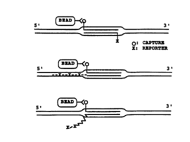
Figure l0 illustrates several embodiments of the use
of one double D-loop structure for the isolation and
ident$fication of correspond~ng sequences in duplex DNA
targets. one probe can be labeled with a capture moiety
(e.g., ag was done with biot~n in Fx~mple 7). The other
probe is then l~beled with a detection moiety, such ~s a
r~dioactive la~el, biotin, or digoxigenin or other modified
bases. The probes are co~ted and hybridized to the nucleic
acid sample that is being tested for the presence of the
taryet sequence. The hybridization reactions can be
deprote~nized or used directly.
The probe labeled with the ca~L~le moiety is tr~pped.
This trapping can be accomplished by, for example, labeling
the probe with a biotin moiety and exposing the reaction
mi~LuLe to streptavidin that h~s been attached to a solid
sup~olL. Alternatively, the capture moiety can be digoxig-
enin and the trapping ca~ be accomplished using ~n anti-
digoxigenin antibody attached to a ~olid support. Addi-
tional ~roups c~n be conveniently ~tt~he~ to the ends of
DNA molecules as follows. The oligonucleotide probe is
combined with digoxigenin-l--dUTP (an analog of dTTP, 2'-
deoxy-uridine-5'-triphosphate, coupled to digoxigenin via
an ll-atom spacer arm, Boehringer Mannheim, Indianapolis
IN) and termin~l deoxynucleotidyl transfer~se (GIBCO BRL,
Gaithersburg, MD). The number of dig~ dUTP moieties
incorpor~ted using this method appeared to be less than 5
(probably only 1 or 2). Alternatively, dig~ dUTP
moieties can be inco~oLated lnto the oligonucleotide
sequence o$ a probe as with ~iotin.
Typically, tbe following combinations of double-
stranded probes are used in the capture detection system:
(i) first probe/capture, labeled w~th biotin or digoxige-
nin, second probe/detectionl radiolabeled; (ii) first
~093/05178 2 131l6 2 1 ~ PCT/JP92/01135
probe/capture, labeled with biotin, second probe/detection,
labeled with digoxigenin; or (iii) first probe/capture,
labeled with d$goxigenin, second probe/detection,labe~ed
with biotin.
one convenient method to ~equester captured DNA is the
use of strepta~idin-conjugated superparamagnetic polysty-
rene beads as described in Example 7. After capture of
DNA, the besds csn ~e retrieved by p~acing the reaction
tubeQ in a magnetic rack.
Alternatively, avidin-coated agarose beads can be
used. Biotinylsted agaro~e beads ~i~mobilized D-biotin,
Pierce) are bound to avldin. Avidln, like streptavidin,
has four bin~ing sltes for biotin. One of these binding
sites is used to bind the avidin to the biotin that is
coupled to the ag~rose beads via a 16 atom spacer arm: the
other biotin binding sites remain available. The beads are
~iY~t~ with hybridization complexes to capture biotinylated
DNA (Exampl- 7). Alternative methods (Harlow ct ~l.) to
the bead capture methods ~ust dsscribed include the
following ~Lle~LaYidinylated or avidinylated supports:
low-protein binding filters, or 96-well plates, or modified
biotin capture methods such as i~inobiotin (Rigas, B ., et
al.)-
For either of the above bead methods, the beads are
isolated and the smount of hybridization complex that has
been ca~ e~ is qu~ntitatcd. The method of quantitation
~pen~-~ on how the ~econd strand DNA probe has been
prepared. If the second probe is radio~ctively labelled
the beads can be counted in a scintillation counter.
Alternatively, the captured DNA may be detected using a
ch~-;iluminescent, fluorescent or colorimetric detection
system.
Many o~ the experiments described above have made use
of radio-labelled oligonucleotides: Example 7 combines the
use of a biotin labeled flrst probe with a radioactively
,
5-
' ' ' ' ~ , , ' ~ , " ~ ~ ~ "
~ ~ ~B~
_ 32
labelled second probe. The techniques involved in radiola-
beling of oligonucleotides have been discussed above. A
specific activity of I08 cpm per ~g DNA is routinely
achieved using standard methods (eg., end-labeling the
S oligonucleotide with adenosine [~-3~P] -5' triphosphate and
T4 polynucleotide kinase). This level of specific activity
allows small amounts of DNA to be measured either by
autoradiography of gels or filters exposed to film or by
direct counting of sample in scintillation fluid.
Radiolabeling and chemiluminescence (i) are very
sensitive, allowing the detection of sub-femtomole quanti-
ties of oligonucleotide, and (ii) use well-established
techniques. In the case of chemiluminescent detection,
protocols have been devised to accommodate the requirements
of a mass-screening assay. Non-isotopic DNA detection
techniques have principally incorporated alkaline phospha-
tase as the detectable label given the ability of the
enzyme to give a high turnover of substrate to product and
the availability of substrates that yield chemiluminescent
or colored products.
For chemiluminescent detection, biotinylated or
digoxigenin-labelled oligonucleotide probes can be detected
using the chemiluminescent detection system "SOUTHERN
LIGHTS ",developed by Tropix, Inc. The basic technique can
be applicable to detect DNA that has been captured on
either beads, filters, or in solution.
Alkaline phosphatase is coupled to the captured DNA
complex. To do this several methods, derived from commonly
used ELISA (Harlow et al.; Pierce, Rockford IL) techniques,
can be employed. For example, the second strand DNA probe
can be end-labelled with digoxigenin-l1-dUTP (dig-11-dUTP)
and terminal transferase (as described above). After the
DNA is captured and removed from the hybridization m.ixture,
an anti-digoxigenin-alkaline phosphatase conjugated
antibody is then reacted (Boehringer Mannheim, Indianapolis
2 1 li~
' ~93/0~17X ' PCT/JP92/01135
33
IN) with the digoxigenin-containing oligonucleotide- ~e
~ntigenic digoxigenin moiety is recognized by the ant~body-
enzyme con~ugate.
CapL~L~d DNA hybridization products ~re detected using
the alk~line r~o~rh~tase-con~ugated ~ntiho~es to digoxige-
nin as follows. One chemiluminescent substrate for
A 1 ~A 1~nephosphatase is3-(2'-spiro~ ntane)-4-methoxy-4-
t3'-ph~ep~oryloxy) phenyl-l,2-dioxetane disodium salt
(AMP~D). ~erhosphorylation of AMPPD results ~n an unstable
com~un~, which decom~o~e~, releasin~ a prolonged, steady
emission o~ light at 477 nm. Light measurement is very
sens~tive and can detect minute quantities of DNA (e.g.,
10~-103 attomoles1-
Colorimetric substrates for the al~aline phosphatase
lS system have ~lso been tested. While the colorimetricsubstrates ~re useable, use of the light emission system is
morc sensit~ve.
An alternative to the above biotin cspture system is
to u~ digoxig~n~n ~n plac!o of biotirl to modi ~y the ~irst
~trand probe: biotin ~s then used to replace the digoxige-
n~n moieties in the above described detection system. In
this arrangement the ant~-digoxigenin antibody is used to
capture the DNA hybridizstion complex. SL~ 6~ Lavidin
conjugated to alkaline phosphatsse is then used to detect
the presence of captured oligonucleotides.
other alternst~ve capture systems include the
following: (i) the use of a DNA binding protein and its
cogn~te binding scquence, where the cognste binding
sequence is the capture ~oiety thst is included as a 5r
terminal seguence in the first strand probe (Kemp et al.);
and (ii) t~e use of hybridization capture where a non-
target-complementsry DNA sequence, such ss poly(T), is
incorporated as a 5'terminal sequence in the first strand
probe, and a complementary nucleic acid, such as poly(A),
is used to cspture the probe and associated nucleic acid by
W093/05178 2 1 1 6 2 ~ ~ PCT/JP92/0113-
34
hybridization. Either of these two methods can be used in
con~unction with a ~olid SUp~G~ L.
Another alternati~e system is to fix one probe to a
membrane (Saiki et ~l.), coat the probe, add target and the
second coated probe, deprotelnize, w~h and detect.
Figure lO illustrates several arrangements for one
doub~e D-loop structure detection. Figure lOA shows one
probe str~nd la~eled wlth a capture moiety, such as biotin,
and the second probe strand labeled with a detection
moiety, such as digoxiqe~in. F~gure ~OB shows one probe
strand labeled with a capture moiety, such as digoxigenin,
and ~he second probe strand having a homologous tail
extension that is labeled with mult~ple detection moieties,
such as biotin. Fig~re lOC illustrates the addition of
detection ~or capture moietie~) to heterologous tails
attached to either and/or bot~ strands of the double-
stranded probe.
C. The USQ of Multiple Double D-loop Structures for
Diagnostic Appl~cations.
In add$tion to exp~oiting one double D-loop structure
complexes in capture/detection systems, multiple double D-
loop structures can be used as well. In cases where
reassociat~on of probe strands may be a problem, for
example with larger probes, the use of multiple double D-
loop structures provides for a reduced background. In this
method two or more sequences need to be known in the target
sequence.
Figure ll illustrates ~everal arrangements for
detection based on two double D-loop structures. Figure
llA shows an example of labeling both strands of a first
duplex probe with the capture moiety and both strands of a
second duplex pro~e with a reporting/detection group. The
capture moiety can be contained in either one or ~oth
strands of the first probe set. In this system the
~"~93/05178 2 1 1 6 2 ~ ~ PCT/JP92/01135
hy~ridiz~tion complexes are captured as descri~ed above,
hou~c~, reassociation of the strands o~ the first duplex
probe gener~tes no ba~.oun~ for the reaction since
neither strand contains a reporterldetection qroup. After
capture, the hybridization complexes are detected on the
basis of the presence of the sQcond duplex probe in the
hybridization complcx. Detection of the reporter group is
acco~pl~s~ed as de~cribed above.
A second embod$ment o~ thls system, based on t~e
presence of multlple double D-loop structures in the target
complex, i~ illustrated in Figure llB. ~n this case only
one strand of the first duplex probe is labeled with the
capture moiety and only one strand of the second duplex
probe is labeled wlth the reporter moiety.
As described above for one double D-loop structure
detection, heterologous t~ils and ~equences homologous to
the target DNA can be added to the duplex probes.
D. T~e Use of Double D-loop Structures in RecA
prote$n Facilitated DNA Amplification Reactions.
DNA ~mpl~flc~tion reactions have been described that
predominantly rely on t~ermal denaturation (Mullis; Mullis
et al.; Scharf et al.) for strand separation in preparation
for continued amplific~tion. Described below are two
detection systems baQed on D~A amplification subsequent to
t~e formation o~ single or multiple double D-loops.
(i) One ampll~ication/detection method of the present
invention utilizes multiple dou~le D-loop structures to
facilitate amplification without the need for thermal
denaturation.
Experiments performed in 5U~U~ ~ of the present
invention have demonstrated that use of two pairs of
complement~ry DNA primers, which are homologous to
d~fferent regions of the double-stranded target, in the
hy~r$dization reactions of the present invention results in
WO93/05178 21~ 36 PCT/JP92/0113~
the formation of two double D-loops in the target DNA.
These double D-loops fiank and define a speci~ic region of
DNA on a native duplex target (~igure 12A): D~801/2 and
D~803/4 ~Table l) are examples of probes/pr~mers that can
participate in th$s reaction.
The sesultlng DNA target structure is recognizable by
various DNA polymerase~ as a substrate for DNA synthesis.
An example of one euch ampll~cation reaction is presented
in Example 8. The substrate for the ampli~ication reaction
described in Example 8 ~s the lambda DNA genome. The
pr~mers ~efine a t~rget region of approximately 500 bp.
Typ~cally, th~ amplifi~tion reactions contain DNA polymer-
ase ~e.g., the Klenow fragment), RecA protein coated
primers, aTP~S (or rATP ~alone and in the presence of a
regenerating system), dATP, GTP~S, and mixes of ATP~S and
rATP or ATP~S and ADP), the target substrate, necess~ry co-
~actors and ~ll four dNTPs (includlng modified or labeled
d~TPs). These re_ct~ons may also conta~n DNA helicase,
topoisomer~se, or other similar D~A unwinding agents and~or
other DNA polymerases.
These reaction cond$tions favor extension of the
primers at their 3' ends with subsequent primer elongation
in the 5' to 3' direction. The 3' end extension of two or
more of the four DNA primers in the structure ~y polymerase
2~ de~ines regions of D~A target ampli~ication ~etween DNA
primers (see Fig. l3). DNA polymerase extensicn of the
other two primers ca~ also occur at their 3' ends, unless
these are chemically or physically blocked $o restrict D~A
amplification to a defined region (~xample 8).
DNA polymerases catalyzin~ 3' extension between primer
pairs may displace primers on the same strand, or, a}terna-
tively, new synthesized product(s) could be lig~ted to the
primer with DNA lig~se, including thermoresistant or
thermosensitive DNA ligases ~picentre Technologies,
MA~i~on, WI). Replication can also be faci~itated by
W093/0~178 PCT/JP92/01135
~ 7'" 2~621~
~ a~f~.iate DNA helicase, topoisomerase, single-
strand bind~ng proteins ~SSB), gene 32 (or other similar)
prote~ns, or Re~R~-enco~e~ enzymes or other proteins, some
of which have associated helicase activlties. Any primers
5prop-rly poslt~oned by Rec~ protein could ~ used for
replication initiation.
Typic~lly, probes u~ed in tbe two-double D-loop ~ecA
protein catalyzed DNA amplirication are approximately 60 to
80 bp in s~z-. Probes larger os smaller can also be used
10as well, but react~on condit~ons ~ay need to be mod~fled,
for example, inclusion of stabi~izing peptides (e.g., SS8
or gene 32 psotein), drugs, antlho~es~ polyamines or
cros~ ng reagents.
Mu~tlple prlmer~ of different size~ can also be used.
15Prlmers c~n also be homologous to the target D~A duplex
along the~r entire length, or they can contain end or
lnternal regions of partial non-homology, such ~s heterolo-
gous ta$1s (see above). The only requirement for these
pr~mers is that the ba~es used for the 3' extens~on of the
20desired ampl~ficstion product ls avail~ble to prime DNA
synthesis. As degcribed above, primers can also contain
mod$fied phosph~te b~honec or bases such as biotin or
dlgoxigenin or appended functions such as DNA modlfying
enzymes and chemica~ agents.
25React$on in the presence of excess RecA protein-coated
primers allows ~ormation of new multiple or double D-loops
on the newly replic~ted and ampllfied DNA. The RecA
prote$n-co~ted pr~mers serve to initiate additional rounds
- of DNA synthesl~, and in this w~y the DNA tarqet is
30~mpl~ried w$thout the need for thermally denaturing the
target DNA to position the ~mplification pri~ers.
DNA amplification reactions c~n use any of a nu~er of
DNA polymerases or polymerase m~xtures, including the
- ~ollowing: Klenow large fragment of ~. col~ DNA polymerase
35I; T7; T4; and/or other viral or cellular DNA polymerases
PCT/JP92/0113
WO93/05178 n~- ~ ~
~ ~ ~1 ~ 38
and their mutants, for example, double-KlenoW mutant
proteins h~ving no exonuclease activity.
The DNA products of two-dou~le D-loop reactions are
defined by the DNA primers used. The left and riqht
primers to the doubl- ~-loop regions to be amp~ified define
the S' ends of the new~y synthesized D~A a~plificat~on
products. When the pri~ers ~re not displaced, the newly
synthesized DNA product can be ligated in a RecA-catalyzed
amplification reaction as illustrated in Flgure 12B.
Using multlple primer sets, it is also possible to
generate DNA ampllricat$on products which have cohesive
ends. The DNA products can then hybridize through their
overlapping cohesive ends and the length of these
associated DNAs ~s subsequently extended (Haase et al.).
Elongation of existing strands, displacement synt~esis, and
RecA-catalyzed base pairing in the overlap regions may
increase the yield of l~rge DNA targets. This can be
important for RecA-catalyzed DNA amplirication in cells in
situ, which may, under eertain condit~ons (Haase et al . ),
require large DNA products or appended groups for retention
in s~tu.
The double D-loop reaction, or multiple double D-
loops, can be st~bly rormed in agarose for in situ
ampllfication reactions. Txpically, the agarose is of the
low-melting temperature v~riety, although mixtures of
d~fferent types of agarose are possible, and the
concentration of ag~ro~e is about 0.4-1%. Under these
conditions the agarose gel prov~des a restricti~e medium
that alQo allows retention of shorter DNA products: this
is particularly useful in in situ reactions ~see below).
The RecA-facilitated DNA ampli~ication reaction can be
carried out at 37~C as well as at elevated temperatures
that are below the thermal denaturation temperatures of
target DNA dup}ex or pri~er:target hybrids, for example 50-
60-C. Use of elevated temperatures in these reactions
39 ~
expands the repertoire of enzymes available for primer
extension and may allow longer tracts of DNA to be
synthesized. The temperature of the amplification reaction
will dictate the choice of reaction components: for
example, wild-type ~. coli RecA-protein coated probes are
added to the target DNA at 37-39~C, DNA synthesis is then
accomplished at 50-55~C with Thermus aquatic~s DNA
polymerase, the temperature of the reaction is then lowered
to 37-39~C and RecA-protein coated probes are added re-
added. Alternatively, at high temperatures temperature-
resistant RecA-like proteins could replace the wild type E.
coli RecA protein (as discussed in WO-A-91/17267).
The two-double D-loop, or multiple double D-loop,
reaction using RecA protein-catalyzed primer positioning
for DNA amplification reactions has important diagnostic
applications for DNA detection and amplification in
solution diagnostics or in situ diagnostics.
(ii) A second detection method exploiting the double
D-loop and DNA amplification uses polymerase addition of
labeled or modified dNTPs for signal amplification.
Extension of the 3' end of a primer in a single (triple-
stranded) D-loop structure using DNA polymerase was
demonstrated by Cheng et al. (1988). ~.fter formation of a
single double D-loop in a target DNA molecule DNA
polymerase, necessary cofactors, and all four dNTPs are
added to the reaction. Strand extension takes place from
the 3'ends of either one or both probe strands of the
double-stranded probe (~igure 13A). Alternatively, the 3'-
end of the strand containing the capture moiety can beblocked to prevent primer extension. One or more of the
dNTPs is labeled for detection by a standard method (e.g.,
biotin, digoxigenin, or, fluorescent or radioactive
moieties). The incorporation of the labeled dNTP results
in the amplification of the detection signal.
CA 0211621~ 1998-03-30
This method can be further exploited by using multiple
double D-loop structures to target the region of interest (as
described above). The 3'-ends of the double-stranded primers
external to the target region can either be blocked (as
illustrated in Figure 13B by an asterisk) or not. This
method of signal amplification can be further enhanced by
using multiple rounds of RecA-facilitated amplification, as
described above, in the presence of a labeled moiety.
Target detection with signal amplification can also
be accomplished as follows. A double-D-loop is formed at the
target sequence using two probe strands, where at least one
of the strands contains a capture moiety. The resulting
double-D-loop complex can be deproteinized and captured via
contacting the capture moiety with a capture medium. The
captured complex is released by heating: the complex can be
released either as dsDNA or ssDNA. If necessary, e.g., if
the complex is released as dsDNA, the complex is denatured
and the target DNA released. To this mixture DNA synthesis
primer(s), which are complementary to a target sequence, are
added. These primers do not contain sequences that were
present in the original double-stranded probe. DNA
polymerase and dNTPs, are then added, under the appropriate
buffer conditions, to synthesize DNA from the hybridized
primer(s). This primer-directed template synthesis can be
carried out in the presence of labeled dNTPs, for example,
radiolabeled dNTPs, biotin-labeled dNTPs, FITC-labeled dNTPs,
or other suitably labeled DNA precursors. The inclusion of
label allows target detection via the appropriate detection
systems, such as, a fluorometer in the case of FITC-labeled
dNTPs.
E. The Use of Double D-loop structures in RecA
Protein Facilitated in si tu Hybridization.
Another detection method which utilizes the double
D-loop structure is in situ hybridization with fixed cells
41
(Example 9): RecA-facilitated in situ hybridization methods
have been described in co-owned PCT International Publication
No. WO 93/05177 "In situ Hybridization Method," published on
March 18, 1993. The in situ hybridization of RecA protein
coated duplex probes provides the ability to localize a
target sequence in an isolated, fixed biological structure or
within a nucleus or nuclear volume relative to other targeted
sequences and/or the nuclear membrane, using a confocal laser
scanning microscope (van Dekken et al. Cytometry 11:579
10 (19go) ) .
One application of the in situ method is described in
Example 9D. In this method, dividing HEp-2 nuclei are fixed
and probed with the RecA/chromosome-X alpha satellite DNA
probe complex, and labeled with FITC-avidin. The pattern of
probe binding in the dividing nucleus is evaluated using
standard light fluorescent or laser scanning microscopic
techniques. To localize the bound probe, the same field is
viewed by phase contrast microscopy, without changing the
focus of the lens. By overlaying the resulting two
photomicrographs, the relative position of the nuclear
membrane and nuclear division plane can be seen with respect
to the probe-labeled chromosomes.
This aspect of the present invention provides
simplified in situ procedures for localizing target
sequence(s) in a biological structure. Typically, fixed
cells or subcellular structures are probed in suspension or
on slides followed by flow-cytometric or microscopic
analysis. The method reduces artifacts by eliminating the
need for a heat denaturation step, and allows more rapid and
specific detection of target sequences. The method can be
applied equally well to unique, low, and/or high-copy number
target sequences.
In particular, the method allows detection of low-copy
sequences without the requirement to first amplify the
sequences. Since most gene mapping and chromosomal studies
are expected to involve specific, unique, or low-copy
WO93/05178 2 ~ ~ 6 2 1 S 42 PCT/JP92/0113~
sequences, the prcsent ~n situ method provides an important
advantage for gene mapping studies, as well as $or
diagnostic applications involving unique or low-copy
numbers of normal, mutant or pathogen se~enceC. Also, the
present method allows for determination of chromosome
content by flow cytometric analysis.
one general ~ "o-Lic appllcation of this in situ
method is for us- in mapping a ~elected gene or regul~tory
sequenc- in a chromosome, and/or in a particular region of
lo the chromosome. The target gene may be one which (a)
generates a selected gene product, tb) is suspected of
performing a critical cell-cGnL~ol function, such as a cell
or viral oncog-n-, ~c) is related to a repeat seguence, (d)
is suspected o~ cont~ning a genetic defect which prevents
expression of an active gene product, (e) may be related in
chromosome position to a marker probe region with ~ known
map position, and/or (f) may represent an integrated vira}
seguence.
The probe strand~ for in situ hybridization can be
labeled in a numb~r of ways including direct l~h~l~n~ with
fluorescent ~oieties like fluorescein~ dUTP (Boehrin~er-
M~nnhP~ urther, individu~l probe strands can be used
to generate a coupled-fluore~cPnce system where, for
example, the emission energy of one fluorescent moiety,
incorpor~ted in one strand) emits light a~ the excitation
energy of the second fluorescent mo~ety, incGL~Lated in
t~e second probe str~nd. Such a coupled-fluorescence
system takes advantage Or the proximity of the probe
strands in the double D-loop complex.
When the DNA probes are d~rected aga~nst specific
cellular pathogens, typic~lly for detecting the presence of
a v~ral or bacterial pathogen in an ~nfected cell, the
fixed labeled cells may be exA~ned by light or
fluorescence microscopy to detect and localize lnfecting
pathogens in ce~ls. Alternatively, cell infection, and
~, ~,,,s ~ ~
s.r~
W,O 93/05178 2 1 1 ~ 2 1 ~ PCI'/JP92/0113~
-
percent cells infected, can be determined by fluorescence
acti~ated cell sorting (FACS) after ~n situ hybrldization
o~ RecA protein coated duplex probes to nuclei or cells in
~uspension tTr~sk et al.).
F. The Use of Double D-loop Structures in Restriction
Enzyme Cleavag- Ba~ed Detection Systems.
The double D-loop structures of the present invention
can be used to detect the presence of target DNA in a
sample by i,-- o~cin~ alterations at the target/double D-
loop complex which modl~y, in a detectable m~nner, the
response of th~s complex to restriction enzyme digestion.
Several examples o~ such detection systems are described
below.
/ One method of detectlon that exploits tbe double D-
loop ~nd restriction enzyme digestion is as follows. A
region ~n the t~rget DNA i8 chosen ~s the double-stranded
probe seguence. The probe sequence is modified to contasn
an internal regtriction s$te t~at is not present in the
tarqet D~A. Such a restriction ~ite can be chosen so as to
minimize base palr m$smatrhin~ between the target and the
probe (Figure 14). The double D-loop is then formed ~nd
the complexes deproteinized. The complexes are then
captured on tbe solid D~G.~ and digested with the
restriction enzyme for which the site has been i-,-Lod~ced
in the probe sc~uence ~in Fi~ure 14A, PYuI~.
Alternatively, the complexes can ~e digested~_with the
restriction endonuclease before capture (Figu~e 14B).
Since the PvuII restriction site is not reconstit~ted when
the probe is hybridized to the target sequence, the
restriction enzyme will only cle~ve renatured probe:pro~e
complexes, not probe:target complexes. The solid support
is washed and examined for the presence of the detection
moiety. Th~s method allows the reduction of any ba~yLound
ssgnal that m~y be generated by probe renatur~tson.
CA 0211621~ 1998-03-30
44
A second detection method works on a similar
principle. In this method the target DNA is un-methylated
and the double-stranded probe DNA is methylated before RecA
protein-coating. The double D-loop complex is formed, the
complex captured and deproteinized. The sample is then
digested with, for example, DpnI which cleaves its
recognition site (SEQ ID NO:2) only when the A residue on
both strands is methylated. Since the methylated restriction
site is not formed when the probe hybridizes to the target
sequence, DpnI cleavage only occurs when the probes are
renatured. The solid support is washed and examined for the
presence of the detection moiety. As above, this method also
allows the reduction of any background signal generated by
probe renaturation.
The methylation state of the DNA can also be exploited
using the target/double D-loop complexes as follows. In this
method either the target DNA is methylated or the double-
stranded probe is methylated prior to RecA protein-coating.
The double D-loop complex is formed, the complex captured and
deproteinized. The captured complexes can be isolated from
the solid support and split into multiple samples. One
sample is digested with a methylase-sensitive or methylase-
dependent restriction enzyme and another is digested with a
methylase-insensitive restriction enzyme: for example, MboI
does not cleave DNA when the A residue is methylated and
Sau3A I cleaves the same restriction site independent of the
A residue's methylation state.
These samples can then be size fractionated (e.g., on
an agarose or acrylamide gel, or by HPLC) and the banding
pattern of the samples compared. This method allows
isolation and subsequent ex~m;n~tion of restriction fragment
length polymorphisms of a chosen fragment between a number of
samples from different sources.
Methylation may also be used to protect a specific
restriction enzyme site from digestion (Nelson et al.).
wog3/os178 2 1 1 6 2 15 PCT/JP92/0113~
-
If, for example, it was des$rable to isol~te a ~boI
fragment sp~n~tn~ a particular region, but internal to the
region an MboI slte existed, the fragment could be isolated
as follow~. A double D-loop structure is formed at the
intern~l restriction s$te in the target DNA using
methyl~ted probes. The target/double D-loop complex is
deproteinized, digested with MboI and captured (Figure
15A). T~is method can be used to (i) examine restriction
enzyme polymorphisms at restriction sites ad~acent to the
protected site in fragments obtained from different
sources, or (i~) capture ~nd clone ~ desired sequence using
a restrict$on enzy~e even when an internal clea~age site is
present for tbat enzyme. The ~act that deproteinized
double D-loop fitructures are susceptible to restriction
lS enzyms cle~v~ge has been demonstrated by PleI restriction
endonuclease slte-spcciflc cleavage of probe:target
complexes formed w~th 5~0-mer probes ~nd 2.9-kb homoloqous
target fragments.
The double-stranded RecA protein-coated probes can
themsel~es be u~ed to protect a specific restric~ion enzyme
site from d$gestion. In ~his case, the complex is
deproteinized. T~e target/double D-loop complex is
digested wit~ the restriction endonuclease and ca~Lu~d
(Figure lSB). Alternat~vely, the double-stranded RecA
protein-coated probes can be used to protect a mod~fication
site, for cxample, ~ methyl~tion site, from modificatio~.
In this case, ~s for the above restriction site protection,
the complex is not d-proteini7ed. The t~rgetrdouble D-loop
complex is tre~ted with the modification reagent and then
deproteinized. Modi~ication target sites within the
target/double D-loop complex are protected and lack the
modific~tion.
The ability to block restriction site cleavage is
useful in geno~ic ~apping. For example, i~ the ~arget DN~
defines a known region in the genome, li~e th~ PvuI site at
WO93/05178 PCT/JP92/0113~
2~1~2~ ~ 46
approximat-ly 26.3 kb on the lambda genome, the known site
is protected by one of the appro~ches described above.
U..~.o~ected and protected lambda genomic DNA is digested
with PvuI. The change in restriction fragment patterns
between the two digests allows one to deduce which
fragmen~s are n~xt to one another -- based on the
di~appearance of bands and the sizes of the new bands in
the sample con~ini~ blocked restriction sites.
Re~triction enzymes, and their re-~pon~e to methylation
states, are commonly available ~nd conditions for their use
are well known in the art (Ausubel ~t ~l.; Maniatis et
al.).
G. The Vse of Double D-loops to Generate Site-
Spec~ ric Clea~age in DNA.
Oligonucleotides have been used to direct cutting
agents to spec~ic singlP ~-~anded and double-stranded
nucleic acids sites ~Corey et ~l.; Dreyer, G.B. et ~l.;
Moser, ~t al.). One advantage of oligonucleotide directed
cleavage is that the experi~enter is no longer ~Pren~Pnt on
existing c'eavage function~: any desired DNA cleavage
function c~n be tailor-made. The RecA protein-coated
double ~L~nded probes of the present invention can be used
to generate site specific cle~va~e in a num~er of w~ys.
For example, a speci~ic target sequence is 6elected and a
doublc ~L~nded DNA probe corresponding to t~e se}ected
sequence i~ generated. An EDTA moiety is attached to one
or both strand~ of the oligonucleotide pro~e. One method
of ~ttachment of EDTA to an oligodeoxyn~cleotide via the C-
5 of thymidine has been described by ~reyer ~ ~l. Theprobe can then be Red protein-coated and the double D-loop
complex formed with target DNA. Cleavage oc~urs in the
presence of oxygen upon ~ddition of Fe(II) and a reducing
agent (usually DTT) to the EDTA-probe:target hybrids.
wos3/0sl78 2 1 1 ~ 2 1 ~ pCT/JP92/01135
47
Alt-rnati~ely, cutting can be ~co~r~ ~ sh~ using
peptide fr~g~ents der~ved rrOm DNA binding/cleaving
proteins (sluka~ 7.P., ~t ~1.) which are att~ched to the
ol~gonucleotide probes. Further, restriction endonucleases
that havQ freguent cut ~$tes or relatively non-specif$c
phosphod~ester~ses, such as staphyloco~cAl nuclease, can be
attached to an oligonucleotide to generate a hybrid
catalyt~c ~gent t~_t has increa~ed sequence speci~ic~ty
~Corey ~t ~1.)'.
0 O~igonucl-otld-s are att~h.~ to the phogphodiesterase
or other cle~ving agcnt elther before R~cA protein-coating
or ~fter doubl- D-loop complex formation and
deprot~;n~7~tion. After ~ssoci~tion of the
phosrho~p~tQrasQ~ r olease or peptide with the double D-
loop complex, the reaction condlt~ons are modified to allow
the hydrolys~s o~ both target DNA str~nds in a site-
sp-cif~c f~h~on. Depending on the activity of the
catalytlc agent either one or both strands of the double-
stranded probe ls modlfled to contain the agent.
H. Use of the Double D-loop in DNA Enrichment.
Thc double D-loop hybr~d~z~tion co~plex of the present
~nventlon can also be used ~or separation and enrichment of
selected t~rget DNA ~equences. For ex~mple, the double-
stranded probe c n be formed cont~ning a capture moiety in
one or both of the probe strands. The double D-loop
co~plex ~s for~ed between the dou~lc ~L~anded probe and
target DNA contA~n~ ~n a mixture of DNA. The double D-
loop complexes t~at contain the probe and target sequence
are then separ~ted from the reaction ~ixture using the
ca~L~e moiety, by, ~or example, attachment to a solid
support. ~he complex can then be dissociated ~y heating to
relea~e the target duplex from the U~Gl ~ and, if
- neoe~ry, the rcleased D~A renatured to regenerate the
original target dup~ex DNA. Alternatively, the entire
WO93/05178 211 62~ ~ PCT/JP92/0113~
48
doubl- D-loop complex may simply ~e rele~ed from the solid
s~o~L.
To test whether the targeted duplex DNA could be
re~ea~ed from the hybrids simply by heating, the thermal
stab$1ity o~ deproteinized double D-loop hybrids formed
wlth ~P-end-labeled 500-mer probes and the 10.1 kb ApaI
tasget fragment wa~ ed: the hybr$ds were completely
stable at 75-C in lxT8E ~u~fer. About half the DNA probe
~tr~nds wers released at 80-C. E~sentially all of the DNA
probe ~trand~ were released from the target at 85~C. This
melting profile i8 similar to that for duplex probe:target
hybr~ds formed by heating and then slowly cooling the
reactant mixture in the ~bsence of RecA protein. The
hybrid melting profile was approxi~ately 10~C lower than
lS that of duplex 500-mer probe, sel~-~nnealed, under
identical ion$c conditions.
The hybridization reaction is potentially useful for
targeting c~romosomal or gene frag~ents identified only by
sequence-tagged sites (STSs) (0180n, et al.) for the
following reasons:
(i) the RecA-mediated hybridization reaction does not
require denaturation of the duplex DNA target for hybrid
formation, and
(ii) targets are rele~sed from isolated hybrids ~y
heating to a te~perature that dissociates the probe from
the probe:target complex but that does not denature the
du~lex tar~et-containin~ analyt~, Q . g ., a chromosomal or
genc _G..~aining fragment. The target DNA analyte can then
be recovered in duplex form.
Further, careful control of the hybrid melting
temperature would permit a Qelection against the hybrids
which might have only partial homology with the probe.
Stringent melting temperature selection may be important
when probes are used with co~plex mixtures of target DNAs.
One 2dvantage of recovery of duplex ~arget DNA versus
CA 0211621~ 1998-03-30
single-strand-denatured target DNA is that duplex DNA tends
to be more resistant to shear forces than totally denatured
single-stranded DNA. The recovery of duplex target DNA, by
the method of the present invention, would allow the
enrichment and isolation of specific duplex gene or genome
segments, including large chromosomal fragments, which can
then be used for further manipulations and/or analysis.
Target DNA duplexes obtained by this method can be
used in DNA amplification reactions (Mullis; Mullis et al . )
or in standard cloning techniques (Ausubel et al.; Maniatis
et al.).
I. Homogeneous Diagnostic Assay
A protocol for a homogeneous diagnostic assay that can
detect a specific native target DNA duplex has been tested.
This assay involves double-stranded target capture using
double D-loop hybrids followed by DNA signal amplification,
as described briefly below.
(i) Capture of double-stranded DNA targets
A technique can be worked out for using double D-loop
hybrids to specifically capture a large double-stranded DNA
such as lambda DNA genome (-50 kb). The reaction is also
applicable for the capture of smaller duplex DNA targets.
The technique uses RecA-coated single-stranded probes
labeled with a capture moiety such as biotin, preferably
averaging 300-500 bases in size. All DNA probes are
preferred to be homologous to sequences within preferably
about 1000 base region of the duplex DNA target. The
biotinylated probes are prepared by nick-translating
preferably about 1000 bp DNA duplex fragment in the
presence of bio-14-dATP. Heat-denatured probe DNA is coated
with RecA protein and then reacted with duplex target DNA.
After probe:target hybrid formation, the hybridization reac-
CA 0211621~ 1998-03-30
tion mixture can be stopped, for example, with 20 mM EDTA
and treated with 0.5 M salt, and hybrids can be captured on
washed magnetic Dynabeads~ M-280 Streptavidin (Dynal).
After capture, beads are washed 3X in buffer containing 1
M salt. It is likely that the high salt conditions at least
partially removed a proportion of the bound RecA protein.
Target DNA capture can be measured by using 32p_ labeled
lambda DNA and by directly counting the radioactivity that
remains associated with the beads after washing. The
results of the capture reaction using a large lambda DNA
genome (-50 kb) are shown in Table 3. The specificity of
the reaction for the capture of biotinylated probe which was
hybridized with double-stranded target was verified by
including three control reactions (Table 3, reactions 2-4).
These reactions showed that: (1) RecA-coated biotinylated
probe was required for specific target DNA capture (reaction
1) since no significant signal was obtained in a reaction
without RecA (reaction 2), (2) RecA inclusion in the
reaction was not the cause of target capture because only
background level DNA capture occurred in the presence of
non-biotinylated non-homologous DNA probe coated with RecA
(reaction 3), and (3) the average background, non-specific,
target DNA capture was approximately 3.5~ (reactions 2-4).
(ii) Signal amplification from captured DNA
A prototype protocol for detecting the bead-
captured target DNA is described briefly below. For this
step, captured target is released from the beads by
heating in a reaction mix containing dNTP precursors,
one or more of which is labeled (e.g. bio-14-dATP, or
dNTP with a directly detectable label, such as FITC-11-
dUTP, etc...) and a ss DNA primer (or primers)
51
not homologous to the original capture probe sequences,
but homologous only to target sequences, is (are) added
and allowed to anneal to the single strands of target
DNA. After annealing, the reaction is cooled and a DNA
polymerase enzyme preferably, DNA polymerase T7 (Seque-
nase~ Version 2.0, USB) and appropriate buffer are
added to allow DNA synthesis by primer extension.
Alternatively, a high temperature polymerase could also
be used and the reaction incubated at a temperature
allowing processive DNA synthesis. During primer
extension, labeled DNA precursor(s) is incorporated
into the newly synthesized DNA strand(s). Because the
DNA primer(s) is not homologous to the previously used
RecA-coated probe(s), label incorporation specifically
indicates the presence of the correct captured target.
Use of a capture moiety either directly associated with
the primer(s), such as poly(A), or able to be added
later, allows subsequent capture of the amplified
target signal on oligo(dT) attached to magnetic beads,
cellulose, etc. Capture is followed by removal of
unincorporated precursor label from the amplified DNA,
and if necessary a blocking agent, such as, for exam-
ple, I-Block (Southern-LightTM, Tropix) is used to
block the non-sepcific binding of the detection moiety
(e.g., AVIDx-AP , Tropix) to the capture matrix. After
blocking, substrate is added to allow specific signal
detection from the amplified target DNA, and signal is
detected by any appropriate means. A schematic outline
of a homogeneous diagnostic double D-loop assay is
shown as follows. Although a specific labeling, cap-
ture and detection scheme is presented, there are
numerous ways in which such an assay could be prac-
ticed.
Schematic outline of a homogeneous diagnostic
assay for duplex DNA targets with Double-D-loop hybrids
52
is shown as follows:
Description of Assay Steps Basic Reactions
RecA-coated ss biotinylated
Formation of DNA probes + Duplex Target DNA
probe:target hybrids ~
Mix and incubate at 37~C
for 4' min-1 hr
+ EDTA and 0.5 M salt
Capture of homologous duplex + Magnetic beads
target DNA on beads
8ead-captured
Probe:target DNA hybrids
Ramoval of non-hydbridized ¦ Washes in lM NaCl buffer
DNA by washing
Washed beads with captured hybrids
Heat destabilize hybrids
Target signal amplification
* Remove Beads
1. Anneal target with primer(s)
Heat target DNA with* poly(A)
r----~-~ ( capture) primer(s)
' [lOO~C, 5 min], cool
Primers annealed with DNA target strands
2. Extend primer(s) with DNA , + polymerase [e.q. T7
polymerase to incorporate ' Sequenase~ Version 2 . 0 ]
biotinylated nucleotide(s) Repeat and reactan~s including
' labeled DNA precursor(s)
L------- [e.g. bio-14-dATP]
Target signal detection
Labeled target DNA
+ oligo dT beads
1. Capture of amplified DNA Washes in 0.5 M NaCl buffer
Captured biotinylated target DNA
Washes in 0.5 M and 0.1 M
NaCl buffers
810cking of beads
2. Reaction with avidin-alkaline + AVIDx-AP~
phosphetase (AP)
AP-labeled DNA on beads
Wash in substrate buffer
+ AP substrate
3. Detection of amplified DNA Signal Production
!
Signal Detection
non-homologous to probe DNA,
homologous to desired target DNA
- 53
The following examples illustrate, but in no way are
intended to limit the present invention.
Materials and Methods
Restriction enzymes were obtained from Boehringer
Mannheim (Indianapolis IN) or New England Biolabs (Beverly
MA) and were used as per the manufacturer's directions.
Generally, oligonucleotides were radiolabeled with [~-
32P]ATP and T4 polynucleotide kinase. Labelling reactions
were performed in the buffers and by the methods recommend-
ed by the manufacturers (New England Biolabs, Beverly MA;
Bethesda Research Laboratories, Gaithersburg MD; or
Boehringer/Mannheim, Indianapolis IN). Oligonucleotides
were separated from buffer and unincorporated triphosphates
using Nensorb~ 20 pre-formed columns (NEN-DuPont, Boston,
MA) as per manufacturer's instructions, and subsequently
dialyzed versus dd H20, if necessary.
Exam~le l
Pre~aration of RecA-co2ted Probes
A series of double- and single-stranded DNA probes znd
primers have been generated. The positions of these probes
and primers relative to a contiguous 500 bzse pair region
WO93/05178 ~ 21~ ~4 PCT/JPg2/0113~
o~ the la~bda phage genome are Chown in ~igure 1: the
nuclQotidQ ~equence of t~is r~glon o~ the lambda geno~e i6
~ s~nted ~n Figure 2. The b~se positions of the S~ and 3~
Qnds of each probe and primer, rel~tive to the lambda viral
genome, are listed in Table 1.
Table 1
.~RnA R~F..C D~r~ PROBE AND p~T~FR DNA ~ CES
~sob~ o~ Bas~s T~ol~ Size
~r~mer S~.quenc-~ (b~) or ~p~
PCR01- ~131-71S5 25
PCR02~ 7606-7630 25
PCR03A- 7351-7390 40
DL80-1 7131-7210 80
DL80-2~ 7131-7210 80
DL80-3 7SS1-7630 80
D~80-4 7551-7630 80
500(ds) 7131-7630 500
280(d~) 7351-7630 280
159(ds) 7472-7630 159
121(ds) 7351-7471 121
Biotin-121 7351-7471 121
121-3~P- 7351-7471 121
79(~s) 7g52-7630 79
2~ Biotin-79- 7552-7630 79
, strand.
~- Opposite strand.
Table 2
BEAD CAPTURE OF PROBE:TARGET HYBRIDS SHOWS
THAT THE RecA-CATALYZED DOUBLE D-LOOP PRODUCT
CONTAINS TWO DNA PROBE STRANDS
Probe Strand(s)32p Radioactive DNA
Counts Captured
32p (Radio- ~ TotalCorrected
Biotin active Counts per~ of
(CaptureReporter MinuteCounts per
Reaction RecA DNA) DNA) Expected*Minutet
1 - + + 4 0
2 + - + 11 0
3 + + - O O
4 + + + 110 98 -~
~,
55a
* See Figure 8 for explanation of sample reactions.
Percentages were calculated from the 32p counts
remaining on the Dynal streptavidin-coated beads
after three washes of each capture reaction with lX
acetate reaction buffer. The total expected 32p
counts per minute were determined by scintillation
counting of DNA in minced gel slices from experiments
identical to those in Figure 8. Since no radioactive
DNA was added to reaction 3 (containing biotin
capture probe only), no radioactive counts were
expected for this reaction.
t Radioactive P counts in DNA were corrected for
nonspecific bead capture of background DNA counts
(i.e., counts from reaction 2 without biotin capture
probe).
Primers PCR01 and PCR02 correspond to the primers
supplied in the "GeneAMP~" DNA Amplification Reagent Kit
(Perkin Elmer Cetus, Norwalk CT).
-
56
Single-stranded primers (PCR01, PCR02 and PCR03A) and
single-stranded probes (DL80-1 through 4, biotin-l2ll
121-32P, and biotin-79) were chemically synthesized using
commercially available phosphoramidite precursors on an
Applied Biosystems 380B DNA synthesizer (Applied
Biosystems, Foster City CA).
DNA molecules were biotinylated by reaction with a
biotin phosphoramidite at the last 5' base (New England
Nuclear-DuPont, Boston, MA) before deblocking. All chemi-
cally synthesized DNA molecules were deblocked according tothe manufacturer's specifications.
Short DNA probes (25-mers) were used without further
purification. Single-stranded 80-mer and 121-mer DNA
probes were purified by polyacrylamide gel electrophoresis
using ~%, and 5% or 8% polyacrylamide gels, respectively.
Full sized DNA products were obtained by excising DNA bands
from the gels that corresponded to the correct size DNA
molecule. The DNA molecules were recovered from gel pieces
by electroelution using the "EL~P" system (Schleicher
and Schuell, Xeene, NH). Both probes and primers were
concentrated by standard ethanol precipitation (Maniatis et
al.). Probe and primer DNA concentrations were determined
based on W absorbance at 260nm of the diluted DNA.
Double-stranded 500 and 280 bp regions of the lambda
genome (Figure 1) were synthesized using primers PCR01 and
PCR02, or PCR03A and PCR02, respectively, Taq polymerase
and standard DNA amplification reaction conditions (Perkin
Elmer Cetus; Mullis; Mullis et al.). The amplification
products were separated from the DNA primers by
electrophoresis through a 0.7% agarose (Sigma Type II,
Sigma, St. Louis MO) gel (500-mer) or a 4% ''NUSIEVE''(FMC
BioProducts, ~ockland, ME) agarose gel (280-mer). The DNA
molecules in the bands corresponding to the amplification
products were electroeluted, concentrated, and their actual
concentrations' determined as described above.
~ ~ ~ 57
Double-stranded 121- and 159-mer probes were obtained
by restriction digestion of purified 280-mer using the
enzyme AluI (New England Biolabs, Beverly MA). The DNA
probes were isolated by gel electrophoresis, electroeluted
5and concentrated as above.
Double-stranded 79-mer probe was obtained from
restriction digestion of the purified 500-mer using the
enzyme HpaII. The digestion products were separated and
purified from uncut DNA by electrophoresis using either 3%
10or 4% "NUSIEVE~" (FMC Bioproducts) gels or 1~ agarose gels.
Specific DNA fragments were recovered from the gels as
described above.
Single-stranded or double-stranded DNA molecules were
5'-end-labeled (Maniatis et a7.) with [~-32P]ATP and T4
15polynucleotide kinase (Promega, Madison WI). When neces-
sary, the DNA molecules were dephosphorylated with alkaline
phosphatase (Boehringer Mannheim, Indianapolis IN~ before
labelling with T4 polynucleotide kinase. Un-incorporated
label was removed using "Nensorb 20" nucleic acid purifica-
20tion columns (NEN-DuPont). The labeled DNA molecules could
be further purifi~d by dialysis against sterile
double-distilled water followed by concentration by
freeze-drying. 32P-labeled 121-, 159- or 79-mer were also
obtained by the appropriate restriction enzyme digestion of
2532p end-labeled 280-mer or 500-mer.
Exam~le 2
Purification of the Wild-Ty~e RecA and Mutant RecA 803
Proteins
30RecA and recA-803 proteins were isolated from the
overproducing strains JC12772 and JC15369 (obtained from
A.J. Clark and M. Madiraju). These strains contain the
RecA and recA-803 coding sequences on plasmids present at
high copy numbers per cell. Analysis of total protein from
3SJC12772 and JC15369 cell extracts by SDS-polyacrylamide gel
-
- 58
electrophoresis, under denaturing conditions, showed that
the 38,000-dalton RecA or recA-803 protein is the major
protein produced in these strains.
RecA and recA-803 proteins were purified by modifica-
tion of established procedures (Shibata et al., 1981;
Griffith et al., 198S) using fast protein liquid chromatog-
raphy (FPLC) using a hydroxylapatite column obtained as a
powder (BioRad) followed by an anion ("MONO Q", Pharmacia)
exchange column.
Protein purification was monitored as follows:
(i) identifying the 38,000-dalton RecA protein by
SDS-PAGE ("PhastGe~" system, Pharmacia, Piscataway NJ);
(ii) assay of the RecA ssDNA-dependent ATPase activity
using [~-32P]ATP and single-stranded DNA (Shibata et al.,
1981). The products of the reaction were separated using
PEI cellulose thin-layer chromatography (EM Science, NJ):
the PEI plates were developed in a solvent of 0.5 M LiCl
and 0.25 M formic acid. Products were detected by autora-
diography.
(iii) assay of DNase activity. DNase activity was
monitored by incubating the RecA protein samples with a
mixture of phiX174 linearized and super-coiled circular
double-stranded RF, and circular single-stranded DNAs in
RecA strand-transfer buffer (Cheng et al., 1988) for 1 hr
at 37~C. DNA nicking and digestion were monitored after
deproteinization by visualizing the DNAs with ethidium
bromide after agarose gel electrophoresis and comparing the
quantities of each DNA type in the RecA incubated samples
with those incubated in buffer without RecA. Only RecA
protein samples showing no detectable DNase activity were
used.
(iv) assay of D-loop activity with 500-mer oligonu-
cleotide probe using a method modified from Cheng et al .
(1988).
7 ~
59
Silver stained SDS-polyacrylamide gel profiles of the
final"MONO Q" -purified RecA and recA-803 proteins showed
a single 38,000-dalton band from each preparation that was
essentially free of other cellular polypeptides.
S
Exam~le 3
RecA Protein Coatina Reactions
RecA protein coating of probes was normally carried
out in a standard lX RecA coating reaction buffer (lOX RecA
reaction buffer: 100 mM Tris acetate (pH 7.5 at 37~C), 20
mM magnesium acetate, 500 mM sodium acetate, 10 mM DTT and
50% glycerol (Cheng et al. 1988). All of the probes,
whether double-stranded or single-stranded, were denatured
before use by heating to 95-100~C for five minutes, placed
on ice for one minute, and subjected in a Tomy centrifuge
to centrifugation (10,000 rpm) at 0~C for approximately 20
seconds. Denatured probes were added immediately to room
temperature RecA coating reaction buffer mixed with ATP~S
and diluent (double-distilled H20), as necessary.
This reaction mixture typically contained the
following components: (i) 2.4 mM ATP~S; and (ii) between
10-40 ng of double-stranded probe. To this mixture either
(i) one ~l of RecA protein, usually at S.2 mg/ml (purchased
from Pharmacia or purified as described above), or (ii) an
e~uivalent volume of RecA storage buffer (20 mM Tris-HCl pH
7.5, 0.1 mM EDTA, 1.0 mM DTT, and 50% glycerol) was rapidly
added and mi~ed. The final reaction volume for RecA
coating of probe was usually about 10 ~l. RecA coating of
probe was initiated by incubating probe-RecA mixtures at
37~C for 10 min.
RecA protein concentrations in coating reactions
varied depending upon probe size and the amount of added
probe: RecA protein concentration was typically in the
range of 6.8 to 51 ~M. When single-stranded DNA probes
were coated with RecA, independently of their complementary
WO93/05178 PCT/JP92/0l135
211621~
probe strands, the concentrations of ATP~S and RecA protein
were e~ch rQducsd to one-half of the concentrations used
w~th double ~L~cnded probes: that is, the RecA protein and
ATP~S con~ntration ratios were kept constant for a given
concentratlon of indi~idual probe strands.
Figure 3 shows an autor~diogram ~llustrating RecA
protein binding to 121-mer ~nd 159-mer DNA probes as
measured by DNA band-shtft assays. Heat denatured
~P-labeled double-stranded 121-mer ~nd 159-mer DNA probes
were reacted with RecA protein as descr$bed above. The
final RecA-DNA reaction mixtures cont~ined 2. 4 mM ATP~S.
RecA protein or RecA storage bu~er was added to e~ch of
four reactions containing 0.01 ~g of either denatured 121-
or 159-mer DNA probe. The final concentration of RecA in
each rsaction was 0, 0.137, ~.37 or 13.7 ~M in lanes 1 and
5, 2 and 6, 3 and 7 or 4 and 8, respec~ely (F~gure 3).
All RecA/DNA probe coating reactions were performed in a
~inal ~olume o~ 10 ~1. RecA binding w~ initiated by
incubating all the reactions ~t 37~C for 10 min. Five ~1
2~ aliquots of each reaction were loaded into a 2% agarose gel
in lX TBE buffer and electrophoreRed ~t 9.2 v/cm for 2
hours. A ~eI~I digest of ~X-174 DNA (GIBCO-BRL,
Ga~thersburg MD) served ~s a double-stranded D~A size
marker tM). Marker DNA was 5' end-labeled with 32p as
described above. The gel was air dried on saran wrap in a
Bioscycler oven at 65~C. The dried gel was then exposed to
X-ray f ilm .
As can be seen from ~igure 3, re~ardation of the
e~ectrophoretic mobility of the DNA probes increases with
increasing RecA concentration.
~ he same conditions as described above were employed
for recA-803 protein.
61
Example 4
Formation of RecA Protein-Mediated Multi~lexes
Probe coating reactions were performed as described in
Example 3. After the coating reactions were complete
target DNA was added to each reaction. The target DNA was
derived from the lambda viral genome and if restriction
enzyme digested, contained DNA fragments homologous to the
probe sequence and nonhomologous ones as well. Typically,
0.66 - l.5 ~g of target DNA was added to each reaction in
lX reaction buffer. The magnesium ion concentration of the
total reaction was adjusted to 12 mM by addition of an
aliquot of 0.2 M magnesium acetate. Final reaction volumes
were usually 20 ~l after the addition of the target DNA.
Probe target mixtures were incubated at 37~C to allow
RecA catalyzed homologous probe:target reactions. After
incubation for 60 minutes the reactions were deproteinized
with proteinase K (l0 mg/ml) at 37~C for 15-20 min,
followed by the addition of sodium dodecylsulfate (SDS) to
a final concentration of 0.5-l.2% (w/v). Aliquots of each
reaction were loaded into wells of 0.7 - l.0% agarose gels
after addition of tracking dye (Maniatis et al.). The gels
were electrophoresed either at room temperature or at 4~C.
The gels were stained with ethidium bromide and DNA
molecules visualized by W light. The gels were photo-
graphed using a red filter and Polaroid 667 black and whitefilm.
When radiolabeled DNA probes were utilized in the
reactions, the probe:target complexes were detected by
autoradiography of either wet or dried agarose gels using
either DuPont "CRONEX QUANTA~ or "LIGHTENING PLUS "
intensifying screens and Kodak "X-OMAT A~R5~ film. For
signal quantitation, target DNA bands showing signal on
autoradiograms were excised from gels, crushed, suspended
in scintillation cocktail ("AQUASOL~-2", DuPont-NEN, Boston
W093/05178 ~ 62 PCT/JP92/0113~
MA), and the radloactiYity eounted in a Packard 2000 CA
Tri-Carb liquid scintiilat$on analyzer.
Doub~e-stranded 280-mer and 500-mer probes (Table l)
were re~cted with a double-stranded linear target DNA
frag~ent (8370 bp l~mbda Dr~I digest fragment that contains
each probe sequence) as described above. Denaturcd probe
D~A molecules were coated with ~ecA protein in the presence
of 2.4 ~M ATP~S ~nd 34.2 ~M ~ecA protein. Den~tured probes
that wer~ not coated with RecA protein were added to the
same react~on cond~tions minu~ t~e RecA protein. After
in~h~t~on fo~ lO min at 37~C, o. 66 ~g of lambda geno~ic
DNA, Whlch had been digested with the restriction enzyme
DraI, was added to each probe ~ixture: the genomic D~A was
suspended ~n lX reaction buffer with an ad~usted Mg~
con~Dntrat$on, a~ described above. The f$nal micromolar
- rat$o of double-stranded probe to homologous double-
stranded target fragment was 10.6:1 for soo-mer and 21.6:1
for 280-mer. T~ h~tion of the probe:target reactions were
carried out as described above.
The reactions were depsoteinized and loaded into a
0.7% agarose gel. T~e DNA was sub~ected to electrophores~s
to ~eparate the probe and t~rget D~A fragments. A photo-
graph of the ethid~um bromlde sta$ned DNA in the gel
containing the~e reactions is shown in Figure 4A. The
three reactions on the left side are control reactions
using pUCl8 double-stranded circular D~A target and RecA
coated s~nglc ~L~nded 69-mer probe (these control
substrates were provided by B. Johnston, SRI, Menlo Park,
CA). The center lane contained l kb mar~er DNA ~GI8CO-
BRL). The four reactions on the right side of the ge~ were
the lambda DraI digeqt t~rget DNAs.
~his gel was dried and exposed to X-ray film as
described above. The resulting autoradiogram is sho~n in
Figure 4B. Both RecA coated 500-mer and 2aO-mer pro~es
specifically hybridized with the correct DraI lAmh~A DNA
~093/05178 2 1 i 6 2 1~ PCT/JP92/0113
63
digest target ~rag~ent. The posit~on of probe:target DNA
homology ls at least 832 bp from the 3~ end o~ the 8370 bp
target fragment. This result demonstrates the formation of
500-mer and 280-mer RecA-f~cilitated DNA hybridization
products that are stable to deproteinization. Generation
o~ the~e stable DNA complexes requires RecA protein.
E~m~le 5
St~le Com~lex Forr~tion Retween S~all Double-Str~nded
Probe~ and r~near Double-Stranded Taraet DNAs
This ex~mple describes the use of small double-
stranded probes to gcnerate complexes with linear double-
stranded DNA thAt are stable to deproteinization.
The following hybridization reactions were carried out
as descrlbed in ExAmple 4. All RecA protein-coating
reAction volumes were 10 ~1. Each reAction contained 2.4
mM ATP~S and 20.5 ~M RecA, unless otherwise noted. Final
reaction~ contAlned 1.3 ~g Drar dige~ted lambda DNA. The
Z0 following reaction conditions correspond to lanes in
Figures SA and 5~: ~anes 1 and 2, 280-~er pro~e with and
without RecA protein, respectively; lane 3, 121-mer probe
with ~ecA protein; lanes 4-6, 79-mer probe with RecA
protein. The reActions in lanes 5 and 6 contained RecA
protein conce,.~lations of 8.54 and 41 ~M during the RecA
pro~e coAtinq reaction. The react~ons were deproteinized
and loaded into a 0.~% agarose gel. The gel was su~jected
to electrophoresis to separate the probe and target DNA
fragments.
Figure SA shows a ~otograph of ethidium bromide
st~ined DNA in an agaro~e gel s~owing the lambda D~a~
digest target fragments f~om the above reactions. Figure
SB shows an autoradiogr~ph of the DNA in an agarose gel
shown in Figure 5A. The arrows indicate thP migration
position of the Dr~I lambda target fragment homologous to
WO93/0~178 2 ~ ~ ~ 2 1 ~ 64 PCT/JP92/01135
the probes used. The results indicate that complexes
~table to deproteinization can be achieved with all of the
RecA-coated D~A probes: 280-mer, 121-mer, and 7g-mer. As
abo~e, the stable complexes were formed ~t least 832 bases
from the end of a linear double-stranded DNA target
moleculc.
~YamDle 6
StAhle Com~lex For~tion Re~u~res a Double-Stranded Probe
A. Requirement for a Dou~le-stranded DNA ~robe.
RecA-coated DNA probes were used for hybridization
with 3 0 yg of lam~da Dr~I d~gested target DNA per reaction
(60 minutes) as described in Example 4. Two chemically
synthesized single-stranded DNA 121-mers were u~ed. ~he
strands were complementary to each other. one str~nd was
biotin-labeled and the other 3~P-labeled.
Th~ following reaction numbers correspond to lznes in
Fi~ures 6A and 6B. Re~ctions 1, 2, 5, 6 and 3, 4 contained
2.4 mM or 4.8 ~M ATPyS, L~e~tively. All reactions used
20 ng of the ~P-labeled DNA pro~e strand and 10.4 ~g RecA
protein: each re~ction contained the same ~2P-specific
acti~ity. Reactlons 3-6 also contained the biotin labe}ed
strand. In reaction~ 3 and 4, both DNA probes were added
to the initial 10 ~1 RecA reactions at the same time. For
reactions 5 and 6, the 32p_ and biotin-labeled probes were
each coated with RecA in separate 10 ~1 reactions, then
one-half of each reaction mix wa~ incllh~ted with target DNA
for 30 min before addition of the missing complementary
RecA coated DNA probe strand. The ~P-labeled DNA strand
was added first to reaction 5 and se~ond to reaction 6.
All reactions were deproteinized with protei~ce K and
SDS before electrophoresis. Figure 6A shows a photograph
of ethidium bromide st~ DNA in an agarose gel showinq
3~ the lambda DraI digest target fragments from the abo~e
CA 0211621~ 1998-03-30
reactions. The reaction conditions are summarized in
Figures 6A and 6B. The autoradiogram of the air dried gel
of Figure 6A is shown in Figure 6B.
Two DNA probe strands are required for the
production of stable deproteinized products formed at a
homologous internal sequence on linear target DNA (lanes 3,
5, and 6, compare lane 2). RecA protein is also required
for product formation (lanes 3, 5 and 6, compare lane 4).
B. A Model of the Double-Stranded Stable Product.
Figure 7 shows a possible model for the
deproteinized double-stranded RecA catalyzed hybridization
product. Figure 7 illustrates a stable double D-loop
multiplex formed with short end-labeled DNA probes and a
double-stranded linear DNA target. The model shown depicts
hybridization on the 8370 bp DraI DNA lambda target, where
probe:target homology begins at least 832 bases from the
short end of the target (3' end with respect to the whole
lambda genome). The DraI fragment includes lambda bases 93-
8462. The exact regions of homology are defined by Table1. A single-stranded RecA protein coated probe does not
yield complexes that are stable to deproteinization (see
Figure 6B above).
Exam~le 7
Probe:Tarqet Capture and DNA Detection
A. Hybridization Reactions.
Complementary 121-mer DNA probes (Table 1) were
individually chemically synthesized. The complementary
strands were differentially labeled using [~_32p] ATP and
biotin (the reporter and capture moieties, respectively).
All probe coating reaction mixes contained 2.4 mM ATP~S,
20 ng single-stranded probe, 5.2 ~g of RecA protein (5.2
~g/~l; Pharmacia), or an equivalent volume of RecA storage
buffer (without RecA protein), per 10 ~1 reaction. For
WO93/05178 2 ~ ; 66 PCT/JP92/0113~
experiments using both biotin- and "P-labeled probes
(reactions 1 and 4), 10 ~1 aliquots of the analogous
biotin- or 32P-labeled probe-coating reactions were mixed
together (to give 20 ~1) beforc these mixture~ were added
to the target DNA mix. To keep all reaction Volu~es
constAnt, reactions with only one fiingle-stranded probe
strand treactions 2 and 3) uaed 10 ~1 of probe mix, 10 ~1
o~ probe reaction bu~fer (i.-., no second probe) and 20 ~1
Or target DNA mix.
A lam~da t~rget DNA mix was prepared as rOllOws: lX
RecA rewtion bu~fer, 1 to 10 dilution of 0.2 M stock Mg
acetate, and Ap~ digeRted lAmbda DNA. Twenty microliters
of the target DNA mix was addeB to each of the 20 ~1 probe
reaction mixtures. These reactions were incubated for 60
1~ ~inutes at 37~C. The 40 ~1 reactions were deproteinized,
divided into two equal aliquots, the two aliguots loaded in
ad~acent wells in ~ 0.7~ agarose gel and the components
fractionated by electrophoresls ~as descrlbed a~ove).
The initial speci~ic acti~ity of all reactions
containin~ the 3'P-labeled strand (1, 2 and 4) were iden-
tical. The ethid'um bromida stained gel with ad~acent
duplicate l~nes of each react~on is shown in Figure 8. The
contents of each reaction are summarized in Figure 8.
The portion of each lane of the gel corresponding to
the 10.1 kb lambda t~rget DNA (Figure 8) was excised from
the gel. Each ~xcised frag~ent was placed into a
microcentrifuge tube and rapidly frozen using dry ice. The
DNA contained in each gel frag~ent w~s recovered by
squeezing the frozen gel between folded parafilm until no
~ore liguid w~ extruded. Th~s DNA-containing liquid was
then carefully removed with an "~ DO~F" micropipette.
B. ~he Capture/Detection Assay.
The presence of two probe strands on the same tarqet
molecule (one biotin-labeled and the other 32p_ labeled) was
CA 0211621~ 1998-03-30
assayed by capturing biotin-containing-probe:target hybrids
on streptavidin-coated paramagnetic beads (Dynal, Oslo,
Norway). The manufacturer's bead storage buffer was removed
before use. The beads were washed in lX RecA reaction
buffer, in 10X RecA reaction buffer, and finally in lX RecA
reaction buffer. Before DNA capture, equal aliquots of
washed beads were added to individual 1.5 ml microcentrifuge
tubes and the final wash buffer was removed. Liquid was
removed from all bead suspensions by placing microcentrifuge
tubes containing the bead mixtures in a magnetic separating
rack (Promega, Madison WI).
The DNA-containing reaction samples from above were
each added to a microcentrifuge tube containing an aliquot of
the washed paramagnetic beads. The samples were mixed, and
incubated at room temperature for 15 min. Since beads settle
with time, the mixtures were shaken several times during
incubation to insure efficient biotin:streptavidin exposure.
After the capture reaction, i.e., the binding of streptavidin
to biotin, the paramagnetic beads in each reaction were
amassed with a magnet and the reaction liquid removed.
Each sample of beads was washed three times with lX
RecA reaction buffer. The presence of 32P-labeled probe
strand was assessed by adding liquid scintillation counting
fluor to the beads and counting the radioactivity of the DNA
captured by each bead reaction. These data are presented in
Table 2.
Table 2
BEAD CAPTURE OF PROBE:TARGET HYBRIDS SHOWS
THAT THE RecA-CATALYZED DOUBLE D-LOOP PRODUCT
CONTAINS TWO DNA PROBE STRANDS
Probe Strand(s)32p Radioactive DNA
Counts Captured
32p (Radio- ~ TotalCorrected
Biotin active Counts per~ of
(CaptureReporter MinuteCounts per
Reaction RecA DNA) DNA) ExpectedMinutet
1 - + + 4 0
2 + - + 11 0
3 + + - O 0
4 + + + 110 98
68a
t Radioactive P counts in DNA were corrected for
nonspecific bead capture of background DNA counts
(i.e., counts from reaction 2 without biotin capture
probe).
In Table 2 percentages were calculated from the 32p
radiolabelled DNA counts minute remaining on the Dynal
streptavidin-coated beads after three washes of each capture
reaction with lX acetate reaction buffer. No P-labeled DNA
was added to reaction 3, which contains biotin capture probe
only: no radioactive DNA counts were expected for this
reaction. The "Total Expected Counts" in Table 2 were
determined as follows. Identical reactions were performed as
described above and the products separated by agarose gel
electrophoresis. Gel fragments corresponding to the 10.1 kb
target DNA were excised from the gel, minced, and placed in
''AQUASOLXll. The amount of P-labeled DNA present in the
samples was determined by liquid scintillation counting.
The results indicate that the hybridization product,
containing two complementary but differentially labeled
69
probes, can be captured using the streptavidin interaction
with the biotin labeled probe strand and subsequently
detected by a label in the complementary probe strand.
This bead capture of stable probe:target hybrids
supports that homologous probe:target complexes catalyzed by
RecA protein actually contained two homologous probe strands
on the same double-stranded target molecule.
Example 8
RecA+ Facilitated DNA Amplification
Without Tarqet DNA Denaturation
Reaction conditions for RecA protein facilitated DNA
amplification have been described in WO-A-91/17267, for
"PROCESS FOR NUCLEIC ACID HYBRIDIZATION AND AMPLIFICATION".
Double-stranded probe/primer pairs corresponding to
DL801/2 and DL803/4 (Table 1) were denatured and coated with
RecA protein as described above. To ensure that elongation
of DNA primers occurs in only the desired direction, the 3'-
ends of the appropriate primers can be terminated by a 2',3'-
dideoxynucleotide. The dideoxynucleotide lacks the 3'-
hydroxyl group present in the conventional dNTPs. The
absence of the hydroxyl group inhibits extension by
preventing the formation of a phosphodiester bond between the
dideoxynucleotide and the succeeding conventional dNTP. The
addition of the dideoxynucleotide to the primer can be
achieved by using the enzyme terminal deoxynucleotide
transferase (Pharmacia, Piscataway, NH).
The probes are then allowed to react with the target
DNA as described above. The product of the above reaction,
consisting of two sets of double D-loops, is then used as the
substrate in a typical DNA amplification reaction. The DNA
reaction can be carried out in buffer containing 10 mM
A
WO93/05178 2 1 1 6 ~1~ PCT/JP92/011~
Tris-HCl (pH 7.5), ~-12 mM MgCl2, and 50 mM NaCl
supplemented with 200-750 ~M d~TPs and DNA polymerase
(e.g., exonuclease-free, DNA polymeraQe I, Klenow, or T7
DNA polymerase). In addition, the reaction ~ay be
supplemented with other enzymes or proteins (e. g . DNA
helicafie, topoiQomQrase, DNA lig~se ~nd single-strand
bin~ng ~SSB) protein) which may facilitate the format$on
o~ the _pecific amplification product. The reaction is
allowed to yLo~e~d ~or a~ long a5 n;~c~ ry ~t 37-C. Vpon
termination, sample~ could be deproteiniZQd (SDS,
Prote~n~ R and/or phenol extracted) and analyzed by gel
electrophoresis. After el~L~o~horetic separation, the
resulting amplif$ed D~A can be ~isu~lized by either
ethidium bromide stA~n~ng of the DNA $n the gel or by DNA
hybridization with a target specif~c DNA probe.
Alternatively, ~mplif1c~tion DNA probes could be
biotinyl~ted and the newly synthes$zed DN~ captured by
a~ iate me~ns ~nd then ~e~ ed and detected
prev~ously described.
DNA synthesis reactionq are lnit~ated by the addit$on
of 1-2 unit(s) of exonuclease-free E. coli DN~ polymerase
~ (U.S. Biochemica~s) and 750 ~M of each dNTP. The reac-
tions are maint~ined at 37-C.
Following the initial addition of polymerase, the
reactions can be supplemented with 1 unit of e.g., Klenow
and/or additional dNTPs, at speclfic inter~als spaced over
the time course of t~e reaction.
Samples are tre~ted with prote~n~ce X, before being
loaded for ele~L~u~l.oretic ~eparation. After
electrophoretic separation the resulting ampl$fied DNA
frag~ents can be visualized by either ethidium bromide
st~ ining of the gel or by hybridization with a target
specific probe.
For hybridization analysis the gel can be transferred
by st~nd~rd protoco~s (Maniatis et al.) onto hybridization
ff ~
71
transfer membrane(Hybond -N, Amersham). The DNA is UV
cross-linked (Stratolinker~, Stratagene) to the membrane.
The W -treated transfer membrane is hybridized with end-
labelled (Boehringer Mannheim) probe PCR03A (Table l):
PCR03A tnucleotides 7351 through 7390 of the native lamkda
genome) is a 40-mer corresponding to an internal DNA
sequence of the 500 base pair lambda template that is the
target of the above amplification reaction. The membrane
is subjected to autoradiography.
Example 9
Tn situ DNA Detection Utilizina the Double
D-loop Reactions
A. Preparation of Probe Complex.
Biotinylated chromosome X alpha satellite DNA probe is
obtained from ONCOR (Gaithersburg, MD). Alternatively,
probes can be biotinylated by standard nick-translation
methods or by polymerase chain reaction (Weier et al
1990) -
The double-stranded probe diluted in sterile ddH20 to
the desired concentration prior to denaturation, is
denatured in a 0.5 ml microcentrifuge tube in a 100~C heat
block for 5 minutes. The tube is immediately placed in an
ice water bath for 1 to 2 min followed by a brief
centrifugation at 4-6~C in a "TOMY" microcentrifuge, and
the tubes are returned to an ice water bath. Approximately
5 minutes prior to addition of denatured DNA probe to the
hybridization mixture the tube containing the probe is
placed in ice in a freezer at -20~C. The probe
hybridization mixture contains the following components in
a broad range of concentrations and is combined in the
order listed: 1 ~l of 10X RecA reaction buffer [10X RecA
reaction buffer:100mM Tris acetate pH 7.5 at 37~C, 20 mM
magnesium acetate, 500 mM sodium acetate, 10 mM DTT and 50%
glycerol (Cheng et al., 1988)]; 1.5 ~l ATP~S from 16.2 mM
WO93/05178 211 b~ 21 '~~ 72 PCT/JP92/0113~
stock) tPharmacia) ~GTP~S, rATP (alone or in the presence
of a rATP regenerating system)~ dATP, mixes of ATP~S and
ra~, or mixes of ATP~S and ADP, may also be used in some
re~ctions]; 0.75 ~1 20 ~M magnesium acetate; 4-60 ng (or
more in some reactions) of denaL~L~d probe in sterile
water; 1.25 ~1 ~.137 mM stock RecA protein when purchased
from P~armacia ~when obtained from other sources or
prep red in thc laboratory the amount (~l's) added varies
according to conce..L.ation of stockl.
10The mixture is incu~ated at 37-C for 10 minutes
followed by add$tion of 0.5 ~l/reaction of 200 mM magnesium
acetate. Final concentrations of react~on components are:
4.0 mM to 10 mM Tris acetate, 2.0 mM to 15 mM magnesium
acetate, 20.0 ~M to 50 mM sodium acetate, 0.4 ~M to 1.0 mM
~5DTT, 2% to 5% glycerol, 1 mM to 2.5 mM ATP~S, 0.005 mM to
0.~2 mM RecA protein.
B. Prcp~ration of HEp-2 Flxed Cell Nucle~.
HEp-2 cells were originally derived from human ma~e
larynx epidermoid carc~noma tissue. HEp-2 is a chromosome
ploidy variable cell lin- (Chen).
The cells are cultured for 24 hours after seeding in
DMEM medium (Whittaker or Gibco-BRL) supplemented with 10%
F~S, sodium pyruvate and a penicillin~streptomYCin
antibiot$c mix t 37~C under standard conditions. Cells
are pelleted by low ~ced centri~ugation and the pellet is
resuspended in 75 mM KCl in a 37-C w~ter ~ath for between
5 and 15 minutes for the desired amount of nuclear swelling
to occur, followed by cell fixation accomplished by the
addition of 3:1 ice cold methano~:acetic acid and
centrifugation at 6~C.
one ml o~ fluid is left in t~e tu~e with the pelleted
ce~ls, additional ice cold methanol:acetic acid $s added,
and the cells mixed by gentle mixing of the tube, followed
3S by centrifugation. Repeated additions of methanol-acetate
CA 0211621~ 1998-03-30
degrades cytoplasm (HEp-2 and other cell types may be fixed
in alternative ways, some of which do not degrade cytoplasm.)
Preparations of isolated nuclei are fixed by
resuspension in 3:1 methanol: acetic acid at a concentration
- 2 x 10⁶/ml and is either dropped by pipette in 10 ~l
aliquots onto clean glass slides which are stored at -20~C,
or the suspended nuclei are stored at -20~C for later use.
C. Hybridization Reactions for fixed Preparations.
Ten ~l of probe mixture/reaction from Example 9A is
applied to the fixed preparation on glass slides. Glass
coverslips are placed over the hybridization areas and sealed
with rubber cement, and reactions are incubated in a moist
container in a 37~C CO2 incubator for between 1-4 hours.
Following incubation, the rubber cement is manually
removed and the slides are washed in coplin jars 3 times for
10 minutes each in 2X SSC (20X SSC: 3 M NaCl, 0.3 M sodium
citrate, pH 7.0 is used in all SSC containing preparations in
these assays) in a water bath at 37~C. Other washing
conditions may also be employed.
The slides are placed in pre-block solution [4X SSC,
0.1~ "TRITON~ X-100", 5~ Carnation nonfat dry milk, 2~ normal
goat serum (Gibco), 0.02~ sodium azide, pH 7.0] for 25
minutes at room temperature (RT), followed by immersion in 5
~g/ml FITC-avidin DCS, cell sorter grade (Vector, A-2011) in
preblock solution for 25 minutes at room temperature. The
slides are successively washed in 4X SSC, 4X SSC and 0.1~
"TRITON~ X-100", and 4X SSC for 10 minutes each at room
temperature, followed by brief rinsing in double-distilled
water. The slides are then dried.
Antifade solution is applied to the slides [100 mg
p-phenylenediamine dihydrochloride (Sigma P1519) in 10 ml
phosphate buffered saline, adjusted to pH 8 with 0.5 M
CA 02ll62l~ l998-03-30
74
carbonate-bicarbonate buffer (0.42 g NaHCO3 adjusted to pH 9
with NaOH in 10 ml ddH2O) added to 90 ml glycerol, and 0. 22
um filtered], and coverslips are placed over the
preparations. Antifade containing a counterstain such as
5 propidium iodide or DAPI solution can be used instead of
antifade alone.
If necessary, signal amplification is performed as
follows: Slides are washed for 5-10 minutes in 4X SSC and
0.1~ "TRITON~ X-100" at RT to remove coverslips and antifade,
followed by incubation in preblock solution for up to 20
minutes. The slides are then incubated with biotinylated
goat anti-avidin antibody (Vector BA-0300) at a concentration
of 5 ~g/ml diluted in pre-block solution for 30 minutes at
37~C. Slides are successively washed for 10 minutes each in
4X SSC, 4X SSC and 0.1~ "TRITON~ X-100" 4X SSC at RT followed
by incubation in pre-block solution for 20 minutes at RT,
then immersed in preblock solution with 5 ~g/ml FITC-avidin
for 20 minutes at RT. Slides are again washed in the 4X SSC
series, briefly rinsed in ddH2O, and on slides mounted with
an antifade or antifade with counterstain.
Specific signals are detected using standard
fluorescence microscopy observation techniques.
D. Detection of Specific Chromosome Sequences in
Fixed Nuclei and Whole Cells.
The hybridization mixture is combined in the following
order: 1 ~1 10X RecA reaction buffer, 1.5~l ATP~S (16.2 mM
stock, Pharmacia), 0.75 ~1 magnesium acetate (20 mM stock),
12 ~1 (Example 8A) containing 20 to 60 or more ng of
denatured probe in ddH2O, RecA (0.137 mM stock, Pharmacia).
The mixture is incubated in a 37~C water bath for 10 minutes
followed by addition of 0. 5 ~1 200 mM magnesium acetate.
CA 02ll62l~ l998-03-30
HEp- 2 cell nuclei or whole cells prepared and fixed
as described above are stored fixed in methanol:acetic acid
3: 1 (or other appropriate fixation solutions) at -20~C at a
concentration of approximately 2-3 x 10⁶/ml. About 0. 5 ml of
the suspended nuclei, approximately 1-1. 5 X 10⁶ nuclei, (or
whole cells) are centrifuged in a "TOMY" centrifuge set at 3
to 6 in a 0. 5 or 1. 5 ml microcentrifuge tube. The nuclei or
whole cells are resuspended, sequentially, in 200 ~1 to 1 ml
of 70~, 85~ and 100~ ice cold EtOH. After the final
centrifugation and removal of 100~ EtOH supernatant the
pellet is resuspended in 200-500 ~1 lX RecA reaction buffer
in a 0. 5 ml microcentrifuge tube, at room temperature, and
centrifuged.
The completed probe mixture is mixed with the pellet,
and the tube is placed in a 37~C water bath for 1. 5-2.5
hours. Incubation is stopped by addition of 250 ~1 of 2X SSC
at 37~C followed by centrifugation. The pellet is
resuspended in 250 ~1 of 37~C 2X SSC and incubated for 5
minutes at 37~C. Following centrifugation the pellet is
resuspended in 500 ~1 blocking solution at room temperature
for 20 minutes, then centrifuged and resuspended in 10 ~g/ml
FITC-avidin in 100 ~1 blocking solution at room temperature
for 20 minutes. The tube is centrifuged and 250 ~1 4 X SSC
mixed with the pellet, again centrifuged, and 250 ~1 4 X SSC
with 0.1 ~ "TRITON~ X-100" mixed with the pellet, and again
centrifuged with 250 ~1 4 X SCC all at room temperature. In
some instances different concentrations of and/or different
washing components are used. After a final centrifugation
the pellet was mixed with approximately 20 ~1 antifade. The
prepared nuclei are mounted on a slide and specific signal is
detected using standard fluorescence microscopy techniques.
CA 0211621~ 1998-03-30
76
Example 10
RecA Mediated Double D-loop Hybridization Reactions
Usinq a Variety of Cofactors
This example describes the formation of the double-
D-loop complex using a number of different cofactors for the
RecA protein coating reactions.
Double-D-loop reactions were carried out in lX
D-loop buffer (lOX buffer: 100 mM Tris acetate (pH 7.5 at
37~C), 20 mM magnesium acetate, 500 mM sodium acetate, 10 mM
DTT and 50~ glycerol (Cheng et al . 1988) ) using 38 ng probe
and containing ATP~S (Pharmacia), rATP (Pharmacia), dATP
(USB) or GTP~S (Pharmacia) at a concentration of 1.2 mM.
The reactions were established with or without a
regenerating system and contained 1.2 ~g A/ApaI target DNA
digest (New England Biolabs, Beverly MA). The probe was the
Lambda 280-mer (Table 1) end-labeled with 32p (Ausubel, et
al.). The Lambda target fragment, i.e., the ApaI fragment
containing sequences homologous to the probes, is the
smaller 10.1 kb fragment indicated by an arrow in Figure 16.
The final concentration of RecA in all RecA containing
reactions was 12.3 ~M. Typically, the final magnesium
acetate concentration was approximately 12 mM in each
reaction (Example 4).
The double-D-loop formation reactions were
deproteinized using 10 mg/ml proteinase K and 0.5~ SDS. The
deproteinized RecA mediated double D-loop hybridization
reactions, containing heat denatured 280-mer probe and the
different cofactors described above, were resolved by
electrophoresis on an agarose gel. The gel was stained with
ethidium bromide (Maniatis, et al . ) and a photograph of the
gel is shown as Figure 16. The gel was dried and exposed to
X-ray film.
Figure 17 is an autoradiograph of the dried gel
shown in Figure 16. In Figure 17, the lanes correspond to
the following reaction conditions. RecA: lane 2, no RecA;
~093/05178 2 1 ~. 6 21~ pcT/Jp92/oll3s
77
lanes 1, 3-7, +Rec~. Cofactors: ATP~S, lanes 1 and 2;
rATP, lanes 3 and 4; dATP, l~nes 5 and 6; GTP~S, lane ~.
Lanes 3 and 5 also conta~nP~ ~n ATP regenerating system ~6
mM creatine phosphate, 10 U/ml phosphocreatine kinase
S (Sigma, st. Louis M0) and 100 mg/ml BSA tPromega, Madison
WI)~. All r-action cond~tions were as described abo~e.
The s~mple oriqin is indicated in Figures 16 ~nd 17.
As can be s~en ftom t~e results ~ cnted in Figure 17,
stable double D-loop complexes were formed in the presence
o~ each cofactor, as indicated by the ~abeled bands
corresponding to the location o~ the 10.1 kb l~d~ tarqet
fragment (arrow).
FY~rnle 1 1
RecA MPt~ ~ted nouble D-!JQo~ ~Ybrld~zation R~ctigns
Contain~n~ ATP~S or ~ Mixture of ATPyS and rATP
' This example describes the formation of the double-D-
loop complex using ATP7S and mixtures o~ ATP~S/rATP as
cofactors for the RecA protein coating reactions.
~he reaction conditions were as descri~ed in Example
10. Ftgure 18 s~ows the photoqraph of an ethidlum ~romide
s~nD~ agarose gel on which components of deproteinized
RecA ~P~ ted double ~-loop hybridization reactions, using
20 ng he~t denatured 500-mer probes (Table 1), were
resolved by electrophoresis. The probes were end-la~eled
w~th ~P as above. ~e gel was dried and exposed to X-ray
~ilm.
Flgure 19 shows an ~utoradiograph of the dried gel in
Figure 18. In Figure 19, the lanes correspond to the
following reaction conditions. Lanes 1, 3 and 5 -- ATP~S
cofactor (1.2 mM). ~anes 2, 4 and 6 -- a co~bination of
ATP~S and rATP cofactors (O.73 mM and 0.5 mM,
respecti~ely). Lanes 1, 2 and 3, 4 -- reactions done with
two di~erent lots of A/AP~I target DNA digest (New England
Biolabs). Reactions in l~nes 5 and 6 used a A/Dr~I target
~ ~ J ~
CA 02ll62l~ l998-03-30
78
DNA digest. All reactions contained 1. 5 ~g of target DNA
mixture. RecA concentrations in all reactions were 6.85 ~M.
All concentrations are based on a final volume of 20 ~1.
All reaction conditions were as above.
The sample origin is indicated in Figures 18 and 19.
As can be seen from the results presented in Figure 19,
stable double D-loop complexes were formed in the presence
of each cofactor, as indicated by the labeled bands
corresponding to the location of the 10.1 kb and 8.4 kb
lambda target fragments (arrows).
While the invention has been described with
reference to specific methods and embodiments, it will be
appreciated that various modifications and changes may be
made without departing from the invention.
Exam~le 12
A homoqeneous diagnostic assaY
A. Labeling of lambda DNA target
To facilitate evaluation of the capture reaction,
lambda DNA (BRL), heated to 65~C for 5 min, was end-labeled
with 32p label using a Klenow fill-in reaction. The labeling
reaction contained 10 ~1 10X Klenow buffer (50 mM Tris HC1
pH 7.5, 5 mM MgCl2, 10 mM ~-mercaptoethanol), 8 ~1 1.25 mM
dNTP mixture, 5 ~1 [~_32p] -dCTP, 18.3 ~1 0.82 ~g/~l lambda
DNA, 2 ~1 5 U/~l Klenow DNA polymerase (Pharmacia) and 56.7
~1 dd H20. After incubation at 37~C for 30 min the reaction
was spun through a Sephadex~ G-50 (Pharmacia) column in a 1
ml syringe, Labeled DNA was precipitated in ethanol in the
presence of 0.3 M NaOAc, resuspended in 20 mM Tris-HCl pH
7.5, 0.1 mM EDTA (TE) and then ethanol precipitated a second
time. The dried DNA pellet was resuspended in 45 ~1 TE
buffer. The DNA concentration was determined by its
absorbance at 260 nm in a spectrophotometer.
79
B. DNA Probe synthesis and biotinylation
A 1000 bp region of the lambda genome was synthe-
sized using standard protocols for thermally cycled
PCR. The reaction used two chemically synthesized
primers PCRO2 and PCR01000 (sequence: 5'
GCGGCACGGAGTGGAGCAAGCGTGA 3', including bases 6631 to
6655 on the lambda genome), all four dNTP precursors
and Taq DNA polymerase (Promega). Synthesized 1000-mer
DNA (including lambda bases 6631 to 7630) was centri-
fuged through a Sephadex G-50 (Pharmacia) column and
the DNA recovered by ethanol precipitation (2X). The
1000-mer DNA was resuspended in TE buffer, and its
concentration was determined by OD measurement at
260 nm and verified with the DNA DipstickTM (Invitro-
gen). Purified 1000-mer was then labeled with bio-14-
dATP (Gibco-BRL) using the ~3RL, Nick Translation Sys-
tem. By slightly modifying the BRL protocol and adding
twice the recommended amount of enzyme mix and incubat-
ing at 15-16~C for 1 hr 15 min, DNA probes with an
average single-strand size of 300-500 bases were ob-
tained. Nick-translated probes were precipitated in
0.3 M NaOAc in ethanol and after resuspension in TE,
DNA concentration was determined with the DNA DipsticTM
(Invitrogen).
C. RecA-coating of probes and probe:target hybri-
dization
The single-stranded nick-translated probe was
coated with RecA protein in a reaction mixture contain-
ing 1 ~l of lOX acetate reaction buffer (Cheng et al,
1988), 1.5 ~11 of 3.24 mM ATPr S (Sigma), O.6 ,ul of recA
(2.76 ,ug/~ul), 4.4 ~1 of sterile ddH20, and 2 ,ul of heat
denatured DNA probe (15 ng/,ul). The DNA probe was heat
denatured at 100~C for 5 min, quick-cooled in an ice-
water bath, centrifuged at 4~C in a Tomy microcentri-
A
2~621~
fuge for 20 sec to collect the liquid, and then the
~ proper aliquot was immediately added to a mixture
containing the other reaction components. The total
volume of the RecA-coating reaction mix after probe
addition was 10 ~1. The probe mix was incubated at
37~C for 15 min followed by addition of 10 ,ul of a
lambda target DNA mix containing 4 ~1 of lOX acetate
reaction buffer, 4 ,ul of 0.2 M Mg(OAc)2, 4 ~ul cf 32p_
labeled whole duplex lambda DNA (280 ng/~l; previously
heated at 65~C for S min) and 28 ~1 of ddH20. The
RecA-mediated hybridization reaction was incubated at
37~C for 60 min, then 1.3 ~1 of 300 m~ EDTA (pH 8.0)
was added to give a final concentration of -20 mM.
This was followed by addition of 21 ,ul of 20 mM Tris-
, acetate buf~er pH 7.5 with 1 M NaCl, to sive a final
salt concentration of 0.5 M. Three control reactions
were run along with the RecA reaction containing lambda
probe. The first control reaction was identical to ~he
RecA reaction except that it did not contain RecA
protein and instead, an equivalent amount of RecA
storage buffer was added. ~he two other control reac-
tions were identical to the experiments con~ainins
lambda probes except that the lambda probes were re-
placed by an equivalent amount of nonlabeled, nick-
2~ translated oX174 DNA RFI DNA (NEBiolabs). Approximate-
ly 5 min before use, 300 ml of ~ynabeads~ (Dynal) were
washed three times in 20 mM Tris-acetate pH 7.5, 1 M
NaCl. Wash buffer was removed by amassing the beads in
a magnetic separating rack (Promega) and each hybridi-
zation reaction (in 0.5 M NaCl) was added to a separatealiquot (100 ~1) of washed beads in a microcentrifuge
tube and incubated with beads at room temperature for
30 min, with occasional gentle shaking of the tubes to
carefully resuspend the beads ln the reaction liquid.
3~ After capture, the liquid was removed and the beads
~IQ~ T~
81
were washed 3X with 100 ~l 20 mM Tris-acetate pH 7.5,
l M NaCl.
The radioactivity on the beads and in the washes
was determined by counting in Safety-Solve (RPI Corp.)
in a Packard Scintillation counter. The results of
these experiments are shown in Table 3.
Table 3. The double D-loop hybridization reaction with
RecA-coated biotinylated complementary lambda DNA
probes allows the specific capture of double-stranded
-50 kb lambda viral target DNA on magnetic beads.
% Capture of
Reaction Single-stranded RecA Protein 32P-Labeled
Probe DNA Lambda
DNA Target
1 lambda + 45.9
2 lambda - 3.7
3 ~X174a + 3.6
4 ~X174a - 3.1
a Non-biotinylated, nick-translated RFI
Legend to Table 3: RecA-mediated double D-loop reac-
tions using biotinylated (nick-translated with bio-14-
dATP) lambda DNA probes, or non-labeled (nick-translat-
ed with dATP) ~X174 RFI probes, and 32P-labeled whole
lambda genomic DNA targets were carried out. The
single-stranded probes obtained by nick-translation
''A
82 2~
averaged 300-500 bases in size and the lambda DNA
probes were all homologous to a contiguous 1000 bp
region of the lambda viral genome. RecA-coated ss
p-obes were reac~ed with lambdz target DNA for 1 hr at
37~C, treated wi~h 20 mM EDTA and 0.5 M NaCl, and
a-finity captured on freshly washed streptavidin-cozted
masnetic Dvnabeads~ (Dynal). Non-captured DNA was
emoved from the reaction mix~ure by washing. m~ he ~ of
32P-labeled lambda DNA rem2ining on ~he beads zfter
wzshing ~'2S determined by scintillztion coun-ing.
Reactions without RecA protein and~or with ~Xi74 DNA
sequences 2s probe, served as controls (see Text and
Methods for details). The results show thzt double D-
loop hybrics fo-med between nick-t~ansl2ted ?robes and
~5 12-ge double-stranded tzrget DNAs c~n be s?eci-ically
ca?tu~ed and detected with masneti~ beads.
. Signal amplification
-or the purpose of detecting captu-ed t2rget DNA
when ~he target is no~ labeled and/or .or amplifying
s-gn21 for detecting low copy number Iz-gets, _he
~-ashed Dynabeads 3 with zttached capt~red DNA, we~e
-~zshed once in lX T7 buffer (lOX buffer: ~00 m~ T-is
HCl pH 7.5, iO0 mM MgC12, 50 mM DTT) and ~hen -esus-
2, pended in 44 ~1 of amplification reaction mix con~ain-
in5 31.1 ul ddH20, 0.5 ~1 each o 10 mM dCT~, dGT? a~d
dTTP, 9.4 ul 0.53 mM bio-1~-dATP (BRL) and 2 ul 0.8 ~M
of poly(A) primer r sequence: 5'A1_ATAC5GCTGAGvTTTT-
CAACGGC 3': lncluded bases (without poly(A) tail), zre
numbe_s 8001 to 8023 on ~he lambda genome]. The prim-
er, tzrget DNA mixture was then hea.ed .o 100~C for
5 min and cooled to -37~C ror -10 min befo_e the -ol-
lowing was added; 5 ~1 lOX T7 buffer, 0.5 ~1 13 U/~l T7
âeGUe~25e~ Version 2.0 (USB), and ddH20 .o a final
_, volume of 50 ~Il. The _eaction was ~hen incuba.ed for
83
1 hr at 37~C before being stopped by addition of 5 ~ul
of 0.3 M EDTA pH 8Ø
E. Signal detection
Incorporation of bio-14-dATP into primer-
synthesized DNA was detected using the Southern-LightTM
(Tropix) chemiluminescence assay. DNA from amplifica-
tion reactions with and without T7 enzyme were diluted
appropriately in TE and 1 ,ul of DNA mix was added to
4 ,ul 200 mM NaOH with 12.5 mM EDTA, incubated at room
temperature for 5 to 10 min, then spotted onto dry
Tropilon-45TM nylon membrane on plastic wrap. DNA
spots were air dried, the dried membrane was trans-
ferred onto 3MM CHR (Whatman) chromatography paper and
20X SSC was dropped onto the 3MM paper around the edges
of the nylon. When the DNA dots were wetted, DNA was
crosslinked to the membrane with a Stratagene Strata-
linker set on "auto". After the nylon was fully wetted
by 20X SSC, the Southern-LightTM (Tropix) biotinylated
DNA detection procedure was used with AVIDx-AP~ (alka-
line phosphatase; Tropix) and AMPPD substrate, accord-
ing to the manufacturer's recommended protocol. Chemi-
luminescence was detected using a Camera Luminometer
(Tropix) and Polaroid 612 film. Comparison of results
from reactions with and without T7 Sequenase~ showed
that biotin had been incorporated into DNA and was
easily detectable by using a chemiluminescence assay.
If detection uses an indirect label detection process
(i.e., biotin label reacted with AVIDx-AP~ for detec-
tion, rather than direct detection of incorporated
label, such as FITC) and the detection step is done on
beads [Dynabead ~, Dynal; oligo(dT) beads, Promega], or
on a matrix [such as oligo(dT) cellulose, Stratagene
Poly(A) QuickTM], the capture matrix must be incubated
with some agent to block the non-specific sticking of
84
the detection reagent to the matrix, I-Block reagent
mix (Blocking Buffer: Southern-LightTM, Tropix) has
been used for this purpose. Washed beads [or oligo(dT)
cellulose], with attached DNA were washed lX in Block-
ing Buffer then incubated in Blocking Buffer for lO to30 min, before washing and addition of AVIDx-AP~ (Tro-
pix). Excess unbound AVIDx-A~ was then removed by
washing according to the Tropix protocol. The capture
matrix was then washed with a buffer compatible with
substrate detection (Assay Buffer, Tropix can be used
for detection by chemiluminescence; alternatively for
detection by fluorescence, the ATTOPHOS system of JBL
Scientific, with its compatible buffers can be used).
Substrate is added and the DNA-bound AP is detected by
the appropriated means.
A
W O 93~05178 2 1 1 6 2 1 ~ PC~r/JP92/0113
S~:O~N~ T ~SSrNG
(1) GENERAL INFORMASION~
~$) APPT2CA~Ss S-na, Fll--~ P.
C~h~u~, Corn~ J.
- ~rllng, D~ld A.
~ll) S2SLE OF r~vEN~ ONs D~-gno~tl~ Appl~tlon- of Double-D-Loop
For~t~on
) PUYR~~ OF SEQU~N OE Ss 2
lv ~ Pi-'S~'OltDFllC~S ADDP~SS s
~A) ~DP~SC~ Law O~flc-~ of P-t-r D-hlinger
lS ~B) SrR~S: 350 C~mbrldg- Av~nu~, Sult- 300
~C) CI~Y~ P~lo Alto
~D) S~AS~I CA
~E) O~Uh~n~S U5A
tF) SSPs 94306
~) CCI~PU~L~ P~'nArlr-~ rORMs
A) MEDSUM TYPEI r~oppy dl-~
~B) COMPVSER: 2BM PC co~,-tl~l-
~C) OP~RASINC SYSSEM: PC-DOS/MS-DOS
lD) SOFrWUREs P~t-ntln Rele~ ~ ~l.0, V~r~on ~1.25
tv~) ~unR~ APPLSCaTlON DA~As
(A) APPLSCATSON NUMBER:
~E) F2LSNC DASE:
~C) CLASS2FSCASSONs
~ll) PR~OR APPLSCASION DA~A:
~A) APPLlCAS20N NUMBER: VS 07/~20,32
(B) F2LSNC DATEs 07-MAY-1990
~A) APPLSCASlON NVM8ERs US 07/755,462
(B) F~LSNC DATEs 04-SE~S-l991
~111~ AS~O~NEY/AcENS lh~ SSON;
(~) NAME: F~b~an, C-ry R.
~B) REGSS~RASSON NUMBERs 33,375
~C) k~ilh~NCE/~G~A~. NUN8ER: 4255-0001.32
W O 93/05178 21i 6~1~ 86 PCT/JP92/01135
(lX~ TESECONMUnSCASSON SNFORMAS~ONs
(A) T~I~P~ONE: ~41S) 324-0880
~B) TELEFAX: (415) 324-0960
~2) ~NFO~AT~ON FO~ SEQ ~D NO:ls
~1~ SEQUENCE C~A~TrPTSSICSs
~A) S~NGTR: 500 b--- p~lr~
~B) ~Y~s nq~ Cld
~C) STPA~l~tlESSs doubl-
tD) SOPOLOGY: l~n--r
~11) M~ JCU~ r~PEs DNA Ig-nomlc
~1~1) h~rO~n~SCAL: NO
~) ORIG~AL SOCRCE:
~A) Or~~TSMs ~AMBDA
) POSlSSON S~ GENONEs
(A) C~ROMOSOME/S~ n~s 500 8AS~ PASRS
~B) MAP POSSSSONs 1131 TO 7630
~C) UNSTS: bp
~xl) ~.g~ r- ~erRrPSSON: SEQ SD NO:l:
GA~GAG~SCG T~-CC~-ACA AC~GCG.AA ~CA~GGCC~, ~CGGGGC~T ~ C 60
TGÇAÇCAÇSC CASGP.C~AA GATGAACSCA ~G~'CCC~ CCGCrCC~ GG~ AACAAC 120
TGAACCGTGA TGTCAGCC~G P,rCGCCACÇA AAG~AGAAcT CGcGC~CC~l &$GGCAGAGC 180
$G~An~Gr,A GCSTGASCAC ACGGATGAAA ~,~CCCC1CA Gr~r~CC~rT CTCAGCCGGG 240
AAAA~C1GC~ G~Cr/GGACA$ GAAAATGAGG SGGGASCAGC c~;crc~ C~G~GA~C 300
SGGATACGSC SGAACSGCTC AC6~,CC~C CAC~GG$GAA GCSGC~SACS CASGCACS$C 360
A~GC~rGCG CGASGAACC$ G$GCCA m G .G~.GCCCGG ~CGGCG~1 CC~Gr~ G 420
W 0 93/0~178 2 1 1 6 2 ~ ~ PCT/JP92/0113~
~7
CCGG~GIGGC AGCCCP~ATG AC~CACCGCG GCC$GGCCAG AATGCAATAA CGGGACGCGC 480
TGTGGC5CA~ TSCGA$AACC ~oo
(2) I~OR~ATSON FOR SEQ lD NO:2:
~l) SEyu h~ CHA~ ~F~Ts5$cs:
(A) LSNG5Hs 4 ba~e pa~o
~) TYPEs ~tcl~ ac~d
~C) STR~nE~NESS: doubl-
(D) TOPOLOGY: llnear
~i) MOS~CUn~ TYPE: DNA (genomlc~
}5 (~) n~ru-A~IC~L: NO
~v~) ORIGINAL SOUROE :
~C) IND~VIDUAL ISOLA~E: Cleavage ~lte for Dpn~
(x~) S~Q~ DESrR~PTSON: SEQ ID NO:2:
CA5C 4
.