Note: Descriptions are shown in the official language in which they were submitted.
2117~20
METHOD AND INSTRUMENTS FOR PERFORMING
ARTHROSCOPIC SPINAL SURGERY
This invention relates to a method and instruments to perform
5 an arthroscopic spinal procedure and, more specifically, a
laminectomy, although the techniques and instruments disclosed
are also useful in performing spinal fusions.
It is the present practice in performing laminectomies on the
10 cpine, whether in the cervical, thoracic, or lumbar regions, to follow
a procedure in which there is some dissection of muscle and tissue
by the surgeon. Although every attempt is made to minimise such
dissection, the fact remains that it is necessary and it is a major
factor in determining rehabilitation time for the patient which can
15 range anywhere from six weeks to three months.
The present invention is specifically directed to applying the
arthroscopic surgical techniques used on knees and shoulders to
spinal surgery and, more specifically, to laminectomies. By utilising
20 arthroscopic techniques, muscle dissection is maintained at an
absolute minimum and in many instances there may be no such
dissection and the only invasion of the body will be the three small
stab wounds necessary to insert the cannulas which provide the
passageways for the procedure. The utilisation of arthroscopic ~ ;
25 concepts in spinal surgery requires instruments not heretofore
available. Specifically, a cannula is utilised which has a tissue
manipulating or moving edge thereon which is used to create a
space for subsequently used instruments. Also, since all of the
surgical steps must be performed by utilising cannulas as
30 instrument passageways, it was necessary to develop new
instrumentation which would pass through cannulas and yet still
perform the necessary tissue and bone removal, nerve movement,
and the final retraction of the herniated portion of the disc.
US Patent No.4,545,374 describes a method and instruments
for performing a percutaneous lumbar diskectomy. In this patent the
lumbar region of the spinal column is accessed by laterally inserting
a cannula through the patient's side. This technique has minimum
::
. . . ~ ~........... .
- . . .
. . ,
. .
2 ' 2117220
utilisation and will not permit the removal of approximately 80
percent of herniated discs in which the disc is sequestered and for
that reason it has not been utilised to any substantial extent in
performing spinal laminectomies.
The present invention relates to a method and instruments for
arthroscopicaily accessing a predetermined area of a patient's
spinal column and for subsequently performing desired surgical
procedures thereon.
Another pùrpose of the invention is to provide a cannula for
use in the surgical procedure described having a tissue moving or
manipulating surface at one end thereof which is utilised to create a
working space for subsequently applied instruments.
According to the invention we provide a cannula for performing
an arthroscopic spinal surgical procedure comprising a tubular body
member having an interior instrument passage and a tissue
manipulating surface at the end thereof which will be inserted into a
patient's body to an area adjacent the spinal column, said tissue
manipulating surface extending radially outwardly from said tubular
bodymember. ;
Another purpose of the invention is to provide rongeur cutting
instruments having a cross sectional area of a size and shape to ~-
pass through cannulas for use in per~orming spinal surgical
procedures utilising arthroscopic techniques.
According to the invention we further provide an instrument for
use in an arthroscopic spinal surgical procedure to both sever and
remove pieces of tissue and/or bone by suction, including a body
having a suction connection at one end thereof, a handle member
attached to said body pro~imate said suction connection, said body
having a rongeur cuUing element at the opposite end thereof, and a
suction passage between said suction connection and said rongeur
cuUing element, a sleeve coaxially mounted on said body for axial
movement relative at one end thereof which cooperates with said
body rongeur cutting element, a second handle member connected
:: `.
. . ... : . . ~ . . . .
~ `-' "' . ' . ' " '
i ~ : :.'
.. . .
' ~ '.: ; . .
,
3. Z117220
to said sleeve and pivotally connected to said first handle mernber .
for causing movement of said sleeve, the exterior of said sleeve
through a major portion of its length having a size and shape to
pass through a cannula for positioning within a working space
adjacent a patient's spinal column. `
Thus we further provide apparatus for use in performing an
arthrosocpic spinal surgical procedure including a cannula having a
body member with an internal generally cylindrical bore, a rongeur
10 having a generally cylindrical cross section of a size to pass through
said cannula, said rongeur having relatively movable cutting
elements and a pair of pivotal handles for effecting relative
movement of said cutting elements.
Another purpose of the invention is to provide a Kerison
rongeur having suction capabilities for removing tissue and/or bone
in the described surgical process.
Another purpose of the invention is to provide a surgical
cutting instrument having a cross section shape and size adapted
for use in surgical techniques in which the instrument must pass
through a cannula and all subsequent manipulation thereof must be
done through the cannula.
Another purpose of the invention is a method of
arthroscopically performing a spinal laminectomy utilising a plurality
of cannulas, each of which provide instrument passages whereby
the entire laminectomy may be performed with minimum body ~
invasion and only requiring three small stab-type wounds for the :
insertion of the cannulas.
According to the invention we provide a method for :
arthroscopically accessing a predetermined area of a patient's
spinal column including the steps of:
inserting a first cannula into a predetermined position relative
to a patient's spine,
inserting ~luid utilising viewing means through said first
cannula, moving muscle and/or tissue through manipulation of the
_ .... . ~ . . ., .... . ; .
, .- . ~ .
.
`.. ~ . ,
4' ' 2117220
first cannula to create a working space and utilising the fluid
associated with the viewing means to maintain the working space,
inserting a second cannula into a predetermined position
relative to the first cannula,
inserting a cutting instrument through the second cannula and
into the working space to incise the ligamentum flavum adjacent the
working space to expose bone in the predetermined area of the
patient's spine, ~: .
inserting a third cannula into a predetermined position relative
to the first and second cannulas, and thereafter conducting a
surgical procedure on the spinal column by manipulating
instruments through said second and third cannulas.
Another purpose of the invention is a method of performing a
spinal laminectomy in which a plurality of cannulas are utilised and
in which the initially inserted cannula has a tissue manipulating or
moving surface thereon which is used to create a working space for -
the subsequently utilised instruments which will pass through the
other cannulas.
~ ~
We further provide a methocl of performing an arthroscopic ~ : :
laminectomy in a predetermined area of a patient's spinal column :~-
including the steps of:
inserting a first cannula having a tissue manipulating surface : .
thereon into a predetermined position relative to a patient's spine,
inserting a fluid utilising viewing means through said first
cannula, ~ :
moving muscle and tissue through manipulation of the first
cannula and its tissue manipulating surface to create a working
space adjacent the spinal column and utilising the fluid associated
with the viewing means to maintain the working space,
inserting a second cannula into a predetermined position
relative to the first cannula,
inserting a cutting instrument through the second cannula
and into the working space to incise the ligamentum flavum adjacent
the working space,
inserting a cutting/suction instrument through the second
cannula and into the working space to remove a portion of the
.
' .: : . ~ . ' '
~ ~ . ` . " ' ~ ` ' ' " ' ' . .' .
" ` : ' . . ; ` '.~' `,
:- : , ~ : . ' '
' ~: , ' : .' ''
tll7220
ligamentum flavum sufficient to expose the nerve and disc area of
the spinal column,
inserting a nerve moving instrument through the second
cannula and into the working space to move sufficient nerves to
5 expose the spinal column disc area,
inserting a third cannula into a predetermined position
relative to the first and second cannulas, and
inserting a grasping instrument through the third cannula to
grasp and remove a herniated portion of the spinal disc.
Another purpose of the invention is to provide techniques for
performing spinal laminectomies requiring the absolute minimum
tissue and muscle dissection.
Another purpose of the invention is to provide a surgical
technique for obtaining access to a predetermined area of the spinal
column utilising a plurality of cannulas as the instrument passages.
Other purposes will appear in the ensuing specification,
drawings and claims.
The invention is illustrated diagrammatically in the following ~
drawings wherein: ~-
Figure 1 is a diagrammatic illustration of a portion of the
lumbar region of the spinal column illustrating the initial step in the
describedsurgical procedure;
Figure 2 is a partial transverse section through the spinal
column illustrating the second step in the described surgical
procedure;
Figure 3 is a transverse section, similar to Figure 2, illustrating
a further step in the described surgical procedure;
Figure 4 is a transverse section, similar to Figures 2 and 3,
illustrating yet a further step in the described surgical procedure;
;~ , . . .
- . .: .
. ;, . ,, ,. , : : . . .
. ~ .
. . . ~
,-. . ... ~ . . . - . ~ ,~
:. , - -
. ~ . . . : .. :
.. :. , . ~ .
;.. ~ .,.
6 ' 2117220 . : `
Figure 5 is a side view, in part section, illustrating a Kerison
rongeur cutting instrument having a suction connection thereon;
. ~
Figure 6 is a view along plane 6~ of Figure 5;
Figure 7 is an enlarged partial perspective illustrating a portion
of the instrument of Figures 5 and 6;
Figure 8 is an enlarged section along plane 8~ of Figure 5;
-,
Figure 9 is a side view of a cannula having a tissue
manipulating surface thereon; and
Figure 10 is an enlarged partial top view of the tissue
manipulating surface of the cannula of Figure 9.
The invention will be described specifically in connection with - n
a iaminectomy in the lumbar region of the spine. It should be ~ -
understood that the surgical technique described, as well as the -
disclosed instruments, may be used in performing spinal
laminectomies utilising arthroscopic techniques on the cervical and
thoracic areas of the spine, as well as performing other surgical
procedures, more specifically, spinal fusions. ~ -
In spinal laminectomies as this surgical procedure is currently
performed, it is necessary to have some dissection of tissue and ~ ~ ~
muscle to access the hemiated disc. Even though every effort is ~ -
made to minimise such dissection, it is necessary in order to obtain ~ ~
access to the disc and to insert the instruments necessary for `
30 removal of the sequestered portion of the disc. It is the dissection of
the muscle and tissue and the associated ~rauma which determines
the patient rehabilitation time. The less invasive the procedure, the
quicker the patient will retum to full activity. Rehabilitation time is
meant tc include not only time in the hospital, but the time before
35 the patient can retum to full time work or other activity.
It is present-day practice to perform arthroscopic surgical
procedures on certain areas of the body, specifically the knee and
7' ~17~2~ . ~
shoulder, and these procedures, since they involve minimal invasion
of the body, usually by puncture wounds, may be done on an
outpatient basis, eliminating time spent in the hospital, and also
substantially reducing the rehabilitation time before the patient
5 returns to full time activity. The present invention is speGifically
directed to utilising the concepts of arthroscopic surgery in
performing spinal laminectomies and/or fusions. Before describing
the surgical procedure, certain instruments which were not
heretofore available will be described, which will lead to a fuller
10 understanding of the surgical techniques.
In Figures 9 and 10 there is a cannula having a body 10 which
is cylindrical in form and may be of a size utilised in arthroscopic
knee and shoulder surgery. Body 10 has an internal cylindrical
15 passageway 12 to accommodate a viewing scope and the fluid
necessary for proper utilisation of the scope. The body 10 may
have an enlarged end 14 and a threaded end 16 for use in attaching
the desired viewing instrumentation. Of specific importance in the
cannula of Figures 9 and 10 is the interior end 18 which will be
20 inserted into the patient's body in the desired location. There is a
cuUing edge 20 which may be termed a tissue manipulation or
moving edge as it will perform more in the nature of tissue ~ .
movement than it will tissue cutting or dissection. The edge 20
extends both radially and axially of the body 10. Looking
25 particularly at Figure 10 it should be noted that the arcuate edge 20
extends radially outwardly of opposite sides of the body 10 and has
walls 22 which extend from the end 20 to the body and provide
strength and integrity to the tip or cutting edge. As shown
particularly in the side view of Figure 9, the cutting edge 20 bends
30 substantially radially beyond the circumference of the body with the
exterior surface of the end flowing smoothly from the body to the
cutting edge 20. There is a wall 24 which in part provides the
opening connecting passageway 12 with the end of the cannula. By
movement of the cannula once inserted, the surgeon can utilise the
35 edge or surface 20 to manipulate the tissue and muscle as will be
described subsequently herein.
.
' . . . : , . ;:.
; .
.
. ... . ..
... . . . .,.. , . .. ~
,, . ,., .. ,~,;, ,; . .-~ . -
8 '211 7220
Figures 5 through 8 disclose a Kerison rongeur suction punch.
Kerison rongeur instruments are known in the art, as are suction
instruments, which are commonly used in a number of surgical
procedures. The instrument of Figures 5 through 8 combines a
Kerison rongeur with a suction adapter so that particles removed by
the cutting action of the instrument may be eliminated from the
working area. Of particular importance is the fact that the cross
sectional area of the Kerison rongeur is of a size and shape to pass
through a cannula. In this connection, the invention encompasses ~.
other types of surgical cutting instruments which do not have suction
connections therefor, but which have a cross sectional area of a
size and shape so that they may pass through working cannulas to
reach the area of interest for the surgeon.
In Figures 5 through 8 the instrument has a body 30 which has
a suction connection 32 at one end thereof and a cutting tip 34 at :
the opposite end thereof. The body 30, which is cylindrical in a
major portion of its length, has an axially extending passage 36
which connects to the suction attachment 32 and, as particularly
shown in Figures 7 and 8, is radially offset from the axis of the body. :
Body 30 has a recess 38 adjacent the cutting end 34 to form a -
trough 40 which functions as a receptacle for severed tissue and/or
bone prior to such particles being drawn through the suction - - `-
passage 36. Body 32 is attached to a fixed handle element 42 by a
pair of fastening elements 44 which connect the platform portion 46 ~.
of the fixed handle 4~ to the underside of the body.
Coaxially mounted on body 30 is a movable sleeve 48 which
has a cutting edge 50 at one end thereof which cooperates with the
cutting edge 34 to sever pieces of tissue and/or bone upon coaxial
movement of the sleeve 48 relative to the body. Sleeve 48, as :
particularly shown in Figures 7 and 8, has a round cross section :
throughout a major porlion of its length so that it may be received .
within a cannula to perform certain surgical procedures while so
positioned. Sleeve 48 has an enlarged portion 52, illustrated in
Figure 8, and which, as shown, may be generally square in cross
section, but could be otherwise. The enlarged portion 52 is pivotally
attached through a slot 54 to an arm 56 of a movable handle 58.
. . ,
,
.
~: .
%1 1 7220
Handle 58 and the fixed handle 42 are pivotally attached together,
for example by a screw connection 60. A pair of cooperating and
interconnected leaf springs 62 and 64 bias the handles 42 and 58 to
the open position of Figure 5. When the handles are squeezed
together, against the action of leaf springs 62 and 64, sleeve 48 will ~ -
slide to the right, in the direction of arrow 66, so that the cutting
edges 50 and 34 are brought together. These edges are sharp and
the bringing together of these edges, as in a typical Kerison rongeur
type of cutting action, will sever whatever is positioned between
them.
Of importance in the instrument of Figures 5 through 8 is the
fact that the cross section of sleeve 48 and that portion of the
instrument where the cuUing action occurs is round or circular so
that it will loosely fit within a working cannula. All functions of the - -
instrument are performed while it is so positioned and it is thus
necessary that the cross section of the major portion of the pistol d
grip Kerison rongeur suction punch described match the interior of
the cannula. Suction attached to fitting 32 is effective to remove any
particles which are severed by the described cutting action.
The surgical procedure is illustrated on a step-by-step basis in
Figures 1 through 4. As indicated earlier, the procedure will be
described in connection with an arthroscopic spinal laminectomy,
although the techniques desuibed essentially provide arthroscopic
access to an area of the spine and a fusion or other procedure may
also be performed using 1he instruments and techniques described.
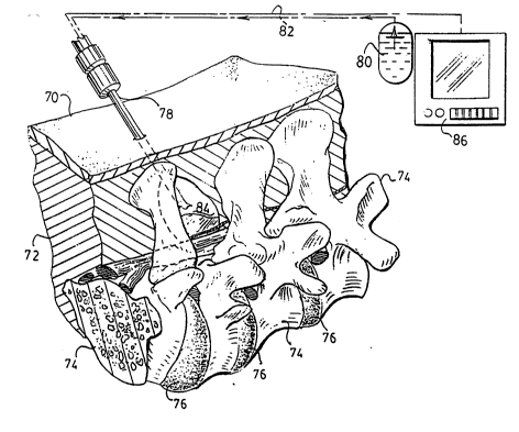
In Figure 1 the outer skin of the patient is indicated at 70 and
muscle and other tissue is indicated at 72. The bone of the spinal
column is indicated at 74 and spinal discs are indicated at 76. The
initial step in the procedure is to insert a cannula 78 which is of the
type illustrated in Figures 9 and 10 through the skin, tissue, and
muscle into an area adjacent the spinal column. A viewing scope
will be passed through the cannula and fluid will be supplied from a
source 80 along a line 82 so that fluid passes through the cannula
80 to an area 84 at the end of the inserted cannula. The surgeon
may utilise a \/iewing screen 86 to have a full picture of the area in
which the interior end of the cannula is working. The purpose of the
10 Z117220
cutting or tissue moving end of the cannula is so that the surgeon
may manipulate it to create the working space 84. This is done by
moving muscle and/or tissue rather than cutting it and the pressure
of the fluid which is utilised in the viewing scope, which is of a type
conventional in arthroscopic surgery, will maintain the space once it ~-
has been created. The pressure of the fluid within space 84 will
keep or maintain the muscle and tissue away from the area in which
the surgeon wishes to work.
Once the space 84 has been created as described, a second :
cannula 88, having an internal diameter slightly greater than the
cannula 78, is inserted as illustrated particularly in Figure 2. The
space 84 which was created as described above exposes the
ligamentum flavum illustrated at 90 in Figure 2 and the next step in
the procedure is for the surgeon to insert a cutting tool, such as that
illustrated at 92, having a cutting end 94 through working cannula
88. Ligamentum flavum is an elastic tissue which spans the space
between adjacent vertebrae as particularly illustrated in Figure 2.
The cutting element or curette 92 will incise the ligamentum flavum,
for example by making a slit at the superior edge of the inferior :
lamina. Once this slit has been made, the Kerison rongeur suction
punch 95 illustrated in Figures 5 - 8 will be inserted through cannula
88, as illustrated in Figure 3, to remove sufficient portions of the
ligamentum flavum to expose the bone beneath it. In some
instances it may be necessary to use the Kerison rongeur suction
punch to actually remove portions of bone, as what is required is
that the ligamentum flavum and/or bone be removed to a sufficient
extent to expose the spinal nerves indicated generally at 96. Note
particularly the opening 98 in the ligamentum flavum in Figure 3.
At this point in the procedure the disc is accessible to the
surgeon and the herniated portion of the disc indicated at 100 can
be removed. The first step in removing the sequestered fragment
100 of the disc is to move nerves 96. This is done by inserting an
instrument 102, illustrated in Figure 4, through cannula 88 and
gently slipping the hooked end 104 beneath the nerves and moving
the nerves a sufficient distance to provide complete access to the
sequestered portion 100 of the disc. Once the nerves have been so
. : ~ , , .
. . .
- - .:. :
:
.
. . . . ..
11 211 7220
moved, a third cannula 105 is utilised. This cannula may be
inserted at any point in the procedure once the nerves and bone
have been exposed by removal of the necessary portion of the `
ligamentum flavum. Cannula 105, again a working cannula and of
5 essentially the same internal diameter as cannula 88, will provide an
access passage for a grabbing or clamping instrument 106 which
has an operating end 108 of the type to grasp the sequestered
portion 100 of the herniated disc and remove it. The herniated
portion of the disc is then withdrawn through cannula 105. The
10 relative positions of the three cannulas will vary depending upon the
exact location of the damaged area of the spinal column. The
positions shown in Figures 1 - 4 are merely illustrative.
Approximately 80 percent of herniated discs are sequestered
15 which means that the herniated portion has actually broken away
from the body of the disc. Even in those instances in which the
herniated portion is not sequestered, it still may be removed as
described. In some instances it may be necessary, prior to
removing the hemiated portion of the disc, to use a knife again
20 inserted through the third cannula 105, to excise any tissue which
may be overlying the disc. The important point, however, is that all
of the described steps in the surgical procedure are performed
arthroscopically through the described cannula passages and the
various tools which may be necessary to first expose the nerves,
25 then move the nerves, and then grasp the hemiated portion of the
disc, will all be utilised in the cannula passages described.
Once the steps described above have been completed and the
hemiated portion of the disc has been removed, all that remains is
30 for the surgeon to withdraw the cannulas and suture the puncture
wounds which were the only invasions of the body necessary for the
entire surgical procedure.
Of importance in the procedure described is the minimal
35 movement of body tissue and muscle and the lack of any incising or
cutting of body tissue and muscle. This substantially reduces
rehabilitation time and will permit the operation to be performed on
an outpatient basis~
``' ' ; . : ~`
: . ' :~.
12 117%20
Although the procedure has been described in connection with `
a laminectomy, it should be clear to one s.~llled in the art that once
the area of the bone is exposed as described, bone particles and/or
bone segments for a fusion may also be inserted through a cannula
and properly positioned for that type of procedure. Again, the
procedure is not limited to access of the lumbar region of the spine,
but may be equally utilised in the cervical or thoracic areas of the ;
spine.
1 0
Whereas the preferred form of the invention has been shown
and described herein, it should be realised that there may be many
modifications, substitutions and alterations thereto.
`` . , ~ ' : ,,,`':
~ . . .
~ ,, ~ ,,,., ' ' ~, ...