Note: Descriptions are shown in the official language in which they were submitted.
WO 93/05832 PCT/US92/0762~
21 1869 3
AUTOMATIC CANNULATION DEVICE
BACKGROUND
TECHNICAL FIELD
This invention relates to blood vessel cannulation
devices and is particularly directed to automatic means for
catheter placement within blood vessels.
BACKGROUND ART
Cannulating blood vessels is a common procedure in
patient care, in order to administer fluids, drugs, blood
or blood products. Heretofore, there have been two basic
types of catheter for accomplishing cannulation. In one
instance, the needle is within the catheter: while in the
other instance, the catheter is within the needle. In both
cases, the needle serves to penetrate the skin and the wall
of the blood vessel and, once the blood vessel has been
entered, the catheter is advanced manually until an ade-
quate position is reached. Unfortunately, such manual
catheter placement involves both of the operator's hands;
one for stabilization of the needle, and the other for
advancement of the catheter. Furthermore, manual catheter
placement is an extremely delicate procedure which can be
SUBSTITUTE SHEET
PCT/US92/07625
WO 93/05832
2
performed only by specially trained and highly skilled
medical personnel and, even then, placement failure is not
uncommon, due to such factors as failure to recognize
penetration of the blood vessel, sequence delays, disrup-
tion of the continuity of the blood vessel, patient anatom-
ical variability, etc.
A search in the United States Patent Office has
revealed the following references:
PATENT NO. INVENTOR ISSUED
4,767,407 S.J. Foran Aug. 30, 1988
4,904,240 R.L. Hoover Feb. 27, 1990
4,944,728 M.W. Carrell et al Jul. 31, 1990
4,966,589 J.M. Kaufman Oct. 30, 1990
Each of these references requires manual advancement
of the catheter and, hence, is subject to the disadvantages
discussed above. Thus, none of the prior art catheter
placement devices has been entirely satisfactory.
BRIEF SUMMARY AND OBJECTS OF INVENTION
These disadvantages of prior art catheter placement
devices are overcome with the present invention and an
SUBSTITUTE SHEET
WO 93/05832 PCT/US92/0762~
3 2~ ~ ~~~3
improved catheter placement device is proposed which auto-
matically advances the catheter, once the blood vessel has
been penetrated, and which may be triggered one-handedly
or, in a preferred embodiment, includes means for sensing
penetration of a blood vessel and for automatically advanc-
ing the catheter in response to such penetration.
The advantages of the present invention are prefera-
bly attained by providing an improved catheter placement
device comprising a needle, a catheter concentric with the
needle, resilient means urging said catheter to an advanced
position, and means for triggering the resilient means upon
penetration of the wall of a blood vessel. The triggering
means may be manual or may include means for sensing pene-
tration of a blood vessel and for automatically advancing
the catheter in response to such penetration.
Accordingly, it is an object of the present invention
to provide an improved catheter placement device.
Another object of the present invention is to provide
an improved catheter placement device which permits one-
handed insertion and placement of a catheter.
An additional object of the present invention is to
provide an improved catheter placement device which permits
automatic advancement of the catheter, once a blood vessel
has been penetrated.
SU9STITllTF SHEET
CA 02118693 1999-07-26
4
A specific object of the present invention is to
provide an improved catheter placement device comprising a
needle, a catheter concentric with the needle, self-pro-
pelled, automatic means for catheter advancement, and means
for triggering said means for self-propelled advancement
upon penetration of the wall of a blood vessel. The trig-
gering means may be manual or may include means for sensing
penetration of a blood vessel and for automatically advanc-
ing the catheter in response to such penetration._.
These and other objects and features of the present
invention will be apparent from the following detailed
description, taken with reference to the figures of the
accompanying drawings.
BRIEF DESCRIPTION OF THE DRAWING
FIGURE 1 is a transverse section through a catheter
placement device embodying the present invention:
FIGURE 2 is a view, similar to that of FIG. 1, show-
ing the catheter placement device is its unlocked position:
FIGURE 3 is a vertical section through the catheter
placement device of FIG. 1, taken on the line Iv-IV of FIG. 1;
FIGURE 4 is a plan view of the latch member of the
WO 93/05832 PGT/US92/0762~
2lId~~3
catheter placement device of FIG. 1;
FIGURE 5 is a transverse section through an alterna-
tive form of the catheter placement device of FIG. 1, shown
in the unarmed condition;
FIGURE 6 is a view showing the catheter placement
device of FIG. 5 in its armed condition, after penetration
of the skin, but prior to penetration of a blood vessel;
FIGURE 7 is a view showing the catheter placement
device of FIG. 5 immediately following penetration of a
blood vessel;
FIGURE 8 is a view showing the catheter placement
device of FIG. 5 following advancement of the catheter;
FIGURE 9 is transverse section through another
alternative form of the catheter placement device of
FIG. 1, shown in its unarmed condition;
FIGURE 10 is a view showing the catheter placement
device of FIG. 9 in its armed condition, after penetration
of the skin, but prior to penetration of a blood vessel;
FIGURE 11 is a view showing the catheter placement
device of FIG. 9 immediately after penetration of a blood
vessel;
SUBSTITUTE SHED'
WO 93/05832 PCT/US92/07625
~1~~~~~3
6
FIGURE 12 is a view showing the catheter placement
device of FIG. 9 following advancement of the catheter;
FIGURE 13 is a transverse section through a further
alternative form of the catheter placement device of FIG.
1, shown in its unarmed condition;
FIGURE 14 is a view showing the catheter placement
device of FIG. 13 in its armed condition, after penetration
of the skin, but prior to penetration of a blood vessel;
FIGURE 15 is a view showing the catheter placement
device of FIG. 13 in its armed condition, after penetration
of the skin, but prior to penetration of a blood vessel;
FIGURE 16 is a view showing the catheter placement
device of FIG. 13 following advancement of the cathe-
ter;
FIGURE 17 is a view, similar to that of FIG. 1,
showing another alternative form of the catheter placement
device of FIG. 1;
FIGURE 18 is a view, similar to FIG. 5, showing
another alternative form of the catheter placement device
of FIG. 5;
SUBSTiTU'1'~ ~NE~
WO 93/05832 PCT/US92/0762~
211~G93
FIGURE 19 is a view, similar to that of FIG. 5,
showing an additional alternative form of the catheter
placement device of FIG. 5~
FIGURE 20 is a view, similar to FIG. 9, showing a
further alternative form of the catheter placement device
of FIG. 9 ;
FIGURE 21 is a view, similar to that of FIG. 20,
showing the catheter placement device of FIG. 20 in its
"armed" position;
FIGURE 22 is a view, similar to that of FIG. 9,
showing another alternative form of the catheter placement
device of FIG. 9;
FIGURE 23 is a view, similar to that of FIG. 22
showing the catheter placement device of FIG. 21 in its
"armed" position;
FIGURE 24 is a view, similar to that of FIG. 22
showing the catheter placement device of FIG. 22 immediate-
ly after penetration of a blood vessel;
FIGURE 25 is a view, similar to that of FIG. 22
showing the catheter placement device of FIG. 22 immediate-
ly after release of the catheter;
SUBSTITUTE SHE~1'
WO 93/05832 PCT/LS92/0762s
~~~~s~~
8
FIGURE 26 is a view, similar to that of FIG. 9,
showing an additional alternative form of the catheter
placement device of FIG. 9:
FIGURE 27 shows an alternative form of the catheter
placement device with an automatic arming rod:
FIGURE 28 shows a cross-section of the automatic
arming rod of FIG. 27;
FIGURE 29 is a view, similar to that of FIG. 27,
showing the catheter placement device of FIG. 27 after skin
penetration and the beginning of the automatic arming
process;
FIGURE 30 is a view, similar to that of FIG. 27,
showing the catheter placement device of FIG. 27 after skin
penetration, in a further stage of the arming process;
FIGURE 31 is a view, similar to that of FIG. 27,
showing the catheter placement device of FIG. 27 immediate-
ly after penetration of a blood vessel;
FIGURE 32 is a view, similar to that of FIG. 27,
showing the catheter placement device of FIG. 27 immediate-
ly after release of the catheter;
FIGURE 33 is a view, similar to that of FIG. 27,
S~~BST1TUT~ SHED
r .._.
WO 93/05832 PCT/US92/0762~
9 2I1~~~~
after the removal of the needle, showing connection of the
catheter with the intravenous tubing;
FIGURE 34 shows an additional alternative form of the
catheter placement device;
FIGURE 35 is a view, similar to that of FIG. 34,
showing the catheter placement device of FIG. 34 immediate-
ly after skin penetration;
FIGURE 36 is a view, similar to that of FIG. 34,
showing the catheter placement device of FIG. 34, immedi-
ately after release of the catheter:
FIGURE 37 is a view, similar to that of FIG. 9,
showing an additional form of the catheter placement device
of FIG.9;
FIGURE 38 is a view, similar to that of FIG. 5,
showing an alternative form of the catheter placement
device of FIG. 5 prior to skin penetration:
FIGURE 39 is a view, similar to that of FIG. 38,
showing the catheter placement device of FIG. 38 immediate-
ly after skin penetration;
FIGURE 40 is a view, similar to that of FIG. 38,
showing the catheter placement device of FIG. 38 immediate-
SIIBSTITUTE SHEET
PCT/US92/0762~
WO 93/05832
211~~~
1~
ly after blood vessel penetration;
FIGURE 41 is a view, similar to that of FIG. 38,
showing the catheter of the catheter placement device of
FIG. 38 placed intravenously;
FIGURE 42 shows the separate four components of a
further alternative form of the catheter placement device;
FIGURE 43 shows the device of fig 42 while being
assembled;
FIGURE 44 shows a further stage on the assembly of
catheter placement device of fig. 42;
FIGURE 45 shows the assembled unit of fig.42 prior to
skin penetration;
FIGURE 46 shows the assembled unit of fig.42 after
penetration into a blood vessel;
FIGURE 47 shows the disengaging of the propelling
unit of fig. 42 from the catheter placed intravenously;
FIGURE 48 shows an alternative form of the catheter
placement device of fig.5;
FIGURE 49 shows a top view of the same catheter
SUBSTITUTE SHEET
r ~.
WO 93/05832 PCT/US92/07625
2I~~6J3
11
device of fig.48;
FIGURE 50 shows the catheter placement device of
fig.48 after blood vessel penetration;
FIGURE 51 shows an alternative form of a semi-auto-
matic catheter placement device;
FIGURE 52 shows the catheter placement device of
fig.51 after blood vessel penetration.
FIGURE 53 shows yet an alternative form of a fully
automatic catheter placement device, where the means for
sensing penetration are represented by an opto-electric
source-sensor pair.
FIGURE 54 show an alternative form of the device of
fig.53 where the represented sensors are temperature sen-
sors.
FIGURE 55 show an alternative form of the device of
fig. 53 where the represented sensors are sensors detecting
the electrical conductivity properties of the blood.
FIGURE 56 shows an alternative form of the device of
fig.53 in which the represented sensor is a flow detector
sensor.
SUBSTITUTE SNFET
WO 93/05832
211~~i93
PCT/US92/0762
12
FIGURE 57 shows an alternative device of fig.53,
where the represented sensor is an acoustic sensor.
FIGURE 58 shows yet an alternative form of the device
of fig.53 in which the represented sensor is a pressure
sensor.
FIGURE 59 shows yet an alternative form of the device
of fig.53 in which the represented sensor is an illustra-
tion of a sensor of chemical properties of the blood, such
as a pH sensor, or of a sensor capable of sensing presence
of blood by detecting presence of a component of the blood,
such as oxygen.
DETAILED DESCRIPTION OF THE INVENTION
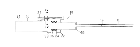
In that form of the present invention chosen for pur-
poses of illustration in FIG. 1, a catheter placement
device, indicated generally at 10, is shown comprising a
needle 12, having a tubular hollow shaft 14 projecting from
a handle portion 16, and a flexible catheter 18 projecting
from a hub 20. As shown, the catheter 18 concentrically
encircles the shaft 14 of the needle 12, while the catheter
hub 20 telescopes over the forward portion of the needle
handle portion 16. However, it will be apparent to those
skilled in the art that, if desired, the needle 12 may be
SUBSTITUTE SHEET
i~lS9Z/ 07 b Z ~
21 186 9 3 13 . -- v. ~?~F~3~~3
made to concentrically telescope over the catheter 18. The
catheter hub 20 has a radial flange 22 formed adjacent the
rear end 24 of the hub 20 and has a lever 26, formed of
resilient material, projecting rearwardly from one side of
the flange 22 and having an opening 28 formed adjacent the
free end of the lever 26, as best seen in FIG. 4. A bridge
member 30 projects radially outward from the needle handle
16 and, as best seen in FIG.3, has an aperture 32 formed
therein to receive the lever 26 and has a stud 34 project-
ing into the aperture 32 which is mateable with the opening
28 of the lever 26 to releasably retain the lever 26 and,
hence, the hub 20 and catheter 18 in the position shown in
FIG. 1. Finally, resilient means, such a spring 36, is
mounted between the rear end 24 of the catheter hub 20 and
the front end 38 of the handle portion 16 of the needle 12
to urge the hub 20 and catheter 18 forwardly.
In use, the catheter 18 and needle 12 are in the
positions seen in FIG. 1, with the catheter 18 encircling
the shaft 14 of the needle 12 and with opening 28 of lever
26 of the catheter 18 engaging the stud 34 of the bridge
member 30 on the handle portion or needle hub 16 of the
needle 12 to releasably lock the catheter 18 in the posi-
tion shown in FIG. 1. When the shaft 14 of the needle 12
penetrates a desired blood vessel, the operator presses the
lever 26 toward the handle portion 16 of the needle 12,
causing opening 28 of the lever 26 to disengage from the
stud 34 and allowing spring 36 to drive the catheter 18
forwardly and, thereby, to advance and position the cathe-
SI~BS~tTUTE SHEET
I~EAIUS
~"' ~~~J:~~ ~~~~~
iF~~~~L 13 APR 1993
ter ~8 - 2 1 18 6 9 3
SUBS T tT~~T~ S~ ~~~~'
IPF~/US
WO 93/05832 PC1'/US92/0762~
~11~69~
14
within the blood vessel. Thereafter, the needle 12 may be
removed and the tubing containing the food, drugs or other
desired material may be attached to the hub 20 of the
catheter 18 for delivery of the desired material into the
patient's blood vessel. It will be apparent that the lever
26 can be operated by the same hand which is holding the
needle handle 16, thus, enabling the operator to position
the needle 12, advance and place the catheter 18 and with-
draw the needle 12 in a one-handed operation, leaving the
operator's other hand free for other purposes.
FIGURES 5-8 show an alternative form, indicated
generally at 39, of the catheter placement device 10 of
FIG. 1 which serves to automatically advance the catheter
when the blood vessel is penetrated. In this form of the
present invention, the catheter 40 encircles the needle
shaft 42 and has a hub 44 formed with an annular recess 46
extending about the interior surface of the hub 44. The
needle 42 has a generally cylindrical hub 48 formed with
openings 50 in the side walls 52 of the needle hub 48 to
receive balls 54, to releasably lock the catheter hub 44 in
the position shown in FIG. 5, as will be more fully ex-
plained hereafter. The rear end 56 of the needle hub 48
carries a radial flange 58 and a cylindrical wall 60 ex-
tends forwardly from the flange 58 to encircle and guide
movement of the catheter hub 44, which is telescopically
SU6STIT1JTE SHEET
WO 93/05832 PCT/US92/0762~
2~~~~~~
slideably between the needle hub 48 and the cylindrical
wall 60. A spring 59 is mounted within the cylindrical wall
60 rearwardly of the catheter hub 44 and serves to urge the
catheter hub 44, and consequently, the catheter 40 forward-
ly. A wall member 62 divides the interior of the needle hub
48 and defines a forward chamber 64 and a rearwardly open-
ing recess 66. Within the forward chamber 64, a piston 68
is slideably mounted and has an annular recess 70 extend-
ing about the periphery of the piston 68, which serves to
receive the balls 54 when the catheter 40 is released, as
hereinafter described. A spring 72 is located between the
wall member 62 and the piston 68 to normally urge the
piston 68 to seat against the forward end of the chamber
64, as seen in FIG. 5, so that the body 74 of the piston 68
serves to force the balls 54 to extend through openings 50.
to engage the recess 46 of the catheter hub 44 and, hence,
to releasably lock the catheter hub 44 in the position
shown in FIG. 5, against the urging of spring 59. The wall
member 62 has a central opening-76 and a post-needle member
78 is mounted in the opening 76 and projects rearwardly as
shown. A capsule 80 is slideably mountable within the
rearwardly opening recess 66 of the needle hub 48 and has a
plug 82 closing the open end 84 of the capsule 80. The
capsule 80 contains a vacuum and the plug 82 is rupturable,
when pressed against the post-needle member 78, to cause
partial retraction of the piston 68, as more fully de-
scribed below.
FIG. 5 shows the catheter placement device 39 in
~t 1~STITUTE SHEET
.-".~" . , __ ~ . 5
21 1 8 6 9 3 16 lPEAIUS ~ 3 APR 1993
preparation for use, but prior to insertion. FIG. 6 shows
the catheter placement device 39 after the needle 42 has
penetrated the skin 86 of a patient, but prior to penetra-
tion of a blood vessel 88. Once the needle 42 has entered
the skin 86, the operator presses the vacuum capsule 80
forwardly, causing the post-needle member 78 to pierce the
plug 82. This causes air from the forward vacuum chamber 64
of the needle hub 48 to enter the capsule 80, which creates
a partial vacuum within the chamber 64 and serves to par-
tially retract piston 68 against the action of spring 72.
This, in turn, creates a vacuum within the chamber 64
forwardly of the piston 68, as seen at region 90. However,
the body 74 of the piston 68 still serves to force the
balls 54 into recess 46 to continue locking the catheter
hub 44 in its retracted position, as seen in FIGS. 5 and 6.
The catheter placement device 39 is now "armed" to automat-
ically advance the catheter 40.
In FIG. 7, the needle 42 has penetrated the wall 89
of the patient's blood vessel 88. The instant that such
penetration occurs, blood from the blood vessel 88 is drawn
into region 90 by the vacuum in the region 90. This drives
the piston 68 rearwardly, against the action of spring 72,
to the point that the annular recess 70 of the piston 68
becomes aligned with openings 50 of the side walls 52 of
the needle hub 48. Consequently, balls 54 can move into the
recess 70 of the piston 68 and out of the recess 46 of the
catheter hub 44. When this occurs, spring 59 drives the
catheter hub 44 and catheter 40 forwardly, automatically,
:,-,. ,
_ ... _.- , :._ ,~:, :y..:,
. , Ji
21 186 9 3 17 ,
v~:~~~':' ~~ ri~R 193
without any actionby the operator, to advance and place
the catheter 40, seen in FIG. 8.
as
In use, the catheter placement device 39 is initially
in the position shown in FIG. 5, with the catheter 40
retracted and with piston 68 urged forwardly by spring 72,
causing balls 54 to pass through openings 50 in the side
walls 52 of the needle hub 48 to enter recess 46 of the
catheter hub 44 and, hence, to lock the catheter 40 in its
retracted position. Once the operator has inserted the
needle 42 into the skin 86 of the patient, the operator
presses the vacuum capsule 80 forward, causing the post-
needle member 78 to penetrate the plug 82. This causes air
from chamber 64 of the needle hub 48 to enter the vacuum
capsule 80 and serves to partially retract the piston 68 to
create a vacuum within region 90 forward of the piston 68,
as seen in FIG. 6, and thereby "arming" the catheter place-
ment device 39. Subsequently, the instant the needle 42
penetrates the patient's blood vessel 88, as seen in FIG.
7, blood from the blood vessel 88 is drawn into region 90
of the needle hub chamber 64. This backflow of blood drives
the piston 68 rearwardly, against the action of spring 72,
until annular recess 70 of the piston 68 becomes aligned
with openings 50 in the side walls 52 of the needle hub 48,
which allows balls 54 to move out of recess 46 of the
catheter hub 44. This automatically unlocks the catheter
hub 44 and allows spring 59 to advance the catheter 40, as
seen in FIG. 8. Because the advancement of the catheter 40
occurs automatically and instantly, in response to penetra-
SUSSTtTUTE SHEET
IPEA/US
T~IIi'~wr- , ~
1~ ~' 1
iFEAI~~ 1 ~ a~~ X993
tion of the
21 1$69 3
~~~:~ ~~f i~;jr~ ~ ~~~~.C~r
IP~~'US
I~'C T' ~~ S '~ ~ / l~ ~ ~ ~ ~
21 1 8 6 9 3 1$ IPEAIUS 13 APR 1993
blood vessel 88 by the needle 42, proper placement of the
catheter 40 is assured and overpenetration or underpenetra-
tion are avoided. Furthermore, pressing of the vacuum
capsule 80 against the post-needle member 78, to "arm" the
catheter placement device 39, can be accomplished by the
operator as a one-handed operation. Thus, the catheter
placement device 39 provides simple and automatic, yet
highly accurate placement of the catheter 40.
FIGURES 9-12 show another alternative form, indicated
generally at 92, of the catheter placement device 10 of
FIG.1 comprising a needle shaft 94 projecting from a gener-
ally cylindrical hub 96 having a flange 98 extending radi-
ally outward from the rear end 100 of the needle hub 96 and
having a cylindrical sleeve 102 extending forwardly from
the flange 98 and spaced from the side wall 103 of the
cylindrical hub 96 to define a space 104 therebetween. A
flexible tubular catheter 106 encircles the needle shaft 94
and projects forwardly from a generally cup-shaped catheter
hub 108 having a side wall 110 which is telescopically
slideable within the space 104 of the needle hub 96. A
spring 112 is positioned within the space 104 between the
flange 98 of the needle hub 96 and the rear end of the side
wall 110 of the catheter hub 108 and serves as a self-
propelling means to urge the catheter hub 108 forwardly.
The side wall 110 of the catheter hub 108 is formed with an
internal annular recess 114
. ~.~ ,,~ ~r . , , ~ .,
_ ,. . . , . r . ; , : s .:~. .. t
..
_.,__,.. _,
2118693 19 ~ ~, ~~,~~,,,,~
and the side wall 103 of the cylindrical needle hub 96 are
formed with openings 116 and balls 118 are seated in the
openings 116 and extend into the recess 114 to releasably
lock the catheter hub 108 in its retracted position and in
turn actuate the self-propelling means 112, as seen in
FIGS. 9, 10 and 11. A piston 120 is slideably mounted
within vacuum chamber 148 delimited by the cylindrical
needle hub 96 and is formed with an annular recess 122,
located adjacent the forward end of the piston 120, and an
axial recess 124, extending forwardly from the rear end 125
of the piston 120. The needle hub 96 has a stud 126 pro-
jecting radially outward adjacent the rear end 100 of the
needle~hub 96 and a trigger member or arming means 128
encircles the stud 126 and is slideably mounted thereon.
The trigger member 128 is formed with a pair of forwardly
projecting flanges 130 and 132. Flange 130 extends forward-
ly from the inner edge 134 of the rear surface 136 of the
trigger member 128 and a spring 127 is seated between the
stud 126 and the trigger member 128 to urge the trigger
member outward causing flange 130 of the trigger member 128
to normally engage the rear end 125 of the piston 120, as
seen in FIG. 9, and serving to normally prevent rearward
movement of the piston 120. Flange 132 extends forwardly
from the inner edge 138 of the front surface 140 of the
trigger member 128. A latch member 142 is supported by a
resilient stem 143, which projects outwardly from the outer
surface of the sleeve 102 of the needle hub 96 adjacent
flange 132 of the trigger member 128. The latch member 142
SUBSTITUTE SHEET
IPEAlUS
_; o ,
.. J , v
2 1 1 8 6 g 3 19~ 2 _:,. ~,_ 1 ~ h~ R 1~9d
has an inclined rear surface 144, which is normally posi-
tinned to partially underlie the end of flange 132 of the
>~BSTtr~~v~ ~~,~.
~.. . r r_ [C'~"
f~,~~'3~. ~_l,~
~ v
21 1 8 6 9 3 2° .. ;~~~%~~~ 13 ~~R 1993
trigger member 128, as seen in FIG. 9. Finally, a spring
146 is located within the cylindrical needle hub 96 for-
wardly of the piston 120 to normally urge the piston 120
rearwardly.
In use, the catheter placement device 92 is normally
in the condition seen in FIG. 9 with the trigger member 128
urged outwardly by spring 127 urging trigger member 128 to
its outward position wherein flange 130 engages piston 120
to hold the piston 120 in its forward position compressing
spring 146 and forcing balls 118 through openings 116 of
the needle hub 96 into recess 114 of the catheter hub 108
to releasably lock the catheter hub 108 in its retracted
position. Once the operator has caused the needle shaft 94
to penetrate the patient's skin 86, as seen in FIG. 10, the
operator presses the trigger member 128 inward toward the
cylindrical sleeve 102 of the needle hub 96, causing flange
130 of the trigger member 128 to disengage from piston 120
and to enter the axial recess 124 of the piston 120, which
allows spring 146 to drive the piston 120 slightly rear-
ward, as seen in FIG. 10, and creating a vacuum in the
chamber 148 forward of the piston 120 within the cylindri-
cal needle hub 96. At the same time, flange 132 of the
trigger member 128 moves inwardly past the inclined surface
144 of the latch member 142, causing the latch member 142
to cam forwardly on the resilient stem 143. When flange 132
has past the inclined surface 144, the resilient stem 143
returns the latch member 142 to its original position, in
which it now overlies flange 132 to lock the trigger
SUBSTITUTE SHEET
IPEAIUS
, ~. . ~ r ,
20.1 , _
member 21 1 8 fi 9 3 . ~PEA~US I3 RPR 1~!~3~--
SUBS'~~TUTE S~~~T
ip~AIUS
.Cr'S92 / 07 ~ z 5
21 ~ . ~~~~'~~~ 13 APR 1993
21 1869 3
128 in its "armed" position, as seen in FIGS. 10, 11 and
12. When the operator causes the needle shaft 94 to pene-
trate the wall 89 of a blood vessel 88, blood from the
blood vessel is instantly drawn into the space 148 within
the needle hub 96, due to the vacuum created therein when
spring 146 drove the piston 120 to its partially retracted
position, as seen in FIG. 10. The vanishing of the vacuum
due to entering or backflow of the blood in the space 148
forces the piston 120 to its fully retracted position, as
seen in FIG. 11, which causes the annular recess 122 of the
piston 120 to become aligned with openings 116 of side wall
103 of the needle hub 96 and allowing balls 118 to move out
of the annular recess 114 of the side wall 110 of the
catheter hub 108. This unlocks the catheter hub 108 and
actuates the spring or the self-propelling means 112 to
drive the catheter hub 108 forward to advance and place the
catheter 106, as seen in FIG. 12. Again, it will be appar-
ent that the operator can use one hand to grasp and posi-
tion the catheter placement device 92, during insertion of
the needle 94 into the patient's skin 86 and to actuate the
trigger member 128 to "arm" the catheter placement device
92 prior to penetration of the blood vessel 88, so that the
catheter placement device 92 can instantly and automatical-
ly advance and place the catheter 106 upon penetration of
the blood vessel 88, without additional effort by the
operator.
FIGURES 13-16 show another alternative form, indicat-
suBSTt~r~~~ s~~Fz
IP~A,r~ss
2 1 1 8 6 9 3 21 ~~= ~ IFEA/US 13 APR 1993
~~""~?'t~S 9 2 / 0 7 r ? ~
ed generally at 150, of the catheter placement device 10 of
,. .
_.
::~ . ~a..
PCT/US92/0762s
WO 93/05832
22
FIG. 1 comprising a needle shaft 152 projecting from a
generally cylindrical hub 154 having a flange 156 extending
radially outward from the rear end 158 of the needle hub
154 and having a cylindrical sleeve 160 extending forwardly
from the flange 156 and spaced from the cylindrical hub 154
to define a space 162 therebetween. A flexible tubular
catheter 164 encircles the needle shaft 152 and projects
forwardly from a generally cup-shaped catheter hub 165
having a side wall 166 which is telescopically slideable
within the space 162 of the needle hub 154. A spring 168 is
positioned within the space 162 between the flange 156 of
the needle hub 154 and the rear end of the side wall 166 of
the catheter hub 165 and serves to urge the catheter hub
165 forwardly. The side wall 166 of the catheter hub 165 is
formed with an internal annular recess 170 and the cylin-
drical needle hub 154 is formed with openings 172 and balls
174 are seated in the openings 172 and extend into the
recess 170 to releasably lock the catheter hub 165 in its
retracted position, as seen in FIGS. 13 and 14. A piston
176 is slideably mounted within the cylindrical needle hub
154 and is formed with an annular recess 178, located
adjacent the forward end of the piston 176, and a rearward-
ly-opening recess 180, extending forwardly from the rear
end 182 of the piston 176. A generally U-shaped capsule 184
is slideably mountable within the rearwardly-opening recess
180 of the piston 176 and has a plug 186 closing the open
end 188 of the capsule 184. The capsule 184 contains a
vacuum and the plug 186 is rupturable, when pressed against
SUBSTITUTE SHEc''
21 1869 3 23 ~ v ~V~~ ?~~J ' ~i~
~:~, . 1 _~ r., R '~
,.~~
~.
.. a post-needle member 190 carried by the piston 176 and
projecting rearwardly therefrom, to create a vacuum within
the space 194 which "arms" the catheter placement device
150. When the operator has inserted the needle shaft 152
into the skin of the patient, the operator presses the
vacuum capsule 184 inward to cause the post-needle member
190 to pierce the plug 186 to "arm" the catheter placement
device 150. Subsequently, the instant the needle 152 pene-
trates the wall 89 of the blood vessel 88, the vacuum in
the space 194 causes blood to backflow into the space 194
and the vanishing of the vacuum due to the blood entering
the space 194 drives the piston 176 to its fully retracted
position, as seen in FIG. 15, wherein the annular recess
178 of the piston 176 is aligned with openings 172 in the
side wall of the needle hub 154, which allows the balls 174
to move out of the annular recess 170 in the side wall 166
of the catheter hub 165. This "unlocks" the catheter hub
165 and a actuates spring 168 to drive the catheter hub 165
forward to advance and place the catheter 164 within the
blood vessel 88, as seen in FIG. 16. Here, again, the
operator can use one hand to insert the needle 152 into the
patient's skin 86 and to press the vacuum capsule 184
inward to "arm" the catheter placement device 150. Subse-
quently, upon penetration of a blood vessel 88, the cathe-
ter placement device 150 will, instantly and automatically,
advance and place the catheter 164 without any additional
action by the operator.
SUt7STq ~' '' ~ '° y ~ sr r~"~
f ~ .''.r . ~, ~.;1"~:~
I
. , .~ '"' ; _~
21 1 8 6 9 3 ; 24 ~ ?P~A~!T,» 13 ASR 1993
FZG. 17 shows an alternative form, indicated general-
ly at 196, of the catheter placement device 10 of FIG. 1.
The catheter placement device 196 is substantially identi-
cal with that of the catheter placement device 10 of FIG.
1, except that the spring 36 of the catheter placement
device 10 is replaced by a pair of magnets 198 and 200 as a
self-propelling means mounted with like poles in opposing
relation, as indicated by arrows 202. In this way, when
lever 26 is released, the magnets 198 and 200 will serve to
drive the catheter hub 20 away from the front end 38 of the
needle 12 and, hence, will serve to automatically advance
the catheter 18.
FIG. 18 shows an alternative form, indicated general-
ly at 204, of the catheter placement device 39 of FIG. 5.
The catheter placement device 204 is substantially identi-
cal with that the catheter placement device 39 of FIG. 5,
except that the spring 59 of the catheter placement device
39 is replaced by a quantity of compressed gas as a self-
propelling means which is supplied from a suitable source,
such as capsule 206. In use, the catheter placement device
204 functions in substantially the same manner as the
catheter placement device 39, except that when the catheter
hub 44 is unlocked the catheter 40 is automatically ad-
vanced by expansion of the compressed. gas from capsule 206,
rather than by expansion of spring 59.
FIG. 19 shows another alternative form, indicated
SUBSTITUTE SHEET
IPF n ~° "~''
. s9~/076C5
21 18 6 9 3 r 2 4 ; ~ I . ~P~A~JS 13 A P R 1993
generally at 208, of the catheter placement device 39 of
~l!BST~ ~ ~~ ~ ~ SHEET
1~~~=J"t1S
WO 93/05832 PCT/US92/07625
''2~ '
FIG. 5. The catheter placement device 208 is substantially
identical with that the catheter placement device 39 of
FIG. 5, except that the spring 59 of the catheter placement
device 39 is replaced by a pair of magnets 210 and 212
mounted with like poles in opposing relation, as indicated
by arrows 214. In use, the catheter placement device 208
functions in substantially the same manner as the catheter
placement device 39, except that when the catheter hub 44
is unlocked, the magnets 210 and 212 will serve to automat-
ically drive the catheter 40 forward for placement, rather
than by expansion of spring 59.
FIGURES 20 and 21 show a further alternative form,
indicated generally at 214, of the catheter placement
device 92 of FIG. 9. The catheter placement device 214 is
substantially identical with that of the catheter placement
device 92 of FIG. 9, except that the locking balls 118,
openings 116 and advancing spring 112 of the catheter
placement device 92 of FIG. 9 have been omitted. Instead, a
cylindrical chamber 216 is provided adjacent the rear end
100 of the needle hub, projecting inwardly from the side
wall 103 of the needle hub 96. The cylinder 216 has an
opening 218 facing toward the rear end 125 of the piston
120 and a piston 220 is slideably mounted in the opening
218. The rear end of the cylinder 216 is connected to a
generally U-shaped duct 222 and a shaft 224 is carried by
the rear end of the catheter hub 108 and projects into the
end 226 of the duct 222. The cylinder 216 and duct 222 are
filled with a suitable hydraulic fluid 228.
_~ ..__~. ~( 1RSTITI!ITF S!-i~E'T __ .
r~~i ~ ~t~~ v ~ '--
2 1 1 8 6 9 3 26 ~~~~~L 13 APR 1993
In use, when button 128 is pressed inwardly, it
releases piston 120, which is driven partially rearward by
spring 146. However, as the piston 120 moves rearwardly, it
creates a vacuum within space 148 which prevents full
rearward movement of the piston 120. Subsequently, penetra-
tion of a blood vessel allows blood to backflow into cham-
ber 148 and the vanishing of the vacuum due to the entering
of the blood into chamber 148 serves to drive the piston
120 rearward. The piston ring or sealing ring 240 assures
that the vacuum in space 148 will be maintained. The
vanishing of the vacuum due to the entering of the blood
into space 148 drives piston 120 farther rearward, which
allows piston ring 240 to expand out of recess 242 in
expanded posterior portion of vacuum chamber 148 and ena-
bles spring 146 to drive piston 120 fully rearward causing
the rear end 125 of piston 120 to engage piston 220 and to
drive the piston 220 rearwardly within cylinder 216. This
forces the hydraulic fluid 228 to flow through duct 222 and
to drive shaft 228 forwardly to release the catheter hub
108.
FIGURES 22, 23, 24 and 25 show yet another alterna-
tive form, indicated generally at 230, of the catheter
placement device 92 of FIG. 9. The catheter placement
device 230 is substantially identical to the catheter
placement device 92 of FIG. 9, except that the locking
balls 118, openings 116 and advancing spring 112 of the
catheter placement device 92 of FIG. 9 have been omitted
Isnwi.'~.~ ~, ~~'T'
i~t1'n~'~ !..~x.y.
i -. .,
, Y ._,~ ~~ ~ r~ V ~.J'
. ~~~S 13 APR ~~ J.
2118693 2'
and the advancing spring 112 is replaced by self-propelling
means comprising a cam 232 which is pivotally mounted in
the side wall 103 of the needle hub 102. The cam 232 has a
trigger portion 234 and an actuator portion 236, which is
engageable with the rear end 238 of the side wall 110 of
the catheter hub 108. Finally, a piston ring 240 is mounted
in a recess 242 of the piston 120 and frictionally engages
the side wall 103 of vacuum chamber or space 148 of the
needle hub 102.
In use, when, after skin penetration, the trigger
button 128 is pressed, it moves arm 130 out of engagement
with piston 120, which allows spring 146 to drive piston
120 partially rearward, to the position seen in FIG. 23.
However, the movement of piston 120 creates a vacuum within
space 148 which opposes the action of spring 146 and limits
the rearward movement of the piston 120. The piston ring
240 assures that the vacuum in space 148 will be main-
tained. Subsequently, penetration of a blood vessel 88 by
the needlg 94, allows blood to flow into space 148, as seen
in FIG. 24. The vanishing of the vacuum due to the entering
of the blood into space 148 drives piston 120 farther
rearward, which allows piston ring 240 to expand out of
recess 242 and enables spring 146 to drive piston 120 fully
rearward to engage the trigger portion 234 of cam 232,
causing cam 232 to pivot and causing the actuator portion
236 of cam 232, as seen in FIG. 25 to drive the rear end
238 of side wall 110 of the catheter hub 108 forward to
release and place the catheter 106.
i
I~~jLTS 9 2 / 0 7 6 2 ~
2 1 1 8 6 9 3 ~ 28 ~ fry=~~~ ~ w
. 1 ~ ~P R x;393
FIGURE 26 shows yet another alternative form, indi-
Gated generally at 244, of the catheter placement device
230 of FIGS. 22, 23, 24 and 25. The catheter placement
device 244 is substantially identical to the catheter
placement device 230 of FIG. 22, except that the cam 232
is omitted and, instead, self-propelling means comprising a
lever 246 is pivotally mounted on the forwardly extending
arm 130 of the trigger button 128 and one side of the
rearwardly extending portion 125 of piston 120 is omitted.
The lever 246 comprises a trigger portion 248, which ex-
tends into the path of movement of the rearwardly extending
portion 125 of piston 120, and an actuator portion 250
which engages the rear end 238 of the side wall 110 of the
catheter hub 108.
In use, the catheter placement device 244 functions
in substantially the same manner as the catheter placement
device 230 of FIGS. 22, 23, 24 and 25. However, when the
blood entering space 146 drives piston 120 rearwardly, the
rearwardly extending position 125 of piston 120 engages the
trigger portion 248 of the lever 246, causing the lever 246
to pivot, driving the trigger portion 248 rearwardly to the
position seen in dotted lines in FIG. 26, and causing the
actuator portion 250 of lever 246 to move forwardly, as
seen in dotted lines in FIG. 26, to drive the rear end 238
of the catheter hub 108 forwardly to~release and place the
catheter 106.
SUBSTITUTE ~HEE~:
Sa2; ~7 6 z 5
29 ~rc~tJ~ 13 APR 1993
21 1869 3 ~1
FIGURES 27 - 33 show a fully automatic form of cathe-
ter placement device, indicated generally at 260; wherein
the means for arming the sensing means which responds to
blood vessel penetration is automatically actuable upon
skin penetration by the needle. The catheter placement
device 260 is essentially similar to the device of FIG. 9
with the following important differences: the arming means,
which in FIG. 9 was represented by the trigger member 128,
is represented in the device 260 by a cylinder or sleeve
302 surrounding the catheter and telescopically slideable
on the catheter, of sufficient thickness, particularly in
correspondence of its distal end 304 to not be permitted to
follow the needle shaft 308 and catheter shaft 306 at the
site of skin penetration, and to be retained as soon as the
distal end of the cylinder is in contact with the skin. The
retention of the cylinder, while the needle and catheter
are being advanced inside the skin, will result in a back-
ward movement of the cylinder relatively to the needle and
the catheter. The backward movement of the cylinder will
force the tooth 298 against a non-steep side of the notch
310 where the tooth was resting in its initial position,
displacing it outward. In turn, the tooth 298 will force
the resilient hook 296 outwardly causing the disengagement
of the hollow piston 272, to which the resilient hook is
anchored, from the edge 320 of a hale 294 formed in the
front end of an anterior fold 322 of the needle hub con-
necting the internal needle hub 324 with the external
needle hub 326.
~L~~i~~f~~~~~ i~c SHE~~'
/ ~ 1
2 9j ~ ~
2 1 1 8 6 9 3 . ~P~S 13 APR 199
The piston 272 , no longer retained, will be urged
backwa-rd
j
_.
CA 02118693 1999-07-26
- 30
by the action of spring 290. However, the movement of
piston 272 creates a vacuum within space or vacuum chamber
328 which opposes the action of spring 290 and limits the
rearward movement of the piston. Subsequently, penetration
of a blood vessel 88 by the needle 94 allows blood to flow
into space 328, as seen in FIG. 31. The pressure of this
blood drives piston 272 farther rearward, which allows
flange 288 of piston 272 to outwardly displace the resil-
ient hook 280, which is anchored to the catheter hub 312,
out of the hole 278 formed in the external needle hub 326.
Resilient hook 280 is the equivalent of the ball members of
FIG. 9. The outward displacement of resilient hook 280 will
result in a disengagement of the catheter hub 312, to which
the hook 280 is anchored, from the needle hub 330. Such a
disengagement will no longer retain the catheter hub 312
from the action of spring 266, and the catheter 306 will be
propelled into an advanced position relatively to the
needle. In FIG. 33, the catheter 306 is shown in its final
intravenous position, after the removal of needle 308 and
needle hub 284, connected to adaptor 263a of intravenous
tubing 263.
FIGURES 34 - 36 show another alternative form of a
catheter placement device, indicated generally at 320. The
catheter placement device 320 is essentially similar to the
device of FIGS. 27 - 33 with the following important dif-
ferences: the self-propelling catheter advancement is
PCT~'uS~2/ 07625
2 1 1 8 6 9 3 31 . ~P~S 13 APR 1993
obtained by the expansion of expandable material 358 and
the omission of catheter automatic arming. The needle hub
342 of the needle 340 contains a chamber 356 filled with a
thermally expandable material 358, such as mercury. Chamber
356 has in its proximal portion a hollow piston 364 with
flanges 366 interposed between the inner face of the cathe-
ter hub 336 and the front of the needle hub 324. A lever
322 with its resilient segment 326 attached to flange 332
of piston 330 protrudes through an opening 372 of catheter
hub 336 and opening 370 of needle hub 342. This lever 322,
in its resting position, locks hollow piston 330 in its
advanced position, not allowing spring 328 to displace the
piston 330 rearwardly. Once the tip of the needle 340
penetrates skin 86, the lever 322 is lifted by the opera-
tor, thus disengaging piston 330 to a rearward position,
resulting in creation of a vacuum within the hollow needle
shaft 340 and spaces 374 and 360. Upon penetration of
needle 340 into a blood vessel 89, blood will rush into the
spaces 374 and 360, via the hollow needle shaft 340. The
blood, at 37 degrees Centigrades or warmer, will cause the
thermally expandable material 358 to expand, provoking the
rapid advancement of piston 364 which, acting upon catheter
hub surfaces 336, will push forward, via its flanges,
catheter 338 further inside blood vessel 89.
Figure 37 shows yet another alternative form, indi-
Gated generally at 400, of the catheter placement device of
~i ' ~ - ,.
r
~~~~~j j ~i ~~
~:~-_. ; ~ , , " ~~ ~ 2 5
21 1869 3 32
IPEAIUS 13 APR 1993
FIG. 9. The catheter placement device 400~is essentially
identical to the catheter placement device 92 of FIG. 9
except that a membrane 402 of deformable material is inter-
posed between spring 146 and piston 120. The dome 401 of
membrane 402 is securely attached, via a screw or pin 404,
to the anterior surface 403 of piston 120. The lateral
segments 406 of membrane 402 are airtightly fitting the
space 408 between the sidewall 103 of needle hub 96 and
hollow cylindrical structure 405,in order to create an air-
tight compartment 407. Catheter placement device 400 basi-
cally functions as catheter placement device 92. When the
catheter has penetrated the skin and the device is armed,
spring 146 is released, causing deformation of dome 401 of
membrane 402 and increasing the volume of the air-tight
compartment 407, to create a vacuum within needle hub 92.
Upon blood vessel penetration, the piston 120, with
attached membrane 402, no longer retained by the vacuum in
space 407, will be carried rearwardly by the action of
spring 146 until ball 118 enters annular recess 122. The
catheter shaft 106, as described for catheter placement
device 192 in FIGS.11 and 12, is free to be displaced
forward within the blood lumen by spring 112.
FIGURES 38 - 41 show yet another alternative form,
indicated generally at 500, of the catheter placement
device 39 of FIG. 5. In this version, the propelling unit
or housing
SUBSTITUTE SHEET
IPEq~US
' , ~ ' ~ ~1
3 3 __ ~.
1 1 8 6 9 3 ' _ ~ ~, :'.iJ.~
510 is basically identical to needle hub 48 of catheter
placement device 39 of fig. 5.The difference between the
two devices lies on the fact that the catheter hub 40 of
device 39 of fig.9 has been replaced by a separate unit,
the interface member 505, while catheter 540 has an ordi-
nary hub 544. Said interface member 505, of a generally
cylindrical shape, has an adaptor 502 protruding from the
front end 503 which fits into the ordinary catheter hub 544
to which is connected.
The needle 542, on the contrary, in this version, is
still an integral, non detachable~part of the propelling
unit 510.
The sequence of operations for unit 500 is similar to
that described for catheter placement device 39 of fig.5.
In summary, after needle 542 of unit 500 has pene-
trated the skin 86, a vacuum is created in front of piston
68 by the arming of the device carried out by forward
pressing of vacuum capsule 80 against post needle member 78
with resulting piercing of plug 82 and rearward aspiration
of piston 68 with partial retraction of said piston.
Upon blood vessel penetration, blood from blood
vessel 88 is drawn into vacuum space 90 in front of
piston 68 causing vanishing of such vacuum and full rear-
ward retraction of piston 68 to permit ball members 54 to
enter recess 70 of piston 68. Interface member 505, not
SUBS'FtTil~'E SHEET
~PE~',IIJS
r~~~uJ~~/ u~ bc~
i~
2 1 1 8 6 9 3 34 , w~ ~'~ -~ - 1 ~ ~~~ ;~9?
any longer retained by balls 54 engaged in annular recess
546 and opening 50, will be urged to advance by spring 59,
driving catheter 540, via adaptor 502 connected to catheter
hub 544, further into the blood vessel.
After catheter 540 is securely placed in blood vessel
88, propelling unit 510 with its interface member 505 is
extracted and the catheter left in place as in fig. 41.
FIGURES 42-47 show another alternative form of cathe-
ter placement device, indicated generally at 600, of the
catheter placement device 39 of FIG S.
In this version, needle 642 and catheter 640 with
their respective ordinary hubs 648 and 644, are separate
units from the propelling unit or housing 610.
Propeller unit 610 is basically identical to needle
hub 48 of device 39 of fig.5.
An interface member 605 has replaced catheter hub 44
of device 39 of fig.5:
Fig. 42 shows separately the four components of the
device 600 prior to assembly and use: propelling unit or
housing 610, ordinary needle 642, ordinary catheter 640,
interface member 605.
Figures 43, 44 and 45 show progressive stages of the
assembly process.
Fig. 43 shows needle 643 with ordinary hub 648 con-
nected to adaptor 603 of propelling unit 610 via a screw
type of mechanism 602, while member interface 605 and
catheter 640 are shown in line ready to be connected.
SUBSTITUTE SHEET
IPEAIUS
~~T,f~;rs r=~ ~ r ~ 7 6 z 5
21 1 8 6 9 3 34 r 1 1FE~/v~ 13 APR ~99~
Fig.44 shows catheter 640 and interface member 605
connected to each other via the fitting of the adaptor 608
SUBS~'tT~JT~ S~iE~T
iPEAIUS
-,~ ,. ~~ r
2 1 1 8 6 9 3 35 ! . - .
IPEANS I 3 aP R 199 ',
of interface member 605 into ordinary catheter hub 644 of '
catheter 640.
Fig.45 shows the entire unit assembled and ready to
be used.
The sequence of operation for the placement of
catheter 640 into the blood vessel is the same as the one
described for the device 39 of fig. 5 and device 500 of
fig. 38-41.
Fig.46 shows the advancement of interface member
605 and catheter 640 upon blood vessel penetration.
Fig.47 shows the propelling unit 610 with interface
member 605 extracted from the catheter 640 placed within
the blood vessel.
Figures 48, 49, 50 show yet an alternative form of
catheter placement device 39 of fig. 5.
In this version, generally indicated at 700, the
propelling unit or housing 710 is a separate unit from the
catheter 740 and the needle 742 with their respective
ordinary hubs 744 and 748.
The separate propelling unit 710 can be applied to
the catheter/needle assembly at the moment of use as needed
by the operator. Propeller unit 710 is basically identical
to needle hub 48 of fig.5, and catheter hub 44 has been
replaced by interface member 705. Interface member 705 of
general cylindrical hollow shape is interposed between
outer wall 760 and inner wall 750 of. propelling unit 710.
Interface member 705 has flange 712 to which two crossed
studs
SUBSTITUTE SHEET
IPE,~US
WO 93/05832 PCT/US92/07625
2I~.~693
36
722 are pivoted by pin 720 on flange 712. Arms 724 of studs
722 are applied to catheter hub 744 maintaining a secure
grip on catheter hub 744 via the pressure exerted by re-
leased band spring 714 on proximal crossed segments of stud
722.
Fig.49 is a top view of device 700. The crossed studs
are visualized maintaining firm grip on catheter hub 744
via arms 724.
The sequence of operation is identical to the one
described for devices 39 of fig.5 and device 500 of figs.
38-41.
In fig.50 interface member 710 urged forward by the
release of spring 59 upon blood vessel penetration, will
carry forward crossed studs 722 which will advance catheter
740 into the blood vessel due to the grip of arms 724 on
catheter hub 744. The propell ing unit 710 can be then
removed from the catheter 740 releasing the grip of arms
724 on catheter hub 744 via approximating proximal segment
of studs 722 against released band spring 714.
Fig.51 and fig.52 show an alternative form of the
semiautomatic version of the catheter placement device
generally indicated at 800.
In this version catheter placement device 800 in-
cludes a generally cylindrically shaped propelling unit 810
with an inner hollow cylinder 814 with a front end 816
supporting needle 840 and an outer cylinder 812, concentric
to inner cylinder 814, bearing flange 822 and lever 844 and
SUBSTITUTE SNEET
_...r-~- _ ~ . . ~ , , --. ..
2 1 1 8 6 g 3 ~ 3, ,
lpE,~'JS 13 AP R 1993
enclosing for the purpose of visualizing backflow of the
blood,_a transparent chamber 826 containing vacuum capsule
830.
Catheter placement device 800 includes also catheter
860 with ordinary catheter hub 862 and an interface member
850 interposed between outer cylinder 812 and inner cylin-
der 824, provided with adaptor 852 fitting into ordinary
catheter hub 862.
Fig.51 shows device 800 in its armed position after
skin penetration. Arming is obtained by pressing forward
vacuum capsule 830 against posterior segment 842 of needle
840 causing the piercing of plug 832 of capsule 830 by said
posterior segment 842.
Fig. 52 shows device 800 after blood vessel penetra-
tion.
Upon blood vessel penetration, vacuum 834 within
capsule 830 will draw blood into transparent vacuum capsule
830.
Presence of blood in capsule 830 will alert the
operator of the occurred blood vessel penetration and he or
she will promptly press handle 844 of lever 824 pivoted on
fulcrum 846, causing disengagement of tooth 844 out of
recess 856 of interface member 850.
Disengagement of tooth 844 will permit advancement of
interface member 840 by action of spring 818 and placement
of its connected catheter 860 within blood vessel 89.
Fig.53 through fig.58 show alternative types of
S~BSTtT~1 ~ ,~ S,-~~ET
iPE~,!~~
~59~/076Z5
2 1 1 8 fi 9 3 3' ~ 1
catheter laceme IPE~IUS,13 APR 1993
p nt devices in which means of sensing pene-
SUBSTtTUTE SHEET
IpFA~i ~~
P'~'~' ~~ ? ~ / ~J '7 6
Z 1 1 8 6 9 3 3$ ~ 1PEAlUS 13 APR 1993
tration comprises various types of transducers activated by
backflow of the blood occurring upon penetration of the
blood vessel. In the following figures, transducers have
been represented with a vacuum chamber accelerating the
backflow, however, naturally, transducers can work as well
without the presence of the vacuum chamber.
Fig.53 shows a schematic version of the device in
which an optical-photosensor senses the occurred penetra-
tion of the blood vessel by optically detecting the blood
rushing into the detection chamber. The optical-photosensor
pair source/sensor chosen for representation in fig.53
includes a light emitting device, 950, and a light sensor,
952. Transduction of light stimuli captured by light sen-
sors 952 in response to passage of the blood within detec-
tion chamber 928 may be variously obtained by various
transduction elements, like photovoltaic, photoconductive,
photoconductive junction, photoemissive. Transducers will
in turn activate electromagnet 925 to displace metal lever
932 of fig.53 by attraction toward its pole effacing the
lever, disengaging in so doing lever 932 from its retainer
934, and permitting forward advancement of the catheter 918
by the resiliency of compressed spring 936 acting upon
flange 938 of needle hub 940 and flange 922 of catheter hub
920.
The light sensor 952 of fig 53 may be placed on the
same side as light emitting device 950. In that case the
SUBSTITUTE SHEET
IFE~~US
'~- ' ~ ' ~ 7 ~ ~ $
38r~1 . .. _ _ . _.
2 1 1 8 6 9 3 IPE~VUS 13 APR ~~93'
light sensor 952 will detect a beam reflected from
the opposite wall of the detection chamber, which in that
case
SUBSTITUTE SHEET
.J , t
21 18 6 9 3 39 ~ ,'~ ~=~ ~ . .~'
,~-.=='!':; 13 APR 1,,9~,
acts as reflecting surface, or by the blood itself that
will reflect the light beam by diffusion.
The light sensor 952 placed on the same side as the
light emitting device.
Fig.54 shows a device substantially similar to
device of fig.53, where temperature sensors, rather than
optical sensors, sense the occurred penetration of the
blood vessel by detecting the variation of temperature
induced on temperature sensors 954 and 956 of fig.54 by
the warm blood rushing into the detection chamber. The
temperature sensors chosen for representation in fig.54 are
sensors responding to heat transfer method of conduction
from blood to sensors, however temperature sensors acting
upon heat transfer method of convection as well as sensors
acting upon heat transfer method of radiation (such as
infrared radiation emitters/receivers) can also be used.
Transduction elements may be thermoelectric elements
(such as thermocouples), resistive elements (such as re-
sistance thermometers), oscillating crystals elements, and
others.
Fig.55 shows a device substantially similar to
device of fig.53, where sensors detecting blood as an
electric conductor, rather than optical sensors, sense the
occurred penetration of the needle into a blood vessel
by
SUBS~~ ~ t.!'~ SHFE'
IPEA/US
° ~ ~ / u.,i~ "~ G: / U f ~ G 5
2 1 1 g 6 9 3 ~~. 40 ~ IP~r~;;~ ~ 3 APR 1993
detecting the appearance of an electrical conductor, such
as blood, rushing into the detection chamber.
The sensors chosen in fig.55 to illustrate the detec-
tion of the physical property of conductivity of the blood
include an electromagnet, 958. Detection chamber 928 is
between the poles of electromagnet 958. The blood rushing
into the detection chamber upon needle penetration of a
blood vessel is detected by an application of the principle
that a voltage proportionate to the rate of flow is induced
in a conductor (in this case blood in the detection cham-
ber) moving through a magnetic field at right angles to the
magnetic lines of forces.
Transduction is achieved by inductance, capacitance,
resistance.
Blood could also behave as a dielectric and as such
the rushing of the blood into the detection chamber could
be detected by capacitance transducers where the passage of
blood results in changes of voltages at the electrodes of
the capacitor.
Fig.56 shows a device substantially similar to device
of fig.53, where sensors detect the physical property of
the blood as a fluid.
The sensor chosen in fig.56 to illustrate detection
of blood as a flowing fluid is mechanical flow sensing
element 966, whose angular displacement is caused by the
rushing of the blood into detection chamber 928 upon
blood
°t '.YC.,I ,' 'a!~_ pi ..
'_~ '_.'' 1. . ~ ' . .
~;~ '-,~~~,'S
~1 18fi9 3 ~~5°z1~7~?5
41
~P~s 13 APR 1993
vessel penetration.
Such a displacements may activate a switch which in
turn will activate electromagnet 926.
Other type of sensor detecting the physical property
of the blood as a fluid can be used, such as other types of
flow sensors, fluid density sensors, humidity and moistures
sensors, hygrometers, and others.
Transduction elements are of most various types and
depend upon the type of sensing element used.
Fig.57 shows a device substantially similar to device
of fig.53, where sensors detecting static and dynamic
acoustic properties of the blood.
The sensors chosen in fig 57 illustrate the presence
of the blood via detecting the sound generated by the blood
rushing into detection chamber 928 by acoustic sensor 974.
Sensors based on ultrasound/sonar technics can also
be used to detect blood rushing into detection chamber upon
blood vessel penetration.
Fig. 58 shows pressure/vacuum transducers, 980,
activated by the change in pressure in detection chamber
928 occurring upon blood vessel penetration.
The pressure transducers may be of various types:
capacitative, inductive, piezoelectric, potentiomentric,
reluctive, strain-gage, servo-type, of vibrating elements,
and others.
Fig. 59 shows yet an alternative form of the device
SUBSTITUTE SHEET
IPEAIUS
WO 93/05832 PCT/US92/07625
21~.86~a
of fig.53 in which the represented sensor 990 is an illus-
tration of a sensor of chemical properties of the blood,
such as a sensor able to detect certain ranges of pH,
characteristic of the blood.
Other kinds of chemical properties of the blood are
susceptible to be detected with the appropriate sensors, as
well as physiological properties of the blood.
Blood could also be detected by detecting blood
components with the use of appropriate sensors analyzers,
such as the o2 sensor 990 of fig.59.
Sensors analyzers could identify blood presence by
any of the technics used to analyze blood components.
The blood components that can be used for blood
detection are all the blood components which can be quickly
identified by analyzers.
Obviously, numerous other variations and modifica-
tions can be made without departing from the spirit of the
present invention. Therefore, it should be clearly under-
stood that the forms of the present invention described
above and shown in the figures of the accompanying drawing
are illustrative only and are not intended to limit the
scope of the present invention.
~UBST~T~1T< B ~HE~'t'