Note: Descriptions are shown in the official language in which they were submitted.
2~1~7
, ` '
..
.
.
NUCLEIC ACID ASSAY PROCEDURE
Field of the Invention
This invention relates to the detection of
genetic material with oligonucleotide probes in sandwich
assays. -
Back~round of the Invention
one known strategy for detecting the presence ofa target polynucleotide within a sample is called the
sandwich assay. A sandwich assay employs separate detector
and capture oligonucleotides, each of which are
complimentary to a base pair sequence of the target
polynucleotide of interest. In the presence of the target
polynucleotide, the detector and capture oligonucleotides
each hybridize to a complimentary base pair sequence.
Typically, the detector oligonucleotide will be conjugated
15 to a moiety that is capable of producing a detectable
signal upon contact with a particular signalling substrate.
The capture oligonucleotide is fixed either directly or
through a fixing moiety to a solid support. After removing
excess sample and other reagents from the solid support,
the capture-target-detector complex is exposed to the
signalling substrate. The intensity of the signal
produced by the signalling substrate indicates the
.
,,
.~
` 2.1J~7.~.1
-2-
concentration of the target polynucleotide present in the
sample.
When a signalling moiety is employed, it is
generally attached to either the internal portion of the
polynucleotide (as is the case when radioactive labels are
added to the detector probe through chemical "nicking" of
nucleotides) or to the 5' terminus of the detector. A
common problem that has affected the sensitivity of
sandwich assays is the presence of non-specific signal in
the assay. Non-specific signalling can be due to retention
of unhybridized detector probe in the solution, etc. Non-
specific signal can skew the results of an assay by
increasing the number of false positive results; this
reduces the sensitivity of the test, thereby precluding the
assay from accurately detecting small concentrations of
target nucleotide in the sample.
C. Brakel et al., EPO Appln. 0330221, describes
oligonucleotides with at least one biotin attached at each
end thereof and an assay system for use therewith.
J. Tada et al., Molec. and Cellular Pro~es 6,
489 (1992), describes an assay employing an amplification
primer which is labelled with biotin at the 5' end thereof.
In view of the foregoing, it is a first object
of the present invention to provide a sandwich assay with
reduced non-specific background signal.
A second object of the present invention to
provide a kit suitable for expeditious performance of
sandwich assays with reduced non-specific background
signal.
A third object of the present invention is to
provide probes and probe sets useful for carrying out
sandwich assays with reduced non-specific background
signal.
. "': '
..,
~ .
2 ~ 7
` -3-
Summarv of the Invention
A method of detecting the presence of a target
polynucleotide in a sample is disclosed. The method
comprises the steps of: -
(a) hybridizing the target polynucleotide with
a detector probe, the detector probe comprising a first
oligonucleotide which hybridizes to a first segment of the
target polynucleotide and a detectable group attached to
the 3' terminus of the first oligonucleotide;
~b) hybridizing the target polynucleotide with
a capture probe, the capture probe comprising a second
oligonucleotide which hybridizes to a second segment of the
target polynucleotide and a capture group attached to the
5' terminus of the second oligonucleotide; and
(c) detecting the hybridization of the detector
probe and the capture probe to the target polynucleotide.
A particular embodiment of the foregoing further
comprises the step of attaching the capture probe to a
solid support prior to the detecting step. For example,
where the capture group is a first member of a specific
binding pair, the solid support may have the second member
of the specific binding pair bound thereto, and the
attaching step may be carried out by binding the first and
second members of the specific binding pair to one another.
The detecting step can then be carried out by detecting the
connecting of the detectable group to the solid support
through the hybridization complex.
The foregoing and other objects and aspects of
the present invention are described in detail in the
drawings herein and the specification set forth below.
Brief DescriPtion of the Drawinas
Figure 1 is a schematic illustration of a
sandwich hybridization complex of a target polynucleotide,
a detector probe, and a capture probe in accordance with
the prior art; and
, ', ,~,
i~
` ` ` 2 1
.`
_4_
Figure 2 is a schematic illustration of a
sandwich hybridization complex of a target polynucleotide,
a detector probe, and a capture probe in accordance with
the present invention.
5 Detailed Descri~tion of the Invention
Nucleotide sequences are presented herein by
single strand only, in the 5 I to 3' direction, from left to
right.
For comparative purposes, a hybridization
10 complex formed in a prior art sandwich assay is
schematically depicted in Fiqure 1. The bottom strand of
Figure 1 is the detector probe lo, which r includes a
detectable group 11 on the 5' terminus of an
oligonucleotide 12. The center strand is the target
15 polynucleotide 13. The top strand is the capture probe 14,
which includes a capture group 15 on the 3' terminus of an
oligonucleotide 16. The short vertical lines connecting
The detector probe 10 and the capture probe 1~ to the target
polynucleotide represent the hydrogen bonds formed during
20 hybridization between complimentary base pairs.
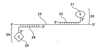
A hybridization complex formed in a sandwich
assay of the present invention is schematically depicted in
Figure 2. This illustration is similar to Fig.1.
Importantly, however, in Figure 2 the top strand is the
25 detector probe 20, and has a detectable group 21 on the 3'
terminus of an oligonucleotide 22. The center strand is
the target polynucleotide 23. The bottom strand of Fig. 2
is the capture probe 24, which has the capture group 25 on
the 5' terminus of an oligonucleotide 26. The aspects of
30 this method are discussed in detail below. ~-
The target polynucleotide can be virtually any
- polynucleotide desired to be detected. For example, the
target can com~rise deoxyribonucleic acid (DNA),
ribonucleic acid (RNA), and analogs and molecular complexes
35 thereof. The target can comprise naturally occurring
polynucleotides or polynucleotides that are produced by ~
:' ~:
I ~ ~ "
-5-
other means, such as recombinant techniques and polymerase
chain reaction methods. The polynucleotide of interest can
comprise virtually all of the polynucleic acids present in
a sample or can comprise any majority or minority portion
thereof. Also, the sample need not contain only
polynucleic acids, but may and generally will contain other
biomaterial. For example, this assay can be used to detect
the presence of specific polynucleotides in whole or
fractionated cells in culture, viri, bacteria, fungi,
algae, yeasts, and other microorganisms. Commonly, these
sources are found in samples taken from the blood, urine,
feces, saliva, pus, semen, serum, or other tissue of an
organism; thus the use of this assay to detect the presence
of polynucleotides specific to one of these sources in a
tissue sample can indicate the presence of the so~rce
itself in the sample. Bacteria such as B-hemolytic
i ~ streptococcz, Haemoph~ ffuenzae, pneumococci, Akycoplasma pneumonu~
rr~ycobac~eria, sa*nonellae, shigellae, Yer~rrua enterocoliti~ca, Escherichia coli,
2 0 Clostridium dif~ile, Campylobacter, Neisseria gonorrhoeae, Treponema pallid~m,
Chlonyd~ ~ochom~, C'los~uun pe~ens can be detected with the
present invention. Exemplary viri suitable for this assay
include influenza A, influenza B, Parainfluenza, respiratory
~yncytial viru8~ adenoviri, rhinoviri, rotaviri, parvoviri,
25 enteroviri, and Herpes simplex virus.
. .
The target polynucleotide should be in single-
stranded form for hybridization, but can be present in the
hybridization solution in double-stranded form as long as
the double strand is denatured to single strands prior to
hybridization of the single strands with the detector
~ ~ probe. Any denaturing technique that is compatible with
i hybridization of the target and detector probe, the
attachment of the target to the solid support, or the
detection of the signalling moiety is suitable for use in
the assay.
r~
.
~3 2 .~ ~ ~ 7 ~ ~
-6-
The target polynucleotide may be an amplicon
produced by any suitable technique, such as strand
displacement amplification or polymerase chain reaction.
See, e.g., G. Terrance Walker et al., Nucleic Acids Res.
20, 1691-1696 (1992); K. Mullis et al., U.S. Patent No.
4,683,202. Thus, the target polynucleotide may be either
natural or synthetic.
The target polynucleotide need only be
sufficiently long to simultaneously bind two different
oligonucleotide probes, with or without overlap of those
probes. Typically the target nucleotide is at least 20
nucleotides in length. The target polynucleotide may be as
long as genomic DNA, but typically the target
polynucleotide is not more than 3,000 nucleotide bases in
length. The first and second segments of the
polynucleotide noted above refer to discreet regions of the
polynucleotide to which a particular probe may hybridize;
the first segment may be positioned in the polynucleotide
either 3' or 5' to the second segment.
A detector probe, as noted above, comprises a
first oligonucleotide which hybridizes to a first segment
of the target polynucleotide and a detectable group
attached to the 3' terminus of the first oligonucleotide.
The length of the first oligonucleotide is not critical so
long as it is capable of binding to a first segment of the
target polynucleotide. The first oligonucleotide is
typically 5, 6, 7, or 8 nucleotide bases in length up to
25, 30, and even 40 or more bases in length. The first
oligonucleotide may be comprised of DNA or RNA, and may be
synthetic or naturally occuring. See generally J.
Goodchild, Bioconjugate Chemistry 1, 165-187 (1990). Any -
suitable detectable group may be employed, examples
including, but not limited to, of enzyme labels (e.g.,
~-alkaline phosphatase, peroxidase such as horseradish
peroxidase, acid phosphatase, ~-D-galactosidase, glucose
oxidase, luciferase), chemiluminescent labels (e.g.,
luminols, lucigenin, acridinium compounds such as
: ::
:~
~ ",i`',`- `'-'.'.',''`. ' : ~',~,' , ~
2 ~
-7-
acridinium esters as described in L. Arnold et al., PCT
Appln W0 89/02896), bioluminescent labels (e-g-,
photoproteins such as aequorin and luciferase), fluorescent
labels (e.g., fluorescein), and electron dense labels
(e.g., ferritin, gold). Typically, the detectable group is
a protein. The detectable group may be attached to the
first oligonucleotide by any suitable technique. See,
e.g., S. Ghosh, Bioconjugate Chemistry 1, 71-76 (1990).
Preferably, the detectable group used is
alkaline phosphatase, and the preferred substrate is a
combination of a dioxetane compound and a fluorescent
compound such as fluorescein, with the reaction carried out
under conditions permitting the activation of the dioxetane
by the alkaline phosphatase and transfer of electronic
energy from the dioxetane to the fluorescent compound, as
described in U.S. Patent No. 4,959,182 to Schapp (the
disclosure of all-U.S. Patent references cited herein is to
be incorporated herein by reference). Suitable dioxetane
and fluorescent substrate systems are commercially
available as LUMIPHOS~ 530 from Lumigen Inc., Detroit,
Michigan, USA.
A capture probe, as also noted above, comprises
a second oligonucleotide which hybridizes to a second
segment of the target polynucleotide and a capture group
attached to the 5' terminus of the second oligonucleotide.
Again, the length of the second oligonucleotide is not
critical so long as it is capable of binding to the second
segment of the target polynucleotide. The second
oligonucleotide, like the first, is typically 5, 6, 7, or
8 nucleotide bases in length up to 25, 30, and even 40 or
more bases in length. Like the first, the second
oligonucleotide may be comprised of DNA or RNA, and may be
synthetic or naturally occuring. Any suitable capture
group may be employed, examples including, but not limited
to, biotin and avidin, antigen and antibody, antibody and
an~ibody binding protein (e.g., Protein A, Protein G,
1~1 ~ ? ~ ~ ~
~`:.~: ,': ~ ' ' .',, .. ~ ~' -, ~ j. , ~ .:-:, ::
~'`. ' ' . ' : : : ... ' ?i' ~ ' ;:;i` ' ;', : '
~?~.'.` " : : : ~ '~ , ;., , : : , `. i ` , . '
' ~ ' ': ~ i: . ~ `', ., : '-'`,' :?' ' .; ', ,. . ~
2 :~ ~ 9
....
-8-
Protein V). In general, the capture group is one member of
a specific binding pair.
The solid support can be any means that permits
the attached capture probe-target-detector probe
hybridization complex to be separated from unbound sample,
detector probe and capture probe. The solid support may
be composed of any suitable material, including glass,
polystyrene, polyethylene, dextran, nitrocellulose, nylon,
and polypropylene. The solid support may take any suitable
form or shape, examples including beads, test tubes,
microwells, membranes, and the like.
It is preferred that the solid support include
a means for binding the capture probe (i.e., have a group
capable of binding, directly or indirectly, to the capture
group of the capture probe attached thereto). Any suitable
technique may be employed. In a preferred embodiment
.
where biotin is used as the capture group on the
oligonucleotide probe, a complex comprising a protein such
as bovine serum albumin (BSA) and biotin is fixed to the
solid support by the protein, streptavidin is bound to the
biotin, and binding sites on the streptavidin remain free
to bind the biotin capture group on the oligonucleotide
probe.
Hybridization of oligogonucleotide probes to
target polynucleotides involves the non-covalent bonding of
a nucleotide strand to a complimentary nucleotide strand
following the Watson-Crick base-pairing of adenine (A) and `
analogs thereof to thymine (T) and analogs thereof or
uracil (U) and analogs thereof, and the pairing of guanine
(G) and analogs thereof to cytosine (C) and analogs
thereof. Hybridization may occur with 100 percent of the
bases of one strand pairing with the proper complimentary
base of the other strand; hybridization may also occur
where only 95, 85, or 75 percent or less of the bases of
one strand are complimentary to the bases of the other
strand. Where two separate probes hybridize to a single
target polynucleotide, as in the present invention, the two
,
;~'~i;:':' : '. :: : - : '':': - :. .: '' , : - ' : ~ , : : -
~'.:,~,~. :,., . ,: ~ : ~: - ,, ,. . :. :. :, : , .. ,- : . --~- ' ,
"~
~;,: .~ . ~ : ,, ,,, ~. , - ,~ ~. ~: , ~- : :, : :
:. ~. , . : . ., . , ,: : ,::.. - : - -
211~7~
probes may hybridize thereto at segments which are either
immediately adjacent or separated by an intervening
segment. The probes themselves may, in some cases,
partially overlap, which is of no consequence so long as
each probe hybridizes to the target polynucleotide.
Conditions which permit hybridization to occur are well
known.
In general, hybrldization may be carried out in
an appropriate hybridization solution, typically an aqueous
solution, in accordance with known techniques. Typically
a hybridization solution will contain 50 percent formamide,
a sodium chloride-sodium citrate mixture, and a small
percentage of DNA (often calf thymus or salmon sperm). The
target, detector probe, and capture probe are added,
denatured if necessary, and allowed to hybridize and attach
to the solid support. Excess reagents and sample are
separated from the hybridized complex for detection. Those
skilled in the art will understand that the steps of
hybridizing the detector probe to the target, hybridizing
the capture probe to the target, and attaching the capture
probe to the solid support can be carried out in any order;
,1 i.e., either hybridization step can precede the other
without affecting the accuracy of the assay, and the
capture probe can be attached first to the solid support
~5 prior to or after it hybridizes with the target. In a
preferred embodiment, the capture probe and the detector
probe are added to the target sample simultaneously and
permitted to hybridize; the solution is added to the solid
support for attachment of this complex through the
attachment moiety of the capture probe.
The detecting step of the assay can be carried
out by known techniques suitable for inducing the
detectable group to produce a detectable signal in
association with the hybridization steps. Generally this
will comprise contacting the detectable group to -a
substrate upon which it can act, and then detecting the
amount of substrate converted by the detectable group. For
~". ' ' ' ' ! ' . : .. ` ,: :~:: ` : - ' ~i : - . . .
,~ . . - - - ~
2 1 ~ .~ 7 ~
' -10-
example, after hybridization in an aqueous hybridization
solution and attachment of the capture probe to a solid
support in contact with the solution, the solution may be
separated from the ~olid support, and the attachment of the
detectable group to the solid support through the
hybridization complex measured (i.e., by bringing a
substrate capable of generating a detectable signal in
contact with the solid support).
Kits for detecting a specific target
polynucleotide in accordance with the present invention may
comprise a detector probe and/or a capture probe, and
optionally includes a solid support as described above.
The components of the kit are typically contained together
in a common package, which may also include a sheet of
instructions for carrying out the method or have such
instructions printed thereon. The kit may also include a -
substrate for the detectable group of the detector probe
capable of producing a detectable signal. -~
The present invention is explained in greater
detail in the following non-limiting examples.
, ~ -.,
EXAMPLE 1 :
Preparation of 5~-Biotinylated CaPtUre
OliqodeoxYnucleotide Pro~e
Oligodeoxynucleotide capture probes were
synthesized as described below. First, a capture oligomer
was prepared using a DNA synthesizer (Model 380B, Applied
Biosystems, Foster City, CA), and Biotin ON~ reagent
(Clonetech, Palo Alto, CA), which produced an oligomer with
three biotin molecules (BBB) at the 5' terminus. The probe
was purified by reverse phase High Pressure Liquid
Chromatography (HPLC, Brownlee Lab Aquapore RP 300 Column -
220 x 4.6 mm, C8 column 7 particle, 300 A pore size) withan ultraviolet (UV) monitor at 254 mm and a gradient of 14
to 44% Buffer B over one hour (Buffer B: 0.lM
Triethylamine-Acetate pH 7 with 50% Acetonitrile; Buffer A:
~1 .: , , , ~ . ,, ~. ,, , , .;, : . : , ,: - ,: ~. ; : ~ , - , ~:
1 ; . . . , ~ - ; ;, ; " i ~; ~ ;,~- ;
:~ ~.' ;.`, , :' '.'!: ` ',, " ` ' '' .' ~ ~' '., ': `~ : `'' ''' ' ' ; i ' ~: ' ' ~- ~ ~ - ': ~ : : '
211~70~
O.lM Triethylamine-Acetate, pH 7) at a flow rate of 1
ml/minute.
EXAMPLE 2
PreDaration of 3'-Alkaline PhosPhatase Detector
OliqodeoxYnucleotide Probe~
Oligodeoxynucleotide detector probes were
synthesized using a DNA synthesizer (Model 380B, Applied
Biosystems, Foster City, CA) and a 3'-amino-modifier column
(Glenn Research, Sterling, VA). This method yielded
oligodeoxynucleotides with 3' amine termini required for
subsequent conjugation with a maleimide derivatized
alkaline phosphatase detector enzyme.
Calf intestine alkaline phosphatase (AP, enzyme
immunoassay grade, Boehringer Mannheim, Indianapolis, ID)
was dialyzed overnight at 4C against 50 mM potassium
phosphate pH 7.5 and subsequently centrifuged to remove
aggregates. The alkaline phosphatase (4 mL, 10 mg/mL) was
combined with a solution of 40 ~L of succinimidyl-4-(p-
maleimidophenyl)butyrate (SMPB, obtained from Pierce,
Rockford, MD, 50 mM) dissolved in N,N'-dimethylformamide
(DMF, Aldrich, Milwaukee, WI) and allowed to react in the
dark at room temperature for 30 minutes. The derivatized
alkaline phosphatase was purified using a NAP-25 column
(Pharmacia, Piscataway, NJ) previously equilibrated with 50
mM potassium phosphate pH 7.5 (degassed and purged with N2).
The absorbances of the NAP-25 column fractions were
measured at 260 and 280 nm and the void volume peak was
pooled. The concentration of derivatized alkaline
phosphatase was determined by absorbance at 280 nm using an
extinction coefficient of 0.75 mL/~mole cm~1. Typically,
about 170 nmoles of SMPB-derivatized alkaline phosphatase
were obtained and stored on ice (less than 2 hours).
The 3' amino-oligodeoxynucleotide (98.4 ~L of
508.2 ~M, 50 nmoles) was diluted in 13.4 ~L of lM potassium
phosphate (pH 7.2) and mixed with 27 ~1 of a solution of n-
succinimidyl-3-(2-pyridyldithio)propionate (50 mM, SPDP,
-- 2~1 97~
-12-
Pierce, Rockford, IL) diluted in DMF. This mixture was
incubated in the dark for 1 hour at room temperature. A
solution of dithiothreitol(DTT, lM) in 50 mM potassium
phosphate (pH 7.5) was added to the SPDP-
oligodeoxynucleotide conjugate/DMF solution (final
concentration of O.lM DTT) and allowed to incubate for-15
minutes at room temperature. Reduction of the SPDP-
derivitized oligodeoxynucleotide with DTT generates a free
thiol group for reaction with SMPB-derivatized alkaline
phosphatase. Excess DTT and 2-thiopyridone were separated
from the derivatized oligodeoxynucleotide by elution over
a NAP-25 column with 50 mM potassium phosphate (pH 7.5).
Within 10 minutes of purification, the reduced
oligodeoxynucleotide was mixed with the SMPB-derivatized
alkaline phosphatase. Rapid mixture of the reduced
oligomer and the SMPB-derivatized alkaline phosphatase can
prevent reoxidation of the thiolated oligomer. The
resulting solution was incubated 2-4 hours at room
temperature, then overnight at 4C, and was then quenched
by addition of l/lOOth the original volume of 50 mM 2-
mercaptoethanol in 50 mM potassium phosphate (pH 7.5). The
crude conjugate was concentrated using a Centriprep 30
centrifugal concentrator (Amicon, Danvers, MA) to
approximately 2 ml. This material was further purified by
HPLC using a DEAE-5PW column (7.5 mm x 7.5 cm), a gradient
of O to 66% Buffer B (Buffer B: 20 mM Tris, lM NaCl pH
7.5, Buffer A: 20 mM Tris pH 7.5) and a flow rate of 1
ml/minute. Absorbance was monitored at 254 mm. Fractions
with A260/A280 equal to 1.0-1.1 correspond to the conjugate
and were pooled. The protein concentration of the
conjugated oligodeoxynucleotide was then determined (BCA
Protein Assay Kit, Pierce, Rockford, IL).
The purified alkaline phosphatase detector
oligodeoxynucleotide probe was diluted to 2 ~M in 20 mM
Tris, lM NaCl, 0.05% sodium azide, 50 ~g/ml sonicated
salmon sperm DNA, pH 7.5, and stored thereafter at 4C.
21I970~
-13-
EXAMPLE 3
5' Detector Probe EnzYme ActivitY
Activity of the alkaline phosphate (AP) detector
oligodeoxynucleotide probes were determined as follows.
The conjugate was diluted to 5 ~g/ml in 50 mM Tris-HCl, 100
mM NaCl, 1 mM MgCl2, 1 mg/ml BSA, pH 7.5. The substrate, 4-
nitrophenylphosphate (pNPP, 5 mM), was dissolved in 1 M
diethanolamine, 1 mM MgCl2, pH 9.8. The conjugate (5 ~
was diluted into 2 ml of the substrate solution at 25 C
and the change in absorbance monitored at 405 nm using a
Hewlet Packard 8452 spectrophotometer. The reaction rates
were calculated from the linear region of the kinetic plots
using the extinction coefficient of p-nitrophenol at 405 nm
(18500 M1cml). The specific activity of the alkaline
phosphatase detector oligodeoxynucleotide probes were
determined to be 850-1300 ~mole/minute/mg.
.
COMPARATIVE EXAMPLE A
Preparation of 3'-BiotinYlated Capture
OliqodeoxYnucleotides
3'-biotinylated capture oligodeoxynucleotides
were synthesized using a DNA synthesizer (Model 380B,
Applied Biosystems, Foster City, CA). 3'Biotin-ON CPG
(controlled pore glass, Clonetech, Palo Alto, CA) was used
to attach a biotin moiety at the 3' terminus of the
oligodeoxynucleotide. Two additional biotins were then
added to the 3' terminus using Biotin-ON phosphoramidite
reagent (also from Clonetech). The oligodeoxynucleotides
were then prepared using standard phosphoramidite reagents
and cleaved from the solid phase to give crude 3'-
biotinylated oligodeoxynucleotides. Purification was done
by reverse phase High Pressure Liquid Chromatography (HPLC)
(Brownlee Lab Aquapore RP 300 Column - 220 x 4.6 mm, C8
column 7 particle, 300 A pore size) with a UV monitor at
254 nm and a gradient of 14 to 44% Buffer B over one hour
(Buffer B~ 0.lM Triethylamine-Acetate pH 7 with 50%
21197~1
-14-
Acetonitrile; Buffer A: O.lM Triethylamine-Acetate, pH 7)
and a flow rate of l ml/minute.
~. -
COMPARATIVE EXAMPLE B
Pre~aration of 5~-~lkaline PhoiPhatase Deteotor
Oliqodeoxvnucleotides
5'-alkaline phosphatase oligodeoxynucleotides
were synthesized from 5'-amino-oligodeoxynucleotides
prepared using a DNA synthesizer (Model 380B, Applied
Biosystems, Foster City, CA). The reagent AminoLink II ~-
(Applied Biosystems, Foster City, CA) was used to place an
amine group on the 5'-end of the oligodeoxynucleotide for
subsequent conjugation with alkaline phosphatase as
described above. The crude conjugate was dialyzed into 20
mM Tris pH 7.5 and concentrated using a Centriprep 30
(Amicon, Danvers, MA) to approximately 2 ml. The
concentrated conjugate was then purified by HPLC using a
DEAE-5PW column (7.5 mm x 7.5 cm) and a gradient of o to
66% Buffer B (Buffer B: 20 mM Tris, lM NaCl pH 7.5, Buffer
A: 20 mM Tris pH 7.5) and a flow rate of 1 ml/minute. -
Absorbance was monitored at 254 nm. The fractions were
collected, the activity of the conjugate was determined,
and the conjugate was stored as described above. ~
.
E)(AMPLE 4
Pre~aration of Coated Microtiter Plates
Biotinylated bovine serum albumin (biotin*BSA)
(Pierce, Rock~ord, IL) was diluted to 5 ~g/ml in 0.3 M
Glycine, pH 9.6 (BRL, Bethesda, MD., prepared using
autoclaved water), pipetted into each well (200 ~l/well) of
microLITEl~ plates (Dynatech, Chantilly, VA), and incubated
at 4C overnigh~. The plates were washed twice (375
~l/wash) using FTA hemagglutination buffer, pH 7.2 (Becton
: Dickinson Microbiology Systems, Cockeysville, MD, prepared
using autoclaved water). Streptavidin (50 ~g/ml) in
hemagglutination buffer was added to the biotin*BSA-coated
microliter wells (100 ~l/well). Plates were covered and
~ '
~ .~ '~;i`.; '~ . :,; ' . ';, .~ ~ ' i ' ~; '
~": ' ~''''''''``''''`'''`~'`''` ' ' ~'-
~ '! "`~, .' ' ,, ~ ; ~ ; ~
2 ~ 0 :1
-15-
incubated for 1 hour at 37~C. Unbound streptavidin was
discarded by inversion and blocking buffer (300 ~1/well)
(hemagglutination buffer pH 7.2, 0.05% weight/volume (w/v)
bovine serum albumin, Sigma Chemical Co., St. Louis, Mo.)
was added. The plates were covered and incubated (30 min,
37C), and the blocking buffer was discarded by inversion.
Plates were washed twice with hemagglutination buffer (375
~l/well), then once using hemagglutination buffer with 2%
w/v trehalose (375 ~l/well) (Fluka, Ronkonkoma, N.Y.).
Plates were dried for approximately 4 hours under vacuum
below 0.5 Torr at 25C, sealed in mylar pouches with
desiccant, and stored overnight at room temperature prior
to use. The plates were stored thereafter at between 2-
8C.
.~
EXAMPLE 5
Microtiter Plate A~ay Procedure
This assay uses synthetic target DNA designed to
imitate the SDA product generated from M. avium/
intracellulare genomic DNA. The target sequence within the
genome was determined from the pMAV29 sequence described in
D. Wirth et al., Molecular and Cellular Probes 4 , 87-105
(1990). A biotinylated (BBB) capture probe binds to the
target DNA and the streptavidin on the microliter plate.
A second detector pro~e conjugated with Alkaline
Phosphatase (AP) binds to the target DNA. The alkaline
phosphatase provides and opportunity for detection by means
of colorimetry, fluorimetry, or chemiluminescence. This
assay evaluates the significance of conjugating the probes
with biotin or alkaline phosphatase at either the 5' or 3'
ends of the oligomers.
The assay used the following probes and target
for the detection of synthetic M. avium/intracellulare
target DNA:
~1) BBB-GGGAACCGGTGACTC (SEQ ID NO:l)(where
¦ 35 each B is biotin): ~
' : ::
, .: '
2~1~7~
. .
-16~
(2) CAAAAACCTTGCGGC-P (~EQ ID NO:2)(where
P is alkaline phosphatase);
(3) P-GGGAACCGGTGACTC (SEQ ID NO:3)(where
P is alkaline phosphatase);
(4) CAAAAACCTTGCGGC-BBB (~EQ ID NO:4)(where ~-
each B is biotin); and
(S) GACCCGACTTGTAAGAGCCGCAAGGTTTTTGGAGTCAC
CGGTTCCCACTCGCAGCCTGCGTCTTTT (8EQ ID NO:5)
SEQ ID NO:l and SEQ ID NO:2 represent a pair of probes of -
the present invention; SEQ ID NO:3 and SEQ ID NO:4
represent a pair of probes of the prior art; SEQ ID No:5
represents a synthetic target DNA. ~-~
Synthetic target DNA (SEQ ID NO:5) was diluted ;
into 0.1 mg/ml sheared salmon sperm DNA (Sigma) in sterile
siliconized tubes to produce solutions having the following
concentrations: 1600, 400, 100, 25, 6.25, o attomoles of
- target DNA/50~1 volume. Diluted synthetic target DNA was
heated to 95C for 3 minutes to denature the DNA. Tubes
were cooled for 5 minutes at room temperature and then 50~1
of the denatured DNA was added to each well. Each level of l;
target DNA was assayed in triplicate. Immediately -~
thereafter, 50~1/well of hybridization mix (lM sodium
phosphate, 0.2% BSA, 40nM capture probe, lOnM detector
probe) was added. The plate was covered and incubated for
45 minutes at 37~C. SEQ ID NO:1 and SEQ ID NO:2 probes
were paired in one set of wells, and SEQ ID NO:3 and SEQ ID
NO:4 probes were paired in a different set of wells. Three
stringency washes (300~1/well) (lOmM sodium phosphate pH 7,
0.1% w/v bovine serum albumin, 0.05% Nonidet P-40 (Sigma)
were performèd at room temperature. Each wash was allowed
to remain in the microliter wells for 1 minute before
removing. Lumiphos~530 (100~1/well, Lumigen Inc., Detroit
Michigan) substrate was added, and the plates were covered
and incubated for 30 minutes at 37C. Luminescence was
read on a microtiter plate luminometer ~LabsystemS,
Research Triangle Park, NC) at 37C, using a 2 second/well
integration time. Results are given in Table 1 below.
,~ ,
~ ' ",''. '''
, :~
' . '
", ,,,, ,, -,- ,~" ~,;,,,,,"~ ",
2~l37a~ ~
-17-
The results indicated as much as a threefold reduction in
background signal for wells assayed with a SEQ ID NO:l/SEQ
ID NO:2 probe set (which utilizes a 5'-biotin moiety and a
3'-alkaline phosphatase moiety) compared to wells assayed
with a SEQ ID NO:3/SEQ ID NO:4 probe set. Because the
probe reagents have identical DNA sequences, the lower
backgrounds can be attributed to differences in terminus
conjugation.
~:
I
. TABLE 1 Avclage Rclativc Light Un~ls (Signal)
Attomoleslrest
Probc Set O :Z5 400 -- CV
SEQ ID NO:I and 8.03 10.88 20.13 49.3 180.5 645.4 3.3-10.1%
SEQ ID NO:2
SEQ ID NO:3 and 24.85 30.97 39.32 56.14 164.33 510.5 1.7-27.4%
SEQ ID NO:4
_ _
~CV%, calculated for each standard (triplicales)
EXAMPLE 6
ARsaY Procedure with Varied Probe Seouences
Using the procedures described above in Examples
4 and 5, probes with a slightly altered sequence were used
to detect the M. avium/intracellulare synthetic target DNA.
Again, the probe sets compared the significance of 3' or 5'
biotin or alkaline phosphatase conjugation. The probes
used in this example were:
~6) B8B-AACCGGTGACTCCA (SEQ ID NO:6)(where
B is biotin);
(7) AAAACCTTGCGGC-P (8EQ ID NO:7)(where P
is alkaline phosphatase);
(8) AAAACCTTGCGGC-BBB (SEQ ID NO:8)(where
B is biotin); and
!' '
i
` -" 2 ~ 3~
-18-
(9) P-AACCGGTGACTCCA (SEQ ID NO:9) (where P
is alkaline phosphatase).
SEQ ID N0:6 and SEQ ID NO:7 represent a probe set of the
present invention; SEQ ID No: 8 and SEQ ID NO:9 represent a
s probe set of the prior art; SEQ ID NO:5 was again the
target DNA. Data gathered in the assay are given in Tiable
2 below.
r
TAB~E2 Avc~gcRc~c~tU~(Si~)
r _ I
Attomol~it l
CV~b I ` ~.
ProbcSet O 6.25 25 1~ 4~ 1~ Range ¦
SEQID NO:6and 9.02 15.96 37.83 125.97 477.7 l79tl 3.1-5.4%
SEQID NO:7
SEQID NO:8and 267.77 ~.33 320.33 433.4 730.03 1936.33 0.35-5.9%
SEQID NO:9 .
, __ ,,
The results again indicate that the probe set consisting of
SEQ ID NO:8 and SEQ ID NO:9 (3'-biotin, 5'-alkaline
phosphatase) show as much as a 30-fold higher backgrounds
and as much as a 16-fold decrease in detection sensitivity
compared to the probe set consisting of SEQ ID NO:6 and SEQ
ID N0:7 (5' biotin, 3' alkaline phosphatase).
EXAMPLE 8
AssaY for M. M~cn~ku~target DNA
~: ::
As in the foregoing Examples, this experiment used
synthetic target DNA. However, the target was designed to
imitate the Strand Displacement Amplification (SDA:G.T.
Walker, et al. ~1992) PNAS 89, 392-396: G.T. Walker, et
al.(1992) Nucleic Acids Res. 20, 1691-1696) product generated
from Mycobactena tuben~osis genomic DNA. The target region within
the genome was derived from the IS6110 sequence obtained
genomic DNA. The target region within the genome was derived
from the IS6110 sequence described in Thierry et al., Nucleic
Acids Research, 18: 188 (1990). The assay methodology
described above was utilized on the following target with the
following probes to assess the detection synthetic M.tb target
DNA:
; ,~ ,.: . : ~ - - . ;; :~ : . - ~ ; ~ , ~
`- ~ 21197~
. . -19-
(l0) TATCCACCATACGGA-BBB (SEQ ID
NO:l0)(where each B is biotin);
(11) P-CGACCTGAAAGACGT (8EQ ID NO:ll)(where
P is alkaline phosphatase);
~12) BBB CCTGAAAGACGTTAT (~EQ ID NO:12)
(where B is biotin);
~13) CCACCATACGGATAG-P (SEQ ID NO:l3)(where
P is alkaline phosphatase); and
~l4) GACACTGAGATCCCCTATCCGTATGGTGGATAAC
GTCTTTCAGGTCGAGTACGCCGTCTTTTT (SEQ ID NO:14).
SEQ ID NO:l0 and SEQ ID NO:ll represent a probe set of the
prior art; SEQ ID NO:12 AND SEQ ID NO:13 represent a probe
set of the present invention; SEQ ID NO:14 represents a
target DNA. The synthetic target DNA was diluted to 2000,
l000, 500, 2 50, 12 5, 62. 5, 3 1.25, and 0 attomoles/50
volume and each probe set was used in an assay of that
target in essentially the same manner as described above.
Table 3 shows the data gathered in the assay.
,.,
. _
TABL~3 Avc~geRcla~cU~tU~(Si~a1) l
. ". :
Attomol~est I
2 0 ~ et 0 31.25 62.5 125 2S0 500 1~0 20~ Range
SEQID NO:10 19 87 25.1 36.9 56.4 104 202 420 856 28- ¦
and 13.39
SEQID NO:11 _ _
SEQID NO:12 4.94 24.13 445 89.6 1707 331.7 721.6 1498 178-
and 9.98
SEQID NO:13
~. l = _ _ _ _ ''''': .:,:
Once again, the probe set with the 5'biotin and 3' alkaline
phosphatase conjugation (SEQ ID NO:12 and SEQ ID NO:13)
showed a significant decrease in background levels and an
3 0 increase in the amount of specific signal generated.
The foregoing examples are illustrative of the
present invention, and are not to be construed as limiting
thereof. The invention is defined by the following claims,
with equivalents of the claims to be included therein.
"
~ ~:
:
7 ~ ~
-20-
SEQUENCE LISTING ~
::
(1) GENERAL INFORMATION:
(i) APPLICANT: Nycz, Colleen M.
Vonk, Glenn P.
Jurgensen, Stewart R. - ~
Myatich, Ronald G. -
(ii) TITLE OF INVENTION: Nucleic Acid Assay Procedure
(iii) NUMBER OF SEQUENCES: 14
(iv) CORRESPONDENCE ADDRESS: -
(A) ADDRESSEE: Richard J. Rodrick
(B) STREET: 1 Becton Drive
(C) CITY: Franklin Lakes
(D) STATE: New Jersey
(E) COUNTRY: USA
(F) ZIP: 07417-1880
(v) COMPUTER READABLE FORM:
(A) MEDIUM TYPE: Floppy disk
(B) COMPUTER: IBM PC compatible
(C) OPERATING SYSTEM: PC-DOS/MS-DOS
(D) SOFTWARE: PatentIn Release #1.0, Version #1.25
(vi) CURRENT APPLICATION DATA:
(A) APPLICATION NUMBER: -
(B) FILING DATE:
(C) CLASSIFICATION:
(viii) ATTORNEY/AGENT INFORMATION:
(A) NAME: Rodrick, Richard J.
(B) REGISTRATION NUMBER: 26,985
(C) REFERENCE/DOCKET NUMBER: P-2652
(ix) TELECOMMUNICATION INFORMATION:
(A) TELEPHONE: 201-847-5317 ;-
(B) TELEFAX: 201-848-9228
(2) INFORMATION FOR SEQ ID NO:1:
(i) SEQUENCE CHARACTERISTICS:
(A) LENGTH: 15 base pairs
:~` (B) TYPE: nucleic acid
(C) STRANDEDNESS: single ,
(D) TOPOLOGY: linear
(ii) MOLECULE TYPE: cDNA
(xi) SEQUENCE DESCRIPTION: SEQ ID NO:1:
GGGAACCGGT GACTC 15
~' ~
rt a ~
.
-21-
(2) INFORMATION FOR SEQ ID NO:2:
(i) SEQUENCE CHARACTERISTICS:
(A) LENGTH: 15 base pairs
(B) TYPE: nucleic acid
(C) STRANDEDNESS: single
(D) TOPOLOGY: linear
(ii) MOLECULE TYPE: cDNA
(xi) SEQUENCE DESCRIPTION: SEQ ID NO:2:
CAAAAACCTT GCGGC 15
(2) INFORMATION FOR SEQ ID NO:3:
(i) SEQUENCE CHARACTERISTICS:
(A) LENGTH: 15 base pairs
(B) TYPE: nucleic acid
(C) STRANDEDNESS: single ..
(D) TOPOLOGY: linear
(ii) MOLECULE TYPE: cDNA .
(xi) SEQUENCE DESCRIPTION: SEQ ID NO:3: .
GGGAACCGGT GACTC 15
(2) INFORMATION FOR SEQ ID NO:4:
(i) SEQUENCE CHARACTERISTICS:
(A) LENGTH: 15 base pairs
(B) TYPE: nucle;c acid :
(C) STRANDEDNESS: single ;.:~
(D) TOPOLOGY: linear .
(ii) MOLECULE TYPE: cDNA :::
(xi) SEQUENCE DESCRIPTION: SEQ ID NO:4:
CAAAAACCTT GCGGC 15
(2) INFORMATION FOR SEQ ID NO:S:
(i) SEQUENCE CHARACTERISTICS:
(A) LENGTH: 66 base pairs
(B) TYPE: nucleic acid
(C) STRANDEDNESS: single
(D) TOPOLOGY: linear
(ii) MOLECULE TYPE: cDNA
(xi) SEQUENCE DESCRIPTION: SEQ ID NO:5:
GACCCGACTT GTAAGAGCCG CAAGGTTTTT GGAGTCACCG GTTCCCACTC GCAGCCTGCG 60
, .
I . :" ` . ~ . , ~ ~ ` ~ - . - , ` `
, ~ ,".',''~-`' .`" ` '~ ' '~ ~ ; ,': " I ` '": ` . ~`'` ~ :~ '
~`,` ~.. ~.. :.. : .... . ` ~ "~ .` .. . . .` `, . .. ..
. ~ .~
211 97~ ~
-22 -
.-
TCTTTT 66
(2) INFORMATION FOR SEQ ID NO:6:
(i) SEQUENCE CHARACTERISTICS:
(A) LENGTH: 14 base pairs : :
(B) TYPE: nucleic acid
(C) STRANDEDNESS: single
(D) TOPOLOGY: linear
(ii) MOLECULE TYPE: cDNA
(xi) SEQUENCE DESCRIPTION: SEQ ID NO:6:
AACCGGTGAC TCCA 14
(2) INFORMATION FOR SEQ ID NO:7:
(i) SEQUENCE CHARACTERISTICS:(A) LENGTH: 13 base pairs
(B) TYPE: nucleic acid
(C) STRANDEDNESS: single
(D) TOPOLOGY: linear
- (ii) MOLECULE TYPE: cDNA
(xi) SEQUENCE DESCRIPTION: SEQ ID NO:7: ~- :
AAAACCTTGC GGC 13
(2) INFORMATION FOR SEQ ID NO:8: ;
(i) SEQUENCE CHARACTERISTICS: .
(A) LENGTH: 13 base pairs . :~
(B) TYPE: nucleic acid
(C) STRANDEDNESS: single ~
(D) TOPOLOGY: linear .
(ii) MOLECULE TYPE: cDNA :
(xi) SEQUENCE DESCRIPTION: SEQ ID NO:8: :~
AAAACCTTGC GGC 13
(2) INFORMATION FOR SEQ ID NO:9:
(i) SEQUENCE CHARACTERISTICS:
(A) LENGTH: 14 base pairs
(B) TYPE: nucleic acid
(C) STRANDEDNESS: single
(D) TOPOLOGY: linear
(ii) MOLECULE TYPE: cDNA
(xi) SEQUENCE DESCRIPTION: SEQ ID NO:9:
2119~01
-23-
AACCGGTGAC TCCA 14
(2) INFORMATION FOR SEQ ID NO:10:
(i) SEQUENCE CHARACTERISTICS: :~
(A) LENGTH: 15 base pairs
(B) TYPE: nucleic acid
(C) STRANDEDNESS: single
(D) TOPOLOGY: linear
(ii) MOLECULE TYPE: cDNA
(xi) SEQUENCE DESCRIPTION: SEQ ID NO:10:
TATCCACCAT ACGGA 15
(2) INFORMATION FOR SEQ ID NO:11:
(i) SEQUENCE CHARACTERISTICS:
(A) LENGTH: 15 base pairs
(B) TYPE: nucleic acid .
(C) STRANDEDNESS: single
(D) TOPOLOGY: linear
(ii) MOLECULE TYPE: cDNA
(xi) SEQUENCE DESCRIPTION: SEQ ID NO:11: ; :~
: CGACCTGAAA GACGT 15 .
(2) INFORMATION FOR SEQ ID NO:12:
(i) SEQUENCE CHARACTERISTICS:
(A) LENGTH: 15 base pairs :
(B) TYPE: nucleic acid
(C) STRANDEDNESS: single
(D) TOPOLOGY: linear
(ii) MOLECULE TYPE: cDNA
(xi) SEQUENCE DESCRIPTION: SEQ ID NO:12:
CCTGAAAGAC GTTAT 15
(2) INFORMATION FOR SEQ ID NO:13:
(i) SEQUENCE CHARACTERISTICS:
(A) LENGTH: 15 base pairs
(B) TYPE: nucleic acid
(C) STRANDEDNESS: single
(D) TOPOLOGY: linear
(ii) MOLECULE TYPE: cDNA
(xi) SEQUENCE DESCRIPTION: SEQ ID NO:13:
. " ~
`
. -24-
CCACCATACG GATAG 15
(2) INFORMATION FOR SEQ ID NO:14:
(i) SEQUENCE CHARACTERISTICS:
(A) LENGTH: 63 base pairs
(B) TYPE: nucleic acid
(C) STRANDEDNESS: single
(D) TOPOLOGY: linear
(ii) MOLECULE TYPE: cDNA
(xi) SEQUENCE DESCRIPTION: SEQ ID NO:14:
GACACTGAGA TCCCCTATCC GTATGGTGGA TAACGTCTTT CAGGTCGAGT ACGCCGTCTT 60 : ~:
TTT 63~
~'
; ~
~ '`
~ ..
: ~
I
~ :
. ~ ,,
.
:,- : ~:.. ~ ., , :.. : ~ ~ -, - ~ ~ . ,.. - . :.~