Note: Descriptions are shown in the official language in which they were submitted.
WO 93/05390 1 21 2 0 c ~ l PCI/US92/07160
AUTOMATED
CAPILLARY ELI~CTROPHO~ESIS APPARATU$
CROS~REEERENCEIO RELATED APPLICATIO~I~
This application is a continuation-in-par~ of application serial no.
07/270,788 filed on NoYember 14, 1988 as a continuation-in-part of
application serial no. 07/125,544 filed on Novernber 25, 1987.
~ACKGROUND OF THE !NVENT!oN
Electrophoresis is a phenomenon in which charged particles
move in a conductive buffer m~dium or fluid across which a potential
difference is applied. The migration is toward an electrode carrying
charge oppocite to that of the particles.
Electrophoresis is one of the most important methods available
for the investigation of biological materials, and probably the most
sfficient procedure for the separation and det~ction of proteins and
other matter.
Electrophoresis separation relies on the diffsrential speeds of
the migration of differently charged particles in an electrical field.
The migration speed is primary a function of the charge on the par-
ticle and tha~fiéld strength applied and the charge on a particle is
dstermined by the pH of the buffer meciium. The most important
application of this technique in biomedical research and clinical
chemistry laboratories, is in the electrophoretic separation of pro-
SUBS l I l ~JTE SHIEET
WO 93/05390 2 1 ~ 0 2 ~j ~ 2 PCr/US92/0716
teins, nucleic acids, their component peptides an~-oligonucleotides,
as well as complex macromolecules such as lipoproteins.
Several different systems are known for practicing elec-
trophoretic separation. For exarnple, one system known as zonai
procedur~s, has advantages but it also has certain limitations. Some
of the most oommon limitations are: The amount of sample required
in order to ravsal the components by the oommon staining proc~dures
is usuaily lar~e, the preparation of the apparatus and complete sys-
tem involved in the electrophoretic sepa.ra~ion is commonly tedious
and time consuming, the tims required to obtain complets separation
of ~he components is often hours, the time required to reveal the
components and to obtain some quantitation of the separatad
substances is also commonly hours, the yield of recovery of the
components as bioiogical actives in most cases is very .ow, the
reproducibility of the slectrophoretic ssparation is not 100 percent
accurate, and the automation to perform`~he entire system operation
is almost lacking.
Capillary electrophoresis has bean shown to be a technique for
obtaining high separation efficiency. For some protsins and small
peptides, ssparation efficiencies of aplproximately one million ~o
about a few million have been demonstrated. In general, this tech-
nique utilizes a fused silica (quar~z) capillary with an inside diame-
ter ranging fro~ about 25 microns to about 200 microns, and a length
ranging from about 10 centimeters to about 100 centimaters. Since
the entire volume of the column is only 0.5 to about 30 microiiters
(yielding probably the smallost total surface area of column
chromatography), the injection volume is usually in the low nanoliters
range. As a consequence, the sensitivity of this technique is quite
SUBSTITUTE SHEET
wo 93/05390 3 2 1 ~ V ,' ~ ~ PCI'/US92/07t60
high and it is possible to obtain quantitation in the order of picomoles
(and probably femtomol~s or attomoles) using fluorescence,
slectrochemical, laser-induced fluorescence, and mass spectrometry
detectors, and to obtain quantitation in the order of nanomoles using
ultraviolet detectors.
In capillary eiectrophoresis, the efficient heat transfer from
small diameter capillaries permits application of unusually high
voltages ranging from about 5,000 volts to about 30,000 volts whils
maintaining a low current, in the rangs of about 10 microamperes to
about 90 microamperes. The application of high voltagss prpmotes
more effective separations and increases the speed of analysis to
record times of about 5 to 40 minutes.
In addition to high separation efficiency (thsoretical plates),
fairly high resolution, high sensitivity quantitation, and small mi-
gration (retention) times, capillary electrophoresis presents a few
more advantages over conventional electrophoresis and, in general,
other chromatographic procedures. Some of these advantages are: a)
application to a wide variety of samples ranging from small ions to
proteins or other macromolecules of molecular weights of approxi-
mately 290,000 daltons or higher (such as DNA fragments, viruses,
and subcellular particles) by using essentially the same column and
probably the same conditions of electrophoretic separation; b~ capil-
laries should p~ovide an ideal system to explore nonaqueous media,
particularly with substances which are highly hydrophobic; c) capil-
laries are reusable many times making the electrophoretic separation
system very practical and economical; d) on-line electronic detection
permits good quantitation and further enhancas possibilities for fully
automatic operation making the capillary electrophoresis system of
SUBSTITUTE SHEET
WO 93J05390 2 1 ~ Q 2 5 1 PCI/US92/07160
higher resolution, greater speed, and better accuracy than
conventional methods.
In th~ prior art, it is generally known that a material, contain-
ing mixtures of substances to be analyzed, can be passed along a
capillary tube and through a detector under the influence of an applied
voltage. The applied voltage charges ~he substances and the charges
on the substances determine their spacing and their speed of passage
along ths capillary tube.
The prior art, U.S. Pat~nts 3,620,958, 3,948,753 and 4,459,198,
show electrophoresis apparatus including a capillary tube connected
between two containers for containing the substance to be analyzed
and having electrical potential applied between the two containers
and across the capillary tube. While the various forms of apparatus
shown in these patents are apparently useful, they require large
concentrations of sampl~s to bs analyzed and nonc is capable of bcing
automated or provides teaching related to automation.
The present invention provides high voltage capillary elec-
trophoresis apparatus including, among other things, means for
feeding small concentrations of sample material into a capillary tube,
automatically applying the proper voltage to cause the components of
the sample to be charged and to flow along the capillary tube through
a d~tector wherein the components are detected and a printed record
is made. Tl~e apparatus can then automatically repeat the process for
the analysis of multiple samples.
The basic apparatus of the invention is susceptible of many
rnodifications in its various parts including the capillary tube portion.
In addiffon, the method of det~ction of samples may be varied and the
SUBSrlTUTE SHEET
wo s3/0s3so 5 2 1 2 0 2 !~i 1 PCl`/US92/07160
collection of samples can be modified. The invention can also be
adapted to measure electroosmotic fiow in a capillary. ;
SUiBSTlTlJTE ~IEET
212~251
WO 93/05390 6 PCT/US92J07160
DESÇRIPTION OF THE ~RAWINGS
Fig. 1 is a perspective view of the rear of apparatus embodying
the invention;
Fig. 2 is a sectional view along the lines 2-2 in Fig. 1;
Fig. 3 is a perspective view of a portion of the apparatus shown
in Fig. 1;
Fig. 4 is a perspective view of a portion of the apparatus of Fig.
,
- , .
Fig. 5 is a sectional, elevational view of a portion of the appa-
ratus of Fig. 1;
Fig. 6 is an enlarg~d perspective view of a portion of the
apparatus of Fig. 1;
Fig. 7 is a front YieW of a portion of the apparatus of Fig. 1;
Fig. 8 is a side elevational view of a modification of a por~ion of
the apparatus of Fig. 1 with portions thereof in section;
Fig. 9 is a front elevational view of à modification of a portion
of the apparatus shown in Fig. 1;
Fig. 10 7s a schematic representation of the electronic control
system used in th~ invention; -
Fig. 11 is a sectional view of a modification of the capillary -
portion of the apparatus of Fig. 1;
SUBSrlTUTE SHEET
wo 93/05390 7 ~ 2 ~ 1 PCr/US92J07160
Fig. 12 is a plan vi~w of a portion of a detec~or used in Fig. 1
with the apparatus of Fig. 11;
Fig. 13 is a schematic representation of a mode o~ operating
multiple pieces of apparatus of the type shown in Fig. 1;
- Figs. 14A and 14B together are a bottom view of a modification
of the base support of the invention;
Fig. 15 is a perspective ~ront ViflW of a modification of the in-
vention;
Fig. 16 is a perspective YieW of a modified capillary cartridge
usable with the apparatus of the invention;
Fig. 17 is a perspective of apparatus embodying modifications of
ths invention;
Fig. 18 is a psrspective enlarged viaw of a switch used with the
apparatus of Fig. 17;
Fig. 19 is a frollt eleva~ional view of a portisn of apparatus of
th~ invention illustrating a modification thereof;
Fig. 20 is a front elevational view of a modification of the ap-
paratus shown in Fig. 19;
Fig. 21 is a front elevationai view of a portion of a capillary
tubs and ma~nifying glass used to analyze electroosmotic flow;
Fig. 22 is a perspective view of a modification of apparatus
embodying the invention;
SUI~STITUTE SHEET
WO g3/0~39~ 2 1 2 ~ 2 5 1 PCr/US92/07160
Fig. 23 is a modification of the capillary tube used with the
inv~ntlon;
Fig. ~4 is a front elevational view illustrating additional appa-
ratus which can be used in the apparatus of the invention;
- Fig. 25 is a perspectivs view of a rnodification of the invention
used with some of ~he apparatus of Fig. 24;
Fig. 26 is a perspective view of a modification of the apparatus
of Fig. 25;
Fig. 27 is a front elevational view of a portion of the apparatus
of the invention illustrating a mode of op*ration thereof;
Fig. 28 shows pulses detected by the method of Fig. 27;
Fig. 29 shows a curve derived from the pulses of Fig. 28;
Fig. 30 is a side elsvational view of a modified capillary in~
cluding means for cleaning the capillary.
Fig. 31 is a side sectional view of the modification of a portion :
of the invention;
Fig. 32 is a side sectional view of the modification of the ap- :
paratus of Fig. 31; ~ -
Fig. 33 i~ a prospective view of another modification of the in-
vention; and
Fig. 34 is a prospective Yiew of a modification of a capillary
tube assembly used in the apparatus of the invention. :
Fig. 35 is a side view of another modification of the invention.
SUBSTITUTE SHEET
WO 93/0s390 9 21 ~ 2 0 2 S l PCI`/US92/07160
l2E~ OF IHE IN~ NTION
The automated electrophoresis apparatus of the invention 10,
shown from the rear in Fig. 1, includes a base support member to
which various pieGes of opera~ing equipment are seGured. The support
rnember 20 is box-like and includas a ~op wall 30, a front wail 40, a
rear wall 50 and end walls 60 and 70 all of which extend downwardly
from the top wall. A bottom oover plate 80 (Figs. 1 and ~) is secured
to th~ support m~mber 20 and provides a flat support surface for the
apparatus 1 0.
The support member 20 is of metal or a plastic and carries on
top wall 30 a left hand box 90 and a right hand box 100 as seen in Fig.
1. The l~ft hand box 90 includ~s an insulating base plate 1109 o'f a
m~al or plastic, secured to top wall 30 and a transparent enolosure,
of plexiglass or th~ like, including (Fi~s. 1 and 2) left and right side
walls 120 and 130, front and rear walls 1~0 and 150 and a top wall
160. The ~op wall 160 is a cover for the box 9û and is adapted to be
lifted off th~ box by means of knob 162 to provide aGcess to the
interior thereof. Th~ enclosure for box 90 is suitably secured to the
base 110.
Box 90 is provid~d with a rotatable horizontal table 170 having
a circular array,of holes or apertures 180 in which fluid sample cups
190 are seated. The table is detachably secured to the upper end of a
vertical post 200 so that tables with different numbers of holes or
differen~ sizes of holes or with other features can be secured to the
post. The post 200 extends through and beneath the top wall 30 of th
support member 20 (Figs. 2 and 3) where it is suitable connected to a
SUBSTITUTE SHEET
WO 93~05390 21 ~ O ~ 51 - 1 o PCI`/US92/07160
small motor 210 which is used to rotat~ the post 200 and table 170.
The motor 210 is secured to the lower surface 32 of the top wall 30
and is of a type which permits the post 200 or an extension thereof to
extend through it to be driven thereby.
The lower end of the post 200 carries a horizontal disk 203 (Fig
3) which rotates with the post and includes a slot 205 which is
adapted to operate with an optical sensor 212 positioned adjacent
~hereto.
Adjacent to the rotatable table 170, referring to Figs. 1, 2 and
6, is a hollow, tubular vertical post 220 having an aperture or slot
230 in its side wail. A horizontal arm 240 has one end inside post
220 and secured to a vertical rod 250 which is suitable mounted so
that it can be driven vertically up and down. The lower end of the
vertical rod 250 or an extension thereof passes through a small motor
260 secured to the lower surface 32 of the top wall 30. The lower
end of the rod 250 carries a laterally projecting arm 2~3 which is
positioned to operate with an optical sensor 280 positioned adjac~nt
thereto.
Referring again to the horizontal arm 240, (Figs. 1, 2, and 6~ ths
outer end thereof terminates in a small solid cylinder 243 which is
oriented vertically and is proYided with two through-holes 247 and
249 which communicate with a hollow tube 248 which extends
downwardly ~r~m the soiid cylinder in alignment with the holes 180
in table 170 and the sample cups therein. The hollow tube 248 is of a
small diameter and is dimensioned so that it can enter a sample cup
190 and extend to about the bottom thereof to enter fluid therein.
SUBSrlTUTE SHEET
wo 93/0~390 1 1 ~ 1 ~ 0 2 5 1 PCI'/US92~07160
The box 100 con~ains the same apparatus as box 90 as described
above. The corr~sponding parts in box 100 carry the same reference
numerals as the parts in the box 90 but primed.
Th~ boxes 90 and 100 include means for applying electrical
potential across the apparatus 10. This means includ~s a first wire
electrode 360 haYing one end secured to a power input terminal 363.
(Figs. 1 and 2) in the rear upwardly in th~ hollow tube 220 adjacent to
the vertical rod 250 and out of the opening 230 in the side wali and
through the hole 249 in cylinder 243 down through the tube 248 to the
end thersof so that it can res~ in a fluid in a sample ClJp when the
apparatus 10 is in operation.
The electrical means also includes a simila~ wire electrode 360'
secured to a power input terminal 365 in the rear wall 50 of the
apparatus 10. This electrode follows a similar path through tube 250'
and cylinder 243' into the tube 248' associated therewith for ultimate
insertiorl into a sample cup. Th~ electrodes 360 and 360' are
preferably of pla~inum or the like and are adapted to oarry the
voltages used in operation of the invention. A power supply 367 is
provided for connsction to terminals 363 and 365 to electrodes 360
~nd 360' for providing the requir~d voltages. Power supply 367 may
also provide whatever other power is needed by the apparatus 10 such
as for the motors 210, 210' and 260, 260'. Other auxiliary power
supplies may be,provided as desired.
In one embodiment of the invention, illustrated in Fig. 2, the
power supply 367 may be of such small size that it can be mounted
within support member 20 at any suitable location so that the appa-
SUBSTITUTE SHEET
wo 93fO5390 2 1 '2 0 2 ~ :1 1 2 PCI/US92/07160
ratus 10 has its own self-contained power supply which may be
manually or computer-controlled.
When a built-in power supply is provided, referring to Fig. 7, a
voltmeter 376 and an amm~ter 378 are secured to the front wall 40
of support member 20 along with a rheostat for adjusting the op-
erating voltage shown on the voltmeter.
A preferred structure for the vertical posts 250, 250', to insure
electrical safety when high Yoltage is applied to electrode 360, is
shown in Figure 8. This embodiment for aase of construction includes
an outer post 420 and slidable inner post- 422 which are both
generally square or rectangular in construction. The outer post in-
cludas a slot 426 in its side wall and a horizontal arm 428 extends
therethrough from the inner post 422. Arm 428 terminates in cylin-
der 243. The cable 360 comes up from the terminal 363 and runs
inside the outer post 420 and terminates in a rigid, relatively large-
ar~a flat motallic electrode 430 positioned perhaps haif-way up the
post to slightly below the slot 426 therein.
Similarly, the slidable inner post 422 carries on its outer
surface a relatively large area electrode 432 which is suitably po-
sitioned so that when the inner post lowered to operating position,
tha two electrodes 430 and 432 are in contact with each other. A thin
platinum 434 runs from the electrode 432 through horizontal arm 428
and into ths cyl~nder 243 and hollow tube 248 as described above.
Referring to Figs. 1 and 2 an optical detector 290 for use in
detecting material passing through an optical detector 290 for use in
detecting materials passing through a capillary tube which extends
through the detectcr is seated on a support frame 300 secured to the
SU~SllTUTE SHEET
wo s3/0s3so 1 3 2 1 2 ~ ~ 5 1 PCI`/US92/07160
top wall 30 of the support member 20 adjacent to the box 100. The
apparatus 10 is designad to use a detector known as an on-column
detector of the type which uses ultraviolet or fluorescent light in the
detection process. Such detectors are made by ISCO of Lincoln,
Nebraska and EM SCIENCE-HITACHI of Cherry Hill, New Jersey.
For use with the apparatus of the invention 10, modifications of
the commercial detectors were made in the cuvette thereof. Other
modifications mioht also be made.
The det~ctor 290 is coupled to other apparatus 294 for provid-
ing a record of the detection operation and one such apparatus is the
ISCO UA-5N4 absorbance/fluorescence variable-wavelength detector
or the EM SCIENCE-HITACHI L-4200/L-4000 UV/visible variable-
wavelength detector which include a strip chart recorder and/or an
integrator.
A rigid holder 308 is provided for supporting a capillary tube for
the apparatus 10 beh~reen box 90 and box 100 (Figs. 1 and 2). This
holder comprises a first hollow rigid tube 310 threadedly secured to
one end of the cuvette of the optical unit of the detector 290 and
supported along its length in a hole 320 in the side wall 130 of the
box 90 and extendin3 into the box 90. A second hollow rigid tube 330
is threadedly secured to the other end of the cuvette of detector 290
and is supported along its length in a hole 320 in the side wall 130 of
the box 100 ~d extending into the box 100. The tubes 310 and 330
are aligned with each other and with the optical s0nsing element
located within tha detector 290.
Preferably, the capillary tube holder 308 is provided with
surrounding rigid tube 309 (Fig. 9) through which a cooling or heating
SUBSTITUTE SHEET
wO 93/05390 2 1 2 0 ~ 5 1 1 4 P~/US92/07160
fluid of any suitable type can be circulated in any suitable manner, to
control the ~emperature of the capillary tube holder and the capillary
tube ther~in.
The apparatus 10 utilizes a small-diameter, fused silica flex-
ible quar~z tube 310 through which ultraviolet light or fluorescent
light used in the detector 290 can pass. The capillary tube, as noted
above, may have insid~ diameter in the range of about 25 rnicrons to
about 200 microns and a langth in the range of about 10 centimet2rs
to about 100 centime~ers. The capillary 350 is supported in the
hoilow rigid holder 308 and extends through the on-column detector
290 and through the optical sensing element or cuvette therein (Fig.
12). At least the portion of the capillary which passes through the
cuvette is transparent to ~he type of light used in the detector. The
input end of ~he capillary 310 in box 90 extends through hole 247 (Fig.
6) in the cylinder 243 and into the tube 248 to the end thereof so that
the capillary can be inserted into a sample cup in tabls 170. The
eutlet end of the capillary 250 in box 100 extends through hole 247' in
the cylinder 243' and into the hollow tube 248'.
Since high voltages are used in operating the apparatus of the
jnvention, it is clear that the capillary tube and the electrodes 360
and 360' should be spaced apart and insulated from each other.
The apparatus 10 includes a timer control 370 for purpose to be
described. ~ timer control is mounted in the front wall of the
support member 20 (Fig. 7) and it includes two rotatable control
wheels 372 and 374 each of which carries digits 0 to 9. The timer
control is used for controlling the time of application of operating
SUBSTITUTE SHEET
.~ . . , ~, . . , ~
wo 93Jo5390 1 5 2 1 2 Q ~ 5 ~ PCI/US92/07160
voltage to the apparatus 10 described below and it may be manually or
computer controlled.
In open-tubular capillary electrophoresis, using potential dif-
ferences of about 5 to about 30 KV, an electroosmotic flow of buffer
is generated in small bore capillaries which transport solute
molecules (analytes) toward a detecting system. Charged analytes
also migrate with or against this flow, depending on their mobilities
and the in~ensity of the el~ctroosmotic flow. In some cases, it is
desirable to eliminate the electroosmotic flow effect and this can be
achieved by providing in the carrisr medium in the capillary certain
substances such as methyl cellulose or certain electrolytes or
polyacrylamide gels. The elimination of the electroosmotic flow
effect permits charged particle migration due to the effect of applied
voltages.
In the following description of the invention, i~ is assumed that
precautions are taken to diminish electroosmotic flow in order to
obtain controllable separations.
In general terms in the electrophoresis process as practiced
with the apparatus 10, the capillary tube 310 is filled with a buffer
solution which has a pH higher than the highest pK of the protein or
other constituent in the sample being analyzed. This provides the
desired negative charging of the capillary and the sample to be ana-
lyzed and th~,'desired resultant flow of negatively charged sample
particles toward the end of the capillary at which positive electrical
potential is applied. In operation of the apparatus 10, ground
potential is applied to electrode 360' and positive potential is applied
to electrode 360. The capillary tube is filled with the desired buffer
SUBSTrrUTE SHEET
go 2 1 2 0 2 ~ 1 1 6 PCI`/US92/07160
solution and then a quantity of a sample is injected into the high
voltage positive (if a positiv~ high voltage power supply is used) end
of the capillary tub~ 350. The components of the sample become
electrically negativaly charged and each component takes on a
different magnitude of charge as determineld by the pH of the buffer
solution and the migration takes place in the direction of the
electroosmotic flow. The charged components of the sample become
spaced apart in the capillary tube and with the proper potentia! ap-
plied, the more highly negatively charged compon~nts pass more
quickly along the capillary through the on-column detector 290. The
detector senses the passage of the chargecl particles and the recorder
284 prints a pulse for each type of charyed particle with the pulse
representing the position of the particles in the flowing stream and
the quantity of the particles thsrein.
More specifically, in op~ration of the! apparatus 10, the follow-
ing steps are performed:
1. First post 250 is raised to provide access to the frse end of
the capillary tube 310, and the capillary tube is filled with buffer
solution of the selected pH by connecting, through a plastic tube
,connector, one end to a suction pump and applying mild suction. To
insure proper electrical operation of the buffer solution, it is de-
gassed by agitation and vacuum, ultrasonic methods or by the intro-
duction of nitrog,en or helium which absorbs oxygen and as an added
advantage prevent bacteria growth. Also, all samples and buffers are
filtered through 0.22 micrometer filters to eliminate large particles
which may clog the capillaries.
SUBSTITUTE SHEET
WO 93/0s390 1 7 ~ 1 2 G '' 5 1 PCr~US92/07160
2. Next, with the buffer-~illed ca,oillary tube properly posi-
tioned in the hollow tube 248, the post 2S0 and arm 240 are raised
and the table 170 is rotatçd to the positiion where the first sample
cup containing a sampl~ to be analyzed is located beneath the tube and
the tube is lowered into the sample in the sample cup.
3. The pow~r supply is connected with its positive output at
terminal 363 on electrode 360 and the other end Is grounded. The
timer 370 is set manually or by a computer-controlled system for the
requirsd number of seconds ne~ded to draw electrokineticaliy a
desired quantity of sample into the input end of the capillary and a
voltage in the range of about 5,000 to 10,000 volts is applied. After
th~ set numb~r of seconds havs elapsed, a quantity of the sample to be
tested is pr~sent in ths input end of the capillary.
4. Next, the voltage is decreased to zero and then the arm 290
is raisad and the table 175 is rotated so that the n~xt sample cup
con~aining bLffer fluid is positioned under the arm and the arm is
lowered so that tube 248 ~nters this sample cup.
5. Now, the voltage is increased to up to about 30,0ûO volts and
this voltage is applied for perhaps 10 to 40 minutes, depending on the
smallest charge assumed to be present on a component of the sample
and to insure that the entire sample passes through the capillary. The
sample is drawn through the capillary and through the on-column
detector 290 to,a sample cup at the other end of the capillary. The
same operation may now be performed with other samples to be
analyzed.
Th~ apparatus of the invention 10 and the foregoing method are
automated and computer-controlled in the system shown in Fig. 10.
SUBSTITUTE SHEET
WO 93/05390 2 1 2 ~ 2 5 1 1 8 PCI~/US~2/07160
The apparatus shown in Fig. 10 includes a computer 400 having a
central processor CPU 404, and a bus 410 which provides pathways
for interconnecting the various operating portions of the invention.
The output of the power supply 367 is connected to the elec-
trodes 360, 360~ and it is connected to the bus 410 and to the CPU.
The timer 370 is also connectsd to the bus and the CPU which controls
the length of time during which power is applied by ths power supply
to the electrodas 360 and 360'. As noted above, this can also be done
manually.
The rotary motors 210 and 210' and the associated sensors 212
and 212' are coupled to the bus and thus to the CPl). Similarly, the up-
down motors 260 and 260' and their sensors are coupled to the bus
and the CPlJ. In addition, the on-colurnn detector 290 is coupled to the
bus and to the CPU and to the recorder.
In automatic operation of the apparatus 10 controlled by a
program set into the computer 400 and CPU 404, first, power is
appiied to the components of the syst~m including the up-down mo-
tors 260, 260' and posts 250 and 2~0' are raised to raise tubes 248
and 248' above the tables 170 and 170' to the desired height as set
into the program. Next, power is applied to the rotary motors 210 and
210' so that the tables 170 and 170' rotate and the disks 203 and 203'
are rotated to a position where the slots 205, 205' therein reach the
sensors 21 2i~212' and complete the light paths across the optical
sensors 212 and 212'. This places the tables with a selected first
hole 180 and sample cups therein under the tubes 248 and 248'. This
is considered the normalized or starting position of the apparatus 10.
SUBSTITUTE SHEET
, . , . , , . , , , . ~
WO~3/0~;390 19 ~ a2~il PCI/US92/071~0
Next, the operator fills the capillary tube 3~0 with buffer so-
lution, for example by attaching one end to a suction pump and
drawing the degassed bu~fer solution slowly into the capillary. After
the capillary is filled with buffer solution, the end is set in place in
the tube 248 or 248' again. At this time, the tube 248 is positioned
over the arbitrarily designated first cup and this cup contains the
first sample to be analyzed.
If the sample is in the next cup, then motor 210 is energized to
move the tabls a distance controlled by the program which automat-
ically places the next cup, which contains sample, under tube 248.
Now, rnotor 260 is energized to lower the arm 240 and the ap-
paratus including tube 248 into the cup containing sample material.
The power supply is manually or automatically turned on to apply a
potential of about 5,000 to 10,000 volts to the electrodes 360 with
electrode 360' grounded and this potential is applied for the number
of seconds s~t by the timer 370 and estimated to be re~uired.
During this period of time, a quantity of sample is drawn into
the end of the capillary tube. At the end of the selected time, the
power is either mechanically or automatically turned off and no
additional sample is drawn into the capiliary tube. Care must be
taken to avoid formation of bubbles in the buffer or any other phe-
nomena which may advarsely affect the normal passage of current.
The analytes ~,~he sample become charged in the buffer solution.
Next, the up-down motor 260 is energized to raise the post 240
and the associated apparatus and the tube 248 is raised above the
table 170. Next, the motor 210 is turned on and the table 170 is
automatically rotated so that the next cup 1~0 containing buffer
SUBSrlTUTE SHEET
WO93/05390 21.2.~ 5 ~ ~ PCr/US92/07160
solution is positioned under the tube 248. Next, the motor 26~ is
en~rgized to lower the post 240 until the motor is stopped by its
sensor 263 at just the point where the tip of the tube 248 is at about
the bottom of the cup 190 or is suitably posi~ioned within the buffer
soiution .
Next, the power supply 367 is manually or au~omatically
switched on to apply a positive potential of about 30,000 volts to the
electrode 360 and this causes the charged components or analytes to
flow in the direction of the electroosmotic flow through the capillary
tube and through the on-column detector 290. The r~corder 194
registers a pulse for each component which passes through it.
As the sample passes through the detector, suitable signals
pass through the systam to energize the appropriate componen~s of
the box 100 to rec~ive the sample and then to rotate the table 170' to
another position if such is required.
After the first sample is run, a second sample may be run in the
same fashion.
In a modification of the invention, illustrated in Figs. 11 and 12
plurality of capillary tubes 310 of the same diameter are provided
in the holder 308 and they are operatad in parallel, or in a bundle, to
provid~ a larger sample handling capacity. Fig. 12 shows multiple
capillaries 310 i~ the detector cuvette 311.
In a modification of this multiple capillary form of the inven-
tion, r~ferring to Fig. 35, a system using multiple capillary tubes 780
has tha outlet ends of the capillaries coupled to a single capillary
SUBSmUTE SHEET
WO 93/û5390 2 1 2 1 2 0 ~ S ' PCI'/US92/0716U
tube 782 by means of a suitable sleeve 784. This modification oYth'b
invention permits a larger amount of sample to be fed to a detector.
As illustrated in Fig. 13, th~ computer and microprocessor can
control the operation of several apparatus 10 simultaneously.
- With respect to the injection of a sample into a capillary, if
desired, the sample may be injected manually or in other sui~able
fashion. In one arrangement, the cups 190 and 190' can be positioned
at different slevations, with cup 190' lower, to permit a quantity of
sample or other fluid to flow into the sample end of the capillary.
As a modification of the invention, the apparatus 10 can be
adapted to include means by which rather than raising and lowering
the posts 250 and 250' and their associatsd apparatus, it raises and
lowers either just specific sample oups or the entire tables 170 and
170'. In this embodiment of the invention, the motors 210 and 210'
would be constructed to both rotate the posts 200 and 200' and to
raise thsm and lower them vertically as required to raise and lower
the tables 170 and 170'. Alternatively, a separate up-down motor
207, 207' (Fig. 3) would be coupled to the table posts 200, 200'.
Since relatively high voltages are used with the apparatus of
the invention, means are provided for disconnecting the electrical
power if the operator may bs exposed to high voltage. This may occur
if ~he operator r~moves one of the covers 160 and 160' on the boxes
90 or 100. Thus, as a sa~ety measure in one arrangement illustrated
in Fig. 2 with respect to box 90, a switch 163 is positioned inside the
box on one of th0 side walls, e.g. wall 120, and close to the cover 160
and adapted to operate with the cover as a power interlock. Thus, for
example, with the switch 163 mounted near-the cover, the cover
SUBSTI~UTE SHEET
WO 93/0539~ 2 1 2 o ~ 2 2 PCr/US92/071ti0
carries a pin or arm which closes the switch when the cover is in
plaoe and opens the switch when the cover is removed and thus
disconnects the high voltage from the apparatus.
In addition, a clamp 164 is coupled to each of the covers 160
and 160' and is hinged to a side wall and positioned to engage the
cover. The clamp must be moved before the cover can be removed.
Thus1 if desired, the switch 163 may be rnounted so tha~ it is operated
by the ciamp or if desired there may be an auxiliary switch (not
shown) asseciated with the clamp.
All of the electrical connections to the switehes and power
supply used with the apparatus of the invention are not shown, since
the conne~tion of switches into the circuit can be easily accom-
plished by those skilled in the art. The lead 166 represents connec-
tions from the switch to the power supply circuit.
It is noted that the use of sniall diameter capillari~s (25-200
microns) reduces the magnitude of temperature (Joule hsat) differ-
ences within a capillary and minilr izes the zone spreading effects
usually found in larger diametar capillarias greater than 200 microns.
îll a modification of the invention shown in Figs. 14A and 14B
and par~ly in Fig. 1, the box 90 and the parts carried thereby are
adapted to slide on the top wall 30 of the support member 20 so that
the space betw,~en the two boxas 90 and 100 can be adjusted to
permit opsration of the apparatus with capillary tubes of different
langths.
In this embodiment of the invention, the top wall 30 of the
support member 20 is provided with a slot 500 over which the base of
SUBSTITUTE SHEET
wo s3/0s3so 2 3 2 1 2 Q 2 ~i 1 PCI/US92J07160
the box 90 is positioned. The motors 210 and 260 are secured to the
lower surface 32 of the base 110 of the box 90 and beneath the slot
500, and in effect inside the base of the apparatus. Ref~rring to Figs.
1 4A and 1 4B, a vertical insulating plate 510 is secured to the lower
surface of the base 1 10 and extends downwardly therefrom. This
plate 510 is disposed between the two motors 210 and 260. On one
surface it carries the sensor 212 for the motor 210 and on the other
surfac~ it carries the sensor for the disk carried by the motor.
In order to block the slot 500 so that foreign rnaterial cannot
fall into the support member, a bellows type member 520 is secured
in place beneath the top surface of the support member. To support
the bellows, a container is provided made up of ~wo L-shaped elon-
gated strips 530 and 540 which are secured to the lower surfaoe of
the support member 20 with two end platas 550 and 560 completing
the container.
A motor 570 for driving the box 90 is secured to the lower
surface of wall 30 of support member 20. The motor 570 carries a
drive wheel 572 which is coupled by means of a drive belt 580 to a
second wheel 582 located remotely therefrom. Motor 570 is posi-
~tioned near the end of the wall 30 beneath box 100 and the wheel 582
is positionsd near the opposite end of the wall 30 ben~ath the box 90.
Wheel 572 is suitably supported on the wall 30 or another wall of the
support memb~r 20. The driv~ belt 580 is secured to the vertical
plate 510 by means of a clamping plate 590 with the belt bstween the
vertical plate and the clamping plate. With this coupling
arrangement, when the motor 570 is turned on, it causes the belt 580
to move the vertical plate 510 and the base 110 of the box 90 to
which it is secured. As the motor drives the box 90 back and forth,
SUBSTITUTE SHEET
wo 93~05390 ~ 2 5 1 2 4 PCI`/US92/07160
the bellows 520 cornpresses and expands as required to maintain the
slot 500 covered.
A modifica~ion of the foregoing embodiment of the invention is
illustrated in Fig. 15. In Fig. 15, capillary electrophoresis apparatus
600 is contructed in modules or sections which can be readily as-
sembled and disasssmbled to vary the length of the apparatus. Thus,
as illustrated in Fig. 15 the apparatus includes end sections 610 and
620 including the boxes 90 and 100 and their associated apparatus
and auxilliary sections 630 having tha sam~ size and shape as the end
sections so that all sections blend together. The auxiliary sections
630, in any desired number, can be inserted betw~en the end sections
with coupling being achi~ved in any suitable manner. In one coupling
arrangement, the various sections 610, 620 and 630 may carry pins
640 in their end surfaces which enter holes 650 in ths adjacent end
surfaces of sections to which they are to be coupled.
In the apparatus shown in Fig. 15, the required electrical con-
nections are made in any suitable manner. For example, the power
supply and the controls ther~fore may all be mounted in one end
scction, or if desired a power supply and controls may be provided in
~each end section. In addition, if desired, means such as pins 654 and
sockets 656 may be provided in the end surfaces of each section so
that when sections are coupled together electrical connections are
made automatially between the adjacent contacting surfaces a~ the
same timeO
The apparatus 600 has the advantage of simplifying the handling
of the various modul~s. The carrying or shipping of several small
modul~s is more convenient than carrying or shipping a single
SUBSTITUTE SWEET
wos3/os3so ~5 21 ,'0 ~i l Pcr/uss2~07160
rela~ively large apparatus. In addition, thls auxiliary sections permit
the apparatus to operate with capillary tubes o~ different lengths.
The apparatus of the invention may also use a modified capillary
which, as shown in Fig. 16, comprises a capillary cartridge 711. The
cartridge includes a capillary cassette which comprises a coiled
capillary tube 713 embedded in a body of m~tal, glass, plastic or the
like. In coiled form, the capillary tube may be of any suitable length
and it may contain various chemistries. Thls capillary cassette is held
in a housing 721 made up of two piates of metal, glass, plastic or the
like coupled by screws or ~he like. A temperature control fluid, which
can be heated or cooled, can be circulated through the housing by way
of inlst and outlet tubes 723 and 725. It is noted that the capillary
cassette can be easily removed from the housing 721 and replaced by
another casset~e of different size or other characteristics.
The utility of the capillary assembly 711 as a readily r2placs-
able cartridge which can provide capillaries of different lengths and
chemistries wilî be clear to those skilled in ths art. A mounting
arrangement for the capiliary assembly or cartridge 711 is illus-
trated in Fig. 17.
In a modification of the invention the electrical apparatus is
modified so that operating volta~es can be pre-set so that by operat-
ing a three position switch, the dlesired pre-set low voltage can be
applied to draw sample into the capillary and then the pre-set high
voltage can be applied to cause the sample to pass down the capillary.
In the third position, zero voltage is applied, the system is off and no
current flows~
SUBSTITUTE SHEET
WO 93/05390 2 1 7 0 2 ~i l 2 6 PCI/U~92/07160
In this embodiment of the invention, as illustrated in Figs. 17
and 18, the front panel of the capillary electrophoresis apparatus 10
described above is modifie~ to include a first potentiometer 604
which is connected to the power supply in the apparatus and is
adapted to be set to a selected low voltage and a second potentiome-
ter 608 is connected to the apparatus power supply to be set to a
selected high voltage. A single, three-position control switch 610 for
applying the voltages set in the potentiometers, in operation of the
apparatus, is also mounted on the front panel accessible to tha
operator. The switch 610, which is shown schematically in Fig. 18
includes a switch arm 618, a low voltage terminal 616, a high voltage
terminal 612 and a zero voltage terminal 613.
Thus, in operation of the apparatus shown in Figs. 17 and 18,
when it is desired to draw a sampie into the capillary 310 by the
application of low voltage across the capillary, the switch 610 is set
to terminal 616 to apply the low Yolta9e set by potsntiometer 604
across the capillary. This operation usually requires seconds of
operating time. When it is desired to apply voltage to cause the
sample to flow down the capillary tube, then switch 610 is set to
contact terminal 612 to apply the high voltage set by potentiometer
608. This operation usuaily occupi@s minutes of time. Switch 610
may be embodied in the computer control system, if such is provided,
for automatic operation. The terminal 613 of the switch applies zero
voltage so t~at the system, in effect is turned off and no current
flows.
In another modification of the invention illustrated in Fig. 19, a
magnifying glass 614 is provided coupled to the capillary tube 310 to
permit the operator to observe the flow of buffer solution into and
SUBSTITUTE SHEET
wo 93/05390 2 7 2 1~ Q 2 ~ 1 PCI/US92/07160
along the capillary and to determine whether gas bubbles are present
in the buffer solution. It also psrmits the operator to determine
whether there is normal flow of liquid through the capillary tube. If
the flow is very slow or if there is no flow at all, this is an
indication of a non-functional column, probably due to blockage of
flow because large macromelecules or aggregates ar~ adsorbed on the
walls of the capiliary or because salt solution has evaporated and
aggregates of salt have formed a wall-like interface which s~ops the
normal flow of fluid.
In the modifioation shown in Fig. 19, the magnifying glass 614
is mounted on an end wall 40 of the housing 20 so that it is acces-
sible to the user. Capillary tube 310 has its left end colJpled through
a plastic connector 617 to a connecting tube 619 of rnetal, glass,
plastic or the lilc~ behind the magnifying glass to a vacuum or peri-
staltic pump 635 and the flow of buffer solution or other fluid
therethrough can be observed.
In a preferred arrangement, the vacuum or peristaltic pump 635
is a miniature pump which can be mounted within the housing 20 as
illustrated in Fig. 20.
After the capillary tube has been filled, tube 619 is removed and
the capillary is set in its operating position in the holder desoribed
above as shown in Figs. 1 and 17 or in any other selected apparatus.
In Figs. 19 and 20, a beaker 800 is shown at the outlet end of
the apparatus to receive samples from the capillary for analysis
purposes.
SUBSTITUTE SHEET
WO 93/05390 21 2 û 2 5 :l PCI/US92/07160
The porous glass joint assembly developed for electrochemical
detection can be used to collect samples and to calculate the elec-
troosmotic flow. As mentioned above, the application of a positive
high voltage generates a bulk flow of buffer and analytes in the di-
rection of the grounded elsctrode.
In another modification of the invention, it is possible to cal-
culate the electroosmotic fiow in two different ways. One way is to
measure the time to generate a drop at the tip of the detection
capillary (the capillary tube after the porous glass joint). Then under
a microscope the drop is aspirated by capi!larity into a piece of
capillary column. Since the distance between the two meniscuses
formed can be measured by a caliper, the total volume loaded into the
piece of capillary tube can now be calculated with the formula of the
volume of a cylinder, knowing the internal diameter of the capillary
tube (see belo~).
It is also possible to measure the eleotroosmotic flow, refer-
ring to Fig. 21, by observing the meniscus formed by the sample with
the magnifier 614 which, for this mode of operation is a high power
magnifying glass, or microscope or the like located at the terminal
~nd of the detection capillary 727. When electroosmotic flow takes
place in the system, if the distance that the meniscus travels be-
tween two points (P1 and P2) is known, one can calculate the volume
of the sample b~ using the equation for a cylinder:
SUBSllTUTE SHEET
WO 93/05390 2 9 ~ 1 2 ~ 2 ~ 1 PCI`/US9~07160
V = 7~ x (d2~4) x I
where V = volume of cylinder, ~c = constant = 3.1416, d = diameter of
the cylinder, and I = the distance between the two points (P1 and P2).
In~ addition if the time needed of the meniscus to travel between the
two points in ~he capillary tube 727 is known, one can calculate the
electroosmotic rate of flow by using the equa~ion:
El~ctroosmotic flow = V/t
where V = volume of the cylinder, and t = time needed of the meniscus
to travel between points P1 and P2.
The electroosmotic flow for several sets of system parameters
is shown in the foliowing Table:
Table. Theoretical Determination of th~ Injection Volume
and the electroosmotic Flow U~ing Fused Silica Capillary of
Variou~ Intern~l Diameters. The Results were Based on Values
Det~rmined Empirically with a 75 ~m x 100 cm Capillary Column for a
Meniscus Migration Time of 60 Seconds. The Applied Voltage was 10
KV (tO0 V/cm)
SUBSTITUTE SHEET
~ 1 ~ 0 2 5 ~ Pcr/usg2/o7l~o
.
Internal Total Net Volume o~ Electroosmotic
Diameter Volum~ Injection Flow
(~m) ~ ) (nl/60 sec) (nl/15 sec) (nl/mm/sec x 103)
,.... ~
0.5 1.68 0.42 8.3
2.0 6.72 1.68 33.3
.:
4.4 14.80 3.70 73-4
100 7.9 26.54 6.64 131.6
200 31.4 105.50 26.38 523.3
~_ .
This computation is made once for the system parameters, i.P.,
constan~ temperature, constant buffer composition, constant capil-
iary co!lJmn dimensions, constant voltaga and amperage pulses, etc.
Alternatively, the distance and the time that the maniscus
travel bstween points P1 and P2 can also be calculated using an
electronic sensor system.
In still ~nother modification of the invention used for dsgassing
the buffer solution, illustrated in Fig. 22, a source 640 of an inert gas
such as helium, nitrogen, argon or the like is provided coupled through
a manifold 641 and tubes 643 which extend vertically from the
manifold 641 to the rotatable table 170. The presence of bubbles in
SUBSTITUTE SHEET
WO 93/053gO 3 1 2 1 2 0 ~ 5 l PCI/US92/07160
the capillary tube will stop the flow of electrons and the system will
not work. Thus degassing of buffers and samples is essential. As the
table 170 is rotated the degassing gas is fed into each of the sample
cups 190. Th~ microinjector tubes 643 carry small amounts of
controllable quantities of gas in order to perform the degassing
syst~m as gsntly as possible without disturbing or contaminating the
sample under study or the buffers n~cessary for the electrophoresis
operation.
In another modification of the capillary tube used in practicing
the present invention, refarring to Fig. 23, a capillary tube 645 in~
cludes several portions or segments of capillary 647, 649 and 650
each of which may have a different internal coating. The various
coatings are selscted to prevent macromolecules adsorption to the
capillary walls and for separating the compon~nts of a sample
wher~by more effici~nt sampls analysis can be achieved. Silane
derivatives are one of th~ suitable coatinlg materials. The coating
materials used in this modification of the invention may be coated
directly on the inner wail of the capillary or it may be provided in
bulk form. In bulk form, masses of the chemicals would be inserted in
the capillary by themselves or coated on an insulating support body of
some kind such as spheres or the like.
The adjacent ends of the tub~ portions may be bùtted end to end
and coupled tog~ther by sleeves 652 which are secured by a suitable
~ement such as epoxy to the outer walls of the capillary portions.
A modification of th~ aspect of the invention shown in Fig. 23 is
shown in Fig. 31 and this modification of the invention is particularly
useful for the analysis of biological fluids and thus for detecting
SUBSTITUTE SHEET
wo 93J05390 2 1 ~ 0 ~ ~- 1 3 2 PCI`/US92/07160
substances in biological fluids such as serum, urine, cerebrospinai
fluid, saliva, tears, etc. The modification now set forth is described
in detail, including the chemistry requirsd for binding of certain
compounds to the capillary wall, in a pap~!r entitled: ~THE USE OF A
CONCENTRATION STEP TO COLLECT URINARY COMPONENTS SEPARATED
BY CAPILLARY ELECTROPHORESIS AND FURTHER CHARACTERIZATION OF
COLLECTFD ANALYTES BY MASS SPECTROMETRY" published in the
Journal of Liquid Chromatography (Volume 14, Number 5, pages 997-
1015, 1991). This paper is incorporated herein by reference.
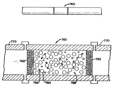
Referring to Figur~ 31, an insert 760, known as an analyte con-
centrator, is provided in a capillary tube 770. The insert 760 includes
porous end platss (or frits) 762 which permit fluid to flow
th~rethrough. The end plates are formed from tiny glass beads. The
ins~rt 760 is a tube which contains glass beads 764, known as con-
trolled-pore glass beads, which are coated with a desired chemical
substance which will provide the desired reaction with a sample
which passes through the analyte concentrator. The controlled pore
glass beads are convei1iently in the range of 200 to 400 mesh in size
(300~ J4)
As noted glass beads 764 are coalted with an antibody 766 (or
antigen) which will attract and hold, with high affinity, a specific
antigen 768 (or antibody) presant in a sample, containing one or
several substan,ces, which passes through the capillary. Subsequent
analysis provides information to the rec;earcher as to the attracted
antigen. As an example, porous glass beads have been coated with
protein A from St~phylococcus aureus which has an affinity for
immunoglobulins.
SUBSTITUTE SHEET
wo 93/05390 3 3 2 1 ~ 0 2 ~i I PCI/US92/07160
A ~urther modification of the foregoing aspect of the invention
using porous glass beads employs an analyte concentra~or 772 which
is a unitary structure which does not requir e porous glass end plates
or frits. This structure shown in Figure 32 includes an annular wall
or sleeve 774 of the diameter suitable to permit the structure to be
coupled to or inserted between two portions of a capillary tube 770.
Secured to the inner wall of the sleevs 774 is branched body 776
made up of a plurality of bodies, beads, pllatelets or the like made of
porous glass material (or polyrneric material having a suitable
chemistry in its surface) and these are interconnected by means of
glass strands 778 which also connect the bodies 780 to ths inner wall
of the sleeve 774. The bodies 780 rnay be of any shape and size for
the intended purpose of being coated with an antibody.
It is noted ttlat the binding of a substance to a surface or wall
or beads or to any other structure within any part of the architec~ure
of the analyte concentrator can be achievcd for chemicals other than
antibodies. These substances may have a high-affinity interaction
(i.e. enzyme-substrate, lectin-carbohydrate, drug-receptor, ion-
chelating agent, etc.), or may ex~rt a strong repulsion to each other
(equal surface charge, etc.).
Fig. 33 shows a modification of the invention wherein a
freeform network of glass or plastic filaments 782 are secured to-
gether and the ,unitary structure thus formed can be coated with an-
tibody (or other chemical substance) and inserted in a capillary tube
784.
In another modification of this aspect of the invention shown in
Fig~ 34, the equivalent of a large number of ba11s in a capillary tube is
SUBSTITUTE SHEET
wO 93/05390 2 ~ ~ O .? ~ 1 3 4 Pcr/uss2/07l60
achieved by an ins~rt 786 in a capiilary tube 770. The insert is
essentiaily a glass or plastic tube having a plurality of small di-
ameter rod passages or through holes 788 which extend through the
body and are coa~ed with a chemical substance as described above.
This form of the invention doas not require porous end plates or frits
to ke~p tha body within the capillary tube.
Fig. 35 shows a modification of an analyte concentrator which
US2S a mesh 801 of a chemical substanc~ or polymer, such as acry-
lamide or agarose or the liket as the matrix for the antibody or anti-
gen.
In operation of the inventions wh~rein structur~s are coated
with an antibody, after a sample has passed through tha structure and
there has been binding of an antigen to the antibody structure, the
capillary is washed to removs ~xcess material and then the trapped
antigens ars removed and processed for study.
In another modification of the inv~ntion illustra~ed schemati-
cally in Fig. 24, the inlet end of a capillary tube (-~aparation capillary)
719 is in a container 723 of buffer solution and th~ outlet end
(detection capillary) is inserted into a porous glass sleeve 725 placed
..
in a container 723' of buffer solution. A fused silica capillary tube
727 extsnds from the end of the capillary 719 out of the buffer
container and sampies flow as droplets out of the end of the capillary
bJbe where~ y are r~coverad.
The apparatus of Fig. 24 can be used as a system for delivering
drugs into the body from container 723. In such use, the end 727 of
the capillary tube would be inserted directly, or would carry a sy-
ringe-like end which can be inserted into the body whereby drug
SUBSTITUTE SHEET
wo 93/~5390 3 5 2 1 2 0 2 S 1 PCr/US92/07160
droplets 729 would be administered or injscted into the cell(s) or
tissue~s) o~ interest.
The porous glass tube 725 is a selective membrane which al-
lows ions to ~scape but the sampl~s to be analyzed pass through the
tubing to be collected.
Th~ principl~s of ~his invantion, used originally for Insertion
in~o the open end of the detection capillary of a carbon fiber electrode
for electrochemical detection of the solute zones, can be used to
provide a fraotion collector. As illustrated schematically in Fig. 25,
the free end of the capillary tube 727 is directed through a vertical
holder 731 coupled to an horizontai arm 733 and positioned over ~he
sample eups 735 in rotatable tabl~ 737. In operation of the apparatus
shown in Figs. 17 and 25, high positive potential is applied to an
electrode in sampie cups 735 in box 90 (autosampler or autoinjector
sid~) and ground potential is applied to an electrods in buffer
container 726 (frae~ion colleGtor side). Sinee ground potential is in
the buffer in container 726, thsre is no potential and no buffer is
required in the sample cups 735 in the table 737 in box 100 and these
portions of the overall apparatus can be handled without concern for
~lectrieal shock.
In still another modification of the inv~ntion last d~scribed and
illustra~ed in Fig. 26, a light sensor 739 is coupl2d to the end of the
tube above tl~'rotatable table and an optieal glass fiber 741 or the
liks is coupled from the sensor to a dstector 743. With this ap-
paratus, the sensor senses the passage of a sample 745 and this is
detected by the deteotor which is used to determine when the table
SUBSTITUTE SHEET
WO 93/~5390 ~ 3 6 PCI/US92/07160
should be ro~ated so that a single sample can be d~posited in each
sample cup 735.
While the method of operating the apparatus 10, as described
above, is satisfactory for many applications and suitable sensitivity
is achieved, another method of operation provides improved spec~rum
analysis and great~r ability to identify samples by providing complete
spectral analysis. Changes in wavel~ngth increments as little as t or
2 mm can b~ us~d in order to maximize sensitivity and cover the
entire spectral range.
In this mode of operation illustrated in Fig. 27, the detector is
set for a first wavelength of operation and, after a sample 630 flows
down the capillary tube to the left to the detector 290 and passes
through the detector and provides an output pulse from the d~tector at
th~ first wavelength, the high voltage polarity is reYersed and the
directicn of the flow of the buffer is reversed, therefore the direction
of the sample is revers~d, and the sample passes to the right through
the detector again, with the detsctor set at a second wavel~ngth.
This causes a second pulse to be provided by th~ det~ctor. Now, the
polarity is reversed again and the sample flows is in the original
rdirection to the left and provides an output with the detector set to
provide a third wavel~ngth of light. This operation of potential
reversal and cycling of the sample back and forth is repeated, at
different detect,or wavelengths of light until a series of pulses is
obtained which, when plotted provides two peak wavelengths, with
the second peak providing accurate identification of the sample.
Almost every pure substance provides such a series of pulses to
permit identification thersof.
SUBSTITUTE SHEET
wo 93/0s3s~ 3 7 ~ 1 2 0 2 5 ~ PCI/US~2/07160
A typical series of pulses 643 which might be obtained to
provide a complete spe~trum for a sample is illustrated in Fig. 28
along with a plot of wavelength versus absorbance, illustrated in Fig.
29 . ~ `
Lights or lamps 700 and 702, shown in Fig. 17 on the front panel
of the apparatus can be used to indicats in which ~rection flow is
taking place at any instant. In addition, a switch 710 is provided for
reversing the high voltage polarity to achieve the cyoling operation
described above.
After many injections of samples, particularly those con~aining
substances which have a tendency to stick to the walls of the
capillary column such as serum or other biological fiuids, it is nec-
essary to restore the capillary column. Since commercially av~ilable
fused-silica capillaries are inexpensive, one way to restore the
capillary column is to replace it entirely.
Another alternative is to recycle the capillary column by a
cleaning procedure. As seen in Fig. 30, a T-shaped connection 747 is
made near the end of the capillary column with a small tube of ma-
terial such as metal, glass, plastic, teflon or the like. This connec-
tion is now part of the capillary column. The connecting tube is at-
tached to a valve 750 and a vacuum pump 752. This system can be
operated through computer control allowing the column to be cieaned
in a coordinated manner, i.e., by purging with potassium hydroxide,
followed by deionized water and buffer aspirated from cups in the
rotatable table 170 or other apparatus and discarded via a teflon port
leading to a fluid trap. The capillary column is then ready for a new
separation test.
SUBSTITUTE SHEET