Note: Descriptions are shown in the official language in which they were submitted.
~NO93/06780 2 ~ 2 0 S 1 6 PCT/Usg2/08380
APPARATUS AND METHOD FOR VASODILATION
Backoround of t~ç Invention
This invention relates to a method and appara~us
s for dilating blood vessels in vasospasm.
Vasospasm is an abnormal and often persistent
contraction of an artery that reduces the caliber of the
artery and may critically reduce blood flow. Vasospasm
can produce a partial or complete obstruction in arteries
that otherwise appear completely normal. Greater or
lesser amounts of dynamic or spastic constriction at the
point of a fixed obstruc~ion can create a severe
reduction of f low even where the f ixed obstruction itself
would be clinically benign.
Vasospasm can occur spontaneously; or it may occur
as the result of certain pharmacological stimuli, such
as, f or exa~ple, ergonovine testing; or of mechanical
stimuli such as contact with a eurgical instrument or a
diagnostic or therapeutic catheter, for example as a
complication of percutaneous transluminal catheter
angioplasty (PTCA); or of environmental stimuli.
Raynaud' 8 phenomenon and Printzmetal angina are two
additional forms of vasospasm. Furthermore, certain
maladies such as subarachnoid hemorrhage can also lead to
vasospasm. In particular, cerebral vasospasm, which is
caused by subarachnoid hemorrhage, and opthal~ic artery
vasospasm may cause severe consequences if not treated
promptly.
Various medications have been tested for the
relief of vasospasms and are only partially effective.
For example, vasospasm in coronary vasculature has been
treated with calcium channel blockers. However, for some
unknown reason that relates to the pharmacological and
anatomical differences between cerebral and coronary
vasculature, these drugs are ineffective against cerebral
W093/06780 PCT/US92/08~0 ; ;
~1211~1~
- 2 -
vasospasm. In addition, mechanical treatment such as
balloon angioplasty is also ineffective against cerebral
vasospasm.
Other non-chemical treatments, e.g., laser
irradiation-induced dilation of the vessels, of vasospasm
have likewise been relatively unsuccessful or plagued
with various problems. For example, laser irradiation-
induced dilation of blood vessels is cumbersome, may
damage surrounding healthy tissue, does not use standard
catheter guide wire techniques, and provides a narrow
margin between the laser energy needed to cause
vasodilation and that needed to perforate the vessel
wall. Moreover, in those laser techniques using low
level constant wave laser radiation, vasospasm resumes as
soon as the radiation ceases.
Furthermore, mechanical dilation treatments, such
as balloon angioplasty, are generally ineffective
because, vasospasm generally resumes after the balloon is
removed, and these treatments are very difficult in
arteries that are hard to catheterize, e.g., the
ophthalmic artery.
Summarv of the Invention
This invention features dilation of blood vessels
in vasospasm through the use of high frequency (on the
order of microseconds) waves, e.g., hydraulic or acoustic
waves, and offers several advantages over known laser
irradiation- or chemical-induced dilation. These
advantages include greater gafety by preventing damage to
the blood vessel walls by the wave generator, e.g., a
laser pulse, increased maneuverability, dilation over a
concentric catheter guide wire, and an increased range of
energy levels that may be safely used for therapy. In
addition, the invention is successful for treating
cerebral vasospasm, which currently is not known to be
susceptible to any mechanical or chemical treatments.
`~093/06780 2 1 2 0 ~ I ~ PCT/US92/08~0
- 3 -
Furthermore, based upon animal studies done to date, we
have found that vasospasm does not resume after treatment
according to the invention. The method of the invention
is suitable for any vasospasm, including any vasospasm
intractable to medication, either functionally, or time
limited.
The invention features an apparatus for dilating a
fluid-filled blood vessel in vasospasm including a
catheter having a lumen containing a fluid, a wave
generator arranged within the catheter lumen for
generating a wave front that propagates through the fluid
in the lumen and is transmitted from the distal end of
the catheter to propagate through the fluid in the blood
vessel, and an energy source connected to the wave
generator to provide energy to produce the wave front.
The wave generator of the invention may be a laser
beam, e.g., ~ pulse, when the energy source is a laser.
i In preferred embodiments, this pulse has a duration of
~, from about 10 nanosec to about 300 ~sec and is of a
wavelength of less than about 600 nm or greater than
about 1000 nm. The laser may be, e.g., a holmium, ultra
violet, or pulsed-dye visible laser.
~t The wave generator also may be a spark generator,
ultrasound agitator, or piezoelectric agitator.
When the distal end of the catheter is open, the
wave front is transmitted from ~he distal end of the
catheter by exiting the ¢atheter and passing into the
vessel. When the distal end of the catheter is sealed
with a membrane, the wave front is transmitted from the
~ 30 di~tal end of the catheter via the membrane.
s~ In any of these embodiments, the wave may be,
e.g., a hydraulic or acoustic wave.
In a preferred embodiment, the invention also
features an apparatus for dilating a fluid-filled blood
3S vessel in vasospasm that includes a catheter having a
"
Aj
~1
.
W093~067~0 PCT/US92/08380 ~ I
' ~20S ~6 4 _
lumen containing a fluid, a laser energy conducting
filament arranged axially within the catheter lumen, the
distal end of the filament being positioned at a distance
fro~ the distal end of the catheter such that laser - ~-
energy emitted from the filament generates a cavitationbubble within the catheter lumen that generates a wave
front that propagates through the fluid in the lumen and
is transmitted from the distal end of the catheter to
propagate through the fluid in the blood vessel, and a
laser energy source connected to the conducting filament
to provide laser energy to produce the wave front.
The invention also features a method of dilating a
fluid-filled blood vessel in vasospasm by propagating a
wave front that induces vasodilation through a fluid in a
blood vessel in need of dilation without generating a
shock wave or cavitation bubble within the blood vessel.
Furthermore, the invention features a method of
dilating a fluid-filled blood vessel in vasospasm by
inserting a catheter into a blood vessel in vasospasm,
the catheter having a lumen containing a fluid,
generating a wave front in the fluid in the catheter,
propagating the wave front through the fluid in the
catheter, transmitting the wave front frsm the distal end
of the catheter, and propagating the transmitted wave
front through the fluid in the blood vessel to induce
va~dilation.
The wave front used in this method may be
generated by a laser pulse that forms a cavitation bubble
within the catheter without forming a laser breakdown-
induced shock wave. In addition, the wave may begenerated ffl other wave generators noted above.
The fluid through which the wave front propagates
may be, e.g., blood, a crystalloid solution such as
~aline or lactated Ringer's solution, or a colloid
solution. When a laser is used, the fluid may be any
V093/06780 2 1 2 Q 5 1~ PCT/US92/08~0
solution that absorbs the incident laser energy. This
fluid i8 in the catheter and, when it is ~omething other
than blood, may be infused into the blood vessel in
vasospasm prior to dilation. - -
Other features and advantages of the invention
w~ll be apparent from the following description of the
preferred embodiments thereof, and from the claims.
Detailed Descrition
The drawings are first briefly described.
~rawinas
Fig. 1 is a schematic of the vasodilator of the
invention.
Fig. 2 is a schematic of a close-ended
vasodilator.
Dilation
The invention utilizes a wave front transmitted
from the end of a catheter to bring about dilation of a
ve~cel in spasm. This wave front is created by a wave
generator, e.g., a laser pulse, a spark generator, an
ultrasound or piezoelectric agitator, or any other
mechanism that can vaporize or displace the fluid rapidly
enough to create a wave front. For example, a laser
pulse may be used to produce a cavitation bubble, i.e., a
vapor bubble, which expands to displace a certain fluid
volume in a column inside the catheter. Any short laser
pulse can be calibrated to give the appropriate dilatory
wave, and the laser energy is adjusted so that a laser-
induced breakdown of the fluid does not occur.
~herefore, essentially no shock wave is created according
to the invention. Only a hydraulic or acoustic wave is
formed, which is on the order of 100 times slower than a
shock wave generated by a laser-induced breakdown. The
physical characteristics of this macroscopic hydraulic or
acoustic wave are significantly different from a
microscopic shock wave.
W093/06780 PCT/US92/08~0
2 ~ 6 -
This displaced fluid serves to create a transient
pressure increase or wave front that propagates coaxially
down the catheter and is transmitted into the occluded
artery. In one embodiment, the catheter is open-ended,
and the wave front is transmitted from the distal end of
the catheter merely by exiting the catheter and passing
into the fluid filling the artery. In other embodiments,
the catheter is alose-ended, and the wave front strikes a
membrane, e.g., a highly pliable polymer film, that
transmits the wave energy to the fluid in the occluded
vessel to generate a transmitted wave that continues on
the outside of the catheter and propagates through the
occluded vessel.
In each embodiment, no destructive shock wave or
cavitation bubble is generated inside the exposed blood
vessel. The only effect on the bl~od vessel is from the
wave that is generated within the confines of the
catheter and then propagates through the fluid within the
vessel and dilates it for some distance beyond the distal
end of the catheter.
Structure
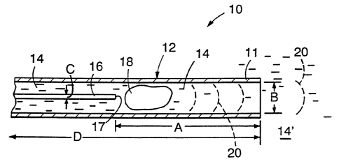
Fig. 1 shows a laser-catheter device 10 that may
be used to generate wave fronts according to the
i invention. The device includes a catheter 12 with an
i 25 inner diameter B and a length D. This catheter is filled
~ with a fluid 14, e.g., blood, a crystalloid solution such
if as ~aline, or a colloid solution. In general, this fluid
i may be any solution capable of absorbing the incident
laser energy. An optical fiber 16, with a diameter C, is !
located within catheter 12. The tip 17 of fiber 16 is
located at a distance A from the distal end 11 of
i catheter 12. When a pulse of laser energy is generated
by a laser (not shown), it is transmitted through fiber
16 and creates a cavitation, or vaporization, bubble 18
in fluid 14. This bubble generates a wave front 20
i
~093/06780 ~ b PT/US92/08~0
(represented by dashed lines in the Figures) that
propagates through the liguid towards the distal end of
the catheter.
This wave front 20 then can either exit the - -
catheter, in the open-ended embodiments, or, as shown in
F~g~ 2, strike a membrane 11~ that ~eals the distal end
11 of catheter 12. Fig. 1 shows that in the open-ended
catheter, the transmitted wave front 20 is the same wave
front 20 inside the catheter. Fig. 2 shows that in the
sealed-end catheter, the transmitted wave front 20'
differs from, but corresponds to wave front 20 inside the
catheter. In both cases, the transmitted wave front
propagates through the fluid 14' that surrounds the
catheter and fills the vessel to be dilated. In other
embodiments, the optic fiber 16 is replaced by another
wave generator to create wave fronts 20.
Use of the Catheter Wave Front Gçnerator
The dimensions and energy of the wave front are
dete D ined by selecting the dimensions A and B of the
catheter and, in the laser system, dimension C of the
optic fiber, as well as the duration and intensity of the
laser pulse. In non-laser embodiments, energy levels of
the ~park from a $park gap or the ultxasound agitation
may be ~elected by standard techniques to achieve the
desired dimensionæ and energy levels.
In particular, the high frequency (preferably on
the order of 75 to 100 ~sec) wave fronts may be created
via a pulsed-dye laser set to deliver a light pulse with
a 15 mJ pulse of 1 ~sec duration into a 200 micron fiber.
This light is disected into a fluid, e.g., blood,
contained well within a catheter inserted in the body
(e.g., A z 25-30 mm or more). Preferably, the laser
pulse is from about 10 nanosec to about 300 ~sec in
duration and is at a wavelength of less than about 600 nm
or greater than about 1000 nm. The preferred lasers for
WOg3/06780 PCT/US92/~3~ ~;
2123S~ 6
- 8 -
use in the invention are holmium, ultra violet, and
pulsed-dye visible lasers. The preferred pulse duration
for a holmium laser is about 250 ~sec.
In addition, the wave front can be created through
the use of an entirely extracorporeal laser delivery
system, with only the catheter being inserted into the
occluded artery, i.e., the dimension A being the length
of the catheter section inserted into the body.
The method of using this catheter wave generator
is as follows. The catheter is advanced in a blood
vessel in the body to an area of vasospasm. once the
catheter is set, a wave front of short duration is
launched down the center of the catheter and either exits
the catheter, or is transmitted via a membrane at the end
of the catheter, into the occluded vessel to effect
dilation. This procedure is repeated as necessary until
the full length of spasm has been dilated.
For example, a Tracker-18 Target Therapeutics (San
Jose, California) catheter has been used successfully,
but smaller, more maneuverable catheters may also be
used. In addition, larger catheters have also been used
successfully, and the proper size may be easily
determined by those skilled in the art depending on the
application.
Animal Studies
We studied intraluminal high frequency waves in
the form of hydraulic waves as a means of reversing
arterial vasospasm in the rabbit model. Vasospasm was
induced in 10 New Zealand white rabbit carotid arteries
in vivo by application of extravascular whole blood
- retained in a silicon cuff. The hydraulic wave consisted
o~ a wave front propagated in an overdamped mode with a
single oscillation (average velocity 30-40 m/s; BW 200
~sec; repetition rate 2 Hz). This wave front was created
~ . 212 ~ S ~ G rCT/USg2/08~
_ g _
in the carotid arteries with a 3 Fr laser catheter
delivered via the femoral artery and coupled to a pul~ed-
dye laser. Carotid artery diameters were assessed by
angiography and measurements made from cut films. - -
Baseli~ne diameters decreased by 37.5% followingapplication of blood. Following delivery of the
hydraulic wave, arterial diameter increased by a mean of
48% over spasm diameter, with no reversion to spasm
dia~eters. Such waves delivered to control vessels not
in spasm had minimal effect on their diameter (mean
change of 5.3~). Scanning electron microscopy revealed
no evidence of perforations or other endothelial damage.
These experiments demonstrate that high frequency
intraluminal wave fronts, e.g., hydraulic waves, can
~5 rapidly reverse arterial vasospasm without loss of
structural or endothelial integrity of arterial walls.
Thera~v
This system can be used for the treatment of any
vasospa~m including cerebral va~o~pasm, ophthalmic
vaso~pasm, acute closure in post PTCA patients, as well
as any spasm intractable to medication, either
functionally, or time limited. --
Other embodiments are within the following claims.