Note: Descriptions are shown in the official language in which they were submitted.
CA 02120731 2003-02-19
60412-3004
- 1
METHODS OF IMPROVING ALLOGRAFT OR XENOGRAFT TOLERANCE BY ADMIN-
ISTRATION OF AN LFA=3 OR CD2 BINDING PROTEIN
TECHNICAL FIELD OF INVENTION
The present invention relates to methods of
improving tolerance of transplanted xenograft tissue or
allograft tissue by administration of LFA-3 or CD2
bindinq proteins in mammals, includinq humans.
EACKGROUND OF THE INVENTION
An allograft is tissue that is transplanted
between genetically nonidentical members of the same
species. Allografts of organs such as the heart,
kidney, liver, pancreas, cornea, bone marrow, lung and
skin have become an increasingly successful and
accepted medical practice for the treatment of various
end stage diseases. The resulting increase in demand
for transplants, unfortunately, has not been matched by
an increase in the present donor supply, and efforts to
increase the supply of human donors are not predicted
to match the rising demand for human orqans. For
example, only 2,000 of the 14,000 patients per year who
are eligible for a cardiac allograft actually receive a
heart transplant in the United States (Rose, "Risks Of
WO 93/06852 2 12 07 2 1 PCT/US92/08754
- 2 -
Cardiac Transplantation", Ann. Thorac. Sura., 47,
p. 615 (1989)).
Consequently, interest has increased in
alternative sources for donor organs. One such
alternative source is xenografts, which are transplants
of tissue from one species to another species.
A problem for both allografts and xenografts
is rejection of the donor graft tissue by the
recipient. Graft rejection is the result of a
complicated and not fully understood chain of events in
the immune system. There are generally two facets of
the immune response: 1) a cell mediated response,
primarily comprising cytotoxic T cells which attack and
kill foreign cells or virus-infected cells; and 2) a
humoral response, comprising the activation of B cells
to plasma cells which secrete antibodies specific for
foreign macromolecules.
Graft rejection is histologically
characterized by the progressive infiltration of
mononuclear cells, including lymphocytes, into the
foreign tissue. The increased presence of these cells
precedes the destruction of the graft by several days.
Sensitized T lymphocytes, therefore, appear to be the
principal initiators of the rejection process.
T lymphocytes play a major role in the immune
response by interacting with target and antigen-
presenting cells. For example, T lymphocyte-mediated
killing of target cells is a multi-step process
involving, initially, adhesion of cytolytic
T lymphocytes (the effector cells) to target cells,
such as graft endothelium. Also, helper T lymphocytes
help initiate the immune response by adhesion to
antigen-presenting cells within the graft tissue.
These interactions of T lymphocytes with
target and antigen-presenting cells are highly specific
õW0 93/06852 2120ry 3{ PC.'I'/US92/08754
- 3 -
and depend on the recognition of an antigen on the
surface of a target or antigen-presenting cell by one
of the many specific antigen receptors on the surface
of T lymphocytes.
The receptor-antigen interaction of
T lymphocytes and other cells is also facilitated by
various T lymphocyte surface proteins, e.g., the
antigen-receptor complex CD3 and accessory molecules
such as CD4, LFA-1, CD8, and CD2. It is also affected
by accessory molecules such as LFA-3, ICAM-1 and MHC
that are expressed on the surface of the target or
antigen-presenting cells.
The interaction between CD2 and LFA-3 remains
poorly understood with respect to activation of T cell
activity. Recent studies have suggested that there is
a specific interaction between CD2 (a T lymphocyte
accessory adhesion molecule) and LFA-3 (a target cell
and antigen presenting cell accessory molecule) which
mediates T lymphocyte adhesion to the target or antigen
presenting cell. This cell-cell adhesion has been
implicated in the initiation of T lymphocyte functional
responses (Dustin et al., "Purified Lymphocyte Function
Associated Antigen 3 Binds To CD2 And Mediates
T lymphocyte Adhesion," J. Exp. Med., 165, pp. 677-92
(1987); Springer et al., "The Lymphocyte
Function-associated LFA-1, CD2, and LFA-3 Molecules:
Cell Adhesion Receptors of the Immune System", Ann.
Rev. Immunol., 5, pp. 223-52 (1987)). The LFA-3/CD2
interaction also plays a role in mediating T lymphocyte
interactions with thymic epithelial cells, in antigen-
independent and dependent conjugate formation and in
T lymphocyte rosetting with erythrocytes (see, e.g.,
Seed et al., "Molecular Cloning Of The CD2 Antigen, the
T-Cell Erythrocyte Receptor, By a Rapid Immunoselection
WO 93/06852 PCT/US92/08754
2120731
- 4 -
Procedure", Proc. Natl. Acad. Sci. USA, 84, pp. 3365-69
(1987)).
LFA-3, which is found on the surface of a
wide variety of cells, including human erythrocytes,
has become the subject of a considerable amount of
study to further elucidate its role in various
T lymphocyte interactions (see, e.g., Krensky et al.,
"The Functional Significance, Distribution, and
Structure of LFA-1, LFA-2, and LFA-3: Cell Surface
Antigen Associated with CTL-Target Interactions",
J. Immunol., 131(2), pp. 611-16 (1983); Shaw et al.,
"Two Antigen-Independent Adhesion Pathways Used by
Human Cytotoxic T-cell Clones", Nature, 323, pp. 262-64
(1986)). Two natural forms of LFA-3 have been
identified. One form of LFA-3 ("transmembrane LFA-3")
is anchored in the cell membrane by a transmembrane
hydrophobic domain. cDNA encoding this form of LFA-3
has been cloned and sequenced (see, e.g., Waliner
et al., "Primary Structure of Lymphocyte
Function-Associated Antigen-3 (LFA-3)", J. Exp. Med.,
166, pp. 923-32 (1987)). Another form of LFA-3 is
anchored to the cell membrane via a covalent linkage to
phosphatidylinositol ("PI")-containing glycolipid.
This latter form has been designated "PI-linked LFA-3",
and cDNA encoding this form of LFA-3 has also been
cloned and sequenced (Wallner et al., PCT publn.
WO 90/02181).
The human CD2 (Til) molecule is a 50 kD
surface glycoprotein expressed on >95% of thymocytes
and virtually all peripheral T lymphocytes.
Biochemical analyses using specific monoclonal
antibodies have suggested that CD2 is T lineage-
specific and exists on the cell surface in several
differentially glycosylated forms (Howard et al., "A
35. Human T Lymphocyte Differentiation Marker Defined by
CA 02120731 2003-02-19
6041.2 -30Q4
- 5 -
Monoalonal Antibodies that block 1;-Rosatts Formation",
2X=o1. , 126, pp. 3117-32 (1961) r Brown at al., in
Iaukoq,vta Tvcinq ZII, ad. KcKichasl, Oxford University
Prass, pp. 110-12 (1987); Sayra =t al.,
I "Molecular cloning and expression of Til cDNAs reveal a
receptor-like structure on human. T lymphocytes", Proc. Natl.
]-cad, flci. USa, 64, pp. 2941-45 (1987)). Tha sequence
of a human C07 qans has been reported (8aad and Aru!!o,
"Molecular Cloninq of the CD2 Antiqon, the T-co7.1
Erytarocyta Receptor, by a Rapid smaunoa+slaction
Procedure", prea. Natlõ A,cse! Acl. U81-, a{, pp. 3365-69
(1987); Sayra at al., iUM (1997). 8o2ubZ= CD2
Dolypaptidas havinq an LFA-3 bindinq domain hava been
roperted (PCT pub2. WO 90/04187).
Monoclonal antibodies to C03, a.9, T53/16,
T11l , Til=; T113, and to L'PA-3=, 0.9. , TS2/9, have also
been reported (sea, e.g., Muqhes ot ai., "Ths Endo-
thelial Ca21 as a itsqulator of T-Ca11 Function",
isssunol. Bsviy,va, 117, pp. 89-103 (1990) f Ksuar, "An
..~..--
Alternative Pathway o! T-Coil Aotivations A lruactional
Rols !or the 50 kd T1l f9hsap lrythrocyts Receptor Pro-
tain", sjlõy, 36, pp. 897-906 (1984)1 Sanchsz-Madrid
at aY., "Three Distinct Antiqans Asaociatad with Human
T-Lyaphocyts-lssdiatad Cytolysis t LtA-Z, LI'71-Z, and
Z'J Lt11-30 , iõt1. kca:d. Sci. t1SA, 79, pp. 7489-93
(1952) )r Sroabarq at al., :*r,,,_aõn_,_; sntatism, sl, pp. 219-
135 (1991) 1 EP 0 260 880 h1.
Suppression of tha immwsa response to prsvsnt
graft rejection has previously been effected by dzvqs,
such as prodnisons, eyclosporins, asathioprins or
cyclophoaphamide, vhich nonspe4ilically block oall-
7adiatsd rssponsos. rrradiation has also basn used to
destroy T and s lymphocytes that aould react aqainst
the tranaplanted qralt tissue. Imunosuppression with
the above techniques, however, oanaot produc= antiqan-
CA 02120731 2003-02-19
60412--30Q4
- 6 -
spaeific tolerance and, therefore, greatly increases
the pationtts susceptibility to opportunistic
infection. In addition, other detrimental side effects
will occur with chronic uio of the above
immunosuppression techniques, For example, chronic
cyclosporine treatment is associatsd with a high
incidenc= of renal toxicity, hypertension and malignant
neoplasm.
Cytotoxic T lympKooyte mediated rosponses are
controlled by cyclosporine or prednisone, but imune
suppressivt therapy is ineffectual for humoral
rejection episodes. Currently, there is no therapeutic
intervention for huawral rejection.
To date, th4refore, conventional methods and
therapeutic agents have not proved to be satisfactory
for improving tolerance of xenoqrafts or allografts.
Accordingly, the need still exists for a process which
avoids the disadvantages of thr conventional methods
and agents whil= providing an effective method for
decreasing the severity of rejection of qraft tissue.
Si]NIl-"Y OP TgE I?NENTION
The present invention generally solves many
of the probleaa retarred to above. It, for the first
time, providea a method for improvinq tolerance of
transplanted allograft tissue or xenoqraft tissue in a
aamal. The method of this invention compris4s the
steps of adYinistering to a sasmal, preferably a human,
a qraft tissue and an LFA-3 or CD2 bindinq protein.
The methods of the invention will preferably be used to
improve tolerance of cardiac and renal xenoqrafts and
allografts. The methods of this invention are superior
to previously available therapies for improving graft
tolerance for many reasons, includinq avoidance of
undesirable side effects such as increased
CA 02120731 2004-06-30
60412-3004
- 6a -
According to one aspect of the present invention,
there is provided use of a soluble lymphocyte function-
associated antigen-3 (LFA-3) polypeptide-immunoglobulin
fusion protein to improve tolerance of transplanted
allograft tissue in a mammal.
According to another aspect of the present
invention, there is provided use of a monoclonal anti-LFA-3
antibody produced by a hybridoma selected from hybridomas
having accession numbers ATCC HB 10693 (1E6), ATCC HB 10694
(HC-1B11), ATCC HB 10695 (7A6) and ATCC HB 10696 (8B8) or is
the monoclonal antibody TS2/9, to improve tolerance of
transplanted allograft tissue in a mammal.
According to a further aspect of the present
invention, there is provided a medicament for use in
improving tolerance of transplanted allograft tissue in a
mammal comprising a soluble lymphocyte function-associated
antigen-3 (LFA-3) polypeptide-immunoglobulin fusion protein
and a pharmaceutically acceptable diluent.
According to still a further aspect of the present
invention, there is provided a medicament for use in
improving tolerance of transplanted allograft tissue in a
mammal comprising a monoclonal anti-lymphocyte function-
associated antigen-3 (anti-LFA-3) antibody produced by a
hybridoma selected from hybridomas having accession numbers
ATCC HB 10693 (1E6), ATCC HB 10694 (HC-1B11), ATCC HB 10695
(7A6) and ATCC HB 10696 (8B8) or the monoclonal antibody
TS2/9.
WO 93/06852 2120731 PC'T/US92/08754
- 7 -
susceptibility to opportunistic infection, renal
toxicity, hypertension and malignant neoplasm.
BRIEF DESCRIPTION OF THE DRAWINGS
Figures 1 and 2 illustrate T cell dependent
B cell activation assay results for two baboons
injected with an anti-LFA-3 monoclonal antibody (1E6)
and one baboon injected with a non-specific isotype
matched control monoclonal antibody (MOPC21).
Immunoglobulin production as measured by OD units in an
ELISA assay is reflected on the y axes. The number of
days after the initial injection of anti-LFA-3
monoclonal antibody is illustrated on the x axes.
DETAILED DESCRIPTION OF THE INVENTION
Definitions
As used herein, an "LFA-3 binding protein" is
a protein comprising one or more polypeptides capable
of binding to LFA-3. LFA-3 binding proteins include
immunoglobulin light chains, immunoglobulin heavy
chains and antigen-binding fragments thereof. The
component polypeptides of an LFA-3 binding protein
composed of more than one polypeptide may optionally be
disulfide-bound or otherwise covalently crosslinked.
Accordingly, LFA-3 binding proteins include intact
immunoglobulins of types IgA, IgG, IgE, IgD, IgM (as
well as subtypes thereof), wherein the light chains of
the immunoglobulin may be of types kappa or lambda.
Such binding proteins also include portions of intact
immunoglobulins that retain LFA-3-binding specificity,
for example, Fab fragments, Fab' fragments, F(ab')2
fragments, F(v) fragments, heavy chain monomers or
dimers, light chain monomers or dimers, dimers
consisting of one heavy and one light chain, and the
like.
CA 02120731 2003-02-19
6041.2 -3004
8
Also contemplated within the term "LFA-3
binding protein" are soluble CD2 polypeptides and
derivatives thereof, including fusions, that bind to
LFA-3. As used herein, a "soluble CD2 polypeptide" is
a CD2 polypeptide incapable of anchoring itself in a
cell membrane. Such soluble polypeptides include, for
example, CD2 polypeptides that lack a sufficient
portion of their membrane-spanning domain to anchor the
polypeptide or are modified such that the membrane-
spanning domain is nonfunctional. Soluble CD2
polypeptides bind to a naturally occurring LFA-3
polypeptide and are encoded by (a) a naturally
occurring mammalian CD2 DNA sequence (e.g., SEQ ID
NO:5), (b) a DNA sequence degenerate to a naturally
occurring CD2 DNA sequence or (c) a DNA sequence that
hybridizes to one of the foregoing DNA sequences under
conditions equivalent to about 209C to 276C below Tm
and 1 M sodium chloride. Such soluble CD2 polypeptides
are well known. For example, several are described in
PCT WO 90/08187.
As used herein, a "CD2 binding protein" is a
protein comprising one or more polypeptides capable of
binding to CD2. CD2 binding proteins include
immunoqlobulin light chains, immunoglobulin heavy
chains and antigen-binding fragments thereof. The
component polypeptides of a CD2 binding protein
composed of more than one polypeptide may optionally be
disulfide-bound or otherwise covalently crosslinked.
Accordingly, CD2 binding proteins include intact
immunoglobulins of types IgA, IgG, IgE, IgD, IqM (as
well as subtypes thereof), wherein the light chains of
the immunoglobulin may be of types kappa or lambda.
Such binding proteins also include portions of intact
immunoglobulins that retain CD2-binding specificity,
WO 93/06852 21 20 '~ 3~ PCT/US92/08754
õ...
- 9 -
for example, Fab fragments, Fab' fragments, F(ab')2
fragments, F(v) fragments, heavy chain monomers or
dimers, light chain monomers or dimers, dimers
consisting of one heavy and one light chain, and the
like.
Also contemplated within the term "CD2
binding protein" are soluble LFA-3 polypeptides or
derivatives thereof, including fusions, that bind to
CD2. As defined herein, CD2 binding proteins include
fusions of soluble LFA-3 polypeptides and
immunoglobulin regions, such as LFA3TIP (described
infra). As used herein, a "soluble LFA-3 polypeptide"
is a LFA-3 polypeptide incapable of anchoring itself in
a cell membrane. Such soluble polypeptides include,
for example, LFA-3 polypeptides that lack a sufficient
portion of their membrane-spanning domain to anchor the
polypeptide or are modified such that the membrane-
spanning domain is nonfunctional. Soluble LFA-3
polypeptides bind to a naturally occurring CD2
polypeptide and are encoded by (a) a naturally
occurring mammalian LFA-3 DNA sequence (e.g. SEQ ID
NO:1 or SEQ ID N0:3, (b) a DNA sequence degenerate to a
naturally occurring LFA-3 DNA sequence or (c) a DNA
sequence that hybridizes to one of the foregoing DNA
sequences under conditions equivalent to about 20 C to
27 C below Tm and 1 M sodium chloride. Such soluble
LFA-3 polypeptides are well known. For example,
several are described in United States patent
4,956,281, which is herein incorporated by reference.
As used herein, a "humanized recombinant
antibody" is an antibody, produced by recombinant DNA
technology, in which some or all of the amino acids of
a human immunoglobulin light or heavy chain not
required for antigen binding have been substituted for
WO 93/06852 2120731 PC,'1'/US92/08754
- 10 -
the corresponding amino acids from a nonhuman mammalian
immunoglobulin light or heavy chain.
As used herein, a "chimeric recombinant
antibody" is an antibody produced by recombinant DNA
technology, in which all or part of the hinge and
constant regions of an immunoglobulin light chain,
heavy chain or both, have been substituted for the
corresponding regions from another immunoglobulin light
chain or heavy chain.
As used herein, "improving tolerance" of
transplanted graft tissue is decreasing the severity of
or eliminating one or more of the general
characteristics of graft rejection. Such
characteristics evidence immune response directed
against the graft (foreign) tissue and include, for
example, progressive infiltration of mononuclear cells,
such as lymphocytes, into the foreign tissue,
production of lymphocytotoxic antibodies, cytolysis,
necrosis, vasculitis, hemorrhage and fibrosis. Another
readily observable indication of improved tolerance
will be prolonged survival of transplanted graft tissue
in a recipient as compared to a non-immunosuppressed
recipient (control).
Graft Tissue
The methods of this invention are useful in
improving tolerance in mammals, including humans, of
transplanted allograft tissue or xenograft tissue.
They comprise the steps of administering to the mammal
a graft tissue and an LFA-3 or CD2 binding protein.
Such grafts include allografts and xenografts of
tissues derived from sources including the heart,
kidney, liver, pancreas, cornea, bone marrow, lung,
skin and blood. Such tissues include portions of the
organs mentioned above and subfractions of blood.
WO 93/06852 PCT/US92/08754
2120731
- 11 -
Preferably, the methods of this invention are used for
= cardiac allografts and xenografts, and renal allografts
and xenografts. The methods of the invention can be
practiced on any mammal, preferably humans.
In selecting graft tissue, a variety of
factors should be considered. These include, for
example, a minimization of genetic disparity to the
extent possible, ABO blood group compatibility, HLA
compatibility, the availability of donor tissue, the
immune status of the patient and size of the donor
organ. Specifically, in the case of cardiac and renal
allografts or xenografts, the donor organ should be
anatomically compatible and physiologically competent
to support the organ function requirements of the
recipient. Surgical protocols used for various graft
transplants are well known.
While not wishing to be bound by theory,
applicants believe that the LFA-3 and CD2 binding
proteins used in the methods of this invention are
prophylactic and therapeutic for inducing tolerance of
the xenografts or allografts because they inhibit
T cell activation. This inhibition typically occurs
when the LFA-3 or CD2 binding protein inhibits the
LFA-3/CD2 interaction. However, certain LFA-3 and CD2
binding proteins used in this invention may inhibit
T cell activation without inhibiting the LFA-3/CD2
interaction.
Preferred LFA-3 and CD2 binding proteins for
use in the methods of this invention are effective to
inhibit T cell activation.
The utility in the methods of this invention
of specific LFA-3 or CD2 binding proteins may easily be
determined by assaying their ability to inhibit the
LFA-3/CD2 interaction, their ability to inhibit T cell
activation or both.
WO 93/06852 PCT/US92/08754
- 12 -
The ability to inhibit the LFA-3/CD2
interaction may be assayed, for example, using a simple
cell binding assay that permits visual (under
magnification) evaluation of the ability of the
putative inhibitor to inhibit the interaction between
LFA-3 and CD2 on cells expressing these molecules.
Jurkat cells are preferred as the CD2+ substrate and
sheep red blood cells or human JY cells are preferred
as the LFA-3+ substrate. The binding characteristics
of binding proteins useful in this invention may be
assayed in several known ways, such as by radiolabeling
the binding protein (e.g., with 35S or 125I) and then
contacting the labeled binding protein with CD2+ or
LFA-3+ cells, as appropriate. Binding characteristics
may also be assayed using an appropriate enzymatically
labelled secondary antibody. Rosetting competition
assays, such as those described in Seed et al., Proc.
Natl. Acad. Sci. USA, 84, pp. 3365-69 (1987) may also
be used.
The ability of LFA-3 and CD2 binding proteins
to inhibit T cell activation may be determined in any
number of conventional T cell activation assays. These
include, for example, assays which assess the ability
of the binding protein to inhibit T cell proliferation
or cytokine secretion in response to mitogens or
activating monoclonal antibodies directed to other cell
surface proteins (see, e.g., Moingeon et al., "The
Structural Biology of CD2", Immunological Rev., 111,
pp. 111-44 (1989)).
LFA-3 and CD2 Binding Proteins
Many types of LFA-3 and CD2 binding proteins
are useful in the methods of this invention, including
monoclonal antibodies, recombinant antibodies, chimeric
recombinant antibodies, humanized recombinant
. _-.._.~..,~....~.w._.w.. u..w...~_._.....,... . _.__... __...._ _.___.~_..
._ . _.... . _ _
WO 93/06852 PCT/US92/08754
2120731
- 13 -
antibodies, soluble LFA-3 and CD2 polypeptides and
LFA-3 and CD2 mimetic agents, as well as derivatized
(e.g., fused to another polypeptide) or truncated forms
of any of the foregoing.
A. Antibodies
The LFA-3 and CD2 binding proteins useful in
this invention include monoclonal antibodies,
recombinant antibodies, chimeric recombinant
antibodies, humanized recombinant antibodies, and
antigen binding portions thereof. Preferably, the
antibodies are monoclonal antibodies.
It is more preferable to use a monoclonal
anti-LFA-3 antibody produced by a hybridoma selected
from the group of hybridomas having accession numbers
ATCC HB 10693 (1E6), ATCC HB 10694 (HC-1B11), ATCC HB
10695 (7A6), and ATCC HB 10696 (8B8), or the monoclonal
antibody known as TS2/9 (Sanchez-Madrid et al., "Three
Distinct Antigens Associated With Human T-Lymphocyte-
Mediated Cytolysis: LFA-1, LFA-2 and LFA-3", Proc.
Natl. Acad. Sci. USA., 79, pp. 7489-93 (1982)). Most
preferably, the monoclonal anti-LFA-3 antibody is
produced by the hybridoma having accession number ATCC
HB 10693 (lE6).
Among the anti-CD2 antibodies, preferable
monoclonal antibodies include monoclonal antibodies
known as the T111 epitope antibodies, including TS2/18
(Sanchez-Madrid et al., supra, (1982)).
The technology for producing monoclonal
antibodies is well known. Briefly, an immortal cell
line (typically myeloma cells) is fused to lymphocytes
(typically splenocytes) from a mammal immunized with a
preparation comprising a given antigen, and the culture
supernatants of the resulting hybridoma cells are
screened for antibodies against the antigen. See
WO 93/06852 21v0~, ~31 rC.T/US92/08754
~
- 14 -
generally, Kohler et al., "Continuous Cultures Of Fused
Cells Secreting Antibody Of Predefined Specificity",
Nature, 256, pp. 495-97 (1975). Useful imoaunogens for
the purpose of this invention include LFA-3-expressing
or CD2-expressing cells, as well as cell free
preparations containing LFA-3, CD2, or counter
receptor-binding fragments thereof (i.e., CD2 fragments
that bind to LFA-3 or LFA-3 fragments that bind to
CD2). Also useful are derivatized forms of LFA-3, CD2
or portions thereof, such as fusion proteins consisting
of a soluble LFA-3 polypeptide fused to at least
portions of immunoglobulin hinge and constant domains
(e.g., LFA3TIP, described infra).
Immunization may be accomplished using
standard procedures. The unit dose and immunization
regimen depend on the species of mammal immunized, its
immune status, the body weight of the mammal, etc.
Typically, the immunized mammals are bled and the serum
from each blood sample is assayed for particular
antibodies using appropriate screening assays. For
example, useful anti-LFA-3 and anti-CD2 antibodies may
be identified by testing the ability of the immune
serum to block sheep red blood cell rosetting of Jurkat
cells, which results from the presence of LFA-3 and CD2
on the respective surfaces of these cells, screening
for the ability to inhibit T cell activation in vitro
or screening for both. The lymphocytes used in the
production of hybridoma cells typically are isolated
from immunized mammals whose sera have already tested
positive for the presence of the desired antibodies
using such screening assays.
Typically, the immortal cell line (e.g., a
myeloma cell line) is derived from the same mammalian
species as the lymphocytes. Preferred immortal cell
lines are mouse myeloma cell lines that are sensitive
WO 93/06852 21 -! 2Ut1-731 PCT/US92/08754
-.TM 6
- 15 -
to culture medium containing hypoxanthine, aminopterin
and thymidine ("HAT medium").
Typically, HAT-sensitive mouse myeloma cells
are fused to mouse splenocytes using polyethylene
glycol (PEG 3350). Hybridoma cells resulting from the
fusion are then selected using HAT medium, which kills
unfused and unproductively fused myeloma cells (unfused
splenocytes die after several days because they are not
transformed). Hybridomas producing a desired antibody
are detected by screening the hybridoma culture
supernatants, for example, for the ability to bind to
LFA-3 or CD2, or for their ability to block Jurkat cell
adhesion to sheep red blood cells. Useful hybridomas
may also be identified by screening for the ability to
inhibit T cell activation. Subcloning of the hybridoma
cultures by limiting dilution is typically performed to
ensure monoclonality.
To produce anti-LFA-3 and anti-CD2 monoclonal
antibodies, hybridoma cells that tested positive in
such screening assays are cultured in a nutrient medium
under conditions and for a time sufficient to allow the
hybridoma cells to secrete the monoclonal antibodies
into the culture medium. Tissue culture techniques and
culture media suitable for hybridoma cell culture are
well known. The conditioned hybridoma culture
supernatant may be collected and the desired antibodies
optionally further purified by well known methods.
Alternatively, the desired antibody may be
produced by injecting the hybridoma cells into the
peritoneal cavity of a Pristane-primed [2,6,10,14-
tetramethylpentadecane (Aldridge Chemical Co.,
Milwaukee, Wisconsin)] mouse. The hybridoma cells
proliferate in the peritoneal cavity and secrete the
antibody which accumulates in ascites fluid. The
CA 02120731 2003-02-19
60412-3004
- 16 -
antibody may be harvested by withdrawing the ascites
fluid from the peritoneal cavity with a syringe.
LFA-3 and CD2 binding proteins useful in the
present invention may also be recombinant antibodies
produced by host cells transformed with DNA encoding
immunoglobulin light and heavy chains of a desired
antibody, or LFA-3 or CD2-binding portions thereof.
Recombinant antibodies may be produced by well known
genetic engineering techniques. See, e.g., United
States patent 4,816,397.
For example, recombinant antibodies may be
produced by cloning cDNA or genomic DNA encoding the
immunoglobulin light and heavy chains of the desired
antibody from a hybridoma cell that produces an
antibody useful in this invention. The cDNA or genomic
DNA encoding those polypeptides is then inserted into
expression vectors so that both DNA sequences are
operatively linked to one or more transcriptional and
translational expression control sequences.-- The
expression vector and expression control sequences are
chosen to be compatible with the expression host cell
used. Typically, both DNA sequences are inserted into
the same expression vector, although the two DNA
sequences aay also be inserted into different
expression vectors.
Prokaryotic or eukaryotic host cells may be
used as expression hosts. Expression in eukaryotic
host cells is preferred because such cells are more
likely than prokaryotic cells to assemble and secrete a
properly folded and immunologically active antibody.
However, any antibody produced that is inactive due to
improper folding may be renaturable according to well
known methods (Kim and Baldwin, "Specific Intermediates
in the Folding Reactions of Small Proteins and the
WO 93/06852 PCT/US92/08754
2120731
- 17 -
Mechanism of Protein Folding", Ann. Rev. Biochem., 51,
pp. 459-89 (1982)). It is possible that the host cells
will produce portions of intact antibodies useful in
this invention, such as light chain dimers or heavy
chain dimers.
It will be understood that variations on the
above procedure are useful in the present invention.
For example, it may alternatively be desired to
transform a host cell with DNA encoding either the
light chain or the heavy chain (but not both) of an
anti-LFA-3 or anti-CD2 antibody. Recombinant DNA
technology may also be used to remove some or all of
the DNA encoding either or both of the light and heavy
chains that is not necessary for LFA-3 or CD2 counter
receptor binding. The molecules expressed from such
truncated DNA molecules are useful in the methods of
this invention. In addition, bifunctional antibodies
may be produced in which one heavy and one light chain
are specific for LFA-3 or CD2 and the other heavy and
light chain are specific for an antigen other than
LFA-3 or CD2, or for another epitope of LFA-3 or CD2.
Chimeric recombinant antibodies may be
produced by transforming a host cell with a suitable
expression vector comprising DNA encoding the desired
immunoglobulin light and heavy chains in which all or
some of the DNA encoding the hinge and constant regions
of the heavy and/or the light chain have been
substituted with DNA from the corresponding region of
an immunoglobulin light or heavy chain of a different
species. When the original recombinant antibody is
nonhuman and the anti-LFA-3 or anti-CD2 antibody will
be administered to a human, substitution of
corresponding human sequences is preferred. An
exemplary chimeric recombinant antibody has mouse
variable regions and human hinge and constant regions.
WO 93/06852 2129731 PCT/US92/08754
- 18 -
See generally, United States patent 4,816,397 and
Morrison et al., "Chimeric Human Antibody Molecules:
Mouse Antigen-Binding Domains With Human Constant
Region Domains", Proc. Natl. Acad. Sci. USA, 81,
pp. 6851-55 (1984).
Humanized recombinant anti-LFA-3 or anti-CD2
antibodies may be produced by transforming a host cell
with a suitable expression vector comprising DNA
encoding the desired nonhuman immunoglobulin light and
heavy chains in which all or some of the DNA encoding
amino acids not involved in antigen binding have been
substituted with DNA from the corresponding region of a
desired human iminunoglobulin light or heavy chain. See
generally, Jones et al., "Replacing The
Complementarity-Determining Regions In A Human Antibody
With Those From A Mouse", Nature, 321, pp. 522-25
(1986) and European patent publication 0 239 400.
Anti-LFA-3 and anti-CD2 antibodies that are
not intact are also useful in this invention, and may
be derived from any of the antibodies described above.
For example, antigen-binding fragments, as well as
full-length monomeric, dimeric or trimeric polypeptides
derived from the above-described antibodies are
themselves useful. Useful binding proteins of this
type include Fab fragments, Fab' fragments, F(ab')2
fragments, F(v) fragments, heavy chain monomers or
dimers, light chain monomers or dimers, dimers
consisting of one heavy and one light chain, and the
like.
Antibody fragments may also be produced by
chemical methods, e.g., by cleaving an intact antibody
with a protease, such as pepsin or papain, and
optionally treating the cleaved product with a reducing
agent. Alternatively, useful fragments may be produced
by using host cells transformed with truncated heavy
CA 02120731 2003-02-19
60412-3004
- 19 -
and/or light chain genes. Heavy and light chain
monomers may be produced by treating an intact antibody
with a reducing agent, such as dithiothreitol, followed
by purification to separate the chains. Heavy and
light chain monomers may also be produced by host cells
transformed with DNA encoding either the desired heavy
chain or light chain, but not both. See, e.g., Ward
et al., "Binding Activities Of A Repertoire Of Single
Immunoglobulin Variable Domains Secreted From
Escherichia goli", Naturg, 341, pp. 544-46 (1989);
Sastry et al., "Cloning Of The Immunological Repertoire
in Escherichia coli For Generation Of Monoclonal
Catalytic Antibodies: Construction Of A Heavy Chain
Variable Region-Specific cDNA Library", Proc. Natl.
Acad. Sci. USA, 86, pp. 5728-32 (1989).
B. Soluble CD2 and LFA-3 Polypeptides
The LFA-3 and CD2 binding proteins useful in
the methods of the present invention include soluble
CD2 and LFA-3 polypeptides. Soluble LFA-3 polypeptides
are preferred.
Soluble LFA-3 polypeptides may be derived
from the transmembrane form of LFA-3, particularly the
extracellular domain (e.g., AAj-AA187 of SEQ ID NO:2).
Such polypeptides are described in United States patent
4,956,281 and published application W092/16622. Preferred
soluble LFA-3 polypeptides include polypeptides
consisting of AA1-AA92 of SEQ ID NO:2, AAi-AASO of SEQ ID
NO:2, AASO-AASS of SEQ ID NO:2 and AA20-AAgo of SEQ ID
NO:2. A bacteriophage comprising a DNA sequence
encoding SEQ ID NO:2 (i.e., SEQ ID NO:1) is deposited
with American Type Culture Collection, Rockville,
Maryland, under the accession number ATCC 75107.
CA 02120731 2003-02-19
6041.-30Q4
- 20 -
Soluble LFA-3 polypeptides may also be
derived from the PI-linked form of LFA-3, such as those
described in PCT patent application WO 90/02181. A
vector comprising a DNA sequence encoding PI-linked
LFA-3 (i.e., SEQ ID N0:3) is deposited with American
Type Culture Collection, Rockville, Maryland, under the
accession number ATCC 68788. Since the PI-linked form
of LFA-3 and the transmembrane form of LFA-3 have
identical amino acid sequences through the entire
extracellular domain, the preferred soluble LFA-3
polypeptides derived from PI-linked LFA-3 are the same
as those derived from the transmembrane form of LFA-3.
Soluble CD2 polypeptides may be derived from
full length CD2, particularly the extracellular domain
(e.g.,, AA1-AA1S5 of SEQ ID NO:6). Such polypeptides may
comprise all or part of the extracellular domain of
CD2. Suitable soluble CD2 polypeptides are described
in PCT WO 90/08187.
The production of the soluble polypeptides
useful in this invention may be achieved by a variety
of methods known in the art. For example, the
polypeptides may be derived from intact transmembrane
LFA-3 or CD2 molecules or an intact PI-linked LFA-3
molecule by proteolysis using specific endopeptidases
in combination with exopeptidases, Edman degradation,
or both. The intact LFA-3 molecule or the intact CD2
molecule, in turn, may be purified from its natural
source using conventional methods. Alternatively, the
intact LFA-3 or CD2 may be produced by known
recombinant DNA techniques using cDNAs (see, e.g., U.S.
Patent 4,956,281 to Wallner et al.; Aruffo and Seed,
Proc Natl. Acad. Sci. USA, 84, pp. 2941-45 (1987);
Sayre et al., Proc. Nati. Acad. Sci. USA, 84,
pp. 2941-45 (1987)).
WO 93/06852 ~~ ~'= ~1 ~'J i PCT/US92/08754
- 21 -
Preferably, the soluble polypeptides useful
in the present invention are produced directly, thus
eliminating the need for obtaining an entire LFA-3
molecule or an entire CD2 molecule as a starting
material. This may be achieved by conventional
chemical synthesis techniques or by well-known
recombinant DNA techniques wherein only those DNA
sequences which encode the desired polypeptides are
expressed in transformed hosts. For example, a DNA
sequence which encodes the desired soluble LFA-3
polypeptide or soluble CD2 polypeptide may be
synthesized by chemical means using an oligonucleotide
synthesizer. Such oligonucleotides are designed based
on the amino acid sequence of the desired soluble LFA-3
polypeptide or soluble CD2 polypeptide. Specific DNA
sequences coding for the desired polypeptide also can
be derived from the full length DNA sequence by
isolation of specific restriction endonuclease
fragments or by PCR synthesis of the desired region.
The soluble LFA-3 and CD2 polypeptides may be
isolated from the fermentation or culture of
transfected host cells and purified using any of a
variety of conventional methods. One of skill in the
art may select the most appropriate isolation and
purification techniques.
While recombinant DNA techniques are the
preferred method of producing useful soluble CD2
polypeptides or soluble LFA-3 polypeptides having a
sequence of more than 20 amino acids, shorter CD2 or
LFA-3 polypeptides having less than about 20 amino
acids are preferably produced by conventional chemical
synthesis techniques. Synthetically produced
polypeptides useful in this invention can
advantageously be produced in extremely high yields and
can be easily purified.
WO 93/06852 ~ 2 1 0 7 ~. ~ 1 PCT/US92/08754
'
- 22 -
C. LFA-3 And CD2 Mimetic Agents
Among the LFA-3 and CD2 binding proteins
useful in the methods of this invention are LFA-3 and
CD2 mimetic agents. These agents are peptides,
semi-peptidic compounds or non-peptidic compounds which
bind to CD2 (LFA-3 mimetic) or to LFA-3 (CD2 mimetic)
and inhibit the CD2/LFA-3 interaction, inhibit T cell
activation or both.
Such mimetic agents may be produced by
synthesizing a plurality of peptides (e.g., 5-20 amino
acids in length), semi-peptidic compounds or non-
peptidic, organic compounds, and then screening those
compounds for their ability to inhibit the CD2/LFA-3
interaction or for their ability to inhibit T cell
activation or both. See generally United States patent
4,833,092; Scott and Smith, "Searching for Peptide
Ligands with an Epitope Library", Science, 249,
pp. 386-90 (1990); and Devlin et al., "Random Peptide
Libraries: A Source of Specific Protein Binding
Molecules", Science, 249, pp. 404-07 (1990), which are
herein incorporated by reference.
D. Derivatized LFA-3 And
CD2 Binding Proteins
Also useful in the methods of this invention
are derivatized forms, including fusions or hybrids, of
the foregoing LFA-3 and CD2 binding proteins in which,
for example, any of the LFA-3 or CD2 binding proteins
described herein are functionally linked (by chemical
coupling, genetic fusion or otherwise) to one or more
of the same or different LFA-3 and CD2 binding
proteins, to pharmaceutical agents, or to both.
One type of derivatized binding protein is
produced by crosslinking two or more LFA-3 or CD2
binding proteins (of the same type or of different
WO 93/06852 PCT/US92/08754
212 07 3 1
- 23 -
types). Suitable crosslinkers include those that are
heterobifunctional, having two distinctly reactive
groups separated by an appropriate spacer (e.g.,
m-maleimidobenzoyl-N-hydroxysuccinimide ester) or
homobifunctional (e.g., disuccinimidyl suberate). Such
linkers are available from Pierce Chemical Company,
Rockford, Illinois.
Another possibility for cross-linking takes
advantage of the PI linkage signal sequence in PI-
linked LFA-3, or fragments thereof. Specifically, DNA
encoding the PI-linkage signal sequence (e.g., AA162-
AA212 of SEQ ID NO:4) is ligated downstream of DNA
encoding a desired polypeptide, preferably a soluble
LFA-3 polypeptide. If this construct is expressed in
an appropriate eukaryotic cell, the cell will recognize
the PI linkage signal sequence and will covalently link
PI to the polypeptide. The hydrophobic property of the
PI may then be exploited to form micellar aggregates of
the polypeptides.
Also useful are LFA-3 and CD2 binding
proteins linked to one or more pharmaceutical agents
(e.g., a fusion or hybrid protein). Useful
pharmaceutical agents include biologically active
peptides, polypeptides and proteins, such as antibodies
specific for a polypeptide other than LFA-3 or CD2.
Other useful pharmaceutical agents include
immunosuppressants, for example, cyclosporine A,
prednisone, FK506, methotrexate, steroids, and
retinoids.
Preferred derivatized binding proteins
include recombinantly produced polypeptides in which a
soluble LFA-3 polypeptide, soluble CD2 polypeptide, or
a peptidyl CD2 or peptidyi LFA-3 mimetic agent is fused
to all or part of an immunoglobulin heavy chain hinge
region and all or part of an immunoglobulin heavy chain
CA 02120731 2003-02-19
60412-30Q4
- 24 -
constant region. Such fusion proteins are expected to
exhibit prolonged serum half-lives and to facilitate
binding protein dimerization.
Preferred polypeptides for preparing such
fusion proteins are soluble LFA-3 polypeptides, most
preferably a soluble LFA-3 polypeptide selected from
the group consisting of AA1-AA92 of SEQ ID NO:2, AA1-AA$0
of SEQ ID NO:2, AASO-AA65 of SEQ ID NO:2 and AA'0-AA80 of
SEQ ID NO:2.
A bacteriophage comprising a DNA sequence
encoding SEQ ID NO:2 (i.e., SEQ ID NO:1) is deposited
with the American Type Culture Collection, Rockville,
Maryland, under the accession number ATCC 75107.
The most preferred fusion proteins of this
type contain the amino terminal 92 amino acids of
mature LFA-3, the C-terminal 10 amino acids of a human
IgGI hinge region containing the two cysteine residues
thought to participate in interchain disulfide bonding,
and the CH2 and CH3 regions of a human IqGI heavy chain
constant domain (e.g., SEQ ID NO:8). This fusion
protein is referred to herein as "LFA3TIP." A plasmid,
pSAB152, encoding an exemplary LFA3TIP is deposited
with American Type Culture Collection, Rockville,
Maryland, under the accession number ATCC 68720. The
DNA sequence of the pSAB152 insert is SEQ ID NO:7.
one way of producing LFA3TIP for use in the
methods of this invention is described in
published application W092/16622. Generally,
conditioned culture medium of
COS7 cells transfected with pSAB152 was concentrated
using an AMICON S1Y30 spiral cartridge system (AMICON,
Danvers, Massachusetts) and subjected to Protein A-
Sepharose 4B (Sigma, St. Louis, Missouri)
chromatography. The bound proteins were eluted and
*Trade-mark
CA 02120731 2003-02-19
6041.2-30Q4
- 25 -
subjected to Superose-12 (Pharmacia/LKB, Piscataway,
New Jersey) gel filtration chromatography.
~
Superose-12 fractions containing LFA3TIP with
the least amount of contaminating proteins, as
determined on SDS-PAGE gels and by Western blot
analysis, (see, e.g., Towbin et al., Proc. Natl. Acad.
Sci. USA, 74, pp. 4350-54 (1979); Antibodies: A
Laboratory Manual, pp. 474-510 (Cold Spring Harbor
Laboratory (1988)), were pooled and concentrated in a
YM30 Centricon (AMICON). LFA3TIP was detected on
Western blots using a rabbit anti-LFA-3 polyclonal
antiserum, followed by detectably labeled goat anti-
rabbit IgG. The purified LFA3TIP of COS7 cells was a
dimer of two monomeric LFA-3-Ig fusion proteins,
connected by disulfide bonds.
Pharmaceutical Compositions And
Methods According To This Invention
The methods according to this invention
improve tolerance of transplanted allograft tissue or
xenograft tissue by administering to a mammal the graft
tissue and one or more LFA-3 or CD2 binding proteins,
including derivatized forms thereof. The LFA-3 or CD2
binding proteins may alternatively be administered as
part of a pharmaceutical composition.
Useful pharmaceutical compositions will
comprise one or more LFA-3 or CD2 binding proteins,
including derivatized forms thereof, typically in a
pharmaceutically acceptable carrier. By
"pharmaceutically acceptable carrier" is meant a
carrier that does not cause an allergic reaction or
other untoward effect in patients to whom it is
administered.
Suitable pharmaceutically acceptable carriers
include, for example, one or more of water, saline,
phosphate buffered saline, dextrose, glycerol, ethanol
*Trade-mark
CA 02120731 2003-02-19
6041-2-30Q4
- 26 -
and the like, as well as combinations thereof.
Pharmaceutically acceptable carriers may further
comprise minor amounts of auxiliary substances such as
wetting or emulsifying agents, preservatives or
buffers, which enhance the shelf life or effectiveness
of the LFA-3 or CD2 binding protein.
The LFA-3 or CD2 binding proteins or
compositions useful in this invention will preferably
be administered in an "effective amount," meaning an
amount capable of improving tolerance to an allograft
or xenograft as defined herein.
It will be apparent to those of skill in the
art that the effective amount of LFA-3 or CD2 binding
protein will depend, inter alia, upon the
administration schedule, the unit dose administered,
whether the LFA-3 or CD2 binding protein is
administered in combination with other therapeutic
agents, the immune status and health of the patient,
the therapeutic or prophylactic activity of the
particular LFA-3 or CD2 binding protein administered
and its serum half-life.
The pharmaceutical compositions may further
be used in conjunction with general immunosuppressive
agents. These include, for example, cyclosporine,
azathioprine and steroids, such as Depo-Medrol
(methylprednisolone acetate), Solumederol
(aethylprednisolone sodium succinate), and prednisone,
administered in amounts effective to suppress immune
response in the mammal being treated. For example,
cyclosporine may be administered at 2-25 mg/kg/day p.o.
starting the day before surgery, azathioprine may be
administered at 50-200 mg/day, Solumederol may be
administered at 125 mq i.v. at the time of
transplantation and on the first post-operative day,
prednisone may be administered at 1 mg/kg/day p.o.
*Trade-mark
WO 93/06852 PCT/US92/08754
2120'731_
- 27 -
starting on the second post-operative day or
Depo-Medrol may be administered at 0.8 mg/kg/day i.m.
starting on the second post-operative day. The above
dosages will, of course, be varied by the practitioner
depending upon factors well known to those of skill in
the art. In general, when used in conjunction with an
LFA-3 or CD2 binding protein, it will be desired to use
the lowest possible effective concentration of such
immunosuppressive agents.
The pharmaceutical compositions may further
comprise other therapeutic or prophylactic agents. The
LFA-3 or CD2 binding protein and the other active agent
may be in the form of a single conjugated molecule.
Conjugation of the two components may be achieved by
standard cross-linking techniques well known in the
art. A single molecule may also take the form of a
recombinant fusion protein.
The additional immunosuppressive, therapeutic
or prophylactic agents may be administered in single
dosage form with the LFA-3 or CD2 binding protein, in a
multiple dosage form separately from the LFA-3 or CD2
binding protein, but contemporaneously, or in a
multiple dosage form wherein the components are
administered separately but sequentially. Such
combination therapies may advantageously utilize lower
dosages of the immunosuppressive, therapeutic or
prophylactic agents.
The pharmaceutical compositions or LFA-3 or
CD2 binding proteins may be in a variety of forms.
These include, for example, solid, semi-solid and
liquid dosage forms, such as tablets, pills, powders,
liquid solutions, dispersions or suspensions,
liposomes, suppositories, injectable and infusible
solutions. The preferred form depends on the intended
mode of administration and therapeutic application.
WO 93/06852 PCT/US92/08754
2120731
- 28 -
The preferred form is injectable or infusible
solutions.
Typically, the LFA-3 or CD2 binding protein
will be suspended in a sterile saline solution for
therapeutic uses. The pharmaceutical compositions may
alternatively be formulated to control release of the
active ingredients or to prolong their presence in a
recipient's system. Numerous suitable drug delivery
systems are known and include, e.g., hydrogels,
hydroxymethylcellulose, microcapsules, liposomes,
microemulsions, microspheres, and the like.
In accordance with this invention, a mammal
that is to receive transplanted graft tissue and an
LFA-3 binding protein is administered a dose between
about 0.01 and about 10 mg LFA-3 binding protein per kg
body weight, more preferably between about 0.1 and
about 5 mg LFA-3 binding protein per kg body weight,
and most preferably between about 0.1 and about 2 mg
LFA-3 binding protein per kg body weight.
A mammal that is to receive transplanted
graft tissue and a CD2 binding protein is administered
a dose between about 0.01 and about 10 mg CD2 binding
protein per kg body weight, more preferably between
about 0.01 and about 2 mg CD2 binding protein per kg
body weight, and most preferably between about 0.01 and
about 1 mg CD2 binding protein per kg body weight.
The LFA-3 or CD2 binding protein or
composition should be administered about once per day
until, within the judgment of the practitioner, the
danger of rejection of the allograft or xenograft
tissue has diminished. The length of administration of
the LFA-3 or CD2 binding protein or composition is
dependent upon the mammal's acceptance of the graft
tissue. General clinical indications of rejection will
vary with the particular organ transplanted. However,
WO 93/06852 PCr/US92/08754
2 19W 07,31
- 29 -
fever, malaise and organ dysfunction are typical
clinical indications of rejection. Symptoms of organ
dysfunction depend upon the organ transplanted, but are
characterized by well known and recognized indicia to
those of skill in the art.
The success of the treatment may be measured
by a variety of methods including biopsies, such as
incisional myocardial biopsy or percutaneous
endomyocardial biopsy to determine the extent of
lymphocyte infiltration, blood assays to determine the
extent of lymphocytotoxic antibody production or a
mixed lymphocyte reaction (see, e.g., Krensky et al.,
J. Immunol., 131, pp. 611-16 (1983); Bradley, "Mixed
Lymphocyte Responses", in Selected Methods in Cellular
Immunology (Mishell and Shiigi, eds.), pp. 162-64 (W.H.
Freeman and Co., San Francisco 1980)). In the case of
renal transplants, biopsies can be taken to determine
the extent of mononuclear cell infiltration and
proliferation, or necrosis of the arterial endothelium
and media in the graft tissue. (Cosimi et al.,
J. Immunol., 144, pp. 4604-12 (1990)).
The method of the present invention, in a
preferred embodiment for allograft tissue, comprises
administering the LFA-3 or CD2 binding protein once per
day for two consecutive days before the transplant and
once per day for one to ten consecutive days after the
transplant. More preferably, the LFA-3 or CD2 binding
protein is administered once per day for two
consecutive days before the transplant and once per day
for two consecutive days after the transplant.
The method of the present invention, in a
preferred embodiment for xenograft tissue, comprises
administering, before the transplant, an LFA-3 or CD2
binding protein contemporaneously with tissue from the
xenograft source. As used herein, "contemporaneously"
WO 93/06852 PCT/US92/08754
- 30 -
when referring to the administration of tissue from a
xenograft source (other than the graft tissue) and an
LFA-3 or CD2 binding protein, will mean that their
administration occurs near enough in time to allow the
binding protein to bind to the tissue from the
xenograft source at an effective level to inhibit a
significant immune response. Preferably, the binding
protein is bound to the tissue from the xenograft
source at saturating levels. In a preferred embodiment
of this invention, administration of one occurs within
approximately zero to six hours of the other. Most
preferably, the tissue from the xenograft source and
the LFA-3 or CD2 binding protein are administered
within approximately zero to one hour of each other.
Either may be administered first. It is preferable,
however, that the binding protein be administered prior
to tissue from the xenograft source.
In an alternate embodiment of the present
invention, the contemporaneous administration is
followed by the administration of LFA-3 or CD2 binding
protein before the transplant.
More preferably, the LFA-3 or CD2 binding
protein is administered before the xenograft transplant
once per day for two consecutive days, then
contemporaneously with tissue from the xenograft source
once per day for one day, and then once per day for one
to ten consecutive days. If the xenograft source
species and recipient species are unusually discordant,
it may be necessary to administer the LFA-3 or CD2
binding protein contemporaneously with tissue from the
xenograft source once per day for two consecutive days
according to the above schedules. In a preferred
embodiment, the binding protein is administered once
per day for five to ten consecutive days after the
contemporaneous administration and before the
PCT/US92/08754
WO 93/06852 2 120 731
- 31 -
transplant according to the above schedules. Most
preferably, the contemporaneous administration of the
LFA-3 or CD2 binding protein and tissue from the
xenograft source is simultaneous.
Although not wishing to be bound by theory,
applicants administer tissue from the xenograft source
to the mammal contemporaneously with LFA-3 or CD2
binding protein with the intent of inhibiting the
development of a population of activated cells
specifically reactive against that tissue. The
contemporaneous administration of LFA-3 or CD2 binding
proteins induces tolerance to the specific subset of
antigens carried by cells from the specific xenograft
source. Accordingly, it will be understood that any
tissue from the xenograft source may be appropriate,
however blood cells from the xenograft source are
preferred. Such tissue should be administered in an
amount sufficient to elicit an immune response. The
preferred method of administration of tissue from the
xenograft source is intravenous. The administration of
between about 1 x 10⁶ to about 1 x 10⁸ whole blood cells
most preferably will serve as the tissue from the
xenograft source. It will be recognized, however, that
lower or higher dosages and other administration
schedules may be employed.
The LFA-3 or CD2 binding protein or
pharmaceutical composition may be administered
intravenously, intramuscularly, subcutaneously,
intra-articularly, intrathecally, periostally, orally,
topically or by inhalation. Ordinarily, intravenous
(i.v.) or intramuscular (i.m.) administration will be
preferred, however, more localized administrations in
the area of transplantation may be more desirable in
some cases due to the wide range of cells in the body
that express LFA-3.
WO 93/06852 2129731 PGT/US92/08754
- 32 -
In a preferred embodiment of the method of
the present invention, the graft tissue is perfused
with an effective amount of LFA-3 or CD2 binding
protein before implantation into the mammal. Most
preferably, the graft tissue is perfused with enough
LFA-3 or CD2 binding protein to saturate all CD2 or
LFA-3 sites on the graft tissue before implantation
into the mammal.
In order that this invention may be better
understood, the following examples are set forth.
These examples are for purposes of illustration only,
and are not to be construed as limiting the scope of
the invention in any manner.
EXAMPLES
Examole 1
Purification Of Anti-LFA-3 Monoclonal
Antibody 1E6 and Monoclonal Antibody MOPC21
1E6 hybridoma cells (ATCC HB 10693) were
grown in RPMI 1640 medium supplemented with 2% fetal
calf serum, 150 g/mi streptomycin and 50 g/ml
gentamicin (GIBCO Life Technologies, Gaithersburg,
Maryland) in three 40 liter stirred glass vessels
(Bellco, # 196536000) at 37 C for 7 - 10 days. The
conditioned media was pooled and collected into 100
liter carboys (NALGENE). Sodium azide was added to
make the pooled suspension 0.02% final concentration.
The cell debris was removed through a 5 filter
cartridge (Polygard, #CN5001EO6, Millipore, Bedford,
Massachusetts) followed by a 0.3 filter cartridge
(Polygard, #CN0301E06, Millipore, Bedford,
Massachusetts) at room temperature. The clarified
supernatant was concentrated 50 to 100 fold using a
YM30 S10 spiral filter cartridge (AMICON, Danvers,
Massachusetts) at 4 C. The concentrate from 50 liters
WO 93/06852 PC,'I'/US92/08754
21?0 7 31
- 33 -
of conditioned media was diluted with two volumes of
equilibration buffer (3 M glycine, 1.5 M sodium
chloride, pH 8.9) and passed through 90 ml of Protein
A-Sepharose (Schleicher and Schuell, Keene, New
Hampshire) overnight by gravity at 4 C.
The column was washed with equilibration
buffer and the bound proteins were subsequently eluted
with 100 mM sodium citrate, pH 3Ø The eluted
fractions were collected into 1/10 fraction volume of 1
M HEPES, pH 7.8. A280 readings of the fractions were
taken and the fractions containing the eluted protein
were pooled and stored at -70 C. Protein A-purified
1E6 was prepared from a total of about 200 liters of
conditioned media. The various pools were thawed,
combined and concentrated to about 10 mg/ml protein in
a 2 liter Amicon stirred cell using a YM30 filter
(AMICON, Danvers, Massachusetts). The concentrated
material was divided into five 100 ml aliquots. Each
aliquot was passed through a 1 liter Superose-6 gel
filtration column (Pharmacia, Piscataway, New Jersey)
developed in phosphate buffered saline at room
temperature. The peak fractions containing 1E6 were
pooled and stored at -70 C. When all the material was
processed, the pools were thawed, combined and adjusted
to 2-3 mg/ml protein with phosphate buffered saline.
The final material was divided into 15 ml aliquots and
stored at -70 C until use.
MOPC21 was purified from ascites purchased
from the Sigma Chemical Corporation (St. Louis,
Missouri) by diluting the ascites into the "Protein A
loading buffer" of 3 M glycine, 1.5 M sodium chloride,
pH 8.9, and passing it over 25 ml of Protein A-
Sepharose (Schleicher and=Schuell, Keene, New
Hampshire) at room temperature. The column was washed
with the loading buffer until the optical density at
CA 02120731 2003-02-19
6041.2-30Q4
- 34 -
280 rnm returned to a baseline level. The bound IgG was
eluted with 50 mM sodium acetate, pH 3.0, at room
temperature and dialyzed overnight against 50 volumes
of phosphate buffered saline at 4 C. After dialysis,
the MOPC21 was passed through a 1 liter Superose-6 gel
filtration column (Pharmacia, Piscataway, New Jersey)
developed in phosphate buffered saline at room
temperature. The peak fractions containing MOPC21,
were pooled, adjusted with phosphate buffered saline to
a final concentration of 2 mg/ml protein and stored in
30 mg aliquots at -70 C until use. All preparations
contained less than 10 units/ml endotoxin as determined
using the commercially available kit Chromogenic LAL*
(Whittaker M.A. Bioproducts, Walkersville, Maryland).
Except as otherwise noted, all purification steps were
performed at room temperature.
Examale 2
Effect Of Administration Of Anti-LFA-3
Monoclonal Antibody 1E6 On Lvmphocyte Function
A. Administration And Samaling Protocols
Two outbred, adult baboons A and B (Papio
anubis) were given bolus injections of 1.45 mg/kg of
the purified anti-LFA-3 monoclonal antibody 1E6, i.v.,
by portacatheter once daily for five consecutive days.
Baboon A weighed 12 kq. Baboon 8 weighed 9.5 kg. As a
control, another adult baboon C, 9.4 kg, was injected
with equal amounts of the non-specific, isotype-matched
mouse monoclonal antibody MOPC21 (Sigma Chemical Corp.,
St. Louis, Missouri). Blood was drawn from the baboons
once or twice before the first injection of antibody
and then, daily for five days, four hours after each
injection. Blood-was also drawn on day 8, day 11 and
day 14, where day 1;is the day of the first injection.
This administration and sampling protocol was used for
*Trade-mark
WO 93/06852 PCT/US92/08754
2120731
- 35 -
all of the assays described in this example, unless
otherwise stated.
B. Toxicology Study with
Anti-LFA-3 Monoclonal Antibody 1E6
The general toxicity of anti-LFA-3 monoclonal
antibody 1E6 and the potential effect on the physical
condition, hematology and blood chemistry of baboons
was evaluated. The general physical condition of the
baboons remained unchanged throughout the study. No
obvious or immediate side effects could be observed.
Hematology and blood chemistries generally remained
normal. In particular, Na+, C1-, K+, creatine, blood
urea nitrogen and liver enzymes AST and ALT levels all
remained with normal limits. In addition, blood cell
counts, including hematocrit, white blood cells,
lymphocytes, monocytes, secgmented neutrophils and
eosinophils, generally stayed within normal ranges.
However, baboon B showed a substantial decrease in
segmented neutrophils after day five.
C. Serum Levels of Anti-LFA-3 Monoclonal
Antibody 1E6 and Control MOPC21
Serum was prepared from blood drawn four
hours after antibody injection. For the baboons
injected with 1E6 (baboons A and B), additional serum
was collected at the 24 hour time point, just before
the antibody injections on days one to five. Serum was
also collected on days 8, 11 and 14. Serum levels of
MOPC21 and 1E6 were determined by measurement of mouse
IgG levels with an ELISA using microtiter plates coated
with goat anti-mouse IgG (Jackson Immunoresearch,
Malvern, Pennsylvania). These ELISAs were standardized
using MOPC21 and 1E6 purified as described in
Example 1. Serum levels of 1E6 capable of binding to
LFA-3 (i.e., "active" 1E6) were measured with an ELISA
CA 02120731 2003-02-19
60412-30Q4
- 36 -
using microtiter plates coated with a soluble LFA-3
polypeptide consisting of AA1-AA1&4 of LFA-3 (see U.S.
patent 4,956,281).
This ELISA was also standardized with 1E6
purified as described in Example 1. In all of the
above ELISA assays, binding of 1E6 or MOPC21 to
microtiter plates was detected using a second goat
anti-mouse antibody that was labelled with alkaline
phosphatase (Jackson Immunoresearch, Malvern,
Pennsylvania). The bound immunoqlobulin was quantified
by the colorimetric conversion of the alkaline
phosphatase * substrate pNPP to its colored product using
a Thermomax (Molecular Devices, Palo Alto, California).
The ELISA reader was at a wavelength of 405 nm. (Data
not shown.)
Serum levels of lE6 and MOPC21 peaked between
day four and day five (about 40-80 g/ml antibody) and
returned to prs-injection levels between day eight and
day eleven. Serum levels of 1E6, 24 hours after
injection, consistently decreased between 50% and 80%
of the level at four hours after injection for serum
collected on days 1-5. In comparison, MOPC21 levels
decreased only between 10% and 20% after 24 hours. The
percentage of active 1E6 in serum varied between 40%
and 70%. 1E6 serum levels were higher in baboon B as
compared to baboon A (9.5 kg compared to 12 kg body
weight), possibly as a result of different tissue space
distribution.
The titer of anti-1E6 antibodies in the
treated baboon serum was determined by ELISA. Purified
1E6 was coupled to microtiter plates and serum from
each bleed was assayed at increasing dilutions. (Data
not shown.)
In both 1E6 injected baboons A and B,
anti-1E6 antibodies were detected after the injection
*Trade-mark
CA 02120731 2003-02-19
6041.2-30Q4
- 37 -
as early as day eleven. Anti-MOPC21 titers were
detected using anti-mouse IgG coated assay plates and
showed the same kinetics as anti-1E6. (Data not
shown.)
D. T cell Activation Assays In Vitro
To determine the effect of 1E6 injections on
T cell acti'vation in vitro, peripheral blood
lymphocytes were isolated from antibody-injected
baboons and assayed for T cell dependent B cell
activation and for T cell proliferation in response to
phytohemagglutinin or activating anti-CD2 monoclonal
antibodies. For each of these assays, peripheral blood
lymphocytes were isolated on Ficoll-Hypaque*(Pharmacia,
Piscataway, New Jersey), according to the
manufacturer's suggested protocol. Peripheral blood
lymphocytes were stored overnight in tissue culture
medium containing 10% fetal calf serum at room
temperature prior to each assay.
1. T cell DeQandent B-Cell Activation Assav.
The T cell dependent B cell activation to
immunoglobulin secretion can be blocked by anti-LFA-3
antibodies (MOPC21 is used as a control).
Peripheral blood mononuclear cells were
purified from whole blood on Ficoll Hypaque density
medium (Pharmacia, Piscataway, New Jersey), according
to the manufacturer's instructions. Adherent
macrophages were removed by incubating the mononuclear
cells on plastic dishes for 45 minutes at 37 C. The
nonadherent lymphocytes were washed in a
physiologically compatible culture medium (RPMI 1640,
GIBCO Life Technologies, Gaithersburg, Maryland),
determined to contain minimal macrophages by FACS
analysis on a FACStar*(Becton Dickinson Corporation,
*Trade-mark
WO 93/06852 2120731 PCT/US92/08754
- 38 -
Mountainview, California) using fluorescently labelled
antibodies specific for macrophage/monocyte cell
surface antigens and cultured in 96-well round bottom
plates (RPMI 1640 supplemented with 10% fetal calf
serum, 2 mM glutamine, 5 x 10⁻⁵ M B-mercaptoethanol and
nonessential amino acids (GIBCO Life Technologies,
Gaithersburg, Maryland)).
In this culture, T cells activate B cells to
secrete immunoglobulin. The B cells are not activated
in the absence of T cells. The immunoglobulin secreted
into the culture medium was measured by sampling
culture medium on day seven and day twelve after the
initiation of the culture. The supernatant (cell free)
samples were analyzed for baboon immunoglobulin using
an ELISA in which the assay plates were coated with
goat anti-human immunoglobulin (Jackson Immunoresearch,
Malvern, Pennsylvania), which also recognizes baboon
immunoglobulin, but does not bind to immunoglobulin
present in the fetal calf serum or to mouse
immunoglobulins. The immunoglobulins from the culture
supernatants that were bound to the goat anti-human
immunoglobulin-coated plates were detected using a
second goat anti-human immunoglobulin reagent to which
an enzyme, alkaline phosphatase, had been coupled
(Jackson Immunoresearch, Malvern, Pennsylvania). The
bound immunoglobulin was quantified by the colorimetric
conversion of the alkaline phosphatase substrate pNPP
(para-nitrophenylphosphate) to its colored product.
Substrate conversion was measured in a Thermomax
(Molecular Devices, Palo Alto, California) ELISA reader
at a wavelength of 405 nm.
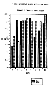
The results of these experiments are shown in
Figures 1 and 2. Figure 1 displays relative absorbance
units at 405 nm from the ELISA assay for assays
performed on baboon B (1E6) lymphocytes from days 0,
WO 93/06852 PCT/US92/08754
2120731
- 39 -
1-5, 8, 11 and 14. Figure 2 displays relative
absorbance units at 405 rim from the ELISA assay for
assays performed on lymphocytes from baboons A(lE6)
and C (MOPC21) on days 0, 1-5, 8 and 11.
For baboon B, T cell dependent B cell Ig
production decreased on the second day of 1E6
injections and remained at about 35% of the day zero
value through day eleven (Figure 1).
For baboon A, Ig production was higher on
days 1-11 as compared to the level before the
injection. This is likely due to the lower lE6 serum
level achieved in baboon A versus baboon B. If Ig
production levels observed on days one through four are
taken as a base value, then a 40% inhibition of Ig
secretion was observed on day five, and a 20%
inhibition on day eleven (Figure 2).
In baboon C, after injection with MOPC21,
peripheral blood lymphocytes showed increased levels of
Ig production between days two and eleven as compared
to the level on day zero.
2. T cell Proliferation Assay
In a T cell proliferation assay, we measured
the ability of activating anti-CD2 monoclonal
antibodies or phytohemagglutinin ("PHA") to cause
proliferation of T cells isolated from baboons A, B and
C on days 0, 1-5, 8, 11 and 14. 1 x 10⁵ peripheral
blood lymphocytes per well were incubated (1) with
anti-CD2 monoclonal antibodies Till and T113 at a 1:900
dilution of ascites fluid, (2) in medium alone, or
(3) with PHA (Sigma Chemical Corporation, St. Louis,
Missouri) (10 g/ml) for three days. After three days,
cells were labelled with 1 Ci/well 3HdT for 18 hours
and then harvested. (Data not shown.)
WO 93/06852 PCT/US92/08754
- 40 -
Peripheral blood lymphocytes from baboon B
showed no increase of 3HdT incorporation in response to
activating anti-CD2 monoclonal antibodies and very low
proliferative activity in medium on days zero to
fourteen.
Peripheral blood lymphocytes from baboon A
responded to anti-CD2 monoclonal antibodies and PHA.
After day four, proliferation in response to those
agents was inhibited about nine fold and remained low
until at least day fourteen.
Peripheral blood lymphocytes from baboon C,
the MOPC21 control, showed very low proliferative
activity at all time points tested, under all
conditions.
The significance of the data obtained is not
clear because of irreproducibility of T cell
proliferation in baboon C and day zero results for
baboons A, B and C.
Example 3
Effect Of Administration Of
LFA3TIP On Lymphocyte Function
A. Administration And Sampling Protocols
Two outbred, adult baboons (4.6 and 7.4 kg)
(Papio anubis) were given bolus injections of 3 mg/kg
of purified LFA3TIP (obtained from Biogen, Inc.,
Cambridge, Massachusetts), i.v., by portacatheter once
daily for five consecutive days. Blood was drawn from
the baboons once before the first injection of antibody
and then, daily for five days, 24 hours after each
injection. Blood was also drawn on day 8, day 10, day
15, and day 22, where day 1 is the day of the first
injection. This administration and sampling protocol
was used for all of the assays described in this
example, unless otherwise stated.
WO 93/06852 2 120731 PCr/US92/08754
- 41 -
B. Toxicology Stu dV With LFA3TIP
The general toxicity of LFA3TIP and its
potential effect on the physical condition, hematology
and blood chemistry of baboons were evaluated. The
general physical condition of the baboons remained
unchanged throughout the study. No obvious or
immediate side effects could be observed. Hematology
and blood chemistries generally remained normal. In
particular, Na+, C1-, K+, creatine, blood urea nitrogen
and liver enzymes AST and ALT levels all remained
within normal limits. In addition, blood cell counts,
including hematocrit, white blood cells, lymphocytes,
monocytes, segmented neutrophils, and eosinophils,
generally stayed within normal ranges. The ratio of
CD4/CD8 expressing cells also stayed within normal
ranges.
Plasma levels of LFA3TIP 10 days after the
last injection were still about 32% of the LFA3TIP
levels immediately following the last injection, which
indicates a much longer half-life than generally
observed with murine monoclonal antibodies.
Fluorescent labeling of CD4 and CD8 expressing cells
indicated that about 10% of CD4+ cells and about 90% of
CD8 + cells were still coated with LFA3TIP 10 days after
the last injection.
Example 4
Baboon Cardiac Allograft Model
A. 1E6 Treatment
An experimental primate cardiac allograft
model where baboon hearts were transplanted
heterotopically in a nonfunctioning position into the
necks of ABO-matched outbred baboons ( a'o anubis) was
used to assess the effect of anti-LFA-3 monoclonal
WO 93/06852 PCT/US92/08754
- 42 -
antibody 1E6 on allograft rejection. The protocol used
was substantially as described in Michler et al.,
"Techniques For Primate Heterotopic Cardiac
Xenotransplantation," J. Med. Primatol., 14, pp. 357-62
(1985), except that an allograft not a xenograft was
perf ormed .
Purified lE6 prepared as described above was
injected into one adult baboon (weight 32 kg) at a dose
of 5 mg/kg, starting on day one, for 2 consecutive days
before the transplant. On the third day, a cardiac
heterotopic allograft transplant was performed with a
heart from a young, 3 kg baboon. One dose of 5 mg/kg
of 1E6 was injected on the day of the transplant and
then once a day for ten consecutive days. Blood
samples were collected two days before transplantation,
prior to injection. Blood samples were also collected
coincident with transplantation and on the fifth,
tenth, sixteenth, nineteenth and twenty-first day after
transplantation. An assay for total 1E6 serum levels
and the proportion of active 1E6 in the serum, i.e.,
the percentage of lE6 capable of binding to LFA-3, was
performed as described in Example 2C. No general
immunosuppressive agents were administered to the
baboon.
The graft was palpated on a daily basis and
monitored by palpation and visual assessment of heart
beat. Electrocardiograms were performed on a weekly
basis. A percutaneous endomyocardial biopsy was
performed on the sixteenth day after transplantation.
All blood chemistry and cell counts performed on the
above described blood samples were within the normal
limits.
Untreated control cardiac allografts in this
model system were rejected a mean of 9 3 days (n=5)
after implantation in non-immunosuppressed baboons
WO 93/06852 PCF/US92/08754
21 20731
- 43 -
(Rose et al., "Cardiac Xenotransplantation", Progress
In Cardiovascular Diseases, 33, pp. 105-17 (1990)).
Rejection is defined, for the purposes of this model
system, as swelling and hardening of the heart, and
cessation of heart beat as measured by an
electrocardiogram. In addition, progressive
infiltration of lymphocytes in the myocardium,
production of lymphocytotoxic antibodies and reaction
to donor peripheral blood lymphocytes are monitored.
Survival of a graft in this system for longer than nine
days, without immunosuppressive therapy, indicates an
increased level of tolerance.
In the 1E6 treated baboon, the transplanted
allogeneic heart was still beating twenty-three days
after the transplant. Thus, 1E6 dramatically improved
tolerance for a cardiac allograft.
B. LFA3TIP Treatment
Using procedures substantially as described
in Example 4A, the effect of LFA3TIP on cardiac
allograft rejection is assessed. Purified LFA3TIP
(described suDra) is injected into one adult baboon at
a dose of 3 mg/kg on day one for 2 consecutive days
before the transplant. On the third day a cardiac
heterotopic allograft transplant is performed with a
heart from a young baboon. One dose of 3 mg/kg LFA3TIP
is injected on the day of the transplant and then once
a day for nine consecutive days.
The schedule of blood sample collection and
analysis, and assessment of allograft rejection, is
substantially as described in Example 4A.
Survival of the graft in the baboon that is
treated with LFA3TIP is extended, compared to graft
survival in untreated baboons, indicating increased
graft tolerance due to LFA3TIP.
WO 93/06852 PCT/US92/08754
- 44 -
Deposits
Murine hybridoma cells and antibodies useful
in the present invention are exemplified by cultures
deposited under the Budapest Treaty with American Type
Culture Collection, Rockville, Maryland, U.S.A., on
March 5, 1991, and identified as:
Designation ATCC Accession No.
1E6 HB 10393
HC-1B11 HB 10694
7A6 HB 10695
8B8 HB 10696
E. coli JA221 transformed with plasmid
pSAB152 (encoding LFA3TIP) was deposited under the
Budapest Treaty with American Type Culture Collection
on October 1', 1991 and identified as:
Designation ATCC Accession No.
pSAB152 68720
A bacteriophage carrying a plasmid encoding
transmembrane LFA-3 was deposited under the Budapest
Treaty with In Vitro International, Inc., Linthicum,
Maryland, U.S.A., on May 28, 1987. This deposit was
transferred to American Type Culture Collection on
June 20, 1991 and identified as:
Designation ATCC Accession No.
AHT16[Agt10/LFA-3] 75107
E. coli transformed with a plasmid encoding
PI-linked LFA-3 was deposited under the Budapest Treaty
with In Vitro International, Inc. on July 22, 1988.
This deposit was transferred to American Type Culture
Collection on June 20, 1991 and identified as:
Designation ATCC Accession No.
p24 68788
WO 93/06852 PCT/US92/08754
2120731.
- 45 -
Seauences
The following is a summary of the sequences
set forth in the Sequence Listing:
SEQ ID NO:1 DNA sequence of transmembrane LFA-3
SEQ ID NO:2 Amino acid sequence of transmembrane LFA-3
SEQ ID NO:3 DNA sequence of PI-linked LFA-3
SEQ ID NO:4 Amino acid sequence of PI-linked LFA-3
SEQ ID NO:5 DNA sequence of CD2
SEQ ID No:6 Amino acid sequence of CD2
SEQ ID NO:7 DNA sequence of LFA3TIP
SEQ ID NO:8 Amino acid sequence of LFA3TIP
While we have hereinbefore described a number
of embodiments of this invention, it is apparent that
our basic embodiments can be altered to provide other
embodiments that utilize the processes of this
invention. Therefore, it will be appreciated that the
scope of this invention includes all alternative
embodiments and variations which are defined in the
foregoing specification and by the claims appended
hereto; and the invention is not to be limited by the
specific embodiments that have been presented herein by
way of example.
WO 93/06852 t46_ PCT/US92/08754
SEQUENCE LISTING
(1) GENERAL INFORMATION:
(i) APPLICANT: WALLNER, Barbara P.
BENJAMIN, Christopher D.
(ii) TITLE OF INVENTION: METHODS OF IMPROVING ALLOGRAFT OR
XENOGRAFT TOLERANCE BY ADMINISTRATION OF LFA-3 OR
CD2 BINDING PROTEINS
(iii) NUMBER OF SEQUENCES: 8
(iv) CORRESPONDENCE ADDRESS:
(A) ADDRESSEE: c\o FISH & NEAVE
(B) STREET: 875 Third Avenue
(C) CITY: New York
(D) STATE: New York
(E) COUNTRY: U.S.A.
(F) ZIP: 10022
(v) COMPUTER READABLE FORM:
(A) MEDIUM TYPE: Floppy disk
(B) COMPUTER: IBM PC compatible
(C) OPERATING SYSTEM: PC-DOS/MS-DOS
(D) SOFTWARE: PatentIn Release #1.0, Version #1.25
(vi) CURRENT APPLICATION DATA:
(A) APPLICATION NUMBER:
(B) FILING DATE:
(C) CLASSIFICATION:
(vii) PRIOR APPLICATION DATA:
(A) APPLICATION NUMBER: US 07/772,705
(B) FILING DATE: 07-OCT-1991
(viii) ATTORNEY/AGENT INFORMATION:
(A) NAME: Haley Jr., James F.
(B) REGISTRATION NUMBER: 27,794
(C) REFERENCE/DOCKET NUMBER: B162CIP
(ix) TELECOMMUNICATION INFORMATION:
(A) TELEPHONE: (212) 715-0600
(2) INFORMATION FOR SEQ ID N0:1:
(i) SEQUENCE CHARACTERISTICS:
(A) LENGTH: 753 base pairs
(B) TYPE: nucleic acid
(C) STRANDEDNESS: single
(D) TOPOLOGY: linear
~. _ _...... ... r
~ll~f7~1
WO 93/06852 _47_ PC.'I'/US92/08754
(ix) FEATURE:
(A) NAME/KEY: CDS
(B) LOCATION: 1..750
(ix) FEATURE:
(A) NAME/KEY: sig_peptide
(B) LOCATION: 1..84
(ix) FEATURE:
(A) NAME/KEY: mat_peptide
(B) LOCATION: 85..750
(ix) FEATURE:
(A) NAME/KEY: misc_feature
(B) LOCATION: 1..750
(D) OTHER INFORMATION: /note- "Human transmembrane LFA-3"
(ix) FEATURE:
(A) NAME/KEY: misc_feature
(B) LOCATION: 646..714
(D) OTHER INFORMATION: /note- "Transmembrane domain"
(xi) SEQUENCE DESCRIPTION: SEQ ID N0:1:
ATG CTT GCT GGG AGC GAC GCG GGG CGG GCC CTG GGG GTC CTC AGC GTG 48
Met Val Ala Gly Ser Asp Ala Gly Arg Ala Leu Gly Val Leu Ser Val
-28 -25 -20 -15
GTC TGC CTG CTG CAC TGC TTT GGT TTC ATC AGC TGT TTT TCC CAA CAA 96
Val Cys Leu Leu His Cys Phe Gly Phe Ile Ser Cys Phe Ser Gln Gln
-10 -5 1
ATA TAT GGT GTT GTG TAT GGG AAT GTA ACT TTC CAT GTA CCA AGC AAT 144
Ile Tyr Gly Val Val Tyr Gly Asn Val Thr Phe His Val Pro Ser Asn
10 15 20
GTG CCT TTA AAA GAG GTC CTA TGG AAA AAA CAA AAG GAT AAA GTT GCA 192
Val Pro Leu Lys Glu Val Leu Trp Lys Lys Gln Lys Asp Lys Val Ala
25 30 35
GAA CTG GAA AAT TCT GAA TTC AGA GCT TTC TCA TCT TTT AAA AAT AGG 240
Glu Leu Glu Asn Ser Glu Phe Arg Ala Phe Ser Ser Phe Lys Asn Arg
40 45 50
GTT TAT TTA GAC ACT GTG TCA GGT AGC CTC ACT ATC TAC AAC TTA ACA 288
Val Tyr Leu Asp Thr Val Ser Gly Ser Leu Thr Ile Tyr Asn Leu Thr
55 60 65
TCA TCA GAT GAA GAT GAG TAT GAA ATG GAA TCG CCA AAT ATT ACT GAT 336
Ser Ser Asp Glu Asp Glu Tyr Glu Met Glu Ser Pro Asn Ile Thr Asp
70 75 80
ACC ATG AAG TTC TTT CTT TAT GTC CTT GAG TCT CTT CCA TCT CCC ACA 384
Thr Met Lys Phe Phe Leu Tyr Val Leu Glu Ser Leu Pro Ser Pro Thr
85 90 95 100
2.
l.I1~+0731
WO 93/06852 -48- PCT/US92/08754
CTA ACT TGT GCA TTG ACT AAT GGA AGC ATT GAA GTC CAA TGC ATG ATA 432
Leu Thr Cys Ala Leu Thr Asn Gly Ser Ile Glu Val Gln Cys Met Ile
105 110 115
CCA GAG CAT TAC AAC AGC CAT CGA GGA CTT ATA ATG TAC TCA TGG GAT 480
Pro Glu His Tyr Asn Ser His Arg Gly Leu Ile Met Tyr Ser Trp Asp
120 125 130
TGT CCT ATG GAG CAA TGT AAA CGT AAC TCA ACC AGT ATA TAT TTT AAG 528
Cys Pro Met Glu Gln Cys Lys Arg Asn Ser Thr Ser Ile Tyr Phe Lys
135 140 145
ATG GAA AAT GAT CTT CCA CAA AAA ATA CAG TGT ACT CTT AGC AAT CCA 576
Met Glu Asn Asp Leu Pro Gln Lys Ile Gin Cys Thr Leu Ser Asn Pro
150 155 160
TTA TTT AAT ACA AGA TCA TCA ATC ATT TTG ACA ACC TGT ATC CCA AGC 624
Leu Phe Asn Thr Thr Ser Ser Ile Ile Leu Thr Thr Cys Ile Pro Ser
165 170 175 180
AGC GGT CAT TCA AGA CAC AGA TAT GCA CTT ATA CCC ATA CCA TTA GCA 672
Ser Gly His Ser Arg His Arg Tyr Ala Leu Ile Pro Ile Pro Leu Ala
185 , 190 195
GTA ATT ACA ACA TGT ATT GTG CTG TAT ATG AAT GGT ATT CTG AAA TGT 720
Val Ile Thr Thr Cys Ile Val Leu Tyr Met Asn Gly Ile Leu Lys Cys
200 205 210
GAC AGA AAA CCA GAC AGA ACC AAC TCC AAT TGA 753
Asp Arg Lys Pro Asp Arg Thr Asn Ser Asn
215 220
(2) INFORMATION FOR SEQ ID N0:2:
(i) SEQUENCE CHARACTERISTICS:
(A) LENGTH: 250 amino acids
(B) TYPE: amino acid
(D) TOPOLOGY: linear
(ii) MOLECULE TYPE: protein
(xi) SEQUENCE DESCRIPTION: SEQ ID N0:2:
Met Val Ala Gly Ser Asp Ala Gly Arg Ala Leu Gly Val Leu Ser Val
-28 -25 -20 -15
Val Cys Leu Leu His Cys Phe Gly Phe Ile Ser Cys Phe Ser Gln Gln
-10 -5 1
Ile Tyr Gly Val Val Tyr Gly Asn Val Thr Phe His Val Pro Ser Asn
10 15 20
Val Pro Leu Lys Glu Val Leu Trp Lys Lys Gln Lys Asp Lys Val Ala
25 30 35
WO 93/06852 -49- PCT/US92/08754
2120731
Glu Leu Glu Asn Ser Glu Phe Arg Ala Phe Ser Ser Phe Lys Asn Arg
40 45 50
Val Tyr Leu Asp Thr Val Ser Gly Ser Leu Thr Ile Tyr Asn Leu Thr
55 60 65
Ser Ser Asp Glu Asp Glu Tyr Glu Met Glu Ser Pro Asn Ile Thr Asp
70 75 80
Thr Met Lys Phe Phe Leu Tyr Val Leu Glu Ser Leu Pro Ser Pro Thr
85 90 95 100
Leu Thr Cys Ala Leu Thr Asn Gly Ser Ile Glu Val Gln Cys Met Ile
105 110 115
Pro Glu His Tyr Asn Ser His Arg Gly Leu Ile Met Tyr Ser Trp Asp
120 125 130
Cys Pro Met Glu Gln Cys Lys Arg Asn Ser Thr Ser Ile Tyr Phe Lys
135 140 145
Met Glu Asn Asp Leu Pro Gln Lys Ile Gln Cys Thr Leu Ser Asn Pro
150 155 160
Leu Phe Asn Thr Thr Ser Ser Ile Ile Leu Thr Thr Cys Ile Pro Ser
165 170 175 180
Ser Gly His Ser Arg His Arg Tyr Ala Leu Ile Pro Ile Pro Leu Ala
185 190 195
Val Ile Thr Thr Cys Ile Val Leu Tyr Met Asn Gly Ile Leu Lys Cys
200 205 210
Asp Arg Lys Pro Asp Arg Thr Asn Ser Asn
215 220
(2) INFORMATION FOR SEQ ID N0:3:
(i) SEQUENCE CHARACTERISTICS:
(A) LENGTH: 723 base pairs
(B) TYPE: nucleic acid
(C) STRANDEDNESS: single
(D) TOPOLOGY: linear
(ix) FEATURE:
(A) NAliE/KEY: CDS
(B) LOCATION: 1..720
(ix) FEATURE:
(A) NAME/KEY: sig_peptide
(B) LOCATION: 1..84
2120731
WO 93/06852 -50- PCT/US92/08754
- (ix) FEATURE:
(A) NAME/KEY: mat_peptide
(B) LOCATION: 85..720
(ix) FEATURE:
(A) NAME/KEY: misc_feature
(B) LOCATION: 1..720
(D) OTHER INFORMATION: /note- "Human PI-linked LFA-3"
(ix) FEATURE:
(A) NAME/KEY: miscfeature
(B) LOCATION: 568._720
(D) OTHER INFORMATION: /note- "Signal sequence for
PI-linkage"
(xi) SEQUENCE DESCRIPTION: SEQ ID NO:3:
ATG GTT GCT GGG AGC GAC GCG GGG CGG GCC CTG GGG GTC CTC AGC GTG 48
Met Val Ala Gly Ser Asp Ala Gly Arg Ala Leu Gly Val Leu Ser Val
-28 -25 -20 -15
GTC TGC CTG CTG CAC TGC TTT GGT TTC ATC ACC TGT TTT TCC CAA CAA 96
Val Cys Leu Leu His Cys Phe Gly Phe Ile Ser Cys Phe Ser Gln Gln
-10 -5 1
ATA TAT GGT GTT GTG TAT GGG AAT GTA ACT TTC CAT GTA CCA AGC AAT 144
Ile Tyr Gly Val Val Tyr Gly Asn Val Thr Phe His Val Pro Ser Asn
10 15 20
GTG CCT TTA AAA GAG GTC CTA TGG AAA AAA CAA AAG GAT AAA GTT GCA 192
Val Pro Leu Lys Glu Val Leu Trp Lys Lys Gln Lys Asp Lys Val Ala
25 30 35
GAA CTG GAA AAT TCT GAA TTC AGA GCT TTC TCA TCT TTT AAA AAT AGG 240
Glu Leu Clu Asn Ser Glu Phe Arg Ala Phe Ser Ser Phe Lys Asn Arg
40 45 50
GTT TAT TTA GAC ACT GTG TCA GGT AGC CTC ACT ATC TAC AAC TTA ACA 288
Val Tyr Leu Asp Thr Val Ser Gly Ser Leu Thr Ile Tyr Asn Leu Thr
55 60 65
TCA TCA GAT GAA GAT GAG TAT GAA ATG GAA TCG CCA AAT ATT ACT CAT 336
Ser Ser Asp Glu Asp Glu Tyr Clu Met Glu Ser Pro Asn Ile Thr Asp
70 75 80
ACC ATG AAG TTC TTT CTT TAT GTG CTT GAG TCT CTT CCA TCT CCC ACA 38/s
Thr Met Lys Phe Phe Leu Tyr Val Leu Glu Ser Leu Pro Ser Pro Thr
85 90 95 100
CTA ACT TGT GCA TTG ACT AAT GGA AGC ATT GAA GTC CAA TGC ATG ATA 432
Leu Thr Cys Ala Leu Thr Asn Gly Ser Ile Glu Val Gln Cys Met Ile
105 110 115
CCA GAG CAT TAC AAC AGC CAT CGA GGA CTT ATA ATG TAC TCA TGG CAT 480
Pro Glu His Tyr Asn Ser His Arg Gly Leu Ile Met Tyr Ser Trp Asp
120 125 130
2120731
WO 93/06852 -51- PGT/US92/08754
TGT CCT ATG GAG CAA TGT AAA CGT AAC TCA ACC AGT ATA TAT TTT AAG 528
Cys Pro Met Glu Gln Cys Lys Arg Asn Ser Thr Ser Ile Tyr Phe Lys
135 140 145
ATG GAA AAT GAT CTT CCA CAA AAA ATA CAG TGT ACT CTT AGC AAT CCA 576
Met Glu Asn Asp Leu Pro Gln Lys Ile Gln Cys Thr Leu Ser Asn Pro
150 155 160
TTA TTT AAT ACA ACA TCA TCA ATC ATT TTG ACA ACC TGT ATC CCA AGC 624
Leu Phe Asn Thr Thr Ser Ser Ile Ile Leu Thr Thr Cys Ile Pro Ser
165 170 175 180
AGC GGT CAT TCA AGA CAC AGA TAT GCA CTT ATA CCC ATA CCA TTA GCA 672
Ser Gly His Ser Arg His Arg Tyr Ala Leu Ile Pro Ile Pro Leu Ala
185 190 195
GTA ATT ACA ACA TGT ATT GTG CTG TAT ATG AAT GGT ATG TAT GCT TTT 720
Val Ile Thr Thr Cys Ile Val Leu Tyr Met Asn Gly Met Tyr Ala Phe
200 205 210
TAA 723
(2) INFORMATION FOR SEQ ID N0:4:
(i) SEQUENCE CHARACTERISTICS:
(A) LENGTH: 240 amino acids
(B) TYPE: amino acid
(D) TOPOLOGY: linear
(ii) MOLECULE TYPE: protein
(xi) SEQUENCE DESCRIPTION: SEQ ID N0:4:
Met Val Ala Gly Ser Asp Ala Gly Arg Ala Leu Gly Val Leu Ser Val
-28 -25 -20 -15
Val Cys Leu Leu His Cys Phe Gly Phe Ile Ser Cys Phe Ser Gln Gln
-10 -5 1
Ile Tyr Gly Val Val Tyr Gly Asn Val Thr Phe His Val Pro Ser Asn
10 15 20
Val Pro Leu Lys Glu Val Leu Trp Lys Lys Gln Lys Asp Lys Val Ala
25 30 35
Glu Leu Glu Asn Ser Glu Phe Arg Ala Phe Ser Ser Phe Lys Asn Arg
40 45 50
Val Tyr Leu Asp Thr Val Ser Gly Ser Leu Thr Ile Tyr Asn L.eu Thr
55 60 65
Ser Ser Asp Glu Asp Glu Tyr Glu Met Glu Ser Pro Asn Ile Thr Asp
70 75 80
WO 93/06852 2 52 0731 PCT/US92/08754
Thr Met Lys Phe Phe Leu Tyr Val Leu Glu Ser Leu Pro Ser Pro Thr
85 90 _95 100
Leu Thr Cys Ala Leu Thr Asn Gly Ser Ile Glu Val Gln Cys Met Ile
105 110 115
Pro Glu His Tyr Asn Ser His Arg Gly Leu Ile Met Tyr Ser Trp Asp
120 125 130
Cys Pro Met Glu Gln Cys Lys Arg Asn Ser Thr Ser Ile Tyr Phe Lys
135 140 145
Met Glu Asn Asp Leu Pro Gln Lys Ile Gln Cys Thr Leu Ser Asn Pro
150 155 160
Leu Phe Asn Thr Thr Ser Ser Ile Ile Leu Thr Thr Cys Ile Pro Ser
165 170 175 180
Ser Gly His Ser Arg His Arg Tyr Ala Leu Ile Pro Ile Pro Leu Ala
185 190 195
Val Ile Thr Thr Cys Ile Val Leu Tyr Met Asn Gly Met Tyr Ala Phe
200 205 210
(2) INFORMATION FOR SEQ ID N0:5:
(i) SEQUENCE CHARACTERISTICS:
(A) LENGTH: 1056 base pairs
(B) TYPE: nucleic acid
(C) STRANDEDNESS: single
(D) TOPOLOGY: linear
(ix) FEATURE:
(A) NAME/KEY: CDS
(B) LOCATION: 1..1053
(ix) FEATURE:
(A) NAME/KEY: sig_peptide
(B) LOCATION: 1..72
(ix) FEATURE:
(A) NAME/KEY: mat_peptide
(B) LOCATION: 73..1053
(ix) FEATURE:
(A) NAME/KEY: miscfeature
(B) LOCATION: 1..1053
(D) OTHER INFORMATION: /note- "Human CD2"
(ix) FEATURE:
(A) NAME/KEY: miscfeature
(B) LOCATION: 628._702
(D) OTHER INFORMATION: /note- "Transmembrane domain"
_ ._....._ . _._.. . __.. . T
073 ) I
WO 93/06852 _53_ PCT/US92/08754
(xi) SEQUENCE DESCRIPTION: SEQ ID NO:5:
ATG AGC TTT CCA TGT AAA TTT GTA GCC AGC TTC CTT CTG ATT TTC AAT 48
Met Ser Phe Pro Cys Lys Phe Val Ala Ser Phe Leu Leu Ile Phe Asn
-24 -20 -15 -10
GTT TCT TCC AAA GGT GCA GTC TCC AAA GAG ATT ACG AAT CCC TTG GAA 96
Val Ser Ser Lys Gly Ala Val Ser Lys Glu Ile Thr Asn Ala Leu Glu
-5 1 5
ACC TGG GGT GCC TTG GGT CAG GAC ATC AAC TTG GAC ATT CCT AGT TTT 144
Thr Trp Gly Ala Leu Gly Gln Asp Ile Asn Leu Asp Ile Pro Ser Phe
15 20
CAA ATG AGT GAT GAT ATT GAC GAT ATA AAA TGG GAA AAA ACT TCA GAC 192
Gln Met Ser Asp Asp Ile Asp Asp Ile Lys Trp Glu Lys Thr Ser Asp
25 30 35 40
AAG AAA AAG ATT GCA CAA TTC AGA AAA GAG AAA GAG ACT TTC AAG GAA 240
Lys Lys Lys Ile Ala Gln Phe Arg Lys Glu Lys Glu Thr Phe Lys Glu
45 50 55
AAA GAT ACA TAT AAC CTA TTT AAA AAT GGA ACT CTG AAA ATT AAG CAT 288
Lys Asp Thr Tyr Lys Leu Phe Lys Asn Gly Thr Leu Lys Ile Lys His
60 65 70
CTG AAG ACC GAT GAT CAG GAT ATC TAC AAG GTA TCA ATA TAT CAT ACA 336
Leu Lys Thr Asp Asp Gln Asp Ile Tyr Lys Val Ser Ile Tyr Asp Thr
75 80 85
AAA GGA AAA AAT GTG TTG GAA AAA ATA TTT GAT TTG AAG ATT CAA GAG 384
Lys Gly Lys Asn Val Leu Glu Lys Ile Phe Asp Leu Lys Ile Gln Glu
90 95 100
AGG GTC TCA AAA CCA AAG ATC TCC TGG ACT TGT ATC AAC ACA ACC CTC 432
Arg Val Ser Lys Pro Lvs Ile Ser Trp Thr Cys Ile Asn Thr Thr Leu
105 110 115 120
ACC TGT GAG GTA ATG AAT GGA ACT GAC CCC GAA TTA AAC CTG TAT CAA 480
Thr Cys Glu Val Met Asn Gly Thr Asp Pro Glu Leu Asn Leu Tyr Gln
125 130 135
GAT GGG AAA CAT CTA AAA CTT TCT CAG AGG GTC ATC ACA CAC AAG TGG 528
Asp Gly Lys His Leu Lys Leu Ser Gln Arg Val Ile Thr His Lys Trp
140 145 150
ACC ACC AGC CTG AGT GCA AAA TTC AAG TGC ACA GCA GGG AAC AAA CTC 576
Thr Thr Ser Leu Ser Ala Lys Phe Lys Cys Thr Ala Gly Asn Lys Val
155 160 165
AGC AAG GAA TCC AGT GTC GAG CCT CTC AGC TGT CCA GAG AAA GGT CTG 624
Ser Lys Glu Ser Ser Val Glu Pro Val Ser Cys Pro Glu Lys Gly Leu
170 175 180
WO 93/06852 54_ PCT/US92/08754
GAC ATC TAT CTC ATC ATT GGC ATA TGT GGA GGA GGC AGC CTC TTG ATG 672
Asp Ile Tyr Leu Ile Ile Gly Ile Cys Gly Gly Gly Ser Leu Leu Met
185 190 195 200
GTC TTT GTG GCA CTG CTC GTT TTC TAT ATC ACC AAA AGG AAA AAA CAG 720
Val Phe Val Ala Leu Leu Val Phe Tyr Ile Thr Lys Arg Lys Lys Gln
205 210 215
AGG AGT CGG AGA AAT CAT GAG GAG CTG GAG ACA AGA GCC CAC AGA GTA 768
Arg Ser Arg Arg Asn Asp Glu Glu Leu Glu Thr Arg Ala His Arg Val
220 225 230
GCT ACT GAA GAA AGG GGC CGG AAG CCC CAC CAA ATT CCA GCT TCA ACC 816
Ala Thr Glu Glu Arg Gly Arg Lys Pro His Gln Ile Pro Ala Ser Thr
235 240 245
CCT CAG AAT CCA GCA ACT TCC CAA CAT CCT CCT CCA CCA CCT GGT CAT 864
Pro Gin Asn Pro Ala Thr Ser Gln His Pro Pro Pro Pro Pro Gly His
250 255 260
CGT TCC CAG GCA CCT AGT CAT CGT CCC CCG CCT CCT GGA CAC CGT GTT 912
Arg Ser Gln Ala Pro Ser His Arg Pro Pro Pro Pro Gly His Arg Val
265 270 275 280
CAG CAC CAG CCT CAG AAG AGG CCT CCT GCT CCG TCG GGC ACA CAA GTT 960
Gln His Gln Pro Gin Lys Arg Pro Pro Ala Pro Ser Gly Thr Gln Val
285 290 295
CAC CAG CAG AAA GGC CCG CCC CTC CCC AGA CCT CGA GTT CAG CCA AAA 1008
His Gln Gln Lys Gly Pro Pro Leu Pro Arg Pro Arg Val Gln Pro Lys
300 305 310
CCT CCC CAT GGG GCA GCA GAA AAC TCA TTG TCC CCT TCC TCT AAT 1053
Pro Pro His Gly Ala Ala Glu Asn Ser Leu Ser Pro Ser Ser Asn
315 320 325
TAA 1056
(2) INFORMATION FOR SEQ ID N0:6:
(i) SEQUENCE CHARACTERISTICS:
(A) LENGTH: 351 amino acids
(B) TYPE: amino acid
(D) TOPOLOGY: linear
(ii) MOLECULE TYPE: protein
(xi) SEQUENCE DESCRIPTION: SEQ ID NO:6:
Met Ser Phe Pro Cys Lys Phe Val Ala Ser Phe Leu Leu Ile Phe Asn
-24 -20 -15 -10
Val Ser Ser Lys Gly Ala Val Ser Lys Glu Ile Thr Asn Ala Leu Glu
-5 1 5
21 20( 31
WO 93/06852 55- PC'T/US92/08754
Thr Trp Gly Ala Leu Gly Gln Asp Ile Asn Leu Asp Ile Pro Ser Phe
15 20
Gln Met Ser Asp Asp Ile Asp Asp Ile Lys Trp Glu Lys Thr Ser Asp
25 30 35 40
Lys Lys Lys Ile Ala Gin Phe Arg Lys Glu Lys Glu Thr Phe Lys Glu
45 50 55
Lys Asp Thr Tyr Lys Leu Phe Lys Asn Gly Thr Leu Lys Ile Lys His
60 65 70
Leu Lys Thr Asp Asp Gln Asp Ile Tyr Lys Val Ser Ile Tyr Asp Thr
75 80 85
Lys Gly Lys Asn Val Leu Glu Lys Ile Phe Asp Leu Lys Ile Gln Glu
90 95 100
Arg Val Ser Lys Pro Lys Ile Ser Trp Thr Cys Ile Asn Thr Thr Leu
105 110 115 120
Thr Cys Glu Val Met Asn Gly Thr Asp Pro Glu Leu Asn Leu Tyr Gln
125 130 135
Asp Gly Lys His Leu Lys Leu Ser Gln Arg Val Ile Thr His Lys Trp
140 145 150
Thr Thr Ser Leu Ser Ala Lys Phe Lys Cys Thr Ala Gly Asn Lys Val
155 160 165
Ser Lys Glu Ser Ser Val Glu Pro Val Ser Cys Pro Glu Lys Gly Leu
170 175 180
Asp Ile Tyr Leu Ile Ile Gly Ile Cys Gly Gly Gly Ser Leu Leu Met
185 190 195 200
Val Phe Val Ala Leu Leu Val Phe Tyr Ile Thr Lys Arg Lys Lys Gln
205 210 215
Arg Ser Arg Arg Asn Asp Glu Glu Leu Glu Thr Arg Ala His Arg Val
220 225 230
Ala Thr Glu Glu Arg Gly Arg Lys Pro His Gln Ile Pro Ala Ser Thr
235 240 245
Pro Gln Asn Pro Ala Thr Ser Gln His Pro Pro Pro Pro Pro Gly His
250 255 260
Arg Ser Gln Ala Pro Ser His Arg Pro Pro Pro Pro Gly His Arg Val
265 270 275 280
Gln His Gln Pro Gln Lys Arg Pro Pro Ala Pro Ser Gly Thr Gln Val
285 290 295
WO 93/06852 2120731
56- PCT/US92/08754
His Gln Gln Lys Gly Pro Pro Leu Pro Arg Pro Arg Val Gln Pro Lys
300 305 310
Pro Pro His Gly Ala Ala Glu Asn Ser Leu Ser Pro Ser Ser Asn
315 320 325
(2) INFORMATION FOR SEQ ID NO:7:
(i) SEQUENCE CHARACTERISTICS:
(A) LENGTH: 1050 base pairs
(B) TYPE: nucleic acid
(C) STRANDEDNESS: single
(D) TOPOLOGY: linear
(iii) HYPOTHETICAL: NO
(iv) ANTI-SENSE: NO
(ix) FEATURE:
(A) NAME/KEY: CDS
(B) LOCATION: 1..1041
(ix) FEATURE:
(A) NAME/KEY: sig_peptide
(B) LOCATION: 1..84
(ix) FEATURE:
(A) NAME/KEY: mat_peptide
(B) LOCATION: 85..1041
(ix) FEATURE:
(A) NAME/KEY: miscfeature
(B) LOCATION: 85..1041
(D) OTHER INFORMATION: /note- "LFA3TIP"
(ix) FEATURE:
(A) NAME/KEY: misc feature
(B) LOCATION: 360._361
(D) OTHER INFORMATION: /note- "LFA-3/IgG fusion point"
(xi) SEQUENCE DESCRIPTION: SEQ ID NO:7:
ATG GTT GCT GGG AGC GAC GCG GGG CGG GCC CTG GGG GTC CTC AGC GTG 48
Met Val Ala Gly Ser Asp Ala Gly Arg Ala Leu G'Ly Val Leu Ser Val
-28 -25 -20 -15
GTC TGC CTG CTG CAC TGC TTT GGT TTC ATC AGC TCT TTT TCC CAA CAA 96
Val Cys Leu Leu His Cys Phe Gly Phe Ile Ser Cys Phe Ser Gln Gln
-10 -5 1
ATA TAT GGT GTT GTG TAT GGG AAT CTA ACT TTC CAT GTA CCA AGC AAT 144
Ile Tyr Gly Val Val Tyr Gly Asn Val Thr Phe His Val Pro Ser Asn
10 15 20
___ ..__...__ ,._ ...~.. _.._,_., r.... _ _._..._. _.... . _ ... ..__.... ..
... . ._ .. . _
2120'7 31
WO 93/06852 _57_ PCF/US92/08754
GTG CCT TTA AAA GAG GTC CTA TGG AAA AAA CAA AAG GAT AAA GTT GCA 192
Val Pro Leu Lys Glu Val Leu Trp Lys Lys Gln Lys Asp Lys Val Ala
25 30 35
GAA CTC GAA AAT TCT GAA TTC AGA GCT TTC TCA TCT TTT AAA AAT AGG 240
Glu Leu Glu Asn Ser Glu Phe Arg Ala Phe Ser Ser Phe Lys Asn Arg
40 45 50
GTT TAT TTA GAC ACT GTG TCA GGT AGC CTC ACT ATC TAC AAC TTA ACA 288
Val Tyr Leu Asp Thr Val Ser Gly Ser Leu Thr Ile Tyr Asn Leu Thr
55 60 65
TCA TCA GAT GAA GAT GAG TAT GAA ATG GAA TCG CCA AAT ATT ACT GAT 336
Ser Ser Asp Glu Asp Glu Tyr Glu Met Glu Ser Pro Asn Ile Thr Asp
70 75 80
ACC ATG AAG TTC TTT CTT TAT GTC GAC AAA ACT CAC ACA TGC CCA CCG 384
Thr Met Lys Phe Phe Leu Tyr Val Asp Lys Thr His Thr Cys Pro Pro
85 90 95 100
TGC CCA GCA CCT GAA CTC CTG GGG GGA CCC TCA GTC TTC CTC TTC CCC 432
Cys Pro Ala Pro Glu Leu Leu Gly Gly Pro Ser Val Phe Leu Phe Pro
105 110 115
CCA AAA CCC AAG GAC ACC CTC ATG ATC TCC CGG ACC CCT GAG CTC ACA 480
Pro Lys Pro Lys Asp Thr Leu Met Ile Ser Arg Thr Pro Glu Val Thr
120 125 130
TGC GTG GTG GTG GAC GTG AGC CAC CAA GAC CCT GAG CTC AAG TTC AAC 528
Cys Val Val Val Asp Val Ser His Glu Asp Pro Glu Val Lys Phe Asn
135 140 145
TGG TAC GTG GAC GGC GTG GAG GTG CAT AAT GCC AAG ACA AAG CCG CGG 576
Trp Tyr Val Asp Gly Val Glu Val His Asn Ala Lys Thr Lys Pro Arg
150 155 160
GAG GAG CAG TAC AAC AGC ACG TAC CGG GTG GTC AGC CTC CTC ACC GTC 624
Glu Glu Gin Tyr Asn Ser Thr Tyr Arg Val Val Ser Val Leu Thr Val
165 170 175 180
CTG CAC CAG GAC TGG CTG AAT GGC AAG GAG TAC AAG TGC AAG GTC TCC 672
Leu His Gln Asp Trp Leu Asn Gly Lys Glu Tyr Lys Cys Lys Val Ser
185 190 195
AAC AAA GCC CTC CCA GCC CCC ATC GAG AAA ACC ATC TCC AAA GCC AAA 720
Asr. Lys Ala Leu Pro Ala Pro Ile Clu Lys Thr Ile Ser Lys Ala Lys
200 205 210
GGG CAG CCC CGA GAA CCA CAG GTG TAC ACC CTG CCC CCA TCC CGG GAT 768
Gly Gln Pro Arg Glu Pro Gln Val Tyr Thr Leu Pro Pro Ser Arg Asp
215 220 225
GAG CTG ACC AAG AAC CAG GTC AGC CTG ACC TGC CTG GTC AM GGC TTC 816
Glu Leu Thr Lys Asn Gln Val Ser Leu Thr Cys Leu Val Lys Gly Phe
230 235 240
WO 93/06852 _58_ PCF/US92/08754
TAT CCC AGC GAC ATC GCC GTG GAG TGG GAG AGC AAT GGC CAG CCG GAG 864
Tyr Pro Ssr Asp Ile Ala Val Glu Trp Glu Ser Asn Gly Gln Pro Glu
245 250 255 260
AAC AAC TAC AAG ACC ACG CCT CCC GTG CTG GAC TCC GAC GGC TCC TTC 912
Asn Asn Tyr Lys Thr Thr Pro Pro Val Leu Asp Ser Asp Gly Ser Phe
265 270 275
TTC CTC TAC AGC AAG CTC ACC GTG GAC AAG AGC AGG TGG CAG CAG GGG 960
Phe Leu Tyr Ser Lys Leu Thr Val Asp Lys Ser Arg Trp Gln Gln Gly
280 285 290
AAC GTC TTC TCA TGC TCC GTG ATG CAT GAG GCT CTG CAC AAC CAC TAC 1008
Asn Val Phe Ser Cys Ser Val Met His Glu Ala Leu His Asn His Tyr
295 300 305
ACG CAG AAG AGC CTC TCC CTG TCT CCG GGT AAA TGAGTGCGG 1050
Thr Gln Lys Ser Leu Ser Leu Ser Pro Gly Lys
310 315
(2) INFORMATION FOR SEQ ID N0:8:
(i) SEQUENCE CHARACTERISTICS:
(A) LENGTH: 347 amino acids
(B) TYPE: amino acid
(D) TOPOLOGY: linear
(ii) MOLECULE TYPE: protein
(xi) SEQUENCE DESCRIPTION: SEQ ID NO:8:
Met Val Ala Gly Ser Asp Ala Gly Arg Ala Leu Gly Val Leu Ser Val
-28 -25 -20 -15
Val Cys Leu Leu His Cys Phe Gly Phe Ile Ser Cys Phe Ser Gin Gln
-10 -5 1
Ile Tyr Gly Val Val Tyr Gly Asn Val Thr Phe His Val Pro Ser Asn
10 15 20
Val Pro Leu Lys Glu Val Leu Trp Lys Lys Gln Lys Asp Lys Val Ala
25 30 35
Glu Leu Glu Asn Ser Glu Phe Arg Ala Phe Ser Ser Phe Lys Asn Arg
40 45 50
Val Tyr Leu Asp Thr Val Ser Gly Ser Leu Thr Ile Tyr Asn Leu Thr
55 60 65
Ser Ser Asp Glu Asp Glu Tyr Glu Met Glu Ser Pro Asn Ile Thr Asp
70 75 80
Thr Met Lys Phe Phe Leu Tyr Val Asp Lys Thr His Thr Cys Pro Pro
85 90 95 100
, w__...._.
2120731
WO 93/06852 _59_ PCT/US92/08754
Cys Pro Ala Pro Glu Leu Leu Gly Gly Pro Ser Val Phe Leu Phe Pro
105 110 115
Pro Lys Pro Lys Asp Thr Leu Met Ile Ser Arg Thr Pro Glu Val Thr
120 125 130
Cys Val Val Val Asp Val Ser His Glu Asp Pro Glu Val Lys Phe Asn
135 140 145
Trp Tyr Va1 Asp Gly Val Glu Val His Asn Ala Lys Thr Lys Pro Arg
150 155 160
Glu Glu Gln Tyr Asn Ser Thr Tyr Arg Val Val Ser Val Leu Thr Val
165 170 175 180
Leu His Gln Asp Trp Leu Asn Gly Lys Glu Tyr Lys Cys Lys Val Ser
185 190 195
Asn Lys Ala Leu Pro Ala Pro Ile Glu Lys Thr Ile Ser Lys Ala Lys
200 205 210
Gly Gln Pro Arg Glu Pro Gln Val Tyr Thr Leu Pro Pro Ser Arg Asp
215 220 225
Glu Leu Thr Lys Asn Gln Val Ser Leu Thr Cys Leu Val Lys Gly Phe
230 235 240
Tyr Pro Ser Asp Ile Ala Val Glu Trp Glu Ser Asn Gly Gln Pro Glu
245 250 255 260
Asn Asn Tyr Lys Thr Thr Pro Pro Val Leu Asp Ser Asp Gly Ser Phe
265 270 275
Phe Leu Tyr Ser Lys Leu Thr Val Asp Lys Ser Arg Trp Gln Gln Gly
280 285 290
Asn Val Phe Ser Cys Ser Val Met His Glu Ala Leu His Asn His Tyr
295 300 305
Thr Gln Lys Ser Leu Ser Leu Ser Pro Gly Lys
310 315