Note: Descriptions are shown in the official language in which they were submitted.
'wM.. 1
Endovascular Electrolytically Detachable Wire for Thrombus Formation
15
Background of the Invention
1. Field of the Invention
The invention relates to a method and apparatus for endovascular
electrothrombic formation of thrombi in arteries) veins, aneurysms, vascular
malformations and arteriovenous fistulas.
2. Description of the PriorArt
Approximately 25,000 intracranial aneurysms rupture every year in North
America. The primary purpose of treatment for ruptured intracranial aneurysm
is to
prevent rebleeding. At the present time, three general methods of treatment
exist)
namely an extravascular, endovascular and extra-endovascular approach.
The extravascular approach is comprised of surgery or microsurgery of the
aneurysm or treatment site for the purpose of preserving the parent artery.
This
treatment is common with intracranial berry aneurysms. The
methodology'coirrprises
the step of clipping the neck of the aneurysm) performing ~a. suture-ligation
of the
neck, or wrapping the entire aneurysm. Each of these surgical procedures is
performed by intrusive invasion into the body and performed from outside the
aneurysm or target site. General anesthesia, craniotomy, brain retractior. and
arachnoid dissection around the neck of the aneurysm and placement of a clip
are
2
typically required in these surgical procedures. Surgical treatment of
vascular
intracranial aneurysm can expect a mortality rate of 4-8% with a morbidity
rate of 18-
20%. Because of the mortality and morbidity rate expected, the surgical
procedure is
often delayed while waiting for the best surgical time with the result that an
additional percentage of patients will die from the underlying disease or
defect prior
to surgery. For this reason the prior art has sought alternative means of
treatment.
In the endovascular approach, the interior of the aneurysm is entered through
the use of a microcatheter. Recently developed microcatheters, such as those
shown
by Engelson, "Catheter Guidewire ; U.S. Patent 4,884,579 and as described in
Engelson, "Catheter for Guidewire Tracking ; U.S. Patent 4,739,768 ( 1988),
allow
navigation into the cerebral arteries and entry into a cranial aneurysm.
In such procedures a balloon is typically attached to the end of the
microcatheter and it is possible to introduce the balloon into the aneurysm,
inflate it,
and detach it, leaving it to occlude the sac and neck with preservation of the
parent
artery. While endovascular balloon embolization of berry aneurysms is an
attractive
method in situations where an extravascular surgical approach is difficult,
inflation of
a balloon into the aneurysm carries some risk of aneurysm rupture due to
possible
over-distention of portions of the sac and . due to the traction produced
while
detaching the balloon.
While remedial procedures exist for treating a ruptured aneurysm during
classical extravascular surgery, no satisfactory methodology exists if the
aneurysm
breaks during an endovascular balloon embolization.
Furthermore, an ideal embolizing agent should adapt itself to the irregular
shape of the internal walls of the aneurysm. On the contrary) in a balloon
embolization the aneurysmal wall must conform to the shape of the balloon.
This
may not lead to a satisfactory result and further increases the risk of
rupture.
Still further, balloon embolization is not always possible. If the diameter of
the deflated balloon is too great to enter the intracerebral arteries,
especially in the
cases where there is a vasospasm, complications with ruptured intracranial
aneurysms
may occur. The procedure then must be deferred until the spasm is resolved and
this
then incurs a risk of rebleeding.
In the extra-intravascular approach, an aneurysm is surgically, exposed or
stereotaxically reached with a probe. The wall of the aneurysm is then-
perforated
from the outside and various techniques are used to occlude the interior in
order to
prevent it from rebleeding. These prior art techniques include
electrothrombosis,
isobutyl-cyanoacrylate embolization) hog-hair embolization and ferromagnetic
thrombosis.
~.... 3
In the use of electrothrombosis for extra-intravascular treatment the tip of a
positively charged electrode is inserted surgically into the interior of the
aneurysm.
An application of the positive charge attracts white blood cells, red blood
cells)
platelets and fibrinogen which are typically negatively charged, at the normal
pH of
the blood. The thrombic mass is then formed in the aneurysm about the tip.
Thereafter, the tip is removed. See Mullah) 'Experiences with Surgical
Thrombosis of
Intracranial Berry Aneurysms and Carotid Cavernous Fistulas ; S~ Neurosurg.,
Vol. 41)
December 1974; Hosobuchi, 'Electrothrombosis Carotid-Cavernous Fistula ; J.
Neurosurg., Vol. 42, January 1975; Arald et al., 'Electrically Induced
Thrombosis for
the Treatment of Intracranial Aneurysms and Angiomas ; Excerpta Medica
International Congress Series, Amsterdam 1965, Vol. 110, 651-654; Sawyer et
al.,
'Bio-Electric Phenomena as an Etiological Factor in Intravascular Thrombosis';
Am. J.
Physiol., Vol. 175, 103-107 (1953); J. Pitoh et al., "Selective Vascular
Thrombosis
Induced by a Direct Electrical Current; Animal Experiments'; J.
Neuroradiology, Vol. 5,
pages 139-152 ( 1978). However, each of these techniques involves some type of
intrusive procedure to approach the aneurysm from the exterior, of the body.
The prior art has also devised the use of a liquid adhesive, isobutyl-
cyanoacrylate (IBCA) which polymerizes rapidly on contact with blood to form a
firm
mass. The liquid adhesive is injected into the aneurysm by puncturing the sac
with a
small needle. In order to avoid spillage into the parent artery during IBCA
injection,
blood flow through the parent artery must be momentarily reduced or
interrupted.
Alternatively, an inflated balloon may be placed in the artery at the level of
the neck
of the aneurysm for injection. In addition to the risks caused by temporary
blockage
of the parent artery, the risks of seepage of such a polymerizing adhesive
into the
parent artery exists, if it is not completely blocked with consequent
occlusion of the
artery.
Still further, the prior art has utilized an air gun to inject hog hair
through the
aneurysm wall to induce internal thrombosis. The success of this procedure
involves
exposing the aneurysm sufficiently to allow air gun injection and has not been
convincingly shown as successful for thrombic formations.
Ferromagnetic thrombosis in the prior art in extra-intravascular treatments
comprises the stereotactic placement of a magnetic probe against the sac of
the
aneurysm followed by injection into the aneurysm by an injecting needle of
iron
microspheres. Aggregation of the microspheres through the,extravascular magnet
is
followed by interneuysmatic thrombus. This treatment has not been entirely
successful because of the risk of fragmentation of the metallic thrombus when
the
extravascular magnet is removed. Suspension of the iron powder in methvi
methymethacrylate has been used to prevent fragmentation. The treatment has
not
-.
.~,.,., 4
been favored, because of the need to puncture the aneurysm, the risk of
occlusion of
the parent artery, the use of unusual and expensive equipment, the need for a
craniectomy, and general anesthesia, and the necessity to penetrate cerebral
tissue to
reach the aneurysm.
5l Endovascular coagulation of blood is also well known in the art and a
device
using laser optically generated heat is shown by O'Reilly) "Optical Fiber with
Attachable Metallic Tip for Intravascular Laser Coagulation of Arteries,
Yeins,
Aneurysms, Yascular Malformation and Arteriovenous Fistulas ; U.S. Patent
4,735,201
(1988). See also, O'Reilly et al., "Laser Induced Thermal Occlusion of Berry'
Aneurysms: Initial Experimental Results ; Radiology, Vol. 171, No. 2, pages
471-74
( 1989). O'Reilly places a tip into an aneurysm by means of an endovascular
microcatheter. The tip is adhesively bonded to a optic fiber disposed through
the
microcatheter. Optical energy is transmitted along the optic fiber from a
remote
laser at the proximal end of the nucrocatheter. The optical energy heats the
tip to
cauterize the tissue surrounding the neck of the aneurysm or other vascular
opening
to be occluded. The catheter is provided with a balloon located on or adjacent
to its
distal end to cut off blood flow to the site to be cauterized and occluded.
Normally,
the blood flow would carry away the heat at the catheter tip, thereby
preventing
cauterization. The heat in the tip also serves to melt the adhesive used to
secure the
tip to the distal end of the optical fiber. If all goes well, the tip can be
separated from
the optical fiber and left in place in the neck of the aneurysm, provided that
the
cauterization is complete at the same time as the hot melt adhesive melts.
A thrombus is not formed from the heated tip. Instead, blood tissue
surrounding the tip is coagulated. Coagulation is a denaturation of protein to
form a
connective-like tissue similar to that which occurs when the albumen of an egg
is
heated and coagulates from a clear running liquid to an opaque white solid.
The
tissue characteristics and composition of the coagulated tissue is therefore
substantially distinct from the thrombosis which is formed by the thrombotic
aggregation of white and red blood cells) platelets and fibrinogen. The
coagulative
tissue is substantially softer than a thrombic mass and can therefore more
easily be
dislodged.
O'Reilly's device depends at least in part upon the successful cauterization
timed to occur no later than the detachment of the heat tip from the optic
fiber. The
heated tip must also be proportionally sized to the neck of the aneurysm in
order to
effectively coagulate the tissue surrounding it to form a blockage at the
neck. It is
believed that the tissue in the interior of the aneurysm remains substantially
uncoagulated. In addition, the hot melt adhesive attaching the tip to the
optic fibe r
melts and is dispersed into the adjacent blood tissue where it resolidifies to
form free
2~2~779
particles within the intracranial blood stream with much
the same disadvantages which result from fragmentation of
a ferromagnetic electrothrombosis.
Brief Summary of the Invention
5 The present invention provides an apparatus for
forming an occlusion within a body cavity comprising:
a pliable coil wire for disposition near an opening
into said body cavity, said pliable coil wire having a
separable distal tip for disposition into said body
cavity to pack said cavity, said distal tip of said
pliable coil wire having no internal reinforcement,
having at most a loosely deformable helical shape, and
capable of being multiply folded upon itself in said body
cavity to pack said cavity;
means for mechanically forming said occlusion within
said body cavity about said distal tip; and
means for separating said distal tip from said wire
to leave said distal tip in a predetermined place within
said body cavity;
whereby said body cavity is occluded by said distal
tip, and any occlusion formed by packing of said tip
therein.
The wire may include a distinguishable
structure at its distal end, which is termed a tip, in
which case the remaining portion of the wire may be
termed a guidewire. The term "wire" should be understood
to collectively include both guidewires and tips and
simply wires without distinct tip structures. However,
the tip may also simply be the extension of the wire
itself without substantial distinction in its nature. A
distal tip of the wire is disposed into the vascular
cavity to pack the cavity to mechanically form the
occlusion within the vascular cavity about the distal
tip. The distal tip is detached from the guidewire (or
wire) to leave the distal tip within the vascular cavity.
~~~~79 y
'"~' 6
As a result, the vascular cavity is occluded by the
distal tip, and by any thrombus formed by use of the tip.
The present invention also provides an
apparatus for forming an occlusion within a body cavity
comprising:
a wire within a microcatheter adapted for
disposition near an opening into said body cavity, said
microcatheter having a separable distal tip electrode,
said wire having a distal tip adapted for disposition
into said body cavity to pack said cavity to form said
occlusion within said body cavity about said distal tip
by means of application of a current between said
electrode on said distal end of said wire packed into
said cavity and said distal tip electrode on said
microcatheter; and
means for separating said distal tip from said wire
to leave said distal tip in a predetermined position
within said body cavity,
whereby said body cavity is occluded by said distal
tip, and any occlusion formed by use of said tip.
In a further aspect, the present invention
provides a wire for use in formation of an occlusion
within a body cavity used in combination with a
microcatheter comprising:
a core wire; and
a detachable elongate tip portion extending said
core wire for a predetermined lineal extent and adapted
to being multiply folded on itself into said body cavity
at a predetermined position to form said occlusion in
said body cavity and coupled to said distal portion of
said core wire, said elongate tip portion being a
pliable, loosely deformable coil having at most a simple
helical shape,
whereby occlusion of said body cavity can be
achieved.
In a further aspect, the present invention
provides a wire for use in formation of an occlusion
.: :~:ll
within a body cavity used in combination with a
microcatheter comprising:
a core wire; and
an unbiased, pliable, detachable elongate tip
portion extending said core wire for a predetermined
lineal extent adapted to being packed entirely into said
body cavity at a predetermined position to at least slow
blood flow therein to form said occlusion in said body
cavity and coupled to said distal portion of said core
wire,
whereby occlusion of said body cavity can be
performed.
The present invention also provides a
microcatheter system for use in formation of an occlusion
within a body cavity comprising:
a microcatheter having a distal end adapted for
disposition in the proximity of said body cavity, said
distal end having an electrode disposed thereon;
a conductive wire disposed in said microcatheter and
longitudinally displaceable therein, said wire
comprising:
a core wire having a distal portion; and
a separable elongate tip portion extending said core
wire for a predetermined lineal extent adapted to being
packed into said body cavity at a predetermined position
to form said occlusion in said body cavity and coupled to
said distal portion of said core wire, said occlusion
being formed by means of applying a current between said
tip portion and said electrode on said microcatheter when
said tip portion is disposed into said body cavity, said
elongate tip portion being a loosely deformable coil
having no prebiased shape other than at most a simple
helical shape,
whereby occlusion of said body cavity can be
performed.
fit:
~..y'. ,,.,Y-
1A7 ~9
"~.' 7 a
The present invention also provides an
apparatus for forming an occlusion within a body cavity
having blood disposed therein comprising:
a separable body adapted for disposition into and
retention in said cavity at a predetermined position,
said body being flexible, and having no prebiased shape
other than at most a simple helical shape, said body
being disposed substantially entirely within said cavity
and impeding movement of blood in said cavity,
whereby said body cavity is occluded by said body.
The present invention also provides an
apparatus for forming an occlusion within a body cavity
having blood disposed therein comprising:
means for disposing a wire near an opening into said
body cavity, and for disposing a separable distal tip of
said wire into said body cavity at a predetermined
position to pack said cavity to form said occlusion
within said body cavity about said distal tip;
means for forming an electrothrombosis in said body
cavity by applying current through said distal tip; and
means for mechanically detaching said distal tip
from said wire to leave said distal tip within said body
cavity at said predetermined position,
whereby said body cavity is occluded by said distal
tip, and any occlusion formed by use of said tip.
The present invention also provides an
apparatus for forming an occlusion within a body cavity
having blood disposed therein comprising:
means for disposing a wire within a microcatheter
near an opening into said body cavity, and for disposing
a separable distal tip of said wire into said body
cavity, and for disposing a separable distal tip of said
wire into said body cavity at a predetermined position to
pack said cavity to form said occlusion within said body
cavity about said distal tip by applying a current to
said distal tip packed into said cavity; and
1779
7b
means for mechanically detaching said distal tip
from said wire to leave said distal tip within said body
cavity at said predetermined position,
whereby said body cavity is occluded by said distal
tip, and any occlusion formed by use of said tip.
The present invention also provides an
apparatus for forming an occlusion within a body cavity
comprising:
a wire adapted to be disposed near an opening into
said body cavity;
a separable distal top of said wire adapted for
disposition into said body cavity to form said occlusion
within said body cavity about said distal tip;
a selectively detachable mechanical coupling between
said distal tip and said wire characterized by detachment
of said distal tip from said wire without necessarily
displacing either said distal tip or said wire during
detachment to leave said distal tip within said body
cavity with said occlusion being formed within said body
cavity;
whereby said body cavity is occluded by said distal
tip, and an occlusion is formed by use of said tip
without necessarily altering desired placement of said
distal tip during detachment or applying any force by
said distal tip to any surface within said body cavity by
reason of said detachment.
The invention can better be visualized by now
turning to the following drawings wherein like elements
are referenced by like numerals.
.'
2~2~'~'~~
8
Brief Description of the Drawings
Figure 1 is an enlarged partially cross-sectioned side view of a first
embodiment of the distal end of the guidewire and tip of the invention.
Figure 2 is an enlarged longitudinal cross section of a second embodiment of
S the guidewire and tip of the invention.
Figure 3 is an enlarged side view of a third embodiment of the invention with
a
microcatheter portion cut away in a longitudinal cross-sectional view.
Figure 4 is a simplified depiction of the wire of Figure 3 shown disposed
within
a simple cranial aneurysm.
Figure 5 is a depiction of the wire of Figure 4 shown after electrolytic
detachment of the tip.
Figure 6 is a plan view of another embodiment of the guidewire and tip
portion wherein the type is provided with a plurality of polyester filamentary
hairs.
Figures 7 and 8 are a diagrammatic depictions of the use of the invention
wherein position markers have been provided on the catheter and wire to assist
in
proper fluoroscopic manipulation.
Figure 9 is a simplified cross-sectional view of the catheter and wire showing
a
ground electrode disposed on the distal tip of the catheter.
The invention and its various embodiments are best understood by now
turning to the following detailed description.
Detailed Description of the Preferred Embodiments
An artery) vein) aneurysm, vascular malformation or arterial fistula is
occluded
through endovascular occlusion by the endovascular insertion of a platinum tip
into
the vascular cavity. The vascular cavity is packed with the tip to obstruct
blood flow
or access of blood in the cavity such that the blood clots in the cavity and
an occlusion
if formed. The tip may be elongate and flexible so that it packs the cavity by
being
folded upon itself a multiple number of times, or may pack the cavity by
virtue of a
filamentary or fuzzy structure of the tip. The tip is then separated from the
wire
mechanically or by electrolytic separation of the tip from the wire. The wire
and the
microcatheter are thereafter removed leaving the tip embedded in the thrombus
formed within the vascular cavity. Movement of wire in the microcatheter is
more
easily tracked by providing a radioopaque proximal marker ~on the
microcatheter and
a corresponding indicator marker on the wire. Electrothrombosis is facilitate
by
placing the ground electrode on the distal end of the microcatheter and
flowing
current between the microcatheter electrode and the tip.
,...
9
When the tip is separated from the wire by electrolytic separation of the tip
from the wire, a portion of the wire connected between the tip and the body of
the
wire is comprised of stainless steel and exposed to the bloodstream so that
upon
continued application of a positive current to the exposed portion, the
exposed
portion is corroded away at least at one location and the tip is separated
from the
body of the wire.
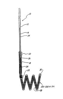
Figure 1 is an enlarged side view of a first embodiment ~ ~f the distal end of
the
wire and tip shown in partial cross-sectional view. A conventional Teflon
laminated
or similarly insulated stainless steel wire 10 is disposed within a protective
microcatheter (not shown). Stainless steel wire 10 is approximately 0.010 -
0.020 inch
(0.254-0.508 mm) in diameter. In the illustrated embodiment, wire 10 is
tapered at its
distal end to form a conical section 12 which joins a section 14 of reduced
diameter
which extends longitudinally along a length 16 of wire 10. Section 16 then
narrows
gradually down to a thin threadlike portion 18 beginning at a first bonding
location 20
and ending at a second bonding location 22.
The stainless steel wire 10, comprised of that portion disposed within the
microcatheter body, tapered section 12, reduced diameter section 16 and
threadlike
section 18, is collectively referred to as a core wire which typically is 50 -
300 cm. in
length.
In the illustrated embodiment the portion of the core wire extending from
tapered section 12 to second bonding location 22 is collectively referred to
as the
grinding length and may typically be between 20 and 50 cm. in length.
Reduced diameter portion 14 and at least pan of sections 12 and first bonding
location 20 may be covered with an insulating Teflon laminate 24 which
encapsulates
the underlying portion of wire 10 to prevent contact with the blood.
A stainless steel coil 26 is soldered to the proximate end of threadlike
portion
18 of wire 10 at first bonding location 20. Stainless steel coil 26 is
typically 3 to 10 cm.
in length and like wire 10 has a diameter typically between 0.010 to 0.020
inch (0.254-
0.508 mm).
The distal end of stainless steel coil 26 is soldered to the distal end of
threadlike portion 18 of wire 10 and to the proximal end of a platinum
secondary coil
28 at second bonding location 22. Secondary coil 28 itself forms a spiral or
helix
typically between 2 to 10 mm. in diameter. The helical envelope formed by
secondan~
coil 28 may be cylindrical or conical. Like wire 10 and stainless steel coil
26,
secondary coil 28 is between approximately 0.010 and 0.020 inch (0.254-0.508
mm) in
diameter. The diameter of the wire itself forming stainless steel coil 26 and
coil 28 is
approximately between 0.001 - 0.005 inch.
""..- . 10
The distal end of secondary coil 28 is provided with a platinum soldered tip
30
to form a rounded and smooth termination to avoid puncturing the aneurysm or
teanng tissue.
Although prebiased to form a cylindrical or conical envelope, secondary coil
28 is extremely soft and its overall shape is easily deformed. When inserted
within
the microcatheter (not shown), secondary coil 28 is easily straightened to lie
axially
within the microcatheter. Once disposed out of the tip of the microcatheter,
secondary coil 28 forms the shape shown in Figure 1 and may similarly be
loosely
deformed to the interior shape of the aneurysm.
As will be described below in greater detail in connection with the third
embodiment of Figure 3, after placement of secondary coil 28 within the
interior of
the aneurysm, a direct current is applied to wire 10 from a voltage source
exterior to
the body. The positive charge on secondary coil 28 within the cavity of the
aneurysm
causes a thrombus to form within the aneurysm by electrothrombosis. Detachment
of
the tip occurs either: (1) by continued application of current for a
predetermined time
when the portion 18 is exposed to blood; or (2) by movement of the wire to
expose
portion 18 to blood followed by continued current application for a
predetermined
time. Ultimately) both threadlike portion and stainless steel coil 26 will be
completely disintegrated at least at one point, thereby allowing wire 10 to be
withdrawn from the vascular space while leaving secondary coil 28 embedded
within
the thrombus formed within the aneurysm.
Figure 2 illustrates in enlarged partially cross-sectional view a second
embodiment of the invention. Stainless steel core 32 terminates in a conical
distal
portion 34. Stainless steel coil 36, shown in cross-sectional view) is
soldered to distal
portion 34 of wire 32 at bonding location 38. The opposing end of the
stainless steel
coil 36 is provided with a soldered, rounded platinum tip 40. In the
illustrated
embodiment, stainless steel core wire 32 is approximately 0.010 inch in
diameter v~ith
the length of stainless steel coil 36 being approximately 8 cm. with the
longitudinal
length of platinum tip 40 being between 3 and 10 mm. The total length of wire
32
from tip 40 to the proximate end is approximately 150 cm.
The embodiment of Figure 2 is utilized in exactly the same manner as
described above in connection with Figure 1 to form a thrombic mass within an
aneurysm or other vascular cavity. The embodiment of Figure 2 is distinguished
from
that shown in Figure 1 by the absence of the extension of stainless core 32
through
coil 36 to tip 40. In the case of the embodiment of Figure 2 no inner core or
reinforcement is provided within stainless steel coil 36. Threadlike portion
18 is
provided in the embodiment of Figure 1 to allow increased tensile strength of
the
v~ire. However, a degree of flexibility of the wire is sacrificed by the
inclusion even of
-. 11
threadlike tip 18, so that the embodiment of Figure 2 provides a more flexible
tip, at
least for that portion of the micro-guidewire constituting the stainless steel
coil 36.
It is expressly understood that the helical secondary coil tip of the
embodiment
of Figure 1 could similarly be attached to stainless steel coil 36 of the
embodiment of
Figure 2 without departing from the spirit and scope of the invention.
Thinned and threadlike portion guidewires disposed concentrically within
coiled portions are well known and are shown in Antoshkiw, 'Disposable
Guidewire ;
U.S. Patent 3,789,841 ( 1974); Sepetka et al., "Guidewire Device ; U.S. Patent
4,832,047
( 1989); Engelson, "Catheter Guidewire ; U.S. Patent 4,884,579 ( 1989); Samson
et al.,
"Guidewire for Catheters ; U.S. Patent 4,538,622 (1985); and Samson et al.,
"Catheter
Guidewire with Short Spring Tip and Method of Using the Same ; U.S. Patent
4,554,929
( 1985 ).
Turn now to the third embodiment of the invention as shown in Figure 3.
Figure 3 shows an enlarged side view of a wire, 'generally denoted by
reference
numeral 42, disposed within a microcatheter 44 shown in cross-sectional view.
Like
the embodiment of Figure 1) a stainless steel coil 46 is soldered to a conical
portion
48 of wire 22 at a first bonding location 50. A thin threadlike extension 52
is then
longitudinally disposed within stainless steel coil 46 to a second bonding
location 54
where stainless steel wire 46 and threadlike portion 52 are soldered to a soft
platinum
coil 56. Platinum coil 56 is not prebiased, nor does it contain any internal
reinforcement, but is a free and open coil similar in that respect to
stainless steel coil
36 of the embodiment of Figure 2.
However, platinum coil 56 is particularly distinguished by its length of
approximately 1 to 50 cm. and by its flexibility. The platinum or platinum
alloy used
is particularly pliable and the diameter of the wire used to form platinum
coil 56 is
approximately 0.001 - 0.005 inch in diameter. The distal end of platinum coil
56 is
provided with a smooth and rounded platinum tip 58 similar in that respect to
tips 30
and 40 of Figures 1 and 2, respectively.
When coil 56 is disposed within microcatheter 44, it lies along the
longitudinal
lumen 60 defined by microcatheter 44. The distal end 62 of microcatheter 60 is
then
placed into the neck of the aneurysm and the wire 42 is advanced, thereby
feeding tip
58 in platinum coil 56 into aneurysm 64 until bonding location 50 resides.in
the neck
of the aneurysm as best depicted in the diagrammatic cross-sectional view~of
Figure 4.
Figure 4 illustrates the insertion of the embodiment of Figure 3 within a
vessel
66 with distal tip of microcatheter 44 positioned near neck 68 of aneurysm 64.
Coil
56 is fed into aneurysm 64 until at least a portion of stainless steel coil 46
is exposed
beyond the distal tip 62 of microcatheter 44. A positive electric current of
approximately 0.01 to 2 milliamps at 0.1 - 6 volts is applied to wire 42 to
form the
,",., ~ 12
thrombus. Typically a thrombus will form within three to five minutes. The
negative
pole 72 of voltage source 70 is typically placed over and in contact with the
skin.
After the thrombus has been formed and the aneurysm completely occluded)
tip 58 and coil 56 are detached from wire 42 by electrolytic disintegration of
at least
one portion of stainless steel coil 46. In the illustrated embodiment this is
accomplished by continued application of current until the total time of
current
application is almost approximately four minutes.
At least one portion of stainless steel coil 46 will be completely dissolved
through by electrolytic action within 3 to 10 minutes, usually about 4
minutes. After
separation by electrolytic disintegration, wire 42) microcatheter 44 and the
remaining
portion of coil 46 still attached to wire 42 are removed from vessel 66,
leaving
aneurysm 64 completely occluded as diagrammatically depicted in Figure 5 by
thrombus 74. It will be appreciated that the time of disintegration may be
varied by
altering the dimensions of the portions of the wire and/or the current.
The process is practiced under fluoroscopic control with local anesthesia at
the
groin. A transfemoral microcatheter is utilized to treat the cerebral
aneurysm. The
platinum is not affected by electrolysis and the remaining portions of the
microcatheter are insulated either by a Teflon lamination directly on wire 42
and/or
by microcatheter 44. Only the exposed portion of the wire 46 is affected by
the
electrolysis.
It has further been discovered that thrombus 74 continues to form even after
detachment from wire 42. It is believed that a positive charge is retained on
or near
coil 56 which therefore continues to attract platelets, white blood cells, red
blood cells
and fibrinogen within aneurysm 64.
Although the foregoing embodiment has been described as forming an
occlusion within a blood-filled vascular cavity by means of electrothrombosis,
the
above disclosure must be read to expressly include formation of the occlusion
by
mechanical mechanisms without resort to the application of electrical current.
A
mechanical mechanism which can be safely disposed into the vascular cavity to
impede, slow or otherwise initiate clotting of the blood or formation of the
occlusion
is within the scope of the invention. The insertion within the vascular cavity
and
maintenance therein of an object with an appropriate blood-clotting
characteristics
can and does in many cases cause the formation of an occlusion by itself.
Depicted in
Figure 6 is an embodiment of the invention wherein such mechanical thrombosis
can
be achieved. Wire 10 has a tapering end portion 14 covered with a Teflon
laminate
24 similar to that described in connection with the embodiment of Figure 1. W
ire 10
is attached by means of a mechanical coupling 100 to a platinum coil 102 which
has a
plurality of filaments or fine hairs 104 extending therefrom. In the
illustrated
n7
13
embodiment, hairs 104 have a length as may be determined from the size of the
vascular cavity in which coil 102 iS to be used. For example) in a small
vessel hair
lengths of up,to 1 mm are contemplated.
(,oil 102 has sufficient length and flexibility that it can be inserted or
coiled
loosely into the vascular cavity. The length ~f coil 102 need not be so long
that the
coil itself is capable of being multiply folded on itself and fill or
substantially fill the
vascular cavity. Hairs 104 extending from coil 102 serve to substantially ,
pac)~ fil l o r
at least impede blood flow or access in the vascular cavity. Hairs 104, which
are
generally inclined backwardly away from extreme tip 106 when delivered) are
thus
easily able to slide forward with little friction through restrictions in the
vessels and
aneurysm. Additionally, hairs 104 do not have sufficient length, strength or
sharpness
to provide any substantial risk or potential for a puncture of the thin
vascular wall.
The plurality of hairs 104) when coiled within the vascular cavity) provide an
extremely large surface for attachment of blood constituents to encourage and
enhance the formation of a mechanical occlusion within the vascular opening.
In the preferred embodiment, coil 102 is mechanically coupled to thin tapered
portion 104 of wire 10 by means of a small drop of polyester 100. Polyester
may be
substituted for the gold solder of the previously described embodiments in
order to
reduce concern or risk of toxic reactions in the body.
Tip portion 104 may also be mechanically separated from wire 10 by means
other than electrolysis. One method is make the connection between tip 104 and
wire
10 by means of a spring loaded mechanical clasp (not shown). The clasps are
retained on tip 104 as long as the clasps remain inside of the catheter, but
spring open
and release tip 104 when extended from the catheter. The catheter and clasps
mad
then be removed from the insertion site.
In another embodiment wire 10 and tip portion 104 screw into each other and
can be unscrewed from each other by rotation of the catheter or wire with
respect to
35
,w . ~ 14 ~~~~779
tip 104. An extendable sheath (not shown) in the microcatheter is advanced to
seize
tip 104 to vrevent its rotation with wire 10 during the unscrewing process.
Even where the occlusion is not formed by electrothrombosis) separation of tip
104 may be effected by electrolysis. In such situations, the electrolysing
current tnay
be concentrated on the sacrificial stainless steel portion of tip 104 by
disposition of an
insulative coating on the remaining platinum portion. For example) tip,144 may
be
provided with a polyethylene coating save at least a portion of the stainless
steel
length. This has the effect of decreasing the time required to
electrolytically
sufficiently disintegrate the steel portion to allow detachment of the
platinum tip,
which is an advantageous feature in those cases where a large aneurysm must be
treated and a multiple number of coils must be deployed within the aneurysm.
Notwithstanding the fact that wire 10 and platinum coil 102 in the
embodiment Figure 6 or wire 10 and platinum coil 28, 36 and 56 in the
embodiments
of Figures 1-5 are radiopaque, there is still some difficulty when
manipulating the
device under fluoroscopy to be able to determine the exact position or
movement of
the probe relative to the aneurysm. This is particularly true when a large
number of
coils are deployed and one coil then radiographically hides another. Figure 7
illustrates an improvement of) for example) the embodiment of Figures 4 and ~.
Microcatheter 144 is positioned so that its distal end 162 within vessel 66 is
positioned
at the opening aneurysm 64. Microcatheter 144 is provided with radiopaque
marker
108 at distal tip 162, a tip marker. Moving toward the proximal end of
microcatheter
144 is a second radiopaque marker 110) a proximal marker. Radiopaque markers
108
and 110 are, for example, in the form of radiopaque rings made of platinum)
approximately 1-3 mm in longitudinal length along the axis of microcatheter
144.
Rings 110 and 108 are typically separated by about 3 cm on microcatheter 144.
Similarly) wire 10 has a radiopaque marker 112 defined on it such that marker
112 on
wire 10 is approximately with aligned with marker 110 on microcatheter 14 when
coil
56 is fully deployed into aneurysm 64. Typically) full deployment will place
the solder
35
,:'
2120'~'~~
or connection point 54 of the order of 2-3 mm past opening 68 of aneurysm 64.
Distal
marker 108 on microcatheter 144 is used to facilitate the location of the
microcatheter tip, which can often be obscured by the coils which have been
previously deployed. The coils are a varying lengths depending on the
application or
5 size of the aneurysm or vascular cavity being treated. Coil lengths of 4-40
cm are
common. Therefore, even though the thinness of coil 56 may make it difficult
to see
under standard fluoroscopy and even though the fineness of Wire 10 may
similarly be
obscured or partly obscured, radiopaque markers 108, 110 and 112 are clearly
visible.
Manipulation of wire 10 to proximal marker 110 can then easily be observed
under
10 conventional fluoroscopy even when there are some loss of resolution or
fluoroscopic
visual obstruction of the coil.
Further, in the previous embodiments, such as that shown in Figures 4 and 5,
when electrothrombosis is used to form the occlusion within vascular aneurysm
64,
coil 56 is used as the electrical anode while the cathode is a large skin
electrode 72
15 typically conductively applied to the groin or scalp. Figure 9 illustrates
an alternative
embodiment wherein microcatheter 144 is supplied with an end electrode 114
coupled to an electrical conductor 116 disposed along the length of
microcatheter
144. Wire 116 is ultimately led back to voltage source 70 so that ring
electrode 114 is
used as the cathode during electrothrombosis instead of an exterior skin
electrode 72.
With the embodiment of Figure 9, the electrical currents and electrical
current paths
which are set up during the electrothrombosis formation are local to the site
of
application which allows even smaller currents and voltages to be used to
initiate
electrothrombosis than in the situation when an exterior skin electrode must
be
utilized. The electrothrombosic current distributions are also better
controlled and
localized to the site of the thrombus formation. The possibility of stray
thrombus
formations occurring at unwanted sites or uncontrolled and possibly unwanted
electrical current patterns being established elsewhere in the brain or body
is
therefore largely avoided.
Many alterations and modifications may be made by those having ordinary
skill in the art without departing from the spirit and scope of the invention.
Therefore) it must be understood that the shape of the tip or distal platinum
coil used
in combination with the wire according to the invention may be provided with a
variety of shapes and envelopes. In addition thereto, the composition of the
micro-
guidewire tip may be made of elements other than platinum including stainless
steel,
beryllium, copper and various alloys of the same with or without platinum.
Still
further, the diameter of the wire) various of the wire described above and the
stainless steel coil immediately proximal to the detachable tip may be
provided with
,.,,
."", 16
differing diameters or cross sections to vary the times and current magnitudes
necessary in order to effectuate electrolytic detachment from the tip. Still
further, the
invention may include conventional electronics connected to the proximal end
of the
wire for determining the exact instant of detachment of the distal tip from
the wire.
5~ Therefore, the illustrated embodiment has been set forth only for the
purposes
of clarity and example and should not be taken as limiting the invention as
defined by
the following claims, which include all equivalent means whether now known or
later
devised.