Note: Descriptions are shown in the official language in which they were submitted.
WO 94/18~86 PCrlUS94/02249
-1- 2l2o9l8
FLOW DIRECTED C.ATHETER
Field of_~Q Invention
The present invention i in the general field
of surgical in~truments and relates ~pecifically to
infusion catheter~ that are uqed in cardiovascular and
endovascular pro~edure~ to deliver diagnostic,
therapeutic or vasoocclu3ive agents to a target site (the
target site being accessible by a tortuou~ path through
the vasculature). The invention also relates to the
process of using the infusion catheter.
Background
Catheters are being used increasingly as a
means for dalivering diagnostic or therapeutic agent~ to
internal target 8ite8 that can be acceQsed through the
circulatory system. ~here are a number of general
approaches for placing catheters within ves els in the
body that are difficult to acces~. In one such
techuique, a torqueable guidewire i8 alternately rotated
and advanced to the target 8ite. With the w~re in place,
the catheter i8 then advanced along the wixe u~til the
distal end of the catheter i~ positioned at the target
site. An example of this technique i~ de3cribed in U.S.
Patent No. 4,884,579. A major draw~ack to this approach
' i8 the time-consuming nature of rotating and advancing
the guidewire and catheter through the vasculature.
A second technique for ad~ancing a catheter to
a target site is to employ a highly flexible catheter
having an inflatable, but pre-punctured balloon at its
WO ~/l~UK PCT~4/OZ~9
-2-
212091~ ~
distal end. In use, the balloon is partially inflated,
and carried by blood flow into the target site. During
placement, the balloon is continually inflated to
replenish fluid leaking from the balloon. Thi~
S technique, too, has major drawbacks including the ~act
that the catheter material is so floppy that it cannot be
pushed without buckling, and instead must be ad~anced
using injected fluid to inflate the balloon in order to
propel the catheter to the target site. Additionally,
10 there i8 a significant risk of rupture of a vessel by a
balloon that has been overinflated.
In order to addre~s some of the above described
problems, another approach has involved the use of
flexible catheters that can be directed to a target site
15 as a result of the blood flowing to that site. In 1991,
Target Therapeutics released a product known as the
"ZEPHYR" flow-assisted infusion catheter. The product
was designed to be introduced into the ~asculature
through a guiding catheter and then allowed to be
20 directed by the blood flow to a target site. The
catheter comprised segments of different materials, a
proximal segment made of nylon, and middle and distal
segments made of a block copolymer of polyamide. The
product proved to be unsuccessful in achieving its
25 desired function as it was not flexible enough to
navigate the tortuous vessel pathway and not strong
enough to withstand the required injection pressure.
~ The present invention is an infusion catheter
j assembly useful for the delivery of diagnostic,
30 therapeutic or vasoocclusive agents to remote portions of
the vascular system, particularly to diagnose or treat
arteriovenous malformations (AVMs). The invention also
3 includes a process for placing the infusion catheter at
the target site and a process for delivering a
i
,,
Wos4/1~6 PCT~S94/0~9
212091~
diagnostic, therapeutic or vasoocclusive agent to the
target site.
Summary of the In~ention
This invention is an infusion catheter fQr
placement within a tortuous, small vessel pathway and a
method for delivery of an agent to a target site. The
infusion catheter is directed to the target site by means
of the flow of blood to that ~ite. The infusion catheter
has an elongate tubular body having proximal and di~tal
ends and a lumen extending between the ends through which
the diagnostic, therapeutic, or vasoocclusi~e agent can
be delivered.
The elongate tubular body is formed of a
relatively stiff tapered proximal segment, a relatively
flexible and strong distal se$ment, and a transition
section between the proximal and distal segments that is
less flexible than the distal segment but more flexible
than the proximal segment. The distal segment has a
burst pressure of at least about 195 psi and is made of a
material that will show a force of about lx10-4 or les~
when ten centimeters of the material i8 deflected 10
from horizontal.
A further aspect of the in~ention i8 a method
for accessing a target site. A guiding catheter is
inserted into the vasculature. An infusion catheter is
then inserted into the guiding catheter. A ~tylet may
optionally be used to ~traighten the soft, flexible
distal end of the infusion catheter for easy in~ertion
into the guiding catheter. If the ~tylet is used, it is
removed once the infusion catheter is inside the guiding
catheter. The infusion catheter is then pushed out of
the guiding ca~heter into the ~asculature. The blood
flow in the vasculature directs the infusion catheter to
the target site.
.
J
W094/1~C PCT~S94/0~9
--4--
212091~
Yet another aspect of the invention is a method
for delivering a diagnostic, therapeutic or vasoocclusive
agent to a target site. The infu~ion catheter i9
inserted into the vasculature by means of a guiding
catheter. The infusion catheter is positioned at the
target site a~ a result of the blood flow to the target
site. The diagnostic, therapeutic or va~oocclusive agent
is then injected through the catheter lumen and infused
into the target site.
Brief Description of the Drawing~
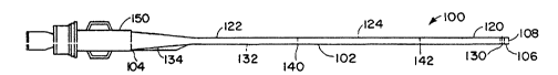
FIG. 1 is a diagram that shows an infusion
catheter constructed according to a preferred embodiment
of the present invention.
FIG. 2 is a diagram that shows the distal end
on one embodiment of the infusion catheter of the present
invention wherein the distal end is formed in an "S n
shaped configuration.
FIG. 3 is a diagram that shows an infusion
catheter, stylet and guiding catheter as~embly.
FIG. 4 is an illustration of a portion of a
tortuou~ path in a soft ti~sue, and the method of guiding
the infusion catheter along this path.
FIG. 5 i~ a graph showing the pounds of force
corresponding to the angle that the distal segment
material of the inventive catheter is deflected a3
compared to the di~tal segment material of a prior art
catheter.
Detailed De~cription of the Invention
FIG. 1 shows an infu~ion catheter 100
constructed according to a preferred embodiment of the
invention. The catheter 100 has an elongate tubular body
102 with proximal 104 and distal 106 ends and an inner
lumen 108 extending between the end~. The elongate
WO 94/18886 PCI~/US94/02249
-5- 21209'1~
tubular body 102 is comprised of three segments; a
relatively flexible and strong distal ~egment 120, a
relatively stiff tapered proximal segment 122 and a
transition section 124 between the proximal and distal
segments that is less flexible than the distal segment
120 but more flexible than the proximal segment 122.
The elongate tubular body 102 has a relatively
flexible and strong distal segment 120 such that the
catheter can easily navigate a tortuous ve~sel pathway.
~3y relatively flexible is meant that a force of about
lx10-4 pounds corresponds to a deflection of the material
that is 10 from horizontal, or only about sx1o~4 pounds
of force to deflect the material about 80 from
horizontal. By relatively strong is meant that the
material has a burst pressure of greater than 195 psi,
more preferably the burst pressure is between about 195
and 220 psi.
The flexible distal segment 120 has an open end
which allows for the infusion of diaynostic, therapeutic
or vasoocclusi~e agents into the target site. The
s flexible distal 9egment 120 is made of a polymer that is
~! ' springy and biologically compatible such as polyurethane,
a block copolymer of polyamide, polyvinyl chloride, or
silicone or blends of the above. The flexible distal
segment 120 carries one or more radiopaque bands 130 or
may be doped with a radiopaque material such as barium
sulfate, bis~th trioxide, bismuth subcarbonate,
tungsten, tantalum or the like 80 that the location of
the distal region of the catheter within the vessel may
be visualized radiographically. The distal segment 120
makes up between about 5 and 25~ of the total length of
the tubular member and i8 between about S and 40 cm long,
preferably between about 10 and 20 cm long. The inner
diameter of the distal segment 120 is between about 0.25
and O.S0 mm, more preferably between about 0.25 and 0.35
7~3~
WO ~/18UK PCT~S94/0~9
2120918 -6-
mm. The outer diameter of the distal segment is between
about 0.S0 and 0.80 mm, more preferably between about
O.60 and 0.70 mm. The wall thickness of the distal
segment 120 is between about 0.1 and 0.3 mm.
S The proximal segment 122 of the elongate
tubular body 102 is relatively stiff such that it can be
easily pushed thus eliminating the need for guidewire
support. The proximal segment 122 i9 made of a polymeric
or metallic material that is relatively stiff and
biologically compatible such as nylon, polyvinyl
chloride, polyethylene terephthalate or other polyester
elastomers or a braided shaft (a polymer outer core with
a metallic mesh inner core). The proximal segment 122
comprises a tapered proximal section 134 for attachment
to the proximal end fitting 150 and a distal section 132.
The proximal section 134 of proximal segment 122 makes up
between about 60~ and 80% of the total length of the
tubular member 102 and i8 between about 90 and 130 cm
long, preferably between about 100 and 120 cm long. The
largest inner diameter of the proximal section 134 (at
the proximal end 104 of the tubular member 102) is
between about 0.40 and 0.60 mm, more preferably between
about 0.45 and 0.55 mm. The outer diameter of the
proximal section 134 at the proximal end 104 of the
tubular member 102 is between about 0.8 and 1.2 mm. The
wall thickness of the proximal section 134 of prox;mal
segment 122 is between about 0.1 and 0.4 mm, more
preferably between about 0.2 and 0.3 mm.
The distal section 132 of proximal segment 122
makes up between 10 and 20~ of the total length of the
tubular body 102 and is between about 20 and 40 cm long,
preferably between about 20 and 30 cm long. The inner
diameter of the distal section 132 of proximal segment
122 is between about 0.20 and O.S0 mm, more preferably
between about 0.25 and 0.35 mm. The outer diameter of
WO g4/18886 PCT/USg4102249
-7- ~ 120~ ~8
the distal section 132 of proximal segment 122 i9 between
about 0.60 and 0.90 mm, more preferably between about
0.60 and 0.70 mm. The wall thicknes~ of the di~tal
~ection 134 of proximal ~egment 122 is between about 0.1
and 0.3 mm.
The tran~ition section 124 of the elongate
tubular body 102 is less stiff than ~he proximal segment
122 but more stiff than the distal segment 120. A
suitable material that i8 biologically compatible i~ a
polymer such as polyurethane, a block copolymer of
polyamide, polyvinyl chloride or ~ilicone with greater
durometer (i.e. that is stiffer) than the flexible distal
segment 120. The transition section 124 may be
radiopaque and thus ob~ervable in the event that the
catheter becomes lodged in a particular portion of the
vasculature or buckle~, and as such the polymeric
material is doped with a radiopaque material such as
barium sulfate, bismu~h subcarbo~ate, bismuth trioxide,
tung~ten, tantalum or the like. The tran~ition section
124 makes up between about 10 and 20~ of the total length
of the tubular member 102 and i8 between about 20 and 40
cm long, preferably between about 25 and 35 cm long. The
transition section 124 may be of constant diameter or may
be tapered. m e inner diameter of the transition section
2S 124 i8 between about 0.20 and 0.50 m~, ~ore preferably
between abou~ 0.20 and 0.35 mm. The outer diameter of
the transition section 124 i~ between about 0.50 and 0.90
mm, re preferably between about 0.60 and 0.70 mm. The
wall thickness of the transition section 124 i9 between
about 0.1 and 0.3 mm.
The proximal ~egment 122, transition ~ection
124 and di~tal ~egment 120 are joined at junctions 140
and 142, respectively. The junctionQ are formed by
flaring, overlapping and heat fusing the materials of the
proximal segment 122 and transition ~ection 124 and the
WO g4/18886 PCTIUS94/02249
2 1 2 09 1 8 -8-
transition section 124 and distal segment 120. The distal
segment 120, tranQition section 124 and distal ~ection
132 of proximal segment 122 may all have approximately
the same out~ide diameter or the transition section 124
and the distal section 132 of the proximal ~egment-122
may be tapered.
A standard proximal end fitting 150 is attached
to the proximal section 134 of the proximal segment 122
by heat fusion with reinforcing tubing.
FIG. 2 shows one embodiment of the distal
segment 120 of the catheter wherein the tip 160 of the
catheter is shaped with ~3team such that the diatal end
106 points to the wall of the ve~sel rather that straight
into the path of blood flow for ease of manipulation
through the tortuous vessel pathway. The particular
embodiment shown i~ an "S" shape, but the tip may be any
shape that allows for access to the particular
vasculature being treated. In this way, if the catheter
becomes lodged against the ves~3el wall, the infusion of
liquid through the catheter propels the distal end 106 of
the catheter away from the vessel wall. A~ the ~tiff
proximal segment 122 is pushed, the distal segment 120
will be carried by the blt~od flood to the target site.
The catheter described above is useful in
2S delivering diagnostic, therapeutic, or vasoocclusive
agent~ to deep tissue.
FIG. 3 shows a catheter as~embly 200 for
placing the infusion catheter 100 at the target site~ An
appropriate guiding catheter 202 i~ inserted into the
30 vasculature using standard placement techniques. A
rotating hemo~tatic valve 204 i~ connected to the guiding
catheter luer adapter 206. The guiding catheter 202 is
continuou~ly flushed with saline. The thumb-screw of the
valve 204 is opened and the infusion catheter 100 is
35 inserted through the rotating hemostatic ~ralve 204.
J
"
WO 94/1~886 PCT/IJS94/02249
21~ 9 :~CC3
Optionally, aq shown in FIG. 3, a teflon-coated stainless
steel stylet 208 is first inserted into the infusion
catheter 100 in order to prevent kinking of the infusion
catheter 100 within the valve 204. The distal end 106 of
the infusion catheter 100 iq advanced proximal to-the tip
of the guiding catheter 202. The stylet 208 is then
remo~ed from the infusion catheter 100. Once the stylet
208 is removed, the infusion catheter 100 is pu~hed out
of the guiding catheter 202. The infusion catheter 100
is gently guided by the flow of blood in the ~asculature
to the target site. Optionally, gentle pushing and
pulling and injection of saline or contrast medium
through the catheter lumen 108 may aid in the placement
of the catheter at the target site.
FIG. 4 shows the method of inserting the
infusion catheter into a tissue region which is reached
by a tortuous path. The figure shows a region of ~oft
tissue 300, such as in the region of the brain,
containing a target site 302. Initially the guiding
catheter, indicated at 202 is fed from a ~ascular access
region~ The infusion catheter 100 i~ inserted into the
guiding catheter 202 and then pushed out of the end of
the guiding catheter. ~lood flow in the vessel then
directs the infusion catheter 100 to the target site 302.
Once the infusion catheter i8 placed at the
target si~e, a ~yringe may be connected to the proximal
end fitting lSO and the diagnostic, therapeutic or
~asoocclusive agent may be infused through the catheter
lumen 108 and into the target site. The injected agent
may include radiopaque agents for viewing blood vessel
anatomy and blood flow characteristics in the target
region, vasoocclusi~e agents which can be u3ed to produce
small-artery ~asoocclusion in the tis~ue region supplied
by the target vessel, and pharmacological agents, such as
anti-tumor drug3 or sclerosing agent~ ~uch as alcohols,
W094/l~UK PCT~4/OZ~9
2 1 2 0 ~ 18 -lo-
which are effective againct identified disease states at
the target site. Vasoocclu~ive agents useful in the
treatment of arteriovenou~ malformations include polymer~
that are activated in the pre~ence of polar solvent~ such
5 as water and include material~ ~uch as n-
butylcyanoacrylate. Other types of vasoocclusive agent
useful in the treatment of arteriovenous malformations
include polymer solutions that coagulate by di~fusion of
the solvent when in contact with blood. Polyvinyl
10 acetate dissolved in dimethylsulfoxide is one ~uch agent.
Alternatively, vasoocclusive coils 304 may be injected
into the infusion catheter and delivered to a target site
to occlude the blood flow at that site.
The following Examples are intended to
15 illustrate the invention but not to limit it in any
manner.
Examples
20 Example 1 - Compari~on of Burst PressurQs
- Prior art catheter~, in particular the "ZEPHYR~
catheter first marketed in 1991 were tested for burst
pressure as were the inventive catheters. Pressure was
applied by injecting liquid with pressures in the range
25 of O to burst in 25-30 psi increments into the proximal
,r end fitting of the catheter- The prior ~rt catheter
s burst at the distal end when approximately 141 p~i of
pressure was applied. This value was a mean value for
- the catheters tested and therefore, statistically, 99.73%
30 (3 sigma) of the values for burst pressure for the prior
'7' art catheters lie between about 97 and 185 psi- The
catheter~ of the present invention burst at the distal
end when a mean value of 207 pci of pressure was applied.
99.73S (3 sigma) of the values for burst pressure for the
35 inventive catheters, therefore, lie between about 195 and
,~,,.
WO 94/10886 PCTIUS94/02249
-11- 2121)918
220 p8i; The inventive catheters, therefore proved to
be ~tronger than the prior art catheters.
Example 2 - Testing of Distal End Flexibility
The flexibilities of the distal ends of the
prior art "ZEPHYR" catheter and the inventive catheters
were compared using a Tinius Olsen bending stiffness
tester. The results are graphically described in FIG. 5.
10 centimeter portions of the distal segments
of each catheter were placed on the steel plate of the
Olsen stiffnesq tester. The material was deflected to
different positions and the corre~ponding pounds of force
recorded. When the inventive catheter was deflected 10,
the stiffness tester ~howed a force of 7xlO-5 pounds,
when it was deflected 50 the force was 3.~x10-4 pounds,
and when the deflection was 80, the force was 4.9x10⁻⁴
pounds. The prior art catheter was deflected 10 and the
stiffness te~ter showed a force of 7.5x10⁻³ pounds, when
it was deflected 50 the force wa~ 8.Sx10-2 pounds, and
when the deflection was 80, the ~orce was 1.23xlO-1
pounds. The inventive catheter, therefore, proved to be
much more flexible than the prior art catheter. Upon
calculation of the slope of the lineq shown in FIG. S,
for the inventive catheter, a 1 deflection corresponds
t 25 to 10-5 pounds of force, and for a prior art catheter, a
~$ 0.3 deflection correspond~ to 10-5 pounds of force.
J
While preferred e~bodiments of the i~vention
; have been described herein, it will be recognized that a
30 ~ariety of changes and modifications can be made without
departing frcm the invention.
tt
"