Note: Descriptions are shown in the official language in which they were submitted.
v
~rV
..:.~~. n
...r', i~.. t.~.. 4
f
. t
M
7 ~ . ~sY.~~, ni.~~:.
..'S,'... ...1..... ~~f.
.. I -iu ,~l. .
..r~..: ~~1n
r, ':. . r . :I r R!'. . d. ..
,.r :" x ,,L w,
"~ .p ~
....
.~~~' ,. , ~ . >.~,1
~'Cl~:....
~ , .. , , .
~ 2 ~ ~ ~ o ~ ~ PCg"/1JS~2/09037
1~V~ 93/0795
DETA ,~'r~ABLE ~ATtL~,Q,.N CATHETER
FOR END4S~:C~PIC TRF~~'1'~'ENT
QF -CIEs 1 CCZTJRET~' A_R ~ ,REFLU~
Backcaround of the Tnvent~.~n
The technical field of this invention is
treatment of urological defects and, in particular,
the treatment of vesicoureteral reflex.
The discharge of urine via the urinary'tract
is essential to the maintenance of healthy kidney
functions. In the normal individual, urine flows
from the kidneys through the ureters into the
bladder. From there it is periodically released via
the urethra. ~The terminus of the ureter at the
bladder normally provides a competent sphincter which
insures that urine flows from tkr~ ureter to the
bladdera lHowever, if this junction is impaired,
vesicoureteral reflex can occur wherein urine from
the bladder can return to the kidney. particularly
during voiding r~r when pressure.is exerted upon the
bladder:
~Tesicour~teral reflex can cause renal damage
or even renal failure either d~.rec~ly as a result of
higy~ p~cessures transmitted to the kidney or
indirectly as a result of infections introduced by
retxomigrati~n of :bacteria into the kidneys. These ,
~r~blems can be particularly acute in newborns and
infants when incoanpetent ureterovesical junctions a're
present. In some children. the task o~ kidney damage
can be reduced by antibi~tic therapies and minor
reflua~-pxoblems will disappear over time with
increased develogmental Pnaturity.
_1_
CA 02122039 2003-03-07
_.
However, when vesicoureteral reflux is
severe, surgery has soften been necessary to repair
the dysfunctional junction. In most of these
approaches, the ureter is dissected from the bladder
and re-implanted to lengthen or otherwise restrict
the submucosal tunnel,.. By reconfiguring the tunnel,
closure of the ureteral lumen can be markedly
improved as a result of intravesical pressure as the
bladder fills.
Unfortunately, these open surgical
procedures always carry risks, including collateral
damage to other urological structures and the
possible introduction of further infectious agents.
These risks are part~.cularly pronounced when surgery
is required on newborns or infants.
The endoscopic treatment of reflux was first
introduced i.n 1981 bar Matouschek when he infected
polytetrafluorethylene (Teflon; paste in the
subureteral region ota patient. In this approach, a
bolus of Teflon paste is introduced into the
subureteral region to restrict ox reshape the
submucosal tunnel. In a manner similar ;.o surgical
procedures, the effective length of the subrnucasal
tunnel is increased and effective closure, as the
bladder fills, is likewise achieved. This technique
was popularized by 0'Donnell and Puri, and has now
been utilized to treat vesicoureteral reflux in over
8000 children.
The use of ':feflori paste in the pediatric
population is not without controversy due to evidence
of Teflon~particle m:~gration to the lungs and nodes
*Trade-mark
_ 2 _
CA 02122039 2003-03-07
and granulorna formation in both animal models and in
humans. Nonetheless, there are definite advantages
in treating these patients endoscopically. The
method is simple and can be completed in less than 15
minutes, it has a success rate of over 85°s with a low
morbidity, and can be performed in an outpatient
basis.
Various other substances have been proposed
as safer alternate implant materials, including
collagen, autologous fat and fibroblast injections,
polyvinyl alcohol foam (Ivalon) and glass; however
each has its disadvantages. Volume loss has been
identified as a problem with collagen, autologous fag
and fibroblast injections. Granuloma formation with:
possible latent carcinogenic effects has been
associated with Ival.on and glass particles as well as
y
Teflon paste.
There exists a need for better methods and
systems far treatment of vesic~oureteral reflux and
related urological disorders. In particular,
approaches that avoid open reconstructive surgery
while providing effective control of urinary reflu~:
would satisfy a lonc;~-fel~ need in the field,
especially in the treatment o:E neonatal birth defects.
*Trade-mark
~. . . . .... , r. ,... .... ,~. r'r~...Jfm yefr:e '~. I ': ' ..J .,.
VNt~ 9~/n?~15 . , ~,, ~ ~ ~ . PCTIlJ592/09037 ! ,
summary_ of the Invention .
Methods and systems for treatment of
vesicoureteral reflux are disclosed in which a
detachable balloon catheter is incorporated into a"~
endoscopic instrument, such as a cystoscopic needle.
The needle is directed through the cystoscope Band
inserted into the subureteral region of the refluxing
bladder to establish a pocket. A catheter ar similar
delivery device can be inserted into this pocket in
the subureteral region, carrying a balloon. The
balloon is then inflated, preferably filled with an
inert biocompati.ble material; and then sealed. After
inflation, the balloon is detached and left in the
pocket such that the function of the bladder and
ureter is reconfigured to~pxevent reflux.
The methods and systems of the present
invention provade implant mat~rials which can be
delivered endoscopically, and which conserve their
volume, and are substantially non-migratary and
non=antigenic.
Tn one aspect of the invention, methods are
disclosed in which a Pocket is established in the
subureteral region, preferably between the mucosal
and submucosal tissue'layers: A balloon structure is
then.intrc~duced into the pocket and inflated (a.g., .
by filling the s ructure;with a biocompatible
material, such.as a polymerizable'soluta,on). The
contents of the balloon preferably are solidified.
and then the balloon is sealed, andl left in place.
~y positioning the balloon within the pocket, the
-,~_
CA 02122039 1999-12-08
ureter is reconfigured to provide a competent
ureterovesical junction.
In another aspect of the invention, systems are
disclosed for performing non-surgical procedures for the
treatment of vesicoureteral reflux, employing a cystoscope,
a positioning means for establishing a pocket in the
subureteral region, and a means for deploying a detachable
balloon structure within the pocket to reposition the
ureteral terminus, thereby alleviating reflux.
In yet a another aspect of the present invention
there is disclosed a system for the deployment of a
detachable balloon, comprising: a cystoscopic system
comprising a sheath, a viewing means for viewing a distal
site and a positioning element extending longitudinally
through the sheath; and an uninflated, detachable balloon
attached to a catheter forming a balloon catheter, said
detachable, uninflated balloon adapted for passage through
a lumen of the positioning element of the cystoscopic
system.
Various cystoscopes can be used in the present
invention and are commercially available from various
sources including, for example, Karl Storz Co. (Culver,
California); and Olympus Corporation of (Wilmington,
Massachusetts) .
The positioning means useful in establishing a
subureteral pocket can be a cystoscopic needle, e.g., a 19
gauge needle which is small enough to fit within standard
CA 02122039 1999-12-08
cystoscopic equipment. In one system, a thin walled
cystoscopic needle was obtained from Cook Urological Co.
(Spencer, Indiana) which had a 19 gauge outer diameter but
had the inner diameter of a standard 18 gauge needle (0.036
inches) .
Balloon structures useful in the present
invention can be formed from silicone or similar
substantially non-antigenic elastic materials. The
uninflated balloons preferably are sized to fit unto the
tip of a catheter which can pass readily through the lumen
of the positioning device (e. g., a cystoscopic needle).
- 5a -
.. ~ '.,1 f ~
......,. :: ~. . :-, ~ . ,. ...; .. ,..... , r' .~.. . .'..n. ~. ..~ 4
'..~,..h ,....'~. . .,..,..~:. ~.~~...'". ~ .,;. ~r,, ,:.,;.-.,:.... .
w~ ~3~o~sm r ~c revsgmo~o~~
The balloon structures can take various
forms but preferably include a sealing mechanism
Which seals the balloon upon inflation. The sealing .
mechanism aan be achieved, for example, by a
constrictive collar, or a lip seal, or both.
The balloon can be delivered by a catheter
which is inserted through the needle or positioning
means to the site where the balloon is to be
inflated. In one preferred embodiment, the catheter
provides a means for not only inflating the balloon
but also means for filling the balloon with a
biocompatible material. Catheters suitable fox use
in the present invention are available from various
sources including, for example, Interventional
Therapeutids (San Francisco, California).
Various materials can be used to fill the
balloon, including collagen, autologous fat or
cellular extracts, or a,n inert polymer. In one
embodimEnt, the balloon is filled with a
polymerizable solution, such as an acrylic solution
which solidifies ,~ : In a preferred embodiment,
the pol~tnerizable-solution is a solution of
hydxozyethyl nnethylacrlrlate (HEM) which is cured to
a solid form 'by the addition of ferrous sulfate and
hydrogen peroxide:
The irav~nt~on will next be described in
connection with certain illustrated embodiments;
however; it should be Clear that those Skilled in the
art can make various mod~.fic~tions, additions and
subtrat;tior~~ with~ut departing from the spirit or
scope of the invention. For example, although the .
CA 02122039 2003-03-07
invention is specifica:Ll.y described i.n connect'.ion with the
treatment of vesicouret.-.eral reflux, i.t. should be clear
that the invention is ,:~pp.Licab:Le to c>tr~er treatment
protocols.
In another asrect, the present invent: ion provides
a system for t=he deploy,~ment of a detacr.able balloon
comprising: a cystosc<:~pic system comprising a sheath, a
viewing means for view.i~rug a distal site and a positioning
element extending longitudinally through the sheath; and a
balloon catheter including an un-~nflated, detachable
balloon attached the ret:,a, said p~s::.tior.ing element hav~~ng
a longitudina~w lumen tlzer~etrroug~., means for ~:assing said
balloon through said l.lmen of said positioning element,
and means for inflating and deta:hing said balloon from
said balloon catheter.
vV~ 93>0~$i5 ~ y ~ r~ ~~ ~~rius9z~o9o3~
Brief Descripi-~ion of the Drawings ,
FIG. 1 is schematic diagrarn of a system
according to the invention;
FIG. 2A is a more detailed schematic diagram
of the distal tip of the system of FIG. 1 prior ~to
inflation of the balloon structure;
FIG. 2B is another diagram of the system of
FIG. 2A in which the balloon means is being inflated
FIG. 2C is anather diagram of the system of
FIG. 2B in which an inflated balhoon.means has been '
detached;
FIG. 3A is a schematic diagram illustrating
an initial stage of a method according to the
invention in which a pocket is established within the
subureteral region; and
FIG. 3B is a further schematic diagram
illustrating a subsequent stage in the method of FIG.
3A in which a ball~on is inflated within the pocket
and then detached:
~~
'V6itD 93J07815 - ~ ~ ~ ~ ~ ~ ~ PC,';i'/US92/09037
,f
Dgtailed Description
FIG. 1 shows a system 10 for treatment of
vesicoureteral reflux including a cystoscope 1~
having a outer sheath 14 and an inner lumen.l5. The
cystoscope includes an eyepiece or other viewing port
(e. g., a video adaptor~ 16 in optical communication
with the~distal tip 18 of the cystoscope. In the
illustrated embodiment, an optical relay mechanisrn
20, including for example, a series of lenslets_22
and a distal cystoscopic lens 24 are disposed within
the lumen 15 of the cystoscope 12.
The cystoscope 12 further includes a
positioning means, e.g., a cystoscopic needle 26 for
positioning a balloon structure 30 in the subur.eteral
region of a reflu$ing,bladder. The balloon structure
is preferably connected to a catheter 28 which passes
through the positioning means 26 and serves to
inflate the ~al3.non structure. In the illustrated
embodiment, the catheter is connected to a
polymerizab~.e solution supply. e.g., a syringe 32.
In FIGS. ~A-2C, the operation of the
positioning means 26 and fiche balloon structure 30 is
illustrated in more detail. As shown in FIG. 2A, the
end of cystoseopic needle 26 is positioned, at a site
where inflation ancl~implantation of the balloon
structure ~.s desiredo' Catheter 28 with balloon 30 at
its tip is then advanced through the needle 26 into
place at the site, eog. in the subureteral region,
and then inflated as shown in FIG. 2B.
A qs
'~V~ 93/07815:~ ~ ~ 2 ~ '~ ~ . . PC'f/~J592/m3037
'~'.
The balloon structure 30 preferably includes
at least one sealing mechanism, such a lip or flap
'v'i seal 34 or a constrictive collar 36, which provide
for self-sealing of the balloon means upon
.,
, 5 inflation. L~uch sealing mechanisms operate to ezpel
.
;
;.
, and/or close the balloon when a certain inflation
state is reached. FIG. 2C shows a fully inflated
balloon which has been detached from the catheter ~8,
such that the catheter 38 and needle 26 can be
withdrawn from the implant site. .
..>
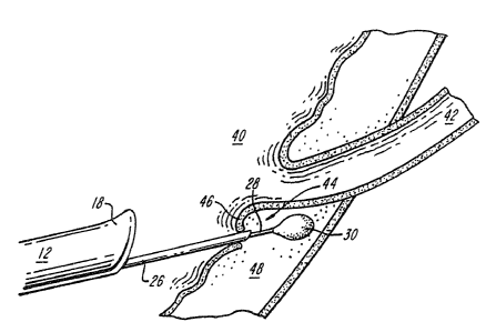
In use, the invention can be practiced by
introducing the cystoscope 12 into the bladder 40
and, as shown in FIG. 3A, inserting the needle 26
into the subureteral region 44 of the refluaing
v' ureter 42 (e. g. between tae mucosal and submucosal
tissue layers 46, 48, respectively). The balloon 30
with the attached delivery catheter 28 then is
inserted through the core of the needle 26 and placed
in the subureteral region. After inflation of the
'pj balloon, e.g.e with a pOlymeriz~ble solution, the
needle is withdrawn from the subureteral tissue
leaving the balloon in place.
;.:::
~
>,y~~5 Hydro~yethyl methylacrylate (HEN1A), a
hydrophylic p~Tymer compatible with silicone and
which solidifies within 60 aninutea after the addition
,;., .
of ferrous sulfate and hydrogen pero~ideo as .
_; ,particularly useful, as, a; filling material ,for the
'.'s30 ballo~n 30. HEMA can be injected through the
catheter 28 to inflate the balloon 30, while
ends~scbpically visualizing the balloon compressive
4 effect on its surrounding tissue. The catheter is
then pulled; leaving the self-sealing detachable
_10..
' :J':n
'WfD 93/0'795 ~ ~ ~ ~ ~ ~ ~ ' i'GTtUS92/09037
;.:';
;;
:balloon in place, as shown in FIG. 3B. The
compressive effect of the inflated balloon 30 is to
reconfigure the ureteral tunnel 42 so as to minimize
the likelihood of refluz.
The invention will next be described in
'connection with certain non--limiting euperimental
::,,:,
protocols.
~..~~ -
,A system similar to that shown in FIG. 1 was
constructed with catheter having a length of about .
100 centimeters and the diameter of about 2.0
French. The balloon design included a small lip seal
'~i valve closure mechanism and had an uninflated
diameter of~about 0.034 inches. A thin walled
'~" ~ cystoscopic needle was obtained from Cook Urological
(Spencero Tndiana) which had ~ 19 gauge outer
1...
diameter but had the inner diameter of a standard 18
gauge needle (0.036 inches). HEMA was used as the
filling material for the balloons: Infused through
the deliver catheter and into the balloon HEhtp.
progresses from a grater like liquid state to a
semi-solid, gel fort and ultimately solidifies within
the balloon shell. Polymerization tame is controlled
,by yary~ing the ingxed$ents necessary for the reaction
to occur. An estimated tire to cure of ~0 minutes
;~
utiiazing a solution composed of
w~is achieved k~y
,
.
64.5% of HA. X2.2% of hydrogen peroxide and 3.25%
of ferrous ammonium sulfate.
pegs were'ch~sen for this study because of
the similarities between porcine and human kidneys.
_ : , . . ,. ~ , .. _. . . ;.. - : .,-.- , . :-- . . -.., .. ~ . . ::. ~ -;
,.:::: . , . .. , . : . : , . . .. . : . . .. ; =.,: : , ~ , :.
. . . . .. . . ..,. . _ . .. ,.::: , .., . . . :. .. . . ... t. . .. ... ....
.. .,:
VV~ 93/078'15 . Pc reuS92eo9o3~
2~~~~~~
~. .,;;
"r The Hanford minipig was used fox the convenience of
.:..;,
its smaller size. Preoperative intravenous
::;
pyelograrns (IVP's) and cystograms with Conray
(Mallinkrodt, Inc., St. Louis. Missouri) were
.::;,
performed in 5 of the 6 minipigs.
. , ,,
~tefluz was created in 6 female Hanford
minipigs by unroof~,ng the ureters bilaterally. This
;;;
was done with the standard technique of open surgery
in two minipigs. However in the other 9 we attempted
y,j and were successful in creating reflux endoscopically
~~utilizing laparoscopic scissors through a 19 French
"., cystoscope.
Four to 6 weeks later the presence of
bilateral reflu~ was confirmed with a cystogram and
the balloon was implanted unilaterally in the
subureteral region. This was done with open surgery
in the first minipig and endoscopically through a 1g
gauge needle and a 1S Fr. cystoscope in 5 minipigs.
A repeat cystograan and IVP were performed 2 to 4
weeks after implantation.
Serial cystograms, ultrasounds, and IVP's
were performed at 4 to 6-week intervals until
sacrifice: The six minipigs- were sacrificed at 9(1),
S(2). 1~(a)::and 24(l)'weeks after balloon
irmplanta~ionThe blaidde~ balloon implant sites were
resected and analyzed macroscopically and
microscopically: ~ Histologic analyses of the bladder,
ureters, regional lymph nodes, kidneys, spleen, liver
and the tissue surrounding the balloon implant sites
here performed-
-1~'
CA 02122039 1999-12-08
. , ..
All minipigs which had preoperative studies
had no evidence of reflux as demonstrated by a
cystogram and no evidence of obstruction as
demonstrated by ultrasonography or IVP's. Four to
sia weeks after unroofing the ureters bilaterally,
cystograms confirmed the presence of bilateral
refluz, and IVP's and renal ultrasonography
demonstrated no evidence of obstruction in each
animal.
Cystography was again performed 2 to 4 weeks
after balloon implantation in all animals. This
demonstrated resolution of reflux in the treated
ureter and persistence of reflux in the opposite
untreated ureter. The serial cystograms,
ultrasounds, and IVP's performed at 4 to 6 week
intervals showed persistence of reflux in the
untreated side and continued effectiveness of the
balloon in the implanted ureter without reflux or
evidence of obstruction.
After sacrifice, gross inspection of the
bladder implant site showed no evidence of extrusion
or abscess formation in any of the minipigs.
Microscopic analyses of the tissues surrounding the
balloon implant showed mild inflammation. A fibrotic
reaction was also evident between the balloon shell
and the ureteral tissue. Tissue sections from the
lymph nodes, kidneys, liver and spleen showed no
evidence of particle migration or inflammatory
reaction.
- 13 -