Note: Descriptions are shown in the official language in which they were submitted.
~ .... WO93/09721 PCT/US92/06186
~ 212~9~S
f'
~:. APPARATUS ~ND METHODS FOR CLAMPING
.TISSUE AND REFLECTING THE SAME
Field of the Invention
This invention relates generally to apparatus and
method~ of clamping tissue, and more particul~rly to
apparatus and methods o~ clamping internal ti~sue during
laparoscopic or other minimally-in~asive medical procedures
on a living being.
ackaround of the Invention
During surgery it is oft~n necessary to reflect
internally located tissue or organs away from a site within
the body of the patient to gain access to that site and/or
for better visualization. Often the surgeon will use a
suture and needle for this purpose, anchoring the free end of
;. the hemostat wi~h a suture to some conveniently adjacent
;3 tissue, or a hemostat alone will be used. While such a
procedure may be generally suitable ~or its intended
purposes, it never the l~ss exhibits several significant
drawbacks. The most significant of those drawbacks is that
the process is time consuming and not conducive to
.;, .
procedures in which only a small incision or puncture is
~ade, e.g., laparoscopy, endoscopy, etc., to provide access
to the interior of the patient's body. ~nother approach to
reflecting internally located tissue entails grasping that
ti~sue by use of some clamp-type device. The problem with
that other approach is that such devices are typically quite
large, and hen~e unsuitable for laparoscopic, endoscopic, or
other small entrianceway, introduction. ~oreover, internally
located tissue is typically quite slippexy, thereby
increasing the difficulty in grabbing and clamping it.
The prisr art includes various tissue clamping or
grasping devices designed for various purposes, e.g.~ to
~taple tissue together to effect an anastomosis. Such
devices are generally not suitable for reflecting internally
~, located tissue during laparoscopic or endoscopic procedures.
Examples of such prior art devices are fsund in the following
patents: ~,890,519 ~Storz, Jr.1; 3,S79,75l (Jonckhee~e Pt
.~
. WO93/09721 PCT/US92/06186
212293~
al~; 3,827,277 (Weston); 4,038,987 ~Komiya~; 4,586,502 (Bedi
et al); 4,5~6,503 (Kirsh et al): 4,741,337 (Smith et al);
4,796,627 (Tucker~; 4,929,240 (Kirsh et al); 4,983,176
(Cushman et al); 5,015,249 (Nakao et al); 5,032,127 (Frazee
et al); 5,035,692 (Lyo~ et al); and 5,049,153 (Nakao et al)~
Various instruments are available commercially for
use with con~entional trocars to reflect internally located
tissue. Such devices typically comprise an elongated device
ha~ing a pair of gra~ping jaws located at the distal end of
the device. The proximal end of the device typically
comprises an actuating assembly, e.g., a pair of finger grips
which are arranged to be squeezed together, to cause the jaws
to close to grasp ti~ue located therebetween. Such de~ices,
while yenerally effective for effecting the reflection of
internally located tissue, still leave much to be desired
from various standpoints. For example, each device re~uires
the use of a conventional trocar for placement thereof. This
factor substantially limits the numbex of devices which can
be used during any operative procedure in order to keep the
operating situs relatively uncluttered. Moreover, since
conventional trocars typically require suturing to close the
incision or puncture created thereby~ it is desirable to
minimize the number of trocar penetrations.
Accordingly, a need exists ~or some means for
gr~sping, without penetrating or damaging, tis~ue located
internally in the body of a living bei~g, where access to the
interior must be achieved through a ve~y small percutaneous
incision or puncture, so that the internally located tissue
can be positioned as desired by use of some means extending
through the same or another percutaneous incision or
puncture, and without necessitating the use of a trocar.
Obiects of the Invention
~ ccordingly, it is a general object of this
invention to provide apparatus and methods of use whi h
overcome the disadvantages of the prior art.
It is a further object of this invention to provide
a clip and method of use which is arranged to be lnserted
~ WO93/09721 PCr/US92/06186
~~ 21229~3~
into the body of a living being via a very ~mall incision or
puncture to clamp a portion of internally located tissue.
It is still a further object of this invention to
provide a clip and method of use which is arranged to be
inserted into the body of a living being via a ve~y-small
incision or puncture to clamp a portion of internally located
tissue to enable the reflection of said tissue from outside
the body of the being.
It is still a further object of this invention to
provide a clip and instrument which is arranged to insert the
clip into the body of a living being via a very small
incision or puncture to clamp a portion of internally located
tissue without penetrating or damaging that tissue.
It is yet a furth~r o~ject of this invention to
provide an instrument which is arranged to be inserted into
the body of a living being via a very small incision or
puncture to retrieve a clip clamped to a portion of
internally located tissue.
It is yet a further object of this in~ention to
provide a method for reflecting, positioning, and/or holding
in position internally locatPd tissue through a small
inci~ion or puncture.
Summary of the Invention
These and other objects of this invention are
achieved by providing a system and method of use for
reflecting tissue located within the body of a living being.
The ~ystem comprises at least o~e clip, an
introducer in trument, and an elongated positioning member.
The introduaer instrument is of a cmall diameter arranged for
delivering the lip through a small percutaneous incision or
puncture to the situs of the tissue. The clip comprising a
pair of jaws and a biasing.member. The jaws have portions
de~ining a mouth therebetween and are moveable between a
first orientation, wherein they are disposed apart, and a
seco~d orientati~n, wherein they ar~ disposed closer
together. The biasing member is coupled to the jaws and is
actuatable via the same or another percutaneous incision or
.s~.j
."'~
WO93/09721 PCT/US92/06186
212293~ 4
puncture to move them from the first orientation to the
second orientation so that at least a portion of the tissue
is trapped within the clip's mouth.
The elongated positioning member has a distal end
portion and a proximal portion, and is of small ~i~meter
arranged to be extended through any of the percutaneous
incisions or punctures from outside the body of the being so
that th~ distal end portion is coupled to the clip, with the
proximal portion of the elongated positioning member being
located outside of the body of the being. The proximal
portion is arranged ts be moved to cause the portion of
tissue trapped within the mouth of the clip to be moved to a
desired position.
The method of this in~ention entails inserting the
clip through a first incision or puncture, locating portions
of the clip~s jaws immediately adjacent the tissue to be
reflected, and then actuating the biasing means to cause the
jaws to move to the second orientation so that a portion of
the tissue is trapped within the clip's mouth.
The elongated positioning element is ex~ended
through the first incision or puncture or a second
percutaneous incision or puncture so that it is coupled to
the clip. Then the elongated positioning element is
manipulated from outside of the body of the being to effect
the reflection of t~e tissue.
In some e~bodiments of the invention the elongated
positioning element comprises a tether which is connected to
the clip as introduced. The tether ~s arranged to be pulled
to effect the movement or positioning of tissue to which the
clip is secured. Plural clips may be coupled to each other
via at least one tether in a pulley arrangement to effect the
movement or positioning of tissue to which one or more of the
clips is clamped. In other embodiments of thP invention the
positioning means additionally comprises a hammock-like
assembly coupled between a pair of clips for holding a
portion of internal tissue thereunder.
~ WO93/D9721 PCT/US92/06186
21229~
. 5
Descri~tion of the Drawin$~
Other objects and many of the attendant advantages
of this invention will be readily appreciated as the same
becomes better understood by reference to the following
detailed description when considered in connection ~ith the
accompanying drawings wherein:
Fig7 1 is a isometric view of a clip and the distal
end of an instrument or tool for applying it, both of which
are constructed in accordance with this invention, and shown
prior to the securement of the clip to internally located
tissue within the body of a living being;
Fig. 2 is a isometric view of a clip and the distal
end of the tool of Fig. 1 shown after to the securement of
the clip to such internally located tissue;
Fig. 3 is a side elevational view, partially in
section, of the clip and the tool ~hown in Fig. 1 prior to
the securement of the clip to internally located tissue;
Fig. 4 is a side elevational view, partially in
section, o~ the clip and the tool shown in Fig. 3 during the
securement of the clip to internally located tissue;
Fig. 5 is a side elevational view, partially in
section, of the clip and th~ tool shown in Fig. 3 after the
~`~securement of the clip to internally located kissue, but
prior to removal of the tool;
~Fig. 6 is a side ele~ational view~ partially in
isaction, of the clip shown in Figs. 1 and 3 after its
securement to the internally located tis~ue, but after the
tool has been remove~;
Fig. 7 is a side elevational view, partially in
section, of an alternative embodiment of the tool shown in
Figs. 1-5;
Fig. 8 is a side elevational view, partially in
section, of another alternative embodiment of the tool shown
in Figs. 1D5;
Fig~ 9 is a isometric view of two clips constructed
in accordance with one aspect of this invention secured to
~i W093/09721 PCT/US92/06186
,
i 2122~3S 6
respective portions of internally located tissue in
accordance with one a~pect of the method of this invention;
Fig. 10 is a isometric view of three clips
constructed in accordance with another aspect of this
in~ention secured to respective portions of internally
located tissue in accordance with another aspect of the
method of this invention;
Fig. 11 is a side elevational view, partially in
section, of another embodiment of a clip and another
embodiment of a tobl for applying it constructed in
accordance with this invention and shown at an initlal point
during the securement of the clip to internally located
tissue;
Fig. 12 is a side elevational view, partially in
section, of the clip and the tool shown in Fig. 11 after ~he
securement of the clip to internally located tissue, but
prior to removal of the tool;
Fig. 13 is a side elevational view, partially in
section, of the clip shown in Fig. 11 after its securement to
the internally located tissue and after the tool has been
removed;
Fig. 14 .is a side elevational view, partially in
section, of the entire tool shown in Fig. 11;
Fig. 15 is an isometric view showing another
alternative embodiment of a clip constructed in accordance
with this invention used wikh a tool like that shown in Figs.
11 - 14,
Fig. 16 is an enlarged side elevational view of the
clip shown in Fig. 15:
Fig. 17 is an end view of clip taken along line 17
- 17 of Fig. 16;
Fig.18 is a side view showing another alternative
embodiment of a clip constructed in accordance with this
invention used, and which is basically constructed like the
clip of Fig. 15, and which is arranged to be used with a tool
like that shown in Figs. 11 - 14 or with a tool like that
.,
. W093/Q9721 2 1 2 2 ~ 3 5 PCT~US~2/06186
shown in Fig. 20 which is also constructed in accordance with
this invention;
j Fig. 19 is a sectional view of clip of Fig. 1
taken along line 19 - 19;
Fig. 20 is a side elevational view, partially in
, section, of the abdomen of a living being shown with interior
i portions thereof being reflected by a system constructed in
accordance with this invention;
Fig. 21 is an enlarged sectional view of the distal
end of the instrument shown in Fig. 20 holding a stack of
clips like shown in Fig. 18 for introduction into the body of
a being to clamp respective portions of tissue located
therein;
Fig. 22 is a plan view of a positioning element
forming a portion of the system shown in Fig. 20 and
constructed in accordance with this invention ~or releasable
coupling to a clip attached to internally located tissue for
effecting the re~lection of such tissue from outside the body
of said being;
Fig. 23 is a plan view, partially in section,
showing a clip removal tool construct~d in accordance with
~: this invention for effecting the remc~al of clips conætr~cted
in accordance with this invention from the body o~ the being;
and
Fig. 24 is a view of the removal tool of Fig. 23
showing the removal of a clip from the body of the being,
~etail~d Description of the Preferred Embodim~nts
Re~erring now in greater detail to the figures
ther2 is shown at 100 in Fig. 1, at 400 in Figs. 11-14~ and
at 400' in Fig. 20 an instrument or tool forming part of a
system for introducing one or more clips 100 and 100' through
a small percutaneous incision or puncture (not shown) in the
body of a patient to attach the clip(s) to some internal
organ or tissue 22 (Figs. 2, 4 6, 9-13, 15, and 20~, so that
the clip(s) can be coupled to ~ome positioning means
ex~ending through that percutaneous incision or puncture or
~i some other percutaneous incision or puncture, whereupon the
WO93/09721 PCT/US92/0618 ~
21229:~
tissue to which the clip(s) is(are) attached can be
reflected, moved or otherwise brought to æome desired
location, by manipulating the positioning means from outside
the body of the patient.
Four exemplary embodiments of clips constructed in
accordance with this învention are shown and descr.ibed
herein. In particular one embodiment of the clip is shown in
Figs~ 1-10 and is denoted by the reference numeral 20.
Another embsdiment of the clip is shown in Figs. 11 - 13 and
is denoted by the re~erence numeral 200. Yet another
embodiment of the clip is shown in Figs. 15 - 17 and is
denoted by the reference numeral 500. Lastly, another
embodiment of the clip is shown in Figs. 18, 19, 20, 21, ~nd
24, That embodiment of the clip is a slight ~odification of
clip 500 and is denoted by the reference numeral 500i.
As will be described in detail later the clips 20,
200, and 500, each include a positioning/holding means in the
form of a flexible filament or tether connected to it when
inserted into the body of the being through a percutaneous
incision or puncture to enable the surgeon to pull on the
tether to move, e.g., reflect, the tissue 22 ~o which the
clip i5 ~ecured to some desired position, e.g., out of the
way of the surgeon's line of sight through the incision or
puncture. ~ will also be described later either of the
clips ~0~ 200, and 500 may be coupl~d to one or more
similarly constructed clips located within the body of the
patient at a different location to form a pulley-like
arrangement for expediting the tissue moving and/or
positioning procedure. Moreover, the positioning/holding
means may additionally comprise a web-like material (also to
be described later) arranged to be coupled to another
internally located clip to form a hammock-like barrier or
xetainer holding a portion of internally located tissue in
position, e.g., out of the way of the operative site.
In the embodiment of Figs. 18 and 19 the clips 500'
do not include any positioning/holding means connected to
them (as is the case of clips 500) when they are inserted
WO93/Og72l PCT/US92/06186
21~293~i
---- 9
into the body of the being. Instead each clip 500' is
constructed and arranged to be coupled to separate
positioning/holding means which extends through any suitable
percutaneous incision or puncture after the clip has be
located in position within the body of the being an* secured
to the tissue to be reflected. The structure and operation
of the clips 500' and of the system for introducing and
positioning them will be described later with reference to
Fig. 20.
As can be seen clearly in Figs. 1 and 2 the clip 20
basically comprises a pair of jaws 30 and 32, connected by
bridging section 34, and a holding member 36. In this
embodiment the holding member comprises a filament or tether,
connected to the bridging section. Each of the jaws 30 and
32 terminates in an angularly extending a free end 38, whose
~ distal edge 40 is serrated. The serrated edges 40 define
3~ what may be called the "mouth" of the clip between the free
3~ ends of the jaws 30 and 32.
The bridging section 34 of the clip 20 is a
generally U-shaped member which cooperates with the jaws 30
and 32 to serve as a biasing means to bias the jaws into a
closed orientation, wherein their free end edges 40 engage
each other to thereby "~lose" the clip's "mouth". In this
regard, and in accordance a preferred embodiment of this
invention, the clip 20 is formed as an integral unit of a
resilient material, e.g., stainless steel, so as to form a
; spring frame wherein the innate resiliency of the frame, and
~; particularly its bridging section 34, naturally biases the
jaw closed.
As will be described later the jaws 30 and 32 also
include cam means thereon which are arranged to cooperate
with other cam means forming a portion of the tool 100 to
open the clip's mouth like that shown in Fig. 3 in order to
enable the clip to clamp or grasp onto the tissue portion 22.
In accordance with a preferred embodiment of this
invention the clip 20 of Fig. 1, the clip 200 or Fig. ll, and
~ he clip 500 of Fig. 15 are each quite small, e.g., 0.180
:
:~
~:
W~93/09721 PCT/U~92/06186..
~l 212293~
1 o
inch (4.57 mm) in diameter by O.7 inch (17.8 mm) long so that
they can be applied using a tool which can fit through a 5 mm
sheath or introducer for use in laparoscopic or endoscopic
surgery. It must, however, be pointed out that the clips 22
~ and 200 of this invention can be of different dimensions,
I e.g., larger, for various types of applications, e.g., manual
securement.
Irrespective of the size or shape of the clips,
each is constructed and arranged to be oriented so its mouth
is open by the time it is adjacent the tissue to be grasped
and its biasing means actuated to cause the clip to snap
closed to grab or grasp the tissue without penetrating it.
As should ~e appreciated by those skilled in the art,
internally located tissue, particularly soft, lightly
pressurized tissue, e.g., a blood vessel, the gallbladder,
the urinary bladder, the colon, a hernia, an aneurysm, etc.,
is typically quite slippery and difficult to grasp. To
overcome that problem, the cli~s of this invention are
designed to snap closed ~irtually instantaneously upon their
"release9' ( as will be described in detail la~er). Such
closing action produces a tissue wave at the edge of the
jaws. T~at wave gets trapped between the jaws, i.e., within
! the mouth of the clip, to result in a firm, reliable, yet
¦ non-pen~trating, grasp on the tissue. Thus, each of the
clips of the subj~ct invention can be said to ~Idynamically~
clamp or txap wet or slippery tissue without the tissue
slipping or popping:away ~rom or out o~ the clip's mouth.
Attention is now directed to Figs. 1 and 3 for a
description of the cam means referred to earlier for
effecting the opening the clip's mouth. Thus, as can be seen
therein each of the jaws 30 and 32 includes a pair of ramps
42. One of the ramps 42 of one pair projects outward and
downward from one marginal edge of the upper jaw 30, while
the other ramp 42 of that pair projects outward and downward
from the other marginal edge of that jaw. In a similar
manner one ramp 42 of the other pair projects outward and
upward from one marginal edge of the lower jaw 32, while the
.i
i
j W093/09721 PCT/US92/06l86
~. 2~2~3~
11
7 other ramp 42 of that pair projects outward and upward from
the other marginal edge of that jaw. Each ramp 42 includes
,~ an inclined cam sur~ace 44 terminating in a "release" surface
l 46. The release surface extends parallel to the central
longitudinal axis 48 of the clip and the applicator tool- 100.
Thus, the cam surfaces on the marginal edge of one side of
the jaw 30 oppose the cam surfaces on the corresponding
marginal edge of the jaw 32 to form a Y-shaped slot 50. The
!,i cam surfaces on the marginal edge of the other side of the
,!l jaw 30 are arranged in an identical manner.
i~. The cam surfaces of each of the Y-shaped slot 50
are arranged to be engaged by respective wedge-shaped cam
members or trunnions 102 forming a portion of the tool 100 to
open the jaws of the clip. This action occurs as the clip 20
is ejected from the tool 100. Before describiny the ejection
~ procedure a description of the tool 100 is in order.
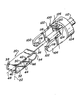
3 Thus as can be seen in Figs. 2 and 3 the tool 100
includes a body portion 104 having a pair of swing jaws 106
~nd 108 mounted at the distal end of the body. The jaws 10~
and 108 basically comprises a pair of elongated m~mbers which
extend generally parallel to the longitudinal axis 48. Th~y
are mounted to pivot or iwing outward about a trans~erse axis
110 extending perpendicular to th~ tool's central
longitudinal axis 48 by means to be described later~ The
clip 20 is arranged to be located in the space between the
;~ ,
swing jaws 106 and 108 as shown in Figs. 1 and 3. A pusher
member 112, in the form of an elongated rod, extends down a
central passageway 114 (Fig. 3) in the tool's body 104 so
thak its distal end 116 is immediately adjacent (or even
abutting) the bridging section 34 of the clip 20. The pusher
member is arranged to be operated by means (not shown) at the
proximal end of the tool to slide it in the distal direction
down the passageway 114 to thereby push the clip 20
longitudinally out from the space between the swing jaws 106
and 108 as shown in Figs 4 and 5.
Each of the wedge-shaped trunnions 102 includes a
pair of cam surfaces 118 which are inclined with respect to
~ WO93/09721 PCT/US92/06186
21~29~5 12
each other and merge together. Each trunnion 102 is project~
from the inner surface of a respective one of the swing jaws
106 and 108 immediately adjacent the distal end thereof and
is located within a respective Y-shaped slot 50 in the clip
20 tsee Fig. 3). - -
The cam surfaces 118 of each trunnion 102 areoriented so that they m~rge together in the proximal
direction. These sur~aces are arranged to engage the
inclined cam surfaces 42 of the clip as the clip is ejected
by the pusher member 112 to pry the jaws 30 and 32 apart in
opposition to the bias force produced by the bridging section
36. In particular, as the pusher member 112 pushes the clip
longitudinally down the axis 48 the cam surfaces 118 on the
trunnions 102 engage respective cam surfaces 44 on the ramps
42. Continued movement of the clip in the proximal direction
causes the cam surfaces i~8 to ride along the cam surfaces
44, and from there they move onto the release surfaces,
thereby progressively opening the clip's jaws 30 and 32
until the mouth of the clip is fully open. This occurs when
the cam surfaces 118 reach the proximal ends of the release
surfaces 46. As soon as the cam surfaces 118 pass the
proximal ends of the release surfaces 46, the jaws 30 and 32
immediately snap together as a result of the bias of the
bridging section 34, thereby instantly closing the clip's
mouth.
The swing jaws 106 and 108 are mounted to pivot
about the axis 110 via a pair of threaded members, e.g.,
screws, 120. In particular each swing Jaw includes an
arcuate proximal~portion 122 of the same outer cylindrical
profile and diameter as the body 104 of the tool 100. The
proximal portion 122 of the swing jaw 106 in turn includes a
pair of diametrically opposed flatted end sections 124. In a
similar manner the proximal portion 122 o~ the swing jaw 108
includes a pair of diametrically opposed flatted end sections
126. Each flatted end section include a hole 128 therein.
The pair of flatted end sections 124 of the swing jaw 106 are
.-- WO93/097~1 PCT/VS92/06186
2:12~33~
13
disposed between the pair of flatted end sections 126 of the
swing jaw 108 and with their holes 128 axially aligned.
The body 104 of the tool lO0 includes a pair of
brackets 130 projecting longitudinally outward from the
proximal end thereof. Each bracket 130 includes a ~hreaded
hole 132 ther~in. The holes 132 in the brackets are aligned
with the holes 128 in the swing jaws 106 and 108. Each of
the screws 120 is extended through a pair of aligned holes
128 in the swing jaws and is threadedly engaged into the
aligned hole 132 in the bracket to pivotally mount the swing
jaws thereon.
The operation of the tool 100 to secure a clip 20
onto a portivn of tissue located within the body of a patient
will now be described. The tool loO with the clip 20 located
therein is introduced through a small percutaneous incision
or puncture (not shown) within the body of the patient using
any suitable conventional tubular sheath ~not shown)
extending through that puncture or incision until the distal
end of the tool is located at the site of the tissue to be
grasped. The pusher member 112 is then operated as described
above to s~art to eject the clip 20 from the tool and at the
same time commence opening the clip's mouth. In particular
the tool is positioned so *hat it engages the tissue 22 to be
grasped when the clip's mouth is fully open so that a portion
24 of that tissue 22 extends slightly into the op~n mouth a~
shown in Fig. 4. Continued operation of the pusher 112
~auses the cam surfaces 118 of the trunnions 102 to clear
the release surfaces of the clip's ramps 42 ~as described
earlier), whereupon the jaws immediately snap clo~ed, ther~by
trapping the portion 24 of the tissue between the serrated
free ends of th~ clip's mouth as shown in Fig. 3.
The tool 100 then is preferably retracted slightly
to enable the surgeon to test if the clip has, in fact,
grasped the tissue. In this regard; since the trunnions 102
will still be located between the clip's jaws, a slight
retraction of the tool will cause the proximal end of each
trunnion 102 to engage the inner surface of the bridging
WO93/09721 PCT/US92/06186
. 2122935 14
section of the clip if the clip has secured itself to the
tissue 24, whereupon the surgeon will notice a slight
resistance to continued retraction of the tool.
In accordance with a preferred aspect of this
invention the clips are precisely constructed so t~at the
amount of clamping force their jaws apply to the tissue
trapped therebetween is controlled and sufficiently low so
that penetration of the tissue does not result.
Once the surgeon has determined that the clip 20
has successfully grasped the tissue 22, such as shown in Fig.
3, the swing jaws 106 and 108 are operated by means ~not
shown) to cause them to swing outward from the longitudinal
axis 48 of the tool. This action frees the clip from the
tool by removing the trunnions l0~ from between the rear. of
the clip's jaws 30 and 32. The tool l00 is then retracted
further until the swing jaws can be pivoted back to the
orientation shown in Figs. 2, yet still clear the clip. Once
this has been accomplished the tool can be removed from the
~heath extending into the puncture by retracting it fully
from the sheath.
At this time the surgeon is ready to reflect the
tissue 22 to a desired position within the patients body,
e.g., to provide a line of site to another internal portion
which was blocked by the presence of the tissue portion 22.
This action is achleved by use of the holding filament or
tether 36. In accordance with a preferred embodiment of this
invention the tether is formed of any suitable flexible
member, e.g., a 2.0 monofilament. The distal end of the
tether extends through a hole (not shown) in the clip's
bridging section 34 and located on xis 48. The tether is
secured in place by a knot 140.
The proximal end of the tether extends from the
clip 22 through the sheath to the outside of the patient's
body. Thus, all that is necessary for the surgeon to do to
effect the desired positioning of the grasped tissue is to
pull on the exteriorly located portion of the tether until
the desired positioning is accomplished. The laprascopic or
-
~ WO93/09721 PCT/US92/06186
212293~
~
endoscopic procedure can then be carried out, with the
reflected tissue now being out of the way to give the surgeon
a good line of sight and to free space within the body at the
operative site at which he/she can work.
The tool 100 is also arranged to effect the removal
of the clip 20 from within the body of the patient after its
use is no longer required or desirable. Like the insertion
action the remo~al action can also be accomplis~ed through
the small percutaneous incision or puncture. In this regard
when it is desired to remove the clip 20 the tool 100 is
reintroduced into the body of the patient through the
puncture until the distal ends of the swing jaws 106 and 108
are adjacent the clip. The swing jaws are then pivoted open
by the same means which had been operated during the clip
securement procedure and the tool positioned so that its
swin~ jaws can be pivoted back to the closed orientation
(like that shown in Fig. 1) but with the trunnions of the
swing jaws located within the Y-shaped slots 50 in the clip.
Once the swing jaws are pivoted back to the orientation such
that the trunnions 102 are within the Y-shaped slots the tool
is then retracted. This action causes the cam surfaces 118
of the trunnions to ride up the cam surfaces 44 of the clip
to thereby open the clip's jaws and release the tissue 24.
Once the cam surfaces 118 o~ the trunnions clear the release
surfaces 46 of the clip, the clip will again snap shut, but
the trunnions will remain in the space bounded by the clip'~
brid~ing section 34. Accordingly, further retraction of the
tool through the puncture will cause ~ho~e trunnions to
engage the inner surface of the bridging section to also
retract the now-freed clip 20. The tool and clip are then
retracted as a unit until they are outside the body of the
patient.
In accordance with one preferred aspect of this
invention two or more of any of the clips 2G, 200, and 500
can be introduced into the body of the patient at different
locations and the clips can be coupled together to form a
pulley-like configuration to expedite the tissue reflection
;,
I W093J09721 PCT/US92/06186
212293~ 16
procedure. One example of such action is shown in Fig. g
where it can be seen that one clip 2OA constructed in
accordance with this invention has been placed so that it has
grasped one portion 22A of internal tissue. The tether 36 of
this clip is extended through the space between the.jaws 30
and 32 and the bridging æection 34 of a second clip 20B. The
second clip 20B has been positioned and op~rated so that it
has grasped another portion 22B of internal tissue. The
proximal end of the tether 36 extends through any suitably
located percutaneous incision or puncture (not shown) to a
point outside the body of the patient. Accordingly, when the
surgeon pulls on the tether in the direction of the arrow Dl,
the direction of the pulling force on the clip 20A (and hence
the tissue grasped by it) will be changed by the passage of
the tether 36 through the clip 2OB to the direction shown by
the arrow D2. Thus, by appropriate placement of one or more
clips 20 the surgeon may position tissue at any desired
location irrespective of the direct~ion of the pulling force
applied to the tether through the puncture or incision.
It should be pointed out at this juncture that each
clip may have its own tether for coupling to one or more
other clips, depending upon the configuration desired.
In accordance with another preferred aspect of this
invention two or more of any of the clips 20, 200, and 500
can be introduced into the body of the patient at different
locations and the clips can be coupled together to form a
system including one or more hammock-like, tissue retainers
to expedite a tissue reflection/retaining procedure. One
example of such action is shown in Fig. 10 where it can be
seen that one clip 20A constructed in accordance with this
invention has been placed so that it has grasped one portion
22A of internal tissue. A second, similar, clip 20B has been
placed so that it has grasped a second portion 22B of
internal tissue. A third, similar, clip 20C has been plac~d
so that it has grasped a third portion 22C of internal
tissue. A section 36A of the tether 36 of the clip 22A is
secured to one end 150 of a hammock-forming web 152. The
~W093/09721 PCT/US92/06186
21~2~3S
17
.
~,other end 154 of the web 152 is connected to a second section
36B of the tether 36 and is extended through the space
between the jaws 30 and 32 and the bridging section 34 of the
second clip 22B. The extending tether section 36B is in turn
connected to one end 156 of a second, hammock-forming web
158. The other end 160 of the web lS8 is connected to a
third section 36C of the tether 36. The tether section 36C
is extended through the space between the jaws 30 and 32 and
the bridging section 34 of the third clip 22C and from there
through a percutaneous incision or puncture (not shown) to a
point outside the body of the patient.
Each of the hammock-like retainer members 152 and
158 is arranged to be spread out to form a barrier wall for
holding or retaining a portion of internal tissue behind it.
In the interests of expediting the insertion of the retaining
system shown in Fig. 10 into the body of the patient by
percutaneous incision the hammock-like retainer members are
each formed of a woven or mesh-like material. This enables
them to be compressed down to a very small external cross
sectional area. Once inserted into the body of the patient
the retainer members can be spread out. Thus, for example,
retainer member 152 can be spread out by the surgeon to the
orientation shown in Fig. 10 to hold back a portion 22D of
r~tissue located between portions 22A and 22B. In a similar
~anner retainer member 158 can be spread out by the surgeon
from the compacted orientation shown in Fig. 10 to hold back
a portion 22E of tissue located adjacent to it. Moreover,
since the clips and retainer members are all coupled
~$
together the pulling on the exteriorly located tether can
enable the surgeon to posi~ion the grasped and retained
tissue as desired.
It should be pointed out at this juncture that
other hammock-like systems can be provided to accomplish
tissue reflection or retention. Thus, for example a hammock
like member formed of a triangular mesh may be spread out and
held in place within the body of the patient to retain tissue
behind it by use of three clips 20, with each clip securing a
!J
~,
!.
. ,1
WO93/09721 PCT/US92/06186
2122935 ' /
18
respective corner of the member to the tissue. Thus, the
system shown in Fig. 10 is merely exemplary.
In Fig. 7 there is shown an alternative embodiment
of the tool lOOA constructed in accordance with this
invention to enable the loading of a stack or column of clips
20 therein. The embodiment of the tool lOOA shown in Fig. 7
is identical to that of tool 100 shown in Fig. 1 except for
the clip loading mechanism (as will be described
~ereinafter). Thus,`in the interest of brevity only that
portion of the tool lOOA will be described.
As can be seen in Fig. 7 the body portion 102 of
the tool lOOA includes a breech port 170 communicating with
passageway 114A. The passageway 114A is similar to
passageway 114 of tool 100 except that it is of a larger
internal diameter to accommodate a stack of breech loaded
clips 20 therein.
The distal end of the passageway 114A opens to the
space between the swing jaws 106 and 108 to deposit the
distal-most clip 20 of the stack of clips into that space.
The pusher member 112 is located in the passageway 114A and
can be moved to a longitudinal position therein so that its
distal end 116 is located proximally of the breech port 170.
Accordingly, in order to load a stack of clips within the
tool lOOA all that is necessary is to retract the pusher
member to that position and then to sequentially feed clips
20 into the port, sequentially using the pusher to position
them in a stack or column until the distal-most clip 20 is
located in the space~between the swing jaws. Each of the
clips 20 includes a tether like that described heretofore.
Such tethers are not shown in Fig. 7 in the interest of
drawing simplicity.
The tool lOOA is used in a similar manner as that
described heretofore, with the most distally located clip
being pushed out of the tool by the next successive clip
under the force applied by the pusher member.
In Fig. 8 there is shown an alternative embodiment
of the ~ool lOOB constructed in accordance with this
WO93/097~1 PCT/US92/06186
`` 212293~
1~
invention to also enable the loading of a stack or column of
clips 20 therein. The embodiment of the tool lOOB shown in
Fig. 8 is identical to that of tool 100 shown in Fig.
except for the clip loading mechanism (as will be described
hereinafter). Thus, in the interest of brevity only that
portion of the tool lOOB will be des~ribed.
As can be seen in Fig. 8 the body portion 102 of
the tool lOOB has a cartridge 172 mounted on the proximal end
thereof. The cartridge 172 is a tubular member having a
central passageway 174 which communicates with a central
passageway 114A in the body portion 102. The passageway 114A
is similar to passageway 114 of tool 100 except that it is of
a larger internal diameter to accommodate a stack of
cartridge loaded clips 20 therein~ The distal end of the
passageway 114A opens to the space between the swing jaws 106
and 108 to deposit the distal- most clip 20 of the stack of
clips in~o that spaceO The pusher member 112 is located in
the passageway 174.
When the cartridge is loaded, i.e., full of a stack
of clips 20, the pusher me~ber is located so that its distal
end 116 is located proximally of the proximal-most clip of
the stack of clips. The cartridge 172, with the stack of
clips and pusher therein is then mounted onto the proximal
~nd of the tool body 104 so that the passageway 174 in the
cartridge is axially aligned with the passageway 114A in the
tool body. ThP pusher 112 is then operated by pressing on
its proximally located head 176 in the distal direction to
push the stack of clips into the passageway 114A until the
distal-most clip of the stack of clips is located in the
space ~etween th2 swing jaws 106 and 108.
The tool lOOB is used in a similar manner as that
described heretofore, with the most distally located clip
being pushed out of the tool by the next successive clip
under th~ forcQ applied by the pusher member.
In Fig. 11 there is shown at 200 an alternative
embodiment of a clip constructed in accordance with this
invention. The clip 200 is arranged to be introduced into
. ,.
W093/09721 PCT/US92/06186
21229~5
the body of the patient in a similar manner as that described
heretofore, except for the u~e of a differ~nt delivery tool.
In this regard the clip 200 is delivered and operated by tool
400 (Fig. 14), the details of which will be described after
describing the construction of the clip 200.
As can be seen in Figs. 11-13 the clip 200 is very
similar in construction to a conventional "alligator~' clip.
Thus it includes a pair of jaws 202 and 204 which are
pivotally connected together by a pivot pin 206. The pivot
pin extends through aligned holes in a pair of tabs 208
located on each side of the jaw 202 at an intermediate point
therealong. A similar pair of tabs 210 is located on each
side of the jaw 204 at a similar intermediate point. The
pair of tabs 210 are disposed between the pair of tabs 208.
The inner surface 212 contiguous with the distal
end of each of the jaws 202 and 204 is serrated. These
portions of the jaws define the "mouth" of the clip 200
therebetween. ~ torsion or compression spring 214 is
interposed between the proximal ends 216 of the jaws 202 and
204. This spring applies a bias force to the proximal ends
of the jaws to bias the distal ends of the jaws closed.
The clip 200 is arranged to be located within a
bore or passageway 402A in the body 402 of the tool 400. A
pusher member 112 (similar to that described heretofore)
extends into the passageway 402A at ~he proximal end of the
tool. The inner diameter of the passageway 402A is large
enough to accommodate the clip 200 therein, but small enough
to constrain the proxim~l ends 216 of the clip's jaws 202 and
204 toward each other so as to hold their distal ends apart
(open~ against the bias of the spring. The clip 200 is held
in this orientation in the distal end of the tool 400, with
the distal snd 116 o~ the pusher member 112 abutting the
proximal end of the clip~
Operation of the tool 400 and clip 200 will now be
described. The distal end of the tool 400 with a clip 200
held therein is inserted into the percutaneous incision or
puncture in a manner similar to that described heretofore.
~ W093/09721 212 2 9 3 5 PCT/US92/06186
21
When the tool is at the desired position, e.g., adjacent an
tissue or an organ like a gallbladder 22 ~Fig. 11), the
pusher member is operated to push the clip out of the distal
end of the tool's body ~02. When the clip has been moved to
the position wherein its the proximal end has cleared the
distal end of the tool's body, as shown in Fig. 12, whereupon
the proximal ends of clip's jaws are no longer constrained by
the passageway 402 so that the bias force of the spring 214
between those ends causes the clip's jaws to immediately snap
closed. This action has the same effect as described
heretofore to trap the tissue 24 within the clip's mouth
without piercing it as shown in Fig. 13. The tool may then
be withdrawn.
In Fig. 14 there is sh~wn the entire tool 400 of
Figs. 11-13. In Fig. 20 there i shown an entire tool 400'
which constitutes a slight modification of the tool 400. The
too~s 40~ and 4Q0' can also be used with the clips 500 and
500' (to be described later). The differences between the
tools 400 and 400' will be considered later, with the common
components of the tools 400 and 400' being given the same
reference numerals in the interests of brevity.
As can be seen in Fig. 14 the proximal end of the
tool 400 is constructed in a manner similar to a conventional
"caulk-tube gun." In particular the tool 400 includes frame
404 including one extending handle member 406 having a finger
hole 408 therein. A second handle or trigger member 410,
having a finger hole 412 is pivotally mounted onto the frame
by a pivot pin 414. When so mounted the finger holes 412 and
408 are adjacent each other, whereupon the surgeon can insert
his/her thumb through hole 408 and his/her index finger
through hole 412 to squeeze (pivot) the trigger 410 toward
the handle 406.
At the upper end of the trigger is a pin 416 which
is adapted to engage a disk-like member 418. The disk-like
member 418 includes a central opening through which the
proximal end portion of the p~sher rod 212 extends. The
disk like mem~er is arranged to frictionally engage the
1 WO93/09721 PCT/US92/06186
3 21229~5 `
,) 22
pusher rod 212 to slide it a predetermined distance in the
distal direction each time that the trigger 410 is pivoted
toward the handle 406.
A helical biasing spring 420 is located within an
opening 422 in the frame 404 so that it bears against or
biases the disk-like member 418 toward the rear (proximally).
~ The pusher rod 212 extends through the spring 420. This
J arrangement insures that the pusher rod will not be moved in
a distal direction until the trigger is operated.
A locking member 424 is pivotally mounted on the
frame 404 by a pin 426, wîth a biasing spring 428 located
between the locking member and the frame~ The locking member
includes a hole through which the pusher rod extends to
frictionally engage the rod and thereby prevent it from
moving in a proximal direction unless the locking member is
pressed inward against the bias of the spring 428. This
latter action is accomplished when it is desired to load the
tool with a stack of clips (as will be described later).
The proximal end of the frame 404 includes a
bayonet connector 430 which is arranged to engage a ~ating
connector 432 on the tool body 402. The passageway 402A in
the tool body 402 is arranged to hold a stack of clips
~ ~ therein. These clips can be of the type previously described
.
and denoted by the reference numeral 200, or may be of the
type denoted by the reference numeral 500 in Fig. 15 or of
the type denoted by reference numeral 500' in Fig. 18. In
Fig. 14 the tool 400 is shown with a stack of clips 500
therein. In either case the distal end of the pusher member
112 is arranged to be located immediately adjacent the
proximal end of the most proximally located olip in the stack
so that when the pusher rod is moved distally by the
operation of the trigger the distally-most located clip is
expelled from the tool to grasp the tissue 22 as described
heretofore with reference to clip 200.
The loading of a stack of clips in the tool 400 is
accomplished by securing a tool body 402 having a stack of
`~ clips within its passageway 402A to the frame portion 404.
'~J
,~
J
W093/0972t PCT/US92/06186
2.122~35
. 23
To that end the surgeon presses the locking member 424 toward
the frame to release the frictional engagement of that member
on the pusher rod. The pusher rod is then withdraw (moved
proximally) until its distal end is located inside the
bayonet connector 430 forming a portion of the frame 404.
The tool body 402 having the stack of clips therein is then
connected to the frame by engaging its bayonet connector 432
with the frame's bayonet connector 430.
The tool 400' is identical in all respects to that
of tool 400, except that in lieu of the bayonet connectors
430 an~ 432 of the tool 400 the tool 400' makes use of a male
luer lock 430 on the frame 404 and a female luer lock 432' on
the body (tube) 402. In addition the tool 400' includes an
end piece 212l on the proximal end of the pusher rod 212.
Figs. 15-17 show the embodiment of the clip 500
mentioned heretofore. That clip is similar in construction
j and operation to clip 200. However, unlike clip 200 but like
clip 20, clip 500 includes means which is actuatable to
enable its mouth to be opened against the bias force tending
to keep the mouth closed so that the tissue trapped within
the mouth may be released when desired. Thus, as can be seen
in Figs. 15 the clip 500 basically comprises a pair of jaws
502 and ~04 which are pivotally connected together by a pivot
pin 5~6. The pivot pin extends through aligned holes in a
pair of tabs 508 locatad on each side of the jaw 502 at an
intermediate point therealong. A similar pair of tabs 510 is
, located on each side of the jaw 504 at a similar intermediate
I point. The pair of tab~ 510 ~re disposed between the pair of
tabs 508.
The distal end of each of the jaws 502 and 504
includes plural serrations forming teeth 512. These portions
of the jaws define the "mouth'l of the clip 500 therebetween.
A torsion or compression spring (not shown in Figs. 16 and 17
but shown at 503 in Fig. 19) is interposed between the
proximal portions of the jaws 50Z and S04. This spring
applies a bias force to the proximal ends of the jaws to bias
the distal ends of the jaws closed.
J
WO93/09721 PCT/US92/06186
2122~3~
24 . .
The proximal end of each of the jaws 502 and 504
terminates in an angularly extending projection 514. These
projections form respective cam surfaces which are arranged
to be engagéd by the open distal end 40~B of the tool body
~ 402 to effect the opening of the clip's mouth to~release
¦ tissue trapped therein as will be described later. A
¦ retraction filament or wire 516 is connected between the free
ends of the projections 514 as shown in Fig. 15.
The clip 500 is arranged to be located within a
bore or passageway 402A in the body 402 of the tool 400 (or
tool 400' for that matter) as described earlier. The inner
diameter of the passageway 402A is large enough to
accommodate the clip 500 therein, but small enough to
constrain the proximal ends of the clip's jaws 502 and 504
~, toward each other so as to hold their distal ends apart
(open) against the bias of the spring. Each clip 500 in the
stack is held in this oxientation in the passageway 402A vf
the tool 400, with the distal end of the pusher memher 112
abutting the proximal end of the proximally-most located
clip.
~peration of the clip 500 is the same as describ~d
with reference to clip 200 and thus will not be reiterated
herein.
Like the c~ips 20, each of the clips 200 and 500,
includes a positionïng tether 36 (not shvwn in the interest
of drawing simplicity) arranged to be pulled as described
heretofore to ~ffect the desired tissue reflection. Thus,
the clips 200 and/or 500 may be used in the same manner as
d~scribed earlier with respect to c~ips 20, e.g., they may be
coupled together via the tether in a pulley like
configuration. They may also include hammock-like retaining
I members like that described earlier for use in the same
3 manner as that describ d earlier.
As mentioned earlier the clip ~00, like clip 20, is
arranged to be readily released from tissue which it has
trapped. Thus, the following discussion will be directed to
the operation of the clip 500 and the tool 400 to release
y
i~
"; i.,,
'r'~'~ WO 93/09721 PCT/US92/06186
! ~ 2 1 ~ 2 9 ~ 5
.~25
.;;,~ trapped tissue from the mouth of the clip. Accordingly, when
,~it is desired to release the clip 500 from tissue 22 it has
trapped within its mouth the tool 400 is positioned so that
'~the open distal end of its body 4 02 is locat~d ad; acent the
;~sproximal end of the clip 500 as shown in Fig. 15. ~ grabber
member 434 is then extended outward from the passageway 402
in the tool 400 so that its hooked end 436 engages or hooks
onto the filament or wire 516 extending between the proximal
ends of the jaws of the clip. The grabber member 434 is then
retracted within the passageway 402A of the tool's body.
`I.'.,J. This action pulls the clip towards the tool and causes the
portions of the tool's body contiguous with the entrance to
the passageway 404A to ride or slide along the cam surfaces
.defined by the clip extensions 516, thereby squeezing the
proximal ends of the clip's jaws 502 and 504 together and
concomitantly opening the clip's mouth. Accordingly, the
`.~tissue portion 24 which had been trapped in the clip's mouth
~' ".1
is ncw released. The clip may then be removed from the
patient in a manner like that described earlier.
.~.As mentioned earlier in Fig. 20 there is shown a
:~. system constructed in accordance with the teachings of this
invention being used to ~ffect the reflection of internally
located tissue during a laparoscopic procedure, e.g.,
laparoscopic bowel resection. A~ is conventional in such
procedures a laparoscope (not ~hown) i5 introduced, via a
!,.,,j trocar (not ~hown~, through the patient's umbilicus into the
insuf~lated abdomen 10 to provide a visual image of the bowel
to be resected. In accordance with the teachings of this
inv~ntion plural clips 500' for securement to portions of the
bowel or other tissue to be positioned by the surgeon, are
introducsd into the abdomen by use of the instrument 400'.
The instrument 400' is extended through a
conv~ ffl ional trocar 14, e.g., a 5 mm trocar, into the
~,patient's abdomen. The surgeon can manipulate the trocar 14
so that the open free end 402B of the instrument's clip
delivery tube 402 is located adjacent a first portion of
,,., ..~
~tissue 12 e.g., a section of bowel, which the surgeon wishes
....
W093/09721 PCT/US92/06186
~ 2~22~)3~
26
to reflect or otherwise position. Once that has been
accomplished the instrument is operated to eject the distal-
most clip 500' from the instrument 400' in the same manner as
described heretofore, whereupon the clip 500' is freed of the
instrument and its mouth closes on the immediately adiacent
tissue portion to trap it therein. Once that tissue portion
is trapped within the mouth of clip, and assuming that the
surgeon deems it necessary to utilize another clip to effect
the tissue reflection, the surgeon can attach a second clip
500' to another portion of the internally located tissue 12.
To achieve that end the surgeon manipulates the trocar 14
through which the introducer instrument 400' extends in the
desired direction so that the free end of the instrument's
introducer tube 402 is aimed at the tissue portion to be
grasped by clip 500'. The instrument 400' is then operated
in the same manner as described heretofore to eject the next
clip 500' from the stack into the abdomen, whereupon it traps
the immediately adjacent tissue portion within its mouth.
Depending upon the procedure being accomplished, other clips
may be secured to different portions of the tissue to be
reflected by merely directing the trocar 14 toward that
tissue and operating the instrument 400' to secure the clips
500' to the tissue. If necessary, another trocar can be
introduced at a different site and the instrument 400'
inserted therethrough to place one or more clips through
that trocar onto internally located tissue adjacent that
trocar.
In order to expedite the expulsion/ej~ction of the
clips 500' from the instrument 400' and to expedite the
coupling of the positioning/holding means to those clips, the
clips 500' of the embodiment of the invention shown in Figs.
18, 19, 20, 21, and 24 are identical to the clips 500 except
for a few structural differences. Thus, in the interests of
brevity all common compo~ents of clips 500 and 500' are given
the same reference numerals. The differences between the
clips 500' and 500 is as follows. Each of the clips 500'
includes two pair of stops 550 and S52. The stops of the
''1! . WO 93/097~1 PCT/US92/06186
122~135
27
pair 550 project perpendicularly from the sides of the
proximal portion 51~ making up jaw 502 of the clip 500, while
the stops of the pair 552 project perpendicularly from the
sides of the proximal portion 514 making up jaw 504 of the
clip.
In accordance with one aspect of this invention the
clips 500' are stacked within the delivery tube 402 of
instrument 400' so that alternat~e clips are oriented about
the central longitudinal axis of the instrument at an angle
of 90 degrees with respect to each other, wher~upon the
distal end of the open jaws 502 and 504 of one clip 500'
engage the pairs of stops 550 and 552 of the immediately
preceding (distally) located clip 500' as shown in Fig. 21.
Accor~iingly, when the instrument 400' is operated the stack
of clips 500' will push one another distally through the
introducer tube 402 without the clips nesting up on each
....
~ther, which action could cause misoperation of the clip
~ ejection process.
,~ Another difference between clips 500'and 500 is
that each clip 500' includes a generally V-shaped retraction
wire 554 secured to the proximal portion 514 of the clip in
lieu of the filament or wire 516 described earlier. In
particular, the ends 560 of the legs 556 and 558 making up
the retraction wire 554 extend through respective holes in
the proximal portions 514 and are crimped in place therein.
Once the clip(s) 500' have been secured to the
internally located tissue 12, the ~urgeon is now ready to
reflect that tissue to the desired position(s). For example,
in order to reflect the tissue to which a particular c}ip
500l is secured to a desired position the surgeon selects an
appropriata site on the skin co~ering the abdomen through
which a positioning/holding device for releasable securement
to the clip can be introduced. To that end and in accordance
with one aspect of this in~ention, a very small diameter
piercing device 700 (Fig. 20), e.g., a standard 13 gauge
hypodermic needle, is used to form a very small percutaneous
incision or puncture 702 through the skin 704 and abdominal
~'i;
r ;
,
j~, .
J~ .X~
.,~'
~.~
i.~. W093/09721 PCT/US92/06186~
212%~3~ i ~
28
wall 706 at the selected site on the abdomen. Similar
~1 devices 700 are used to form similar punctures at other sites
:~ for positioning clips located adjacent those other sites.
The use of such small diameter devices 700, to form the
percutaneous incisions or punctures 702 through w~ich the
: clips positioning/ holding devices are to be introduced (as
will be described later) is desirable since such incisions or
~;q
. punctures are less invasive than those formed by a
conventional 5mm, or greater, trocar. Moreover, the
: incisions or punctures 702 are less traumatic to form, and in
. a~dition will automatically close without loss of abdomen
~: insufflation and without requiring suturing when the device
.~ is rem~ved.
Once each small percutaneous incision or puncture
702 is made by a respective needle 700, a respective
.. ~ positioning/ holding device 800 constructed in accordance
with this invention, is introduced through the associated
needles 700. Each positioning/holding deYice is arranged to
be coupled, e.g., releasably secured, a respective clip
....
having a portion of the internally located tissue trapped
within its mouth. Each positioning/holding device 80 is of
identical construction and will now be described with
reference to Figs. 20 and 22. As can be seen therein the
positioning/holding device is preferably in the form of a
tension cable assembly basically comprising an elongated,
flexible, braided, cable 892, e.g., stainless steel~ having a
distal end portion in the form of a blunt hook 804. The
blunt hook is connected to the cable 802 via a coupling 80~.
An elongated, relatively rigid, e.g., s~ainle s steel, sheath
or sleeve 808 (Fig. 22) is slidable mounted on the cable 802
and is ~rranged to freely slide longitudinally therealong up
to the coupling 806.
The external diameter of the tension cable assembly
800 is smaller than the internal diameter of the needle 700
through which it is introduced so that it can be freely
extended therethrough, into the body of the being to the
position adjacent a clip as shown in Fig. 20. In this
, 1 .
'l
~,WO93/09721 PCT/US92/06186
212293~
29
position the hook 804 is located adjacent the clip, 500'
while the slidable sheath 808 is located immediately adjacent
the coupling 806, so that the distal portion of the sheath is
,~J
located within the insufflated abdomen 10 and the proximal
portion of the sheath is located outside of the bod~ of the
patient. The needle 700 may then be removed from the
percutaneous incision or puncture 702 by sliding it in the
proximal direction along the cable 802.
In order to grab the clip with the tension cable
assembly 800, the surgeon grasps the outwardly extending
pro~imal portion of the sheath 808 to manipulate (aim) it so
that the blunt hook 804 catches onto the retraction wire 554
of the clip. In order to facilitate the securement of the
hook to the clip the sheath 808 may include means (not shown)
for holding it with respect to the cable 802 so that
longitudinal and/or rotational movement of the sheath effects
concomitant movement of the cable. Once the hook of the
cable has been secured to the clip, the sheath 808 is slid
along the cable in the proximal direction away from the
incision or puncture 702 to get it out of the way. In fact,
the sheath 808 may be completely slid off of the cable 802.
: :~
Whether or not the sheath is left in place on the cable is a
function of the desires of the surgeon and depends upon the
technique to be used to remove the clips and the tension
cab~es from the patient after the laparoscopic procedure has
been completed. In any event the movement of the sheath 808
away from the point at which the cable enters the
~;percutaneous incision or puncture 702 into the patient's
abdomen is most desirable to provide a relatively
unobstructed working area for the surgeon.
''~ After the sheath 808 has been slid out of the way
the surgeon may then pull on an externally located portion of
the flexible cable 802 to move the tissue 12 within the
associated clip's mouth to a desired location toward the
abdominal wall 706. When the surgeon is satisfied that the
tissue is in the desired position the surgeon fixes the cable
802 in place with respect to the patientls body by any
.
,.~
/~:
W093/09721 PCT/US92/06186
j 21~2~
suitable means. In the embodiment shown in Fig. 20 a strip
of adhesive tape 810 is used to secure a portion of the
.,
flexible cable 802 of each clip positioning device to the
skin of the patient adjacent the percutaneous incision or
puncture through which the cable extends. Each cabl~ may be
fixed in place by other means, e.g., a standard needle
holder, a clamp, hemostat, etc. (not shown).
As should be appreciated by those skilled in the
art the fixing of the various positioning devices in place on
the abdomen frees the surgeon or other operating room
personnel for performing tasks other than holding the
reflected tissue in place (as has characterized prior art
laparoscopic surgical procedures, wherein the surgeon or
other personnel have the hold the various tissue grasping
devices in position while the surgeon carries out the
procedure).
In accordance with a preferred aspect of this
invention the combined length of the blunt hook 804, the
coupling 806, and clip 500' is very short, e.g., 0.9 inch
(2.29 cm) or less, to enable the tissue 12 trapped in the
clip's mouth to be reflected much closer to the abdominal
wall than that possible with prior art retracting instruments
(since such instruments are used through trocars which
typically extend three to four inches into the body of the
being).
When it is desired to release the reflected tissue
12, such action can be accomplished in several ways. For
example, one technique for releasing the reflected tîssue is
to release, e.g., untape, the assoc~ated cable 802 from the
patient's body, and slide the ~heath 808 (which had been left
on a proximal portion of the cable remote from the
percutaneous incision ~r puncture 702) in the distal
direction back through the incision or puncture until the
distal end portion of the sheath is immediately adjacent the
coupling 806. The surgeon may then manipulate the sheath 808
and cable to unhook the blunt hook 804 ~rom the retraction
wire 554 of the clip 500', thereby releasing the reflected
. . ~ .
!'
,.; WO93/~9721 PCT/US92/06186
~1229~
,.~ 31
... . .
tissue (albeit it with the clip still connected thereto).
The tension cable assembly 800 may then be extracted from the
body of the patient by pulling on it in th~ proximal
directicn through the same incision or puncture 702 through
which it was introduced.
In order to facilitate the extraction of the
tension cable assembly as just described i~ may be desirable
to modify the coupling 806 so that it is positionable with
respect to the blunt hook 804 so that it overlies or covers
the free end portion o~ the blunt hook, to prevent that
portion from snagging on the tissue of the patient contiguous
with the incision or puncture 702.
Another technique for releasing the reflected
tissue is accomplished by removing the sheath 808 from the
tension cable assembly. Then the tension cable assembly is
released by untaping the cable 802 from the patientls
abdomen, whereupon the tissue 12 to which the clip and
tension cable are connected will move to some neutral
1,~2
internal position, carrying the clip and cable with it, and
thereby releasing the tension on the cable. Such action may
result in the automatic disconnection of the tension cable's
blunt hook 504 from the clip's retraction wire 554. If not,
t~e surgeon may manipulate the cable 802 lightly to effe~-t
that disconnect.ion. The tension cable may then be removed
from the body o~ the patiant by extending a retraction tool
~like that to be d~scribed later) through the trocar 14 to
engage (e. g., hook on~o) the blunt hook 804 of the tension
cable. Once that has been accomplished the tension cable ~an
then be withdraw through the trocar 14 out of tAe patient's
body.
If desired clips in accordance with this invention
may be formed of any suitab~e material, e.g., a resorbable
polymer, so that they may be left within the body of the
patient after the laparoscopic procedure has been
accomplished and the tension cable assemblies removed. If,
howaver, removal of the clips from the patient's body is
desired such action may be readily accomplished by use of a
;~
WO93/09721 PCT/US92/061X6
212~35 32
clip removal tool constxucted in accordance with is
invention. One such tool 900 is shown in Fig. 23.
:
As can be seen in Figs. 23 and 24, the tool goo
basically comprises small diameter, e.g., 0.185 inch (4.7
mm), elongated tube 902 terminating at a handle 904 at the
proximal end thereof. The distal end 906 of the tube is
open. An elongated wire 908 extends longitudinally through
the tube 902. The distal end of the wire is in the form of a
blunt hook 910. A slidable plug 912 having a central hole
914 through which the wire 908 extends is located adjacent
the blunt hook 910. The wire 908 is arranged to be slid
longitudinally down the tool to move the blun~ hook from the
retracted position (wherein it is located within the distal
free end of 906 the tube 902 as shown in full in Fig. 23) to
an extended position (wherein it is located outside of the
free end of the tube as shown in phantom therein). To that
end the proximal end of the wire 902 is fixedly secured to a
f
shaft 916 portion of a thumb cap 918. The shaft portion 916
~' extends through the portion of the tube 902 within the handle
904~ A stQp member 920 is fixedly secured within the tube
adjacent the handle 903 and includes a central opening 922
~ through which the wire 908 extends. A helical compression
ii~ spring 924 i5 interposed between the stop member 920 and the
free end of the thumb cap shaft 916 to bias the thumb cap and
the wire 900 attached thereto in the proximal direction to
t ~ the retracted position. When the thumb cap is pressed
inward (distally) with respect to the handle 904 and the bias
force of the spring 924 is overcome the wire 908 is moved
proximally so that its blunt hook 910 is in the extended
position located outside the distal end 906 of the tool 900.
Operation of the retraction tool 900 to effect the
removal of a clip 500' is as follows. The retraction tool
is introduced through the trocar 14 and the trocar aimed so
that the open free end 906 of the retraction tool 900 is
located within the abdomen adjacent a clip ~00' to be
removed. The surgeon then presses on the thu~b cap 918 to
extend the hook 910 to the extended position so that it
.;.,~ .
~;
lv : ~
~ .
~ WO93/09721 PCT/USg2/06186
2 1 2 2 9 3 S
33
.~ . .
catches the V-shaped retraction wire 554 of the clip. Once
this has been accomplished the thumb cap 918 is released,
whereupon the hook 910, with the clip now connected to it, is
pulled into the open end 906 of the retraction tool tube 902.
This action causes inner surface portions of the tube -at the
i~ free end thereof to ride over the legs 556 and 558 making up
the V-shaped retraction wire 554. Accordingly, the proximal
ends 514 of the clips jaws 502 and 504 begin to move toward
each other, thereby initiating the opening of the clip's
mouth. Continued retraction of the clip 500' into the open
free end of the retraction tool causes the inner surface
portions of the tube at the free end thereof to ride over the
cam surfaces formed by angularly extending projections 514 of
the proximal portions of the clip's jaws, thereby further
opening of the clip's mouth, whereupon the tissue which had
been trapped in the clip's mouth is released, leaving the
~$j~ ~ clip partially within the tool as shown in Fig. 24. The
~; tool g00 with the clip therein may then be removed from the
patient's body through the trocar 14. Every other clip can
be removed in the same manner.
It should be pointed out at this juncture that
other instruments, tools, devices, and clips tha~ that
described heretofore can be constructed in accordance with
I the teachings of this invention. Moreover the methods of use
described are not the only methods which can be effected by
sfl use of such device. For example, the clips of this invention
can be used for purposes other than tissue reflection or
; positioning, e.g., they may be used to secure some device or
member to a portion of internally located tissue. Moreover,
the apparatus and methods are not limited to laparoscopic,
endoscopic or other minimally invasive surgery, and can thus
be used for open surgery as well. Thus, it must be kept in
mind that, the structures and methods of use as described
~: heretofore are merely exemplary.
.~, ~
,~
' ~ PCT/U~92/~6186
j WO93/09721
2122~3.~ 34
~ Without further elaboration the foregoing will so
7 fully illustrate our invention that others may, by applying
i~ current or future knowledge, adopt the sa~e for use under
il, various conditions of service.
'~
:i
.
'~,
,~ !
'S~
~'
i