Note: Descriptions are shown in the official language in which they were submitted.
.
;. ''
:';
:. VSO 93/08746
PC.'f/LJS92/09572
.
, ~ ~ ~ ~ ~ ~ ~
:,
HEMOSTATIC'FUNCTURE CLOSURE SYSTEM AND METHOD OF USE
--
w Field of the Invention
This invention relates generally to medical devices
'~:~~;;
and methods of use, and more specifically to a system and
methods of use for sealing percutaneous punctures in blood
..
vessels within the body of a living being.
Backq~round of the Invention.
'''~ Tn United States Letters Patent No. 5, 021, 059,
,',y:which has been assigned to the same assignee as this
7.::,iinvention, there is disclosed a closure device and method of
f;,;~,
use for sealin a small incision or
g puncture in tissue
i
'; separating one portion of the body of a living being from
w;;,
another portion thereof, e.g., a percutaneous puncture in a
artery, to prevent the flow of a body fluid
e.g.
blood
,
,
,
throu h the
g puncture. The closure device is arranged to be
used with (deployed by) an instrument which comprises a
carrier in the form of a tubular member. The tubular member
has a proximally located portion and a distally located
portion. The litter includes an open free end arranged to be
introduced through the incision or puncture. The proximately
located portion of the tubular member is arranged to be
located out of the body of the being when the distally
yr,
~ located portion is extended through the incision or puncture.
'~y,.
.
r The closure device comprises three components,
' namely, an anchor member, a sealing member, and a filament,
e.g., suture. The sealing member is formed of a hemostatic
:1 ~ate~'lWl. e.Ct. . COmDreSSed Collagen fe5am_ Tf'tA anrhnv~ vnesmT,or
~'~a includes a tissue engaging portion configured to pass through
the puncture in one direction but resistant to passage
~~ therethrough' in the opposite direction. The sealing member
includes a tissue engaging portion. The filament is
~~'~ connected between the anchor member and the sealing member in
..
a pulley-like arrangement so that they may be moved relative
~''~~ to each other by the application of a pulling force on the
filament.
The instrument is arranged to expel the anchor
member through the puncture, e.g., into the artery, and to
draw its tissue engaging portion into engagement with the
WO 93/08746 PCT/1JS92/~9572
r,..
,,
2 ._ _.
tissue contiguous with the puncture. The filament extends
through the instrument to a point outside the body of the
being and is arxanged to be drawn in the proximal direction,
whereupon the portion of the filament connecting the anchor
member and the sealing member causes the tissue engaging
portion of the sealing member to move with respect to said '
anchor~member and into engagement with the tissue contiguous
with the puncture on the opposite side thereof from said
anchor member. This action causes the tissue engaging
portion of the sealing member to seal the puncture. from the
,~f
"~ flow of fluid therethrough.
,...
The closure device and deploying instrument in that
patent leave something to be desired from the standpoints of
effectiveness and efficiency of use.
Objects of the Invention
Accordingly, it is a general object of this
invention to rovide a device and methods of use which
,;a P
overcomes the disadvantages of the prior art.
,,;;,
It is a further object of this invention to provide
''' a system including a closure, a deploying instrument, and
; method of use for quickly, easily, safely and effectively
'sealing a percutaneous puncture in a blood vessel within the
'!' body of a living being from another portion.
It is still a further object of this invention to
provide devices and methods for.enabling one to determine the
location of a the wall of a blood vessel or other lumen
through a percutaneous incision or puncture.
Summary of the Invention
,~ These and other objects of this invention are
.~,
a achieved by providing a system for sealing a percutaneous
xr incision or puncture in a blood vessel. The system comprises
Y:,. r
,;.~, carrier means, introducer means, and closure means. The
puncture comprises a tract extending through tissue overlying
the blood vessel. The closure means comprises anchoring
means, sealing means, and filament means. The filament means
~n;.A
,..,
s connected between the anchoring means and the sealing
means. The introducer means comprises a tubular member
WO 93/08746 PCT/US92/09572
3
having a distal free end arranged to be inserted into the
.r:'~''puncture tract and through the puncture. The carrier means
is arranged to be inserted through the introducer means to
t,.~~
:':., expel the anchoring means therefrom and to draw the anchoring
,
;~::;a. .
.
:Y means into engagement with the distal free end of the
: .;
-
~
::, introducer means. The introducer means and the carrier means
:~.:.
.
:.
9 are arranged to be moved together to draw the anchoring means
'vwt
a
Y
,
, into engagement with the interior tissue of the blood vessel
i ~"
,, contiguous with the puncture. The filament means is arranged
''~~ to pull the anchoring means and the sealing means relative to
each other to cause the sealing means to engage tissue
,'
contiguous with the puncture outside of the vessel.
In accordance with one aspect of this invention the
system includes means and a method of use to enable one to
:'~= readily determine the location of the wall of the vessel or
a;
;:; lumen by the percutaneous introduction of such means into the
'~'~'~vessel or lumen. Such means can , be used as part of the
",
~
'~:,
~,~ ystem or method to seal the puncture or incision in a vessel
or lumen or may .be used for other purposes
e.g.
it may be
,
,
used for any application wherein it is desirable to determine
r
the location of a vessel or lumen wall via a percutaneous
~~ incision or puncture.
,
Pxi.ef Description of the Drawinc~,s
Other objects and many of the attendant advantages
of this inventionwill readily be appreciated as the same
a'~
~ becomes better understood by reference to the following
~'di
c' detailed description 'when considered in connection with the
;,:~ accompanying drawings wherein: .
~~p
HS; ~ Fig. l is a side elevational view, partially in
-
section, showing a deploying instrument and a closure device
3
Of the svstem of the subiect invention:
~a. Fig. 2 is an enlarged tap plan view of the closure
5G~' 3
device shown in Fig. 1, with the sealing component shown in
~i,~' an uncompressed state;
Fig. 3 is a top plan view, like that of Fig. 2, but
showing the sealing component in its compressed state ready
for deployment by the instrument of Fig. 1t.
W~ 93/087.46 PCT/US92/~9572
__ ... 4
Fig. 4 is an enlarged top plan view of the anchor
component of the closure device:
' Fig. 5 is an enlarged side elevational view of the
anchor component of clpsure device,
Fig. 6 is a greatly enlarged plan view showing the
''~~ knot used to effect the securement of a filament component of
I
:L,
rP the closure device to the sealing component thereof;
Fig. ? is a top plan view of one embodiment of a
introduces sheath position indicating device forming a
portion of the system of this inventiono
Fig. 8 is an enlarged sectional view taken along
line 8-8 of Fig. 7;
;:. . Fig. 9 is a front elevational view of a torsion
spring used with the deployment instrument:
Fig. 1~ is a side elevational view of the spring
shown ~.n F~.g s ~ f ,
~'~ Fig. 11 is an isometric view of the deployment
:: instrument shown in Fig. 2:
',:'i Fig. 12 is an illustration showing a preliminary
step in the positioning of a conventional introduces sheath
r'~'~~through a percutaneous puncture in an artery using the
.:J:,:
position indicating device shown in Fig. ?:
>x
Fig. 13 is an illustration similar to that of Fig.
12 showing desired position of the introduces sheath within
;,:.
the artery as established by the use of the position
'v'~ ir~d~.cating device shown in Fag. 7;
~,
~
;; Fig. 14 is an illustration showing the introduction
- ~f the deployment instrument into the properly located
,:
,
.
5j ntroduces sheath;
:w
4
;
.. Figs. 35-23 are illustrations, similar t~ Figs. 11
:.
and 12d but showing the sequential steps in the use of the
instrument to deploy the closure device to seal the r
percutaneous puncture in the artery:
~i Fig. 24 is an enlarged illustration ~ showing the
closure device in place after it has sealed the percutaneous
puncture in the artery:
W~ 93/~8746 ~ ~ ~ ~ ~ ~ ~ P~'1US92/09572
g ._ .
Fig. 25 is an isometric view of a position
indicating clip of the system of this invention:
Fig, 26 is an isometric view of a second embodiment
of a introduces sheath position indicating device forming a
portion of the system of this invention;
Fig. 27 is an illustration similar to that of Fig.
12 showing desired position of a conventional introduces
sheath within the artery as established by the use of the
second embodiment of the position indicating device shown in
Fig. 26; .
Fig. 28 is an isometric view of a third embodiment
of a introduces sheath position indicating device forming a
portion of the system of this invention;
Fig. 29 is an illustration similar to that of Fig.
12 showing desired position of a conventional introduces
sheath within the artery as established by the use of the
third embodiment of the position indicating device shown in
Flg. 28;
Fig. 30 is an isometric view of a conventional
dilator:
Fig. 31 is an isometric view of a modified
introduces sheath forming a position indicating device of the
system of this invention;
Fig. 32 is an enlarged sectional view taken along
line 32 ~° 32 of Fig. 31;
Fig. 33 is an illustration similar to that of Fig.
12 showing desired position of the modified introduces sheath
of Fig. 32 3.ocated within the artery;
Fig. 34 is an enlarged top plan view of an
alternative anchor component to that shown in Fig. 4;
Fig. 35 is an enlarged side elevational view of the
alternative anchor shown in Fig. 34; .
Fig. 36 is an enlarged sectional view of an
alternative tamping means to that shown in Fig. 1;
Fig. 37 is an enlarged illustration similar to Fig.
23 but showing the use of the alternative tamping means of
Fig. 36; and
. ..
. -,
.:;
vvo ~3io~~as Pc-rius92~~9s~r~
-w-
6
Fig. 38 is an enlarged illustration similar to Fig.
24 but showing the closure device in place after it has
sealed the percutaneous puncture in the artery using the
alternative tamping means.
Detailed Description of the Preferred EmbodimerSt
gteferring now in greater detail to the various
figures' of the drawings wherein like reference characters
refer to like parts, there is shown at 20 an instrument
forming a portion of a system for deploying a closure device
22 to seal a percutaneous puncture 24 within a blood vessel
26, e.g., the femoral artery, constructed in accordance with
this invention. The puncture 24 includes not only the
opening in the wall of the vessel but also the tract 24A,
i.e.,/ the passageway in the tissue located between the vessel
and the skin of the being formed when the vessel is
punctured.
The instrument 20 and closure device 22 have
particular utility when used in connection with intravascular
procedures, such as angiographic dye injection, cardiac
catheteri~atibns, balloon angioplasty and other types of
recanalizing of atherosclerotic arteries, etc. since the
closure 22 is designed to cause immediate hemostasis of the
blood vessel, e.g., arterial, puncture. However, it is to be
underStO~d that Whlle the des.CrlptlOn Of the preferred
embodiment instrument and closure contained herein is
directed to the closing off of percutaneous incisions or
punctures in arteries, they have much more wide-spread
applications. Thus, the sealing of a percutaneous opening in.
an artery shown herein is merely exemplary.
Hefore describing the closure 22 and the instrument
20 for inserting it to seal the opening, a brief description
of a typical, conventional, intravascular surgical procedure,
e:g., catheter instrumentation of an artery, utilizing a
percutaneous opening will be given to best appreciate the
features of the invention. In such a procedure a cannula of
an instrument, such as an angiographic needle (not shown), is
inserted percutaneously through the skin into the artery,
W~ 93/08746 ~ ~ ~ ~ ~ ~ ~ . PCT/LJS92/09572
._ ..
>r such as the femoral artery, at the sites for the instrument°s
insertion. The needle cannula is held in place and the
wflexible end of a mini-guidewire (not shown) is then passed
through the cannula into the artery to the desired depth
(i.e., longitudinal position therealong). Once tPie mini
guide wire is in place the needle cannula is removed,
leaving' the guidewire in place. An introduces sheath 28
4'.'.I
Ai
(Figs. 12 and 1~) and an arterial dilator (not shown) are
r3
then passed over the guidewire, through the puncture or
incision and into the artery. The guidewire and-then the
:,
,.
dilator are removed leaving the introduces sheath in place.
A catheter, or other intravascular instrument (nat shown) is
r then inserted through the introduces sheath 28 an~1 threaded
r~
down the artery 26 to the desired intravascular location,
e. the sites of the atherosclerotic occlusion.
a g . .
Once the intravascular procedure (e. g.,
angioplasty) has been completed, the catheter is removed.
V's Thereafter, the sheath is removed and the surgeon or other
trained' person applies manual, digital pressure to the
percutaneous puncture until hemostasis has occurred. In
particular, the current standard of care for puncture
hemostasis is to apply digital or mechanical pressure on the
puncture site for twenty minutes to an hour, depending on the
puncture size and the degree of hemolytic therapy. Obviously
this results in wasted time for the physicians and other
catheter lab personnel, and causes inconvenience and
discomfort for the patient. ~ In addition serious
complications arise from persistent bleeding and hematoma
formation in approximately five percent of the patients.
In accordance with the method of this invention the
F'
introduces sheath 28 is left in place within the artery
(although it is anoved so that its distal end is at a desired
position therein, as will be described later). The
deployment instrument 2f3 having the closure device 22 therein
is inserted into the introduces sheath. The closure device
is then deployed (ejected) and operated to immediately seal
the arterial puncture site 24 and plug the tract 24A.
1
VNO 93108746 PCCf/US92109~72
~~.~~994
._ . .
:;a Moreover, as will be appreciated from the description to
follow the closure device 22 is designed to reduce post-
procedure puncture complications, cause minimal inflammatory
reaction and resorb completely within a relatively chart
period of time, e.g., sixty to ninety days.
The details of the closure 22 and instrument 20 for
'r introducing it will be described in detail later. Suffice it
for now to briefly describe the closure and its method of
deployment and use. Thus, as will be seen later the closure
has three basic components, namely, a sealing member 30, an
intraarterial anchor member 32, and a positioning member 34.
,;;8
~a The sealing member is in the form of an elongated rod-like
plug, e~g., a hemostatic, resorbable collagen sponge or foam.
This member is arranged for sealing the puncture. The anchor
a;r.
member 34 is a an elongated, stiff, low-profile, resorbable
member which is arranged to be seated inside the artery
against the artery wall contiguous with the puncture 24. The
anchor member 32 is made of non-hemostatic resorbable polymer
'similar to resorbable suture. The positioning member 34
comprises a filament, e.g., a resorbable suture. The suture
connects the anchor member and the collagen plug (sealing
member) via a pulley-like arrangement which serves to move
the anchor and plug together, to sandwich and lock the artery
wall between the anchor and plug.
The closure device 22 is used after the
interventional procedure is finished. In particular, the
physician inserts the delivery or deployment instrument 20
containing the closure device 22 into the patients~
introduces sheath' 28. On insertion, the anchor member 32
passes out of the distal end of the introduces sheath and
deploys into the artery lumen. The deployment instrument 20
is then withdrawn from the introduces sheath until resistance '
is felt when the anchor member catches on the distal end
thereof. Once this occurs (and assuming that the anchor is '
in the correct orientation when it catches on the end of the
>~
introduces sheath, as will be described later) the
;: deployment instrument and the introduces sheath are then
WO 93/08746 ~ ~ ~ ~ ~ ~ ~ PCT/tUS92/09572
9
v,,.'>.; immediately withdrawn together. This withdrawing action
,:.
~v causes the anchor member 32 to engage (catch) on the artery
wall contiguous with the puncture. Continued withdrawal of
the instrument and introducer sheath causes the pulley-like
,.;;3
configuration of the filament to pull the collagen~plug 30
>~toward the anchor member 32, thereby depositing the plug in
the puncture tract 24A against the exterior of the artery
conti uous with the
g puncture. The pulling on the filament to
..
bring the plug into engagement with the puncture site also
,r~ has the effect of deforming the plug into a larger. diameter
body to aid in holding it in place. Moreover, since the plug
::~.9
is formed of a compressed collagen it also expands
automatically in the presence of blood within the puncture
"=~A tract when deployed, thereby further contributing to the
;~;.i
plug's enlargement. The instrument 20 also includes a tamper
,,,,
(to be described later) which is mounted on the suture and
wln~ich is slidable thereon. The deployment of the plug
,,
_~ member also effects the deployment of the tamper into the
t;~
puncture tract proximally of the plug member. The tamper is
;:r
then used to gently compress and lock the collagen plug on
the outside of the artery.
The closure is now locked in place through the
clotting of the hemostatic collagen plug and by spring
aa:
tension (to be described later) on the filament 34 attached
:.~ ,
to the intraarterialanchor 32. Thus the artery wall is
<'~ sandwiched between the collagen plug 30 and anchor 32.
Within a few hours after deployment, the anchor 32 will be
,;~ ;
~' coated with fibrin and thus attached firmly to the arterial
s..
~y
~; wall, thereby eliminating the possibility of distal
~~x embolization. After approximately thirty days, only a small
deposit of anchor material will remain. In fact, resorption
of all components will have occurred after approximately
sixty days.
The anchor member 32 is non-hemostatic and is sized
to be hemodynamically insignificant in comparison to the size
of the femoral artery. Thus, the resorbable anchor has an
insignificant hemodynamic effect on blood flow.
i~VO 9.3/08746 PCT/U 592/09572
21'~~994
._ _. 10
As will be appreciated by the description to follow
deployment of the closure device 22 by the instrument 2o is
easy, quick and reliable. Anchoring is repeatable, safe, and
effective to deploy the collagen plug. Hemostasis occurs
almost instantaneausly, e.g., in 15 seconds or less, When the
closure device is deployed properly.
Referring now to Figs. 2-5 the details of the
closure device 22 will now be described. As can be seen in
Figs. 2 the sealing member or plug 30 comprises a cylindrical
member formed of a compressible, resorbable, collagen faam,
such as that sold by Collatec, Inc. of Flainsboro, NJ. The
plug 30 is arranged to be compressed from the large diameter
configuration shown in Fig. 2 to the small diameter,
elongated configuration shown in Fig. 3. In the
configuration of Fig. 3 the diameter of the plug is very
small, e.g., 1.32 mm, and therefor suitable for disposition
within the instrument 20 as will be described later. The
plug 30 includes an annular recess 40 extending about its
outer periphery adjacent its proximal end. Three apertures
42, 44, and 46 extend through the plug. In particular, the
aperture 42 is located close t~ the recess 40 and
diametrically through the centerline of the plug. The
aperture 46 is located close to the distal end of the plug
and extends transversely through the plug on one side of the
centerline. The aperture 44 is located between apertures 42
and 44 and extends transversely through the plug on the other
aide of the centerline. These apertures serve as passageways
through which the filament 34 extends to connect. the anchor
member to the plug and are spaced apart to preclude tearing
of the plug.
The manner of connection of the plug to the anchor
will be described later. Suffice it for now to state that
the filament 34 of the closure device 22 serves to couple the
plug component to the anchor component in an arrangement to
effect the movement of the plug component toward the anchor
component, once the anchor component is in its desired
position in the artery at the puncture or incision. In
WO 93/08746 f~.°T/US92/09572~
11
particular the coupling the plug component to the anchor
component simulates a pulley to achieve a desired mechanical
advantage.
4~~' Tn accordance with a preferred embodiment of this
;..,
'~ invention the filament is formed of resorbable, fleXible,
; strong, material, e.g., a resorbable suture.
t
,
,
As can be seen in Figs. 4 and 5 the anchor member
~H 32 basically comprises a thin, narrow, strip or bar of
material, such as a resorbable lactide/glycolide polymer sold
by Medisorb Technologies International L.P. under the trade
designation MEDISORB. The strip is sufficiently rigid such
that once it is in position within the artery (as will be
described later) it is resistant to deformation to preclude
it from bending to pass back through the puncture through
which it was first introduced. The member 32 has a generally
;i
~'
,.;: lanar top surface 48, a generally planar bottom surface 50
and a peripheral side surface 52. Each 2nd of the member 32
~
1~,:~.'
is rounded. The side surface 52 of the anchor member 32
tapers inward from its top surf ace to its bottom surf ace as
shown in Fig. 5 to facilitate the removal of the plug from
the mold for mak~.ng it. A hemispherical projection 54 is
, located at the center of the top surface
,~ 48. The
~ ,
hemispherical projection 54 includes a longitudinally
1f
extending slot 56 disposed perpendicularly to the top surface
48 of the member 32. The bottom 58 of the slot 56 is
arcuate lFict. 5): A cylindrical ooeniner 60 extends
transversely across the member 32 through the projection 54.
A loop 62 of suture-material
extends through
the opening
60.
The loop 62 is a knot 64. The portion of the loop
closed by
,q 62 extending through the opening 60 overlies the bottom 58 of
the slot and forms "pin" about which the filament 34
a
extends. In particular the filament 34 is threaded through
','«~ the slot 56, under the "pin" of the loop 60 and back out the
slot 56 on the other side thereof as shown clearly in Fig. 5.
s
see to connect the plug member 30 to the anchor member 32.
In this regard the pulley-like connection between
the anchor member and the plug member is .effected by
9~V0 93/08746 ~'~C'T/U~92/09572
2~.2~~~4 ..
12
threading the filament 34 from a remote point (which is
located outside the deployment instrument 20 when the closure
device is in place in that instrument) through the transverse
aperture 42, down the plug to the aperture 46, through that
aperture to the opposite side of the plug and from 'there to
the anchor member where it is threaded through the slot 56
and about the '°pin" as described earlier. From there the
filament 34 extends back to the plug where it enters into
aperture 44, passes through the aperture to the opposite side
of the plug, where it terminates in a loop 66 .extending
around the annular recess 40. The loop is secured by a knot
68, whose details are shown in Fig. 6.
In Figs. 34 arid 35 there is shown an alternative
anchor member 32'. That anchor member is virtually identical
to the anchor member 32 except that member 32' includes means
to enable it to be imaged radiographically to facilitate the
placement of the closure at the desired situs within the
patient's body. Thus, as can be seen therein the alternative
anchor member 32' includes a~pair of wells 32A in the top
surface 48 adjacent the respective ends of the anchor member.
A plug or powder of a conventional radiopaque material, which
is preferably biocompatible and which is excretable, e.g.,
solid agents of.sodium diatrizoate, iohexal, etc., is located
within each well 32A. A respective cover or cap 328,
preferably formed of a thin disk of a bioresorbable material,
e.g., pGA, is disposed over each well to seal the material
within the well. each cover is secured to the top surface 48
of the anchor 32' by a seal line extending about the
periphery of the- well< That seal line can be formed in
various ways, e.g., by heat sealing.
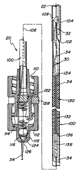
Referring now to Figs. 1 and 11 the details of the
deployment instrument 20 will now be described. As can be
seen the instrument basically comprises a carrier 100 in the
form of an elongated tube 102 formed of a somewhat flexible
material, such as polyethylene or polyvinyl chloride, so that
the carrier may be freely passed through the introducer
sheath into an operative position within the patient's
...,,
'r ~O 93/8746 PCT/US92/09572
- 2~.~2~~~
i
13 ._ _.
:;
artery, notwithstanding any curvature of the introducer
't;~ sleeve which may exist.
:;
In accordance with a preferred embodiment of this
invention the outside diameter of the tubular carrier 100 is
8 French. The distal end of the tube 102 includes a rigid,
r' e.g., stainless steel, sleeve or bypass tube 104 mounted
ey~~ thereon; to enable it to be inserted through a conventional
"' hemostasis valve 28A (Figs. 12-14) forming a portion of the
9
introducer sheath 28, through the sheath, and out the distal
':" end thereof into the artery 26. The distal Enc. of the
flexible tube 102 necks down into a generally hemicylindrical
. configuration (See Fig. 1) which includes a longitudinally
,..;
extending slit (not shown) therein to enable it to be fit
r>
within the bypass tube 104 without buckling.
~''' As can be seen in Fig. 11, the closure device 22 is
v'y
located within the distal end of the tubular carrier 100. In
e particular the anchor member 32 is disposed longitudinally
::~.
within the bypass tube 204 laterally of the central
longitudinal axis 106 of the carrier. The plug member 30 is
located within the tube 102 just behind (proximally) of the
'~. anchor member and on the opposite side of the central
longitudinal axis. In fact the distal end of the plug member
overlies the proximal end of the anchor member. The bypass
,.,;
tube 3.04 includes a reference decent 108 in its periphery
'~ located diametrica2ly opposite to the position of the anchor
>r~
member. The decent 108 serves as a visual guide to help the
user orient the instrument to a proper yaw angle with respect
to the central longitudinal axis for insertion within the
introducer sheath. as will be descr~.bed later.
As can be seen in Figs. 1 and 11, the instrument 20
includes a conventional leer fitting 110. The proximal end
of the carrier tube 102 extends into an opening in the
;r:;~,fitting 110 and is secured in place therein by any suitable
.::.:r,
means. Another conventional luer fitting 112 is threadedly
~y,~ secured to the threaded distal end 114 of the fitting 110.
The fittings 110 and 312 together form a hollow body through
which the proximal end of the filament 34 extends. A
~::.:,
!~~ 93/08746 FCT/US92/0~3S7Z
~12~994
14
;;~ tensioning assembly is located wivhin that body and basically
';~ comprises a ball 126, a cup shaped ball seat 118, a
compression spring 120, and a spring seat 122. The spring
seat is a disk-like member located within an annular recess
~'
d
within the center of the luer fitting 110. The ball seat
;..
includes a conical inner surface 124 having a central opening
126. The spring is a helical member interposed between the
;,;...
spring seat 122 and the ball 116 to bias the ball toward the
conical surface 124 of the ball seat 118. The proximally
.:_:.:3
located portion of the filament 34 extends through _the space
: between the ball 116 and its seat. The amount of force
:::,.,
-~ applied to the ball is established by a spacer sleeve 128
.,
located between the luer fittings 110 and 112. By
''~~ appropriate selection of the width of the sleeve 128 any
desired preload can be applied to the spring.
~,.,
As will be appreciated by those skilled in the art
>~.:;
r~~s' the tensioning assembly just described wil'. tend to hold the
Hyf
filament in glace with respect thereto until the force
applied to the filament exceeds the preload force applied by
1:~
'~°" the compression spring, whereupon the filament will be freed
r' to slide through the instrument.
The carrier 100 also includes a tamping member 130.
This member is --an elongated rod-like member formed of any
suitable material, eg:, polyethylene, and is disposed wi thin
the carrier tube 102 immediately proximally of the plug 32.
,.,
The tamping member 230 includes a central passageway 132
extending down its length from its distal end 134 to its
a.
~,:_ proximal end 136: The filament 34 portion.extending from the
~.~;a anchor member 32 passes through the passageway 132 in the
Y,:.i;.
tamping member and from there into the luer fittings 110 and
,~,.,.~
112, past the tensioning assembly, and out through the hole
~'~ 126 at the proximal end of the instrument 20. A hol ding
~,,~ sleeve or tag 138, e:g., a stainless steel tube, is crimped
Y~
onto the filament so that it engages the proximal end of the .
C~SA '.
fy:n f.. .
~4a,
tamping member 130 to hold that member in place. The tag 138
~f~
is arranged to cooperate with a torsion spring 142 Figs. 9
d5nr
'~c'.~~ and 10) to apply tension onto the filament 34 after the
';:,;
fr
'~V~ 93/(i8746 ~ ~ ~ ~ ~ ~ -~ P~'H'eus9ze~9a7z
i;
,:..~;
._ _. 15
'closure device is in place to enable the instrument 20 to be
removed and the filament severed (as will be described
. ;e
later) .
As mentioned earlier the instrument 20 is arranged
to be inserted into a conventional introduces sheath~28 to
effect the deployment of the closure device 20. Before
describing that operation a brief description of the
introduces sleeve and its method of location with .respect to
the percutaneous puncture is in order. As can be seen in
Figs. 12-14 the sheath 28 includes a body portion i.x~ which a
conventional hemostasis valve 28A is located and a tubular
portion 28,B extending from the body. The tubular portion 28B
terminates in an open distal or free end 28C. The body
portion of the sheath 28 includes a sideport 28D having a
conventional stopcock 28E located therein. The distal end of
the body of the sheath includes an annular groove 28F which
is arranged to receive a position indicator clip 1a0 forming
a portion of the system of this invention, for reasons to be
described later.
Before the instrument can be inserted into the
introduces sheath 28, the sheath itself must be properly
located within the artery. This action is accomplished via a
positioning device 200. That device forms a portion of the
system ~f this invention and is shown in Figs. 7 and 8. As
can be seen the device 200 basically comprises a conventional
dilator whose outer periphery has been modified .to include a
longstudinally extending flat 202. The device 200 ~is
arranged tn be fully inserted within the introduces sheath 28
like sh~wn in Fig. a2: The insertion of the device 200
within the introduces sheath 28 forms a passageway between
the flatted surface 202 of the device 200 and the interior
surface of the tubular portion 288 of the sheath disposed
thereover. The length of the flatted portion 202 is
selected so that when the device 200 is fully with the
introduces sheath, and the distal end of the sheath within
the interior of the artery, the distal end of the flatted
surface extends just beyond the distal end 28C of the
VNO 93/0876 PCT/US92/U9572
~1~~~94 16 .__.
introduces sheath to form a window 204 into which blood may
flow, while the proximal end of the surface 202 is in fluid
;
,
'''~ communication with the interior of the introduces body and
the sideport 28D. Accordingly, blood may flow into the
vi
window 204 through the passageway formed by the flatted
~ surface, into the sideport 28D and prom there to the
:, stopcock 28E when the window 204 is within $hs interior of
.the artery. '
In order to correctly position the introduces
sheath the location of the artery wall must be established.
This is accomplished by inserting the device 200 within the
introduces sheath as just described and then opening the
stopcock 28E to observe the flow of blood therefrom. The
blood will normally flaw out of the opened stop cock by
virtue of the pressure, differential across the lumen wall.
If however, there is insufficient pressure to cause such a
flaw of blood some means (not shown can be used to create
the desired differential pressure, e.g., suction can be used.
In any event once the flow of blood is observed the
introduces sheath with the device therein is then retracted
(moved proximally) until the blood flow through the stopcock
gust stops, a position shown in Fig. 13. This indicates that
''~~ the distal end 28C of the introduces sheath has just left the
.,
;,,
artery lumen. The introduces sheath with the device therein
is then reinserted approximately l0.mm into the puncture to
ensure that the distal end of introduces sheath is at the
desired position within the artery. Blood flow should be
,t
reestablished through the stopcock at this time. Then the
~~;
stopcock is closed. From this point the introduces sheath
must be kept fixed, i.e., it must not move axially relative
~.,
~ to the patient. To achieve that end the user of the system
'1r'd
should provide a continuous grasp on the introduces sheath,
xr, with the patient s groin as a position reference. The
v.4
position indicating device 200 is then removed from the
introduces sheath to ready the introduces sheath for receipt
of the deployment instrument 20 carrying the closure device
~,
22 as will be described later.
., WO 93/08?46 ~ ~ ~ ~ ~ (~ _~ PCT/US92/09S?2
17
In Fig. 26 there is shown a second embodiment of a
positioning device 300 for effecting the proper positioning
a
of the introduces sheath 28 within the artery. As can be
seen the device 300 basically comprises a conventional
obturator whose outer periphery has been modified to 'include
;,r,.
an annular recess 302 extending thereabout. Like the device
200,
the device 300 is arranged to be fully inserted within
iY,
"~
.>;;s ,
the mtroducer sheath 28 as shown in Fig. 27. The insertion
of the device 300 within the introduces sheath 28 forms an
annular passageway between the annular recess 302 of the
. ,,
device 300 and the interior surface of the tubular portion
28B of the sheath 28. A side opening or port 304 is provided
.
,;,,
,yf in the sidewall 28B of the introduces sheath 28 closely
.,,.
,, adjacent its open distal end 28C.
I Ac~
~.~.~xThe length of the annular recess 302 is selected so
;~rx:
~r~c~,
that when the device 30O is fully
with the introduces sheath
,;;
28, and the port 304 in the distal end of the sheath is
loca ted within the interior of the artery, the distal end of
the annular recess 302 extends just beyond the port 304 while
the proximal end of the recess 302 is in fluid communication
..:'/.
with the interior of the introducess
sideport 28D. .
The port 304 forms a window into which blood in the
~c~ artery may flow
when the
distal end
28C of the
introduces
is
located therein. In particular, blood may flow into the
/
window 304 through the . annular passageway foraged between
the
recess 302 and he inner. surface of the tubular portion 28A
~;",~ of the introduces, into the sideport 28D and from there to
~.,;
the stopcock 28E when the window 304 is within the interior
of the artery.
~["~
In Fig. 28 there is shown a third embodiment of a
positioning device 400 for effecting the proper positioning
,.
of the introduc~r sheath 28 within the artery. As can be
seen the device 400 basically comprises a conventional
;~
obturator having a passageway 402 extending longitudinally
down substantially the length of the device. An entrance
port 404 extends radially inward into the device
s.
communicating with the distal end of. the passageway 402,
Vt'4 93/08746 PCT/US92/09572
~,...~,
.. ._ _. 18
while an outlet port extends radially inward into the device
communicating with the proximal end of the passageway 402.
Like the devices 200 and 300, the device 400 is arranged to
be fully inserted within the introducer sheath 28 as shown in
Fig. 29.
The length of the annular passageway 402 is
selected so that when the device 400 is fully with the
introducer sheath 28 and the distal end of the sheath is
''-0 located within the interior of the artery, the inlet port 404
of the~passageway 402 extends just beyond the free end of the
sheath, while the outlet port 406 is in fluid communication
"'' with the interior of the introducer's sideport 28D. The port
404 forms a window into which blood in the artery may flow ,
when the distal end 28C of the introducer is located therein.
Tn Fig. 31 there is shown alternative embodiment
,;>
28' of an introduces sheath. The sheath is similar to sheath
28 described earlier except that its tubular portion 28B
includes a second passageway 502 (Fig. 31) .extending
therethrough. The passageway 502 serves as the passageway
for blood to flow therethrough so~ that the sheath 28',
,; itself, can act as a positioning device for effecting its
proper position~.ng within the artery. As can be seen in Fig.
31 the passageway 5a2 extends longitudinally clown the sheath
rr,
,t~ 28' within gas wall and parallel to the central passageway
;,, .
504 (the central passageway receives the deployment
>~ instrument 20 - o be described later). The distal end of
,,;,
the passageway 502 includes a radially extending port 506.
The proximal end of the passageway 502 (not shown) is in
.' fluid communication with the interior of the introducer's
';: sideport 28D. The introduces sheath 28' is arranged to be
,,,
used with a conventional obturator 600 (shown in Fig. 30).
The positioning of the introduces sheath 28
utilizing either of the devices 300 or 400 or the positioning
of the introduces sheath 28' utilizing the obturator 600 is
similar to that described with reference to the device 200.
Thus, after the introduces sheath is positioned as described
earlier the stopcock 28E is opened to observe the flow of
W4 93/08746 ~ ~ ~ ~ ~ ~ PCT/US92/09572
19 ._ ..
blood therefrom (thereby indicating that the inlet port or
window is within the artery). The intraducer sheath is then
retracted (moved proximally) until the blood flow through the
stopcock just stops, thereby indicating that the distal end
28C of the introduces sheath has just left the arte~"y lumen.
~ The introduces sheath with the device therein is then
;,.A'Y
reinserted approximately 10 mm into the puncture to ensure
that the distal end of introduces sheath is at the desired
position within the artery. Blood flow should be
reestablished through the stopcock at this time. , Then the
"~ stopcock is closed. From this point the introduces sheath
must be kept fixed (as described earlier) and the position
indicating device 300 or 400 (or the conventional obturator
600} removed to ready the introduces sheath for receipt of
.,,
the deployment instrument 20 carrying the closure device 22
;,.
through the central passageway in the particular introduces
:" -
sheath (that passageway is denoted by the reference number
504 in the embodiment 28').
The deployment of the closure will now be described
.y
with reference to Figs. 14-23 and is as follows: The
reference decent 108 on the bypass tube is identified by the
user and the b ass tube
yp grasped by the user and oriented so
that the detent faces up (away from the patient} as shown in
Fig. 14. This ensures that the anchor member is located
towards the patient. The bypass tube is then inserted into
the sheath through the hemostasis valve 28A. The rigid
nature of the bypass tube facilitates the passage of the
carrier 100 through the hemostasis valve and also protects
the closure device from damage. The instrument is then
pushed fully down the introduces sheath so that a stop
surface 110A on the front (distal) luer fitting 110 (Fig. l1)
engages the body of the introduces sheath housing the
hemostasis valve. At this time the distal end of the carrier
will be in the position shown in Fig. 16 and the anchor
member 32 will be located in the artery 26 beyond the distal
end of the introduces sheath. The bypass tube 104 remains
Vd0 93A08746 fCT/US92/09572
,.. 2 0
within the portion of the intraducer sheath housing the
hemostasis valve 28A.
~'rs The position indicator clip 150 is then mounted
onto the annular recess 28F on the introducer sheath 28 as '
shown in Fig. 17. As can be seen in Fig. 25 the flip 150
includes a linear section 150A from which a yoke 150B '
projects perpendicularly. The yoke 150B includes a circular
mouth 1500 for receipt of the annular recess 28F of the
introducer sheath. When mounted in place on the introducer
sheath~the free end 150D of the indicator clap will extend
beyond the distal end of the instrument 20 .(beyond the
; tensioner assembly).
The system 20 is then operated to determine if the
anchor member 32 has been properly deployed. To that end the
,.,.,,,
introduces sheath is then held by the user to prevent axial
;..
y; movement and the instrument 20 is carefully withdrawn from
::
it. This action causes the anchor member 32 to engage or
catch on to the distal end of the introduces. As the anchor
~a' member catches on the distal end of the introduces
, r
resistance will be felt by the user. This resistance must be
,,::
noted by the time the luer fitting 112 housing the tensioner
assembly reaches the free end 150D of the indicator clip 150
r
~,
'art as shown in Fig. 18. If so, then the anchor member will have
~
r
caught on the distal end of the introduc~r at the location of
its hemispherical projection 54 (the desired occurrence).
If, however, no resistance is noted 'by the time
'y= that the luer fitting 112 passes (extends proximally of) the
free arid of the indicator Blip, this will indicate that. the
~i anchor has re-entered the introduces sheath, and that the
v:r~ anehor will not catch onto the artery as required. Thus, if
no resistance is felt at this point, the instrument 20 must
;~...;
be reinserted within the introduces sheath and the foregoing
procedure retried, this time by turning the instrument 20
~~ about its axis 10G by 1/4 turns to each side before it is
.3:::9
again withdrawn.
'F',',1
If the resistance is felt before the leer fitting
,..;
. reaches the free end of the indicator clip this will indicate
f.,
W~ 93/08746 ~ ~ ~ ~ ~ ~ ~ PC.'f/US92/09572
._ _. 21
that one of the curved ends of the anchor member has caught
on the free end of the introduces sheath, an undesired
occurrence. Accordingly, the instrument 20 must be withdrawn
then reinserted within the introduces sheath and the
y foregoing procedure retried, this time by turning the
,a
instrument 20 about its axis 206 by 1/4 turns to each side
before it is again withdrawn.
Once the anchor member has been properly deployed,
as shown in Fig. 18, the collagen plug is deployed. To that
end the introduces sheath 28 and the instrument 20. are held
together and withdrawn as a unit from the puncture, whilst
swinging the unit toward the vertical as shown in Fig. 19.
This action causes the anchor 32 to engage or catch onto the
inner surface of the artery 26 contiguous with the puncture
'' 24. The introduces sheath and the instrument axe pulled
,?j
further outward as shown in Fig. 20. Inasmuch as the anchor
member is trapped against the interior of the artery wall the
I.
continued retraction of the introduces sheath and instrument
causes the filament 34 to pull the collagen plug out of the
carrier tube 102 and into the puncture tract 24A. As the
introduces and instrument come out of the puncture tract,
continuous steady resistance will be felt as the tensioner
assembly described heretofore controls the force on the
filameiat 34 during the retraction procedure. Continued
retraction of the introduces and the instrument brings the
tamping member 1~0 out of the free end of the instrument.
Moreover the pulley arrangement of the filament 24
connecting the anchor member and the plug member ensures that
during the retractir~n of the introduces and the instrument
the plug member is moved into engagement with the exterior of
the artery wall contiguous with the puncture 24. In fact
continued retraction causes the filament to somewhat deform
the plug, i.e., cause it to deform radially outward. The
existence of blood within the puncture tract further
contributes t~ the deformation of the plug member since the
collagen foam expands in the presence of blood.
WO 93/08?4b Pf.'1'/US92/0~~72
22 ._ _.
~~i~ The retraction procedure continues to pull the
introducer and instrument up the filament until the tag 138
is exposed as shown in Fig. 22. At this point the anchor
ej member and collagen plug member have been deployed. At this
,,,
time the collagen plug is tamped by the tamping member 130.
In particular the user quickly compacts the collagen of the
plug by gently tensioning the filament by pulling on the
introducer sheath and instrument in the proximal direction
with one hand. The tamping member is then manually slid dawn
y the f iiament by the user' s other hand so that it enters the
puncture tract 24A and engages the proximal end of the plug
''s~~ member 32. A few gentle compactions are adequate to achieve
the desired result, i.e., to assist the plug member 30 to
conform to the artery contiguous with the puncture and to
,f-:~sassist to lock said the plug in place until hemostasis occurs
(which happens very quickly, thereby locking the closure in
;!~'~ place). It should be noted that during the tamping action
.,.
~'v care must be taken to maintain tension on the filament 34 at
~,,
a load greater .than that used on the tamping member 130 to
ensure that the tamping 'action doesn't propel the plug member
30 into the interi~r of the artery.
After the tamping action is completed the torsion
spring 142 is mounted on the filament 34 as shown in Fig 23.
This action is necessary to maintain appropriate tension on
the filament while the instrument 20 is removed (the filament
.'r,:;severed). In Figs. 9 and 10 the torsion spring is shown. As
can be seen therein the s rin 142 includes a g
,y p g pair of 1e s
,
142A and 1428 projecting outward from a helfcal central
section 1420. Each leg includes a slot 142D at its free end.
One of the slots is arranged to receive the filament 34
::v
sd
therein and to engage the tag 138. The other of.the slots is
arranged to receive the filament 34 therein and to engage the
proximal end. of the tamping member 130. The legs 142A and
142B are biased by the intermediate section 1420 so that when
'~
;i
the spring is mounted on the filament as just described they
will bias the tamping means towards the plug member 30 to
"';' hold it in place so that the filament can be severed (as is
A~VO 93/08746 ~ ~ ~ ~ ~ ~ ~ P~.'~'/US92/09572
23
'y necessary to remove the instrument and the int~oducer from
the closure device). Thus, once the spring is in place the
filament on the proximal side of the tag 138 is cut and the
spring applies a light controlled pressure to the collagen
r; plug and anchor. The clasure is left in this c~nrlition
without being disturbed for approximately 30 minutes. After
that time the spring 142 is removed and the filament is then
' severed at the top of the tamping member 130. The tamping
member 130 is then removed and the remaining portion of the
filament is taped to the skin at 160 as shown in.Fig. 24.
The tape (not shown) should be removed and the filament cut
'r subcutaneously prior to the discharge of the patient.
With the closure in final position as shown in Fig.
24 the anchor member 32 (the only portion within the artery)
does not take up a substantial portion of the interior of the
artery and thus does not block off or otherwise impede the
flow of blood therethrough. Since the components of the
closure are all formed of resorbable materials the closure
can be left in place within the body until it is absorbed.
In Fig. 36 there is shown an alternative embodiment
?00 of tamping means constructed in accordance with this
invention. The tamping means 700 basically comprises an
assembly of two components, whereas the tamping means 130
described earlier is composed of only a single component.
',~ n
Thus, as can be seen in Fig. 36 the assembly 700 comprises a
first tubular component 702 and a second tubular component
?04. The component 702 includes a central passageway 706 and
is formed of any suitable material, e.g., the same material
as used to form the tamping component 130 described earlier.
The second component 704 also includes a central passageway
708 extending therethrough.
The component 704 is mounted on the front or distal
end of the component 702. To that end the component 704
includes an annular recess 710 about its periphery at the
proximal end thereof. This recess is arranged to receive the
distal end 712 of the component 702, with the two passageways
VN~ 93/08746 P~9'1US921~19572
,,. ..
~~.2~~94
._ ... 2 4
;f 706 and 708 axially aligned to enable the filament 34 to
y extend therethrough.
The camponent 704 is preferably formed of a
compressed collagen foam, e.g., the same type of material
'' used for the sealing portion or plug 30 of the closuYe'. The
distal end 714 of the component 704 is arranged to engage the
plug 30 to tamp it down in the same manner as that
accomplished by the distal end 134 of tamping member 130.
f Once the tamping action is completed the torsion spring 142
is mounted on the filament as shown in Fig. 3? so that it is
located between the tag 138 and the proximal end of the
component 702 (in the same manner as described with respect
to tamping member x.30 shown in Fig. 23~. Thus, the filament
on the proximal side of the tag 138 can be cut, while the
7!,
spring applies light controlled pressure to the collagen plug
'130 and anchor 32. The closure is left in this condition in
.y the same manner as described earlier after which time the
spring is removed and the filament severed' at the top
(proximal ends of the tamping component 702. That component
can then be removed, leaving the tamping component 704 within
the puncture tract as shown in Fig. 38. The remaining
(exteriorly extending) portion of the filament is taped to
:a
the skin at 160 as also described earlier.
As should be appreciated by those skilled in the
art the two sections of the filament 34 between the anchor
component 32 atad the plug component 30 effectively form a
a
'°pulley" arrangement to increase the mechanical advantage of
the force applied to the filament to move the two components
~~i toward each other. Accordingly, the closure can be properly
.;,
v~ seated without the application of a high pulling force. The
use of the biased ball and associated seat between which the
filament passes during the placing of the closure ensures
that irrespective of how hard the instrument and the
introducer are withdrawn from the puncture during the
.,
,:,.
deploymewt and seating of the closure, the amount of force
,,;
applied to the filament 34, and hence to the closure device,
;ywill not exceed a predetermined maximum, e.g., one pound.
WO 93/08746 ~ ~-' ~ n~ ;~ ,1.~ PCT1US92/09572
v5
This feature is of cori~i~derable importance to ensure that the
anchor portion of the closure is not pulled through the
opening (e. g., incision or puncture) once it is in place.
As should also be appreciated from the foregoing,
the closure device, the instrument for deploying it, and
their method of use enables the ready, effective and
efficient sealing of a percutaneous puncture in an artery.
Thus, it is expected that the hemostatic puncture closure
device 20 will be a significant advancement in the fields of
cardiology and radiology. The device may allow continuance
of anticoagulation post-procedure, more aggressive use of
thrombolytic agents and safer use of large bore catheters.
It should also reduce discomfort and complication rates for
patients; allow many in-patient procedures to be performed
safely on an out-patient basis; decrease the time and cost of
interventional procedures; and reduce exposure of hospital
personnel to human blood.
Without further elaboration the foregoing will so
fully illustrate our invention that others may, by applying
current or future knowledge, adopt the same for use under
various conditions of service.