Note: Descriptions are shown in the official language in which they were submitted.
~ ` 12~23088
TRa~SPORT CATlIETER
AND ~LTRA801~ND PROBE FOR U8E ~irIT~ ~AME
BACKGROllND OF THE INV~NTION
Field Of The InveIItion
The present invention relates to an improved transport
catheter and to an improved ultrasound probe and method o~
making an ultrasound probe for use in connection with the
multi-lumen transport catheter. More particularly, the
features of the invention relating to the catheter relate
to an improved transport catheter which can accept various
probes for sensing biological conditions-and parameter~ and
which allows high fluid flow rate for introducing fluids
irrespective of the presence of sensing instruments in the
catheter, thereby reducing the risk of patient
complications. The feature of the present invention
relating to the probe relates to an improved ultrasound
probe that allows for more accurate ultrasound readings and
; has a relatively small outer diameter, such that it can be
used in connection with the catheter of the present
invention.
Des¢ription Of Related Art
Numerous catheters exist for sensing, diagnosing and
treating various biologic conditions. For example, there
are cardiac catheters used for angioplasty, for measuring
cardiac output, such as thermodilution catheters, pulmonary
artery wedge pressure monitors, blood flow monitors and
temperature monitors. In use, a transport catheter is
initially introduced into an appropriate vessel or body
cavity. In the case of a thermodilution catheter, for
example, the transport catheter may be introduced into an
appropriate vein. Thereafter, the thermodilution catheter
is inserted and passed through the right atrium and
ventricle and out to the pulmonary artery. After the
catheter is properly positioned and the balloon in~lated,
` 2 2~230~8
various readings can be taken of left heart pressure, for
example, and pulmonary artery temperature. The same
measurements may be taken a number of times while the
catheter is in place. However, if the patient's condition
changes and requires other measurements or diagnosis, or
additional information is desired, æuch as may be required
in view of the results obtained by the thermodilution
measurements, the thermodilution catheter must be removed
and substituted with a different catheter for such
measurements. The subsequent catheter exchange i~creases
the possibility of infection through the introduction of a
second catheter and increases the probability of other
problems such as venous puncture.
Another problem with frequent catheter exchange is that
only physicians are authorized to remove and replace
catheters and probes in the patient's body. However, aft~r
a physician has inserted and positioned the catheter in the
patient's body, a trained nurse is permitted to insert,
position, and replace probes within the catheter, since the
probe does exit the catheter. Therefore, it is desirable
to use a transport catheter in connection with a probe,
such that the probe can be used within the transport
catheter without the removal and replacement of the
transport catheter.
Thermodilution catheters, such as the well known
Swan-Ganz catheters, generally provide for introducing
fluids into the patient through the catheter. However,
some procedures require higher fluid flow rates or
introduction of more viscous fluids than are presently
contemplated with such catheters. Such catheters are
generally not designed for maximum fluid flow or for
efficient flow of relatively viscous fluids.
In the past, multi-lumen catheters were designed wherein
the catheter body was divided into circular sections of
similar size or substantially triangular sections to form
~ 3 21230~ `
the separate lumens. These catheters were generally too
small to accept sensing probes and one or more of the
lumens of such catheters occasionally become constricted at
the seal of the transp~rt catheter. A further disadvantage
of these multi-lumen catheters becomes apparent if an
ultrasound probe was to be used within one of the lumens of
the catheter in order to obtain diagnostic readings. In
- this case, the similar sized lumens surrounding the
probe-carrying lumen contain relatively large amounts of
air space that cause undesirable attenuation of the
ultrasonic signal.
Undesirable signal attenuation is also caused by the
transducer design of the prior art ultrasound probes. For
example, ultrasound probe ~ransducers may be formed of
crystal material, having two leads attached to the crystal
material. The ~irst lead is connected to the inner surface
of the crystal material, and the second lead is connected
to the outer surface of the crystal material. The location
of the second lead on the crystal material causes a "dead"
spot in the attenuation pattern of the ultrasound signal.
Therefore, the ultrasound probe does not provide as
accurate of a reading as desired. Also, the attachment of
the second lead to the outer surface of the crystal
cylinder causes the outer diameter of the ultrasound probe
to increase, making it difficult to fit the ultrasound
probe within the transport catheter. Therefore, a need
exists for an ultrasound probe having a relatively small
outer diameter, and which does not produce dead spots in
the attenuation pattern of the ultrasound signal.
In patients undergoing major surgery or suffering from
serious illness, there is an acute need for a continuous
blood flow measurement, as compared to an inter~ittent
blood flow measurement. Therefore, ultrasonic transducer
probes have been designed to continuously measure cardiac
blood flow.
2 ~ 2 ~
A known method of calculating blood ~low is to multiply
the area of the blood flow times the velocity of ths flow.
Methods have been developed for using ultrasound
transducers to calculate the area and to calculate the
blood flow velocity. For example, it is Xnown to use echo
patterns to determine the cross-sectional area of a blood
vessel, and to use a Doppler technique to determine blood
flow velocity.
However, in order to measure both cross-sectional area
and velocity, two separate and distinct ultrasound
transducer elements were used. A first transducer element
was used to obtain measure~ents of the flow area, and a
second transducer element was used to obtain measurem2nts
of the flow velocity. For example, in one method, several
transducer elements are located at the catheter tip. A
first plurality of transducer elements are activated and
used to calculate the cross-sectional area of the vessel
perpendicular to the catheter tip by echo methods. With
the same catheter, a second distinct annular transducer
element is activated to determine the velocity of the blood
which flows perpendicular to the cross-sectional area. The
velocity is determined by using the Doppler principle,
wherein the Doppler shift created by the movement of the
blood cells is analyzed. The product of the two
measurements provides the blood flow measurement.
~ nother method of determining blood flow includes a
method wherein the transducer generates a single large
uniform cone-shaped beam which extends forwardly into the
pulmonary ar~ery. However, in this method, only a single
cone-shaped beam is analyzed, and a radially-oriented beam
is not utilized to determined the cross-sectional area of
the artery.
With many of the known ultrasound probes, the probe must
be axially aligned within the blood vessel and blood flow
in order to provide an accurate reading. If the probe is
G~
.~.?,.,`,
2 .t 2 ~
not properly aligned, the angle of incidence between the
probe axis and the blood flow adversely affects the
accuracy of the ultrasound probe measurements. Therefore,
a need exists for an ultrasound probe with a single
transducer element that can generate a radially oriented
signal beam and a forwardly oriented beam simultaneously,
and which is not affected by the angle between the axis of
the transducer and the blood flow.
There is also a need for an improved catheter which can
accept a variety of successive probes or sensors or other
instruments, like an ultrasound probe, and which also,
simultaneously, allows for high fluid flow for fluids to be
introduced into the body, as well as the introduction of
relatively viscous fluids. Additionally, a need exists for
lS a multi-lumen catheter that minimizes the sizes of the
lumens which might contain ultrasonic wave attenuating air
in lumens adjacent an instrument containing lumen.
~UMNARY OF THE INVENTION
One object of the present invention is to provide a
transport catheter that provides a plurality of lumens for
accepting various sequential probes through at least one of
the lumens without requiring the insertion and removal of
a different catheter with each probe.
Another object of the present invention is to provide a
transport catheter with a plurality of lumens that allows
for increased fluid flow rate through at least one of the
lumens.
~ still further object of the invention is to provide a
transport catheter with a plurality of lumens that allows
fluid flow through at least one of the lumens with a probe
inserted in the lumen.
Another object of the present invention is to provide a
transport catheter that provides a plurality of lumens for
accepting various sequential probes through at least one of
2~230~8
the lumens without requiring the insertion and removal of
a different catheter with each probe.
Yet anothex object of this invention is to provide a
transport catheter with a plurality of lumens that allows
for an increased accuracy in ult~asound readings by
minimizing the attenuation of the signal caused by the
quantity of air space in the surrounding lumens.
Still another object of the present invention is to
provide an improved pulmonary artery or central venous
transport catheter with a plurality of lumens wherein one
lumen is an inflation lumen used to inflate an inflation
balloon.
Yet another object of the present invention is to
provide a transport catheter with a plurality of lumens
wherein one lumen allows for the passing of a portion of an
instrument along the length of the lumen, such as
thermocouple wires, for example.
These and other objects of the present invention are
achieved through a catheter comprising a catheter body
having an outer edge with an outer dimension and having a
proximal end and a distal end.
One object of the present invention is to provide an
ultrasound probe that provides increased accuracy in
ultrasound readings by reducing dead spots in the sound
wave attenuation pattern.
Another object of the present invention is to provide an
ultrasound probe with a relatively small outer diameter.
A further object of the present invention is to provide
an ultrasound probe that can be used in connection with a
multi-lumen transport catheter.
Still another object of this invention is to provide an
ultrasound probe with a single transducer element that can
generate a radially oriented signal beam and a forwardly
oriented beam substantially simultaneously.
~ 7 ~12308~
Yet another object of the present invention is to
provide an ultrasound probe with a single transducer
element for measuring blood flow that is not affected by
the angle of incidence between the axis of the transducer
and the blood flow.
These and other objects are achieved through an
ultrasound probe for use within a transport catheter,
comprising a probe body having a distal and a proximal end,
and a transducer portion attached to the probe body distal
end. The transducer portion of the probe comprises a
piezoelectric crystal in the form of a hollow cylinder
having a distal end and a proximal end, and further
defining an inner surface and an outer surface. An inside
lead is coupled to the inner surface of the crystal
cylinder, and an outside lead is coupled to the outer
surface of the crystal cylinder. The outside lead has a
first and a second end, wherein the first end is coupled to
the outer surface of the crystal cylinder by a conductive
material, while a substantial portion of the outside lead
remains within the diameter defined by the crystal
cylinder. The preferred transducer portion includes a
layer of acoustically coupling material deposited adjacent
the distal end of the crystal and a layer of acoustically
absorbing material deposited adjacent the proximal end of
the crystal cylinder.
The transducer portion alternately generates a radially
oriented signal beam at a first frequency, and then
generates a forwardly oriented signal beam at a second
frequency. The signal beams are used to calculate
cross-sectional area and blood flow velocity, respectively,
which are then used to calculate blood flow.
The probe is preferably designed for use within a
catheter of the present invention comprising a catheter
body having an outer edge with an outer dimension and
having a proximal end and a distal end. The body also
2~23Q~8
includes walls defining, in transverse cross-section, a
plurality of lumens extending longitudinally substantially
through the catheter body including a first wall de~ining
a first lumen having a first transverse dimension
approximating about half of the dimension of the catheter
body. A second wall defines a curved lumen wherein the
lumen occupies at least a ~arter of an arc around the
catheter body. With this configuration, the catheter can
serve multiple functions. The first lumen can serve not
only as a probe lumen for transporting a suitable probe or
sensor, but also as a lumen for introducing fluids, sensing
fluid pressure and taking fluid samples. The second lumen
is preferably formed so as to maximize the cross-sectional
area for fluid flow while still maintaining sufficient
catheter structural integrity to be reliable under a wide
variety of conditions. The second lumen allows
introduction of fluids at relatively high flow rates, or
fluids with viscosities higher than normal, such as those
which may be more viscous than saline. The first lumen is
preferably circular to accept a wide range of probes.
In a further form of the invention, a third wall is
included defining an inflation lumen for inflating and
deflating an inflation balloon mounted at the distal end of
the catheter body, and a fourth wall is included defining
an instrument lumen for passing a portion of an instrument
along the lumen.
A catheter according to this embodiment of the invention
having an inflation balloon, an inflation lumen and a
fourth lumen can be used as a typical Swan-Ganz type of
catheter for thermodilution measurements. The first lumen
is used for sensing pressure and the like while the curved
lumen serves as an injection lumen. The inflation lumen is
relatively small and serves the standard function, while
the fourth lumen can carry the thermocouple wires. If a
further test or procedure is then necessary, for example as
~ ' ` ~:'' `:. ' . ~ ~ ~' . ' . ` . '
l ,:,.".,."..". ~..., :,.", .
-~ 2~230~8
a result of the outrome of the thermodilution measurements,
a probe can be inserted into the first, probe lumen as
necessary. Additional tests may require other probes, all
of which can in turn be introduced without removing the
catheter, and without signi~icant additional risk to the
patient.
, The above described objects and other objects of the
present invention will now become apparent from a review of
the drawings and the following description of the preferred
embodiments...,
BRIEF DB6CRIPTION OF T~E DRA~ING8 .
FIG. 1 is a perspective view of the transport catheter
of the present invention.
FIG. 2 is a cross-sectional view of the transport
catheter of the present invention taken along line 2-2 of
FIG.1.
FIG. 3 is a perspective view of a catheter according a
. ~ further embodiment of the present invention showing a probe
and an injectate port.
FIG. 4 is a transverse cross-sectional view of the
catheter of FIG. 3 taken along line 4-4.
,. .
FIG. 5 is a longitudinal cross-sectional view of a
portion of the catheter of FI~. 3.
- 25 FIG. 6 is a partial segmented side-sectional view of a
catheter accordinq to a further embodiment of the present
invention.
.
FIG. 7 is partial segmented side-sectional view of an
ultrasound probe of the present invention.
FIG. 8 is a side-sectional view of a transducer of the
~ ultrasound probe of the present invention.
.~ FIG. 9 is a representational view of the ultrasound
probe with a single transducer element generating radially
and forwardly oriented signal beams within a blood vessel.
i-~
~.
'~,'
. . ~
~,.
..,.
:.~
.
.: :, 10
2 ~ 2 3 ~
FIG. 10 is a schematic representation of the signal
source for the ultrasound probe.
DETAILED D~8CRIPTION OF T~E ~RFFERRED E~BODINENTB
Referring now to FIG. 1, a catheter lO for accepting
probes and for introducing fluid through the catheter and
into a body cavity is shown which allows numerous
procedures to be done using a single catheter and which
reduces the likelihood of injury to the patient. In one
preferred embodiment, the catheter 10 is primaxily
comprised of a catheter body 12, an inflation balloon 14,
a plurality of extension tubes 16, and a plurality of
threaded hubs 18. The catheter body 12 has a proximal end
20 and a distal end 22. The inflation balloon 14 is
mounted to the catheter body 12 at the distal end 22 of the
catheter body 12 as would be known to one skilled in the
art. Each of the extension tubes 16 has a respective first
end 24, which is coupled to a corresponding one of a
plurality of lumens (shown in FIG. 2) in the catheter body
12 at the catheter body proximal end 20 at a backform 28.
The extension tubes 16 provide access to each of the
respective lumens. The extension tube corresponding to the
first lumen, described more fully below, also includes
graduations on the outside of the tube to indicate the
depth of insertion of any probe or instrument passed along
the first lumen. The second end 26 of each of the
extension tubes 16 is coupled to a respective one of the
plurality of threaded hubs 18. The threaded hubs 18 each
have a luer taper common in the art for connecting suitable
instruments, such as probe connectors, an inflaticn device
for the inflation balloon and an injection device for the
injectate lumen described more fully below.
Referring now to FIG. 2, a cross-sectional view of the
catheter body 12 is shown taken at an approximate mid-poi~t
of the catheter body. A plurality of walls within the
`i 11 212308~
catheter body 12 define the respective plurality of lumens.
A first wall 34 defines a first lumen or probe lumen 36.
- The cross-sectional configuration of the probe lumen 36 is
preferably circular, and has a diameter in the preferred
embodiment of approximately half the diameter of the
cross-section of the catheter body 12. A second wall 38
defines a second lumen or an injectate lumen 40. In the
preferred embodiment shown in FIG. 2, the cross-section of
the injectate lumen 40 is crescent-shaped. A suitable size
for the second lumen is one where it occupies at least a
quarter of an arc around the cross-section of the catheter
body 12. A third wall 42 defines a third, inflation lumen
44, in cross-section preferably circular, and a fourth wall
46 defines a fourth lumen 48, also preferably circular.
The probe lumen has a large cross-sectional area and
preferably occupies a significant portion of the
cross-sectional area of the catheter so that the catheter
can accept as many different types and configurations of
probe as possible and to permit a wide variety of tasks sr
procedures without having to remove the catheter. The
probe lumen also accepts the improved ultrasound probe of
the present invention herein. The probe lumen i8 also
preferably large enough to permit fluid flow within the
lumen even while a probe or other element is in the probe
lumen. This allows simultaneous instrument sensing and
pressure monitoring or introduction of fluid such as
pharmaceutical through the probe lumen, even with
concurrent introduction or withdrawal of fluid through the
injectate lumen 40. In this manner, removal of the probe
is not required before injectate can be introduced or blood
withdrawn through the probe lumen. Fluid pressure can also
be monitored even while a probe is in place in the lumen
36. For example, the lumen 36 is capable of accepting
hemoglobin oxygen saturation probes, pacing probes, cardiac
output probes, right heart ejection fraction probes, right
`:
~ 12 2123088 ~ ~
heart ejection fraction with hemoglobin oxygen saturation
probes, hemoglobin Ph probes, and ~igh fidelity pressure
monitoring probes. A preferred probe configuration is
circular in external dimension. In one preferred form of
¦ 5 the invention having the four lumens as described, a 7 1/2
French catheter has a 0.056 inch diameter probe lumen and
the probes are preferably arotnd 0.042 inches in diameter.
The advantage of the probe substitution feature of the
probe lumen 36 is apparent from a description of the use of
the catheter 10. In use, the catheter body 12 is first
inserted and properly positioned in the body by a
physician. A selected probe is then inserted through the
probe lumen 36 of the catheter body 12, by either a
physician or a nurse, and the desired procedure is carried
out. Thereafter, another type of probe measurement may be
required such as where the patient's condition changes.
The first probe is then removed from the catheter body 12,
leaving the catheter body 12 in place, and a second probe
is inserted through the probe lumen 36 of the catheter body
12 in order to accomplish a different probe function. The
insertion and removal of the catheter body with each type
of probe i5 avoided, and a nurse is permitted to insert and
remove each of the probes without requiring the physician's
presence. As a result, there is significantly less risk to
the patient of infection from the repeated insertion and
removal of catheters, as well as less risk of venous
puncture or other problems. Moreover, because the
insertion and removal of the probes can be accomplished by
a nurse, the insertion and removal process of the probes is
more convenient and efficient while the physician may
otherwise be occupied.
The large cross-sectional area of the injectate lumen 40
allows for a high fluid flow rate through the lumen, and
also accommodates the flow of relatively viscous fluids.
Therefore, the second lumen 40 is well-suited for
.
Q
13 2123
procedures requiring either high fluid flow rates or the
introduction of relatively viscous fluids. The second,
injectate lumen 40 is even more significant where fluid
must be introduced or withdrawn at the same time the probe
lumen is being used. The cross-section of the injectate
lumen 40 is preferably crescent-shaped, with the
cross-section of the lumen covering or extending around at
least a quarter arc of the catheter body cross-section.
The crescent shape al}ows for maximum fluid flow area
within the catheter body 12 without interfering with the
first lumen 36.
The third lumen 44 is preferably used for inflating and
deflating the inflation balloon 14 to properly position the
catheter, for example where the catheter is used as a
thermodilution catheter. The fourth lumen 48 is preferably
used for instrumentation, such as for passing thermistor
wires or the like along the catheter to a point where a
sensing device is located in the catheter.
The catheter body 12 of the present invention is
preferably formed by any of several well known extrusion
methods. The catheter body 12 may be fabricated from any
of a variety of suitable materials, including, but not
limited to, flexible polyvinyl chloride (PVC),
polyurethane, nylon, or polypropylene. The catheter
body 12 is also preferably coated with heparin.
In the preferred embodiment described herein, the
catheter body 12 has an outer diameter of 0.101 inches
centered on the central axis 50 of the catheter. The total
cross-sectional area of the catheter body 12 is therefore
approximately 0.008 square inches. The first lumen 36
preferably is circular with a diameter of 0.056 inches and
includes within it the central axis 50. The
cross-sectional area of the first lumen 36 is therefore
approximately 0.0024 square inches, which equates to
approximately thirty percent of the catheter body
.
,
: '
~; 14 21 23 08~
cross-sectional area. The cross-sectional area of the
crescent-shaped second lumen 40 is approximately 0.0016
square inches, which is approximately twenty percent of the
total cross-sectional area of the catheter body 12. The
largest distance between oppositely arcing surfaces in the
crescent shape is about 0.024 inches and the radius of
curvature of the ends of the injectate lumen is about O.OlO
inches. To optimize the available area that can be used
for fluid flow, the injectate lumen in the preferred
embodiment is symmetrically placed above the probe lumen
and centered so that an imaginary vertical plane (vertical
- when viewing FIG. 2) through the central axis 50 and the
central axis of the probe lumen bisects both the probe
lumen and the injectate lumen. It should be understood,
however, that where one or the other of the third or fourth
lumens is omitted, the injectate lumen may be for~ed
asymmetrically relative to a line through the central
axis 50 and the central axis of the probe lumen. The third
lumen 44 and the fourth lumen 48 are both preferably
circular, and have a diameter of approximately 0.012
inches. Therefore, the cross-sectional areas of the third
lumen 44 and the fourth lumen 48 are each approximately
O.OOOl square inches, which equates to approximately one
and one-half percent of the total cross-sectional area of
the catheter body 12. The smallest dimension from any of
the lumens radially to the outer edge of the catheter body
is preferably 0.007 inches. The thickness of any wall
between lumens is preferably at least 0.007 inches. The
dimensions of the catheter body 12 and lumens given are
preferred, but they are exemplary only of the preferred
embodiment of the invention.
It should be understood that the cross-sectional
configuration shown in FIG~ 2 is preferred, and extends in
the preferred embodiment substantially the entire length of
the catheter. However, it should also be understood that
:::
2 1 2 3 0 8 8
..~
the inflation lumen 48 terminates at the inflation balloon
14. It should also be understood that the injectate lumen
may open at an injectate port 52 through the outer catheter
wall at a suitable location near the distal end 22 along
the length of the catheter body (FIG. 3). Where the
catheter has an overall usable length of llO centimeters,
the injectate port 52 is typically located about 30
centimeters proximal of the distal end of the catheter, a
standard distance for a thermodiluticn catheter. A
thermistor 54 is exposed to the outside of the catheter
approximately 4 centimeters proximal of the distal end.
Considering the distal-most portions of the catheter in
more detail (FIGS. 4 and 5), fluid flow out the injectate
port is created by placing an injectate lumen plug 56 in
the injectate lumen 40. The plug 56 has a general
transverse cross-section conforming to that of the
injectate lumen and is sealed in place by a suitable
biocompatible filler. The thermistor 54 is potted in an
opening formed in the outer catheter surface. Preferably,
the thermistor is potted in the injectate lumen since the
in~ectate lumen downstream of the plug 56 is otherwise
unused. Thermistor wires 58 from the fourth lumen 48 pass
into the injectate lumen 40 through a cross-over 60 from
the fourth lumen.
In order to reduce the volume of air in the unused and
therefore vacant portion of the injectate lumen, namely the
portion of the injectate lumen distal of the plug 56, a
crescent shaped insert or rod 62 is inserted in the
injectate lumen and fixed with a suitable adhesive 64
between the plug 56 and the cross-over 58. The rod is
preferably formed from the same material as the catheter
and preferably to provide the same flexibility as the
catheter without the plug. A probe 66 is shown in FIGS. 4
and 5 and can be made from plastic, metal, plastic coated
.
: :
~ 16
2~23~8 :
metal, composites or other suitable materials used in
manufacturing probes, sensors or other instrumentation.
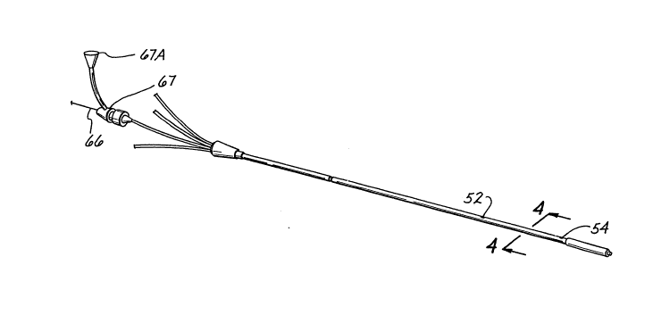
A hemostasis valve 67 is also shown in FIG. 3 through
which the probe passes into the extension tube. An
injection port may also be connected to the valve 67
through an appropriate stopcock, shown schematically at
67A, to which may be connected a conventional pressure
sensor device, a fluid injection device, and the like.
The orientation of the lumens within the catheter body
12 accommodates the four lumens with the probe and
injectate lumens having a relatively large cross-sectional
area. As a result, the cross~sectional areas of the third
and fourth lumens remain relatively small. In use, the
probe and injectate lumens are filled with a liquid, with
only the third and fourth lumens containing any appreciable
air space. The relatively small quantity of air space in
the third and fourth lumens minimizes undesirable 1
attenuation of ultrasonic signals when an ultrasound probe
is used within the probe lumen 36. Therefore, an
20 ultrasound probe used in the probe lumen of the preferred ~
embodiment produces a more accurate result. ~ ;
The accuracy of the ultrasound probe readings within the
transport catheter is also increased when an ultrasound
probe 82 of the present invention is used. The ultrasound
probe 82 is shown in detail in FIG. 7. The use of the
ultrasound probe 82 within the first probe lumen 36 allows
the nurse or physician to reposition the probe 82 until it
is properly positioned, without causing unnecessary tissue
or vascular damage. Moreover, as discussed above, the
physician is not required to position the probe within the
transport catheter, because a nurse is permitted to replace
probes within the catheter. Therefore, the placement of
the probe within the cathetçr body allows for more
convenient and less traumatizing replacement and
repositioning of the probe.
: :
~1 ~
o ~
_:- 17
2123088
Referring now to FIG. 7, an ultrasound probe 82,
accordin~ to one aspect of the present invention, comprises
a probe body 84 having a proximal end 86 and a distal end
88, a connector 90, and a transducer portion 92. The
S connector 90 is coupled to the probe body proximal end 86,
and the transducer portion 92 is connected to the probe
body distal end 88. The connector 90 is of the type known
in the art for use with ultrasound probes.
The transducer portion 92 of the probe is best shown in
FIG. 8. In the preferred embodiment, the transducer
portion 92 is comprised of a ceramic piezoelectric crystal
in the form of a hollow cylinder 94, an inside lead 96, an
outside lead 98, an acoustically absorbing layer 100, an
acoustically coupling layer 102, and a thin sputtered layer
104. The hollow crystal cylinder 94 defines an inner
surface 106, an outer surface 108, a proximal end 124, and
a distal end 126. In manufacturing process of the
transducer portion 92, the crystal cylinder 94 if formed by
extruding or molding a crystal cylinder of a predetermined
length. The inner surface and the outer surface of the
extruded crystal cylinder i5 then plated with nickel or
another type of conductive material in order to improve the
conductivity between the crystal cylinder and the leads.
The extruded crystal cylinder is then cut into
approximately .027 inche5 in length in order to form the
crystal cylinder 94 for the transducer portion 92.
A first end 118 of the inside lead 96 is then co~pled to
the inner surface 106 of the crystal cylinder with an
electrically conductive material. Preferably, the inside
lead first end 118 is coupled to the cylinder inner surface
106 by a silver conductive epoxy 110. The second end 118A
of the inside lead 96 is coupled to the probe driving
circuit 1188 (FIG. lO) through the connector 90 in the
conventional manner. A central portion 119 of the inside
lead 96 extends through the probe body 84.
t' ~ ,~ ;7~ " ~ ~"
_ 18 2123088
The cylinder 94, with the attached inside lead 96, is
then temporarily placed in a tube-shaped casting form or
mold 142. The outside lead 98 is bent at a ninety degree
angle to form an L-shape. It should be noted that various
shapes of the outside lead may function in the same manner.
However, for purposes of reference, the L-shaped lead is
used to describe the preferred embodiment. The L-shaped
lead is defined by a long leg 112 and a short leg 114. The
short leg 114 of the L-shape ends at the fir~ct end 116 of
the lead 98. When the crystal cylinder 94 is positioned in
the casting form, the first end 116 of the outside lead 98
is placed in close proximity to the outer surface 108 of
the crystal cylinder 94, and in an imaginary plane 120
substantially tangential to the outer surface 108 of the
crystal cylinder. In this position, the long leg 112 of
the L~shaped lead 98 then extends toward the probe body 84,
and is located within the imaginary cylindrical shape 122
defined by the outer surface diameter of the cry tal
cylinder 94. Therefore, preferably, no portion of the
outside lead 98 extends outside of the boundaries of the
outer diameter of the crystal cylinder 94.
The outside lead includes a central portion 138, which
is the portion of the lead between the first and second
ends. The central portion 138 extends continuously along
the plane of the long leg 114 of the L-shape. Thus, the
central portion 138 extends through the probe body 84. A
second end 139 (FIG. 10) of the outside lead 98 is coupled
to the probe driving circuit 118B t~rough the connector 90
in the conventional manner.
The layer of acoustically coupling material 102 is then
deposited adjacent the distal end 126 of the cylindrical
crystal 94. The acoustically coupling layer 102 is also
referred to as the matching layer. Preferably, an epoxy
material is used for the matching layer. The epoxy
matexial is selected so as to be acoustically coupled with
~:~``` 19 2123g~8
I the transducer. As shown in FIG. 8, the acoustically
¦ coupling layer 102 is formed in a plug shape, and may
extend into a portion or all of the center of the crystal
hollow cylinder.
The purpose of the acoustically coupling layer 102 is to
generate a solid cone forwardly oriented ultrasound wave
velocity beam 146 (shown in FIG. 9) from the transducer 92.
The sound wave velo~ity beam generated by the transducer
without the acoustically coupling layer resembles a first
cone with a second hollow cone portion in the center of the
first cone. However, with the acoustically coupling layer,
the second hollow cone is filled with sound waves, and the
velocity beam 144 becomes a solid cone. ~he purpose and
function of the forwardly oriented solid cone ultrasound
wave velocity beam is described in more detail herein.
Therefore, the acoustically coupling laye~ 102 allows for
more accurate readings from the transducer.
The layer of acoustically absorbing material ~00 is then
deposited adjacent a proximal end 124 of the crystal
cylinder. Preferably, an epoxy material doped with
approximately eighteen to twenty-six percent rubber powder,
such as HYCAR (TM) type 1422 polymer, available from Zeon
Chemicals, Inc., Illinois, sifted through a 100 mesh
screen, is used so as to act as a sound abscrber. This
layer of epoxy is also known as the backing layer of the
transducer portion 92. The acoustically absorbing layer
100 is preferably formed in a plug shape, and may extend
into a portion or all of the center of the hollow crystal
cylinder 94.
At this point, the transducer portion is removed fro~
the casting form 142. The matching layer, or acoustically
coupling layer is then ground down to a predetermined
length, so as to enable the transducer to produce a 1/4 or
3/4 ultrasound wave length signal.
:
. .
~20 21230~8
¦ After the matching layer 102 is ground, the sputtered
layer 104 is applied in order to make the electrical
connection between the crystal cylinder outer surface 108
- and the outside lead 98. The thin sputtered layer 104 of
conductive material, preferably gold or chromium, is
sputtered along the plane 120 defined by the crystal
cylinder outer surface 108 and the outside lead first end
116. Once the layer 104 of gold or chromium is applied,
the outside lead 98 is then conductive with the crystal
cylinder outer surface 108. Therefore, when current is
applied to the leads 96, 980 current will flow from the
inner surface 106 of the crystal to the outer surface 108
of the crystal or vice versa. ~oreover, because the
outside lead 98 is not directly attached to the outer
surface 108 of the crystal cylinder, the dead spots in the
ultrasound wave pattern are eliminated, and the probe 82
retains its relatively small outer diameter.
The sputtered layer 104 in FIG. 8 is shown covering the
entire outer surface of the transducer portion. However,
the sputtered layer 104 only needs to be applied to the
plane 120 80 as to electrically connect the outer surface
of the crystal cylinder and the outside lead 98. An
electrical isolation layer 140, also referred to as a
conformal coating, is then deposited over the outer surface
of the transducer portion. The electrical isolation
conformal layer 140 is preferably formed of a biocompatible
non-attenuating coating, such as a W curable adhesive
material, for example DYNAX (TM) 201S9 adhesive from Dymax
Engineering Adhesives, Connecticut.
Referring back to FIG. 7, the construction of the probe
body 84 is described. If desired, the probe body 84 may
include a stiffener member 128 that extends from the probe
body proximal end 86 to the probe body distal end 88. The
stiffener member 128 prevents kinks in the probe body 84 in
tight turns, as well as provides strength. The distal end
` 21
2~2~0~ -
88 of the probe body is preferably attached to the
transducer portion 92 by an adhesive layer 136.
As previously described, the second ends of the inside
and outside lead central portions 119, 138 extend toward
the probe body 84. After the stiffener member 128 is
secured to the transducer portion 92 by the adhesive layer
136, the inside and outside lead central portions 119, 138
are twisted and extend to Ihe second ends of the leads,
which are coupled to the driving circuit of the probe
through the connector 90. The twisting of the leads 96, 98
serves to reduce electrical noise to a minimum. A flat
spring wire 132 is coiled around and surrounds the
stiffener member 128 and twisted leads 96, 98.
The probe body 84 also includes a depth or zero
alignment mark 134 on the outer surface of the probe body
84 near the proximal end. The depth mark 134 is visible
through the extension tubes 16 of the catheter 10. The
depth mark 134 is positioned such that the mark 134 is
aligned with a predetermined location on the catheter
extension tube 16 when the probe 82 is properly positioned
within the catheter 10.
For purposes of reference only, the preferred dimensions
of the ultrasound probe 82 are given. The outer diameter
o~ the transducer 94 of the probe 82 i8 preferably
approximately .040 to .047 inches, the wall thickness is
preferably 0.010 inch, and the length is preferably 0.027
inch. The outer diameter of the probe body is preferably
approximately .037 inches. In comparison, the probe lumen
36 has a diameter of approximately .056 inches. Therefore,
the probe and transducer portion diameter is sufficiently
small to allow the probe to fit within the first lumen 36
of the catheter 10, as well as to allow additional fluid
flow through the first lumen 36 if reguired by the
- circumstances. The total length of the probe 82 is
preferably approximately 79.75 inches. The length of the
~.
~ ~` 22
2~23~8
transducer portion 92 of the probe 82, including the
matching layer and backing layer, is preferably
approximately less than .5 inches.
In the preferred embodiment, the ultrasound probe of the
present invention is used in connection with a system that
includes a Doppler unit (not shown) and personal computer
(not shown). The Doppler unit is used to drive the probe
and to house the Doppler electronics. The personal
computer is used to control the Doppler unit and display
and process the signal data. The personal computer
calculates the flow area using two components. The first
component measures flow velocity and the second component
measures flow area. The flow rate is a product of the two
components.
Referring now to FIGS. 9 & 10, in the preferred
embodiment of the ultrasound probe, the single cylindrical
transducer 92 instantaneously analyzes the blood flow area
and the blood flow velocity substantially sim~ltaneously.
It should be noted that the area and velocity are not
measùred precisely si~ultaneously in real time, but, as is
known, the measurements are made essentially
simultaneously, on the order of millionths of seconds.
More specifically, the single cylindrical transducer 92 may
be activated in two distinct modes instantaneously.
Each of the modes is activated at a different frequency.
In the first mode for producing a beam for determining the
area of the pulmonary artery for example, a frequency
generator in the signal source 118B (FIG. 10) generates an
8 Mhz signal in turn causing the transducer to generate an
ultrasound signal in a radial direction, thereby creating
a radially oriented signal beam 146 (FIG. 9). A standard
frequency generator such as an Hewlett-Packard signal
generator may be used to drive the transducer. In the
second mode, for producing a beam for determining the blood
velocity in the pulmonary artery for example, the frequency
,
:;` 23
....... .
generator generates a 2 Mhz sign~ rn causing the
transducer to generate an ultrasound signal in an axial
direction, thereby creating an axially directed signal beam
144 (FIG. 9). The transducer crystal is preferably
designed such that the optimum drivi~g frequency for the
radially directed beam 146 is 8 Mhz while the optimum
driving frequency for the axially directed beam 144 is
2.285 Mhz. More specifically, for signal generator driving
frequencies of 8 MHz and 2 MHz, respectively, the presently
preferred transducer crystal dimensions are an outer
diameter of 0.040 - 0.047 inch, a wall thickness of about
0.010 inch, and a length axially of about 0.027 inch. The
difference between the respective driving frequencies and
the transducer crystal design dimensions results in
decoupling sf the two driving signals for the preferred
design. However, the transducer may be driven at other
frequencies. When the transducer is driven at other
frequencies, the transducer design is preferably modified
- so as to be optimized at the other frequencies, while
preferably keeping the two frequencies for which the
transducer is designed decoupled. The different dimensions
may be determined by modeling.
The transducer crystal is preferably formed from PZT-5H,
a formulation of Pb(Zr,Ti)03 with high electromechanical
coupling coefficient and high dielectric constant.
The dimensions of the crystal are also designed to
generate the axia~y or forwardly directed beam 146 in the
shape of a wide cone. The wider the cone, the closer the
velocity measurement is taken to the point where the area
measurement is taken.
The radially oriented signal beam 146 is used to
calculate the cross-sectional area of the blood flow.
More specifically, the flow area is estimated by measuring
the Doppler power in a narrow Doppler gate of predetermined
area, Pn, for example one centimeter squared, and the power
': ~
. , ~
.,.. " 24 212~0~
from a wider gate of an unknown area, Pw, which corresponds ..
to the unknown flow cross-sectional area. The measured
power from the wider gate Pw corresponds to the number of
red blood cells insonified within the Doppler gate. The
corresponding number of red blood cells insonified is
proportional to the unknown cross-sectional area Pw.
Therefore, if
Pn = 1 cm ; and
Pw = x cm ~ :
- 15 in order to calculate Pw, the unknown cross-sectional are~,
the following equation is used~
2 :
Pw/Pn = x cm
In the second mode, the forwardly oriented signal beam
144 is used to calculate the blood particle velocity. The
blood velocity is calculated directly from the Doppler
frequency shift as measured by the forwardly oriented
signal beam 144.
In order to eliminate the effect of the angle of
incidence a between the axis of the transducer and the
blood flow, the following equation is used to calculate the
blood flow:
Q = V cos (a) * (A/ cos~A))
wherein Q = blood flow; V = blood velocity; A = cross-
sectional area; and a = angle of incidence between
transducer axis and blood fl~w. In the equation for blood
flow, the cos(a)'s cancel out, therefore eliminating the
effect of the angle of incidence on the blood flow
measurement. Because of the perpendicular nature of the
, ~ 25
~;` 212308~
radially and axially directed beams, the estimate of the
volume flow would be self-compensating or independent of
the orientation of the probe with the flow field.
By way of example, in one embodiment of the invention,
the system personal computer screen displays the diameter
mode signal echo pattern in M-mode, wherein the X-axis
represents time, the Y-axis represents depth, and the Z-
axis represents echo amplitude. Simultaneously, t~e second
mode information, the velocity mode, is also preferably
displayed in M-mode on the computer screen. Preferably the
second mode display is color coded so as to clearly
represent the speed of the moving blood particles.
once the information is displayed on the screen, the
user, referring to the first mode, marks the first echo
nearest the probe, and the last Doppler signal furthest
from the probe. These two measurements are used by the
computer to determine the cross-sectional area. In the
second mode, the velocity mode, the user marks the velocity
~ wave. The mean blood flow velocity over the cardiac cycle
is then calculated from this marked information.
The blood flow velocity and cross-sectional area are
then calculated, using the above equation, so as to compute
the blood flow. Therefore, the use of the distinct first
and second modes of the single transducer element provide
for an accurate blood flow measurement, while eliminating
the effects of the angle of incidence between the
transducer axis and the blood flow.
As previously described, the probe 10 is preferably
designed for use with the multi-lumen catheter of the
present invention. In addition to the previously described
advantages of the multi-lumen catheter, the design of the
catheter body 12 also provides the advantage of structural
integrity. The configuration of the lumens and the
thickness of the lumen walls contributes to the structural
integrity and strength of the catheter body, thereby
.,:
^ `:;: `
~ 26 212308~
minimizing the possibility that the catheter may be
constricted or crushed during use. More specifically, in
the preferred embodiment of the invention, a sub~tantial
portion of each of the lumen walls preferably has a
thickness greater than the shortest distance between the
first wall of the probe lumen and the outer edge of the
catheter body 12. Therefore, any possibility that the
catheter body 12 may be pressed or any lumens may be
constricted when the catheter is passed through a seal on
an outer transport catheter is minimized.
Referring back to FIGS 1-4, in a ~urther preferred
embodiment of the invention, a transport catheter includes
the probe and in~ectate lumens 36 and 40, respectively but
omits the inflation balloon and the inflation lumen.
Omitting the inflation lumen allows the injectate lumen to
be made larger if nece~sary by increasing the arcuate
length or arcuate extent of the injectate lumen, thereby
increasing its cross-sectional area and its flow
characteristics. The catheter of this alternative
preferred configuration has a number of applications,
similar to those of the embodiment of FIG. 1, including
sensing, fluid injection and sampling and the like. The
probe lumen i8 still preferably circular in cross-section
and occupies a substantial portion of the catheter
cross-section. The injectate lumen is also preferably
crescent shaped and occupies as much of the remaining
cross-sectional area of the catheter as necessary to
achieve high fluid flow in the lumen or to allow efficient
introduction of more viscous fluids.
An alternative embodiment of a catheter 68 (FIG. 6)
includes a first lumen exit port 70 proximal of the distal
end of the catheter approximately 30 centimeters, in the
embodiment where the catheter length is 110 centimeters.
A round lumen plug 74 is sealed in the circular first
lumen 76 to direct fluid from the first lumen externally of
. '.
27 2~2~ 8
the catheter. The port 70 allows infusion of a fluid
through the first lumen into the body cavity at a
relatively high flow rate. The cross-sectional area of the
port 70 is preferably the Game as that of the first lumen.
The cross-sectional configuration of the oatheter is
preferably the same as that shown in FIG. 2 to allow the
relatively high fluid flow rates in the first lumen and in
the injectate lumen, while also having a relatively small
inflation lumen 78 and a relatively small fourth lumen.
The injectate lumen 78 preferably has the same
cross-sectional configuration as the preferred
cross-sectional configuration of the injectate lumen 40
described above with respect to FIG 2. A portion of the
bottom surface 80 of the injectate lumen is shown as though
the segmented sectional view of FIG. 6 were taken off
center. In a preferred form of the catheter, thermistor
wires from the fourth lumen cross over through the wall
between the first and fourth lumens. The wires extend into
the first lumen near the distal end of the catheter to a
thermistor that is exposed to the outside of the catheter
through the external wall of the first lumen.
Having thus described exemplary embodiments of the
present invention, it should be noted by those skilled in
the art that the within disclosures are exemplary only and
that various other alternatives, adaptations and
modifications may be made within the scope of the
invention. Thus by way of example, but not of limitation,
the relative orientation and dimensions of the lumens
within the catheter body may be altered. Furthermore, the
position and shape of the outside lead may be modified
while still having a substantial portion of the lead within
the diameter defined by the outer surface of the crystal
cylinders. Also the probe body portion of the probe may be
designed with different materials then the stiffener member
and flat spring, yet still satisfy the purpose and function
'~ ~
~ ;~
Q ~
.
~ . 28
21233~8
of the probe body. Accordingly, it is to be understood
that the present invention is not limited to the precise
construction as shown in the drawings and described
hereinabove.
. ..
~h .