Note: Descriptions are shown in the official language in which they were submitted.
~ W094/07567 2 1 2 ~ ~ ~ 1 PCT/US93/07822
A~ONATIC CARDIAC CAPT~RE RE8TORATION AND TERE8~0~D-8EEXING
A~PARAT~8
FIELD OF THE INVENTION
The present invention generally relates to "capture" of
the heart, here defined as the presence of contractions in
the heart in direct response to electrical stimulation
æignals emanating from an artificial pacemaker
("pacemaker"). Also, the present invention relates to
adjusting stimulation signal thresholds for pacemaker energy
efficiency.
BACKGROUND OF THE INVENTION
Generally speaking, a cardiac pacemaker is an
electrical device used to supplant some or all of an
abnormal heart's natural pacing function, by delivering
appropriately timed electrical stimulation signals designed
to cause the myocardium of the heart to contract or "beat".
Stimulation signals usually ha~e well-defined amplitude and
pulse width characteristics which can be adjusted to meet
physiologic and device power conservation needs.
The strength (amplitude) and duration (pulse width) of
the stimulation signals must be of such magnitude that
capture is maintained, to prevent serious complications and
even death. Yet, it is desirable for these magnitudes not
to be higher than is needed for a reasonable safety margin
for longer battery life. Chief among the problems is that
stimulation signal thresholds necessary for maintaining
capture often fluctuate in the short term, and gradually
change in the long term. It has been clinically observed
that the lowest threshold is observed immediately after
implantation of the pacemaker ~the acute threshold).
Inflammation in the tissue around the tip of the stimulation
WO 94/07567 `~1~ 3~0 1 PCT/US93/07822
electrode reguires greater energy to propagate the
stimulation signals, thereby driving the threshold up
sharply during the first two to 8iX weeks to its highest
level (the peak threshold). Some of the inflammation
reduces over the long-term, to lower the threshold below the
peak level--the chronic threshold. However, the chronic
threshold does not reduce to the acute level, since æome
permanent fibrous tissue, requiring greater energy than non-
fibrous tissue for signal propagation, remains around the
electrode tip. In the short-term, thresholds may decrease
with exercise, for example, and may increase with various
activities, including sleep.
Some prior art implantable pulse generators (IPGs)
which serve as cardiac pacemakers have an automatic capture
feature to maintain capture without the need for clinical or
patient intervention. These IPGs typically rely upon
electrical sensors similar to pacing leads (consisting of
insulated conducting wire, electrode tips and a connector
for connecting the lead to the IPG) to sense the presence of
capture in response to the stimulation signals. The
~ fuinction and accuracy of the these sensors have been
;~ adversely affected by one or more of factors including, but
not limited to: myopotentials (electrical signals which are
the product of muscle movement); stray electromagnetic
interference (EMI); problems with the sensor sensitivity
(either too sensitive or not sensitive enough); and
variations of the sensed electrical signals as a result of
changes in thoracic pressure (for example, due to
respiration, coughing or sneezing).
SUMMARY OF THE INVENTION
In view of the foregoing, it is a first object of the
present invention to provide a cardiac pacemaker having an
automatic capture ("auto-capture") feature in wbich its
capture/threshold sensors are unaffected by myopotentials.
~ W094/0~567 2 1 2 3 ~ O 1 PCT/US93/07822
It is a second object of the present invention to
provide a cardiac pacemaker with auto-capture in which its
capture/t~reshold sensors are unaffected by EMI.
It is a third object of the present invention to
provide a cardiac pacemaker with auto-capture in which its
capture/threshold sensors have stable sensitivities.
It is a fourth object of the present invention to
provide a cardiac pacemaker with auto-capture in which its
capture/threshold sensors are unaffected by changes in
thoracic pressure.
In addition to the above, it is a fifth object of the
present invention to provide a cardiac pacemaker with
improved threshold-seeking capabilities following the
restoration of capture.
In order to satisfy the above objects and others, the
present invention provides a cardiac pacemaker system
capable of automatically capturing a heart by adjusting
cardiac stimulation signals, the system at least including:
pressure sensing means coupled to the heart for sensing
pressure indicia related to capture, vel non, in at least
one chamber of the heart; and
capture control means coupled to the pressure sensing
means for, in response to the pressure indicia corresponding
to loss;of capture, controlling the stimulation signals in a
manner to restore capture.
The present invention further provides a cardiac
pacemaker system capable of automatically seeking
stimulation signal thresholds to increase power efficiency,
the system at least including:
pressure sensing means coupled to the heart for sensing
pressure indicia related to heart contractility in at least
one chamber of the heart; and
threshold control means coupled to the pressure sensing
means for controlling the stimulation signals in a manner to
W094/07567 2 123 ~ 1 PCT/US93/0782
seek efficient stimulation signal thresholds in response to
the pressure indicia.
And, the present invention provides a cardiac pacemaker
capable of automatically seeking stimulation slgnal
thresholds to increase power efficiency at least including:
capture detection means for detecting capture of a
heart;
amplitude seeking means coupled to the capture
detection means for seeking amplitude thresholds of the
stimulation signals; and
pulse width seeking means coupled to the capture
detection means for seeking pulse width thresholds of the
stimulation signals;
wherein amplitude thresholds and pulse width thresholds
may be changed contemporaneously.
The details of the present invention will be revealed
in the following description, with reference to the attached
drawing.
BRIEF DESCRIPTION OF THE DRAWING
The various figures of the drawing are briefly
described as follows:
Figure l is a schematic block diagram of a multi-
sensor, rate-responsive, single chamber IPG capable of
subsuming the present invention;
Figure 2 is a typical strength-duration curve for
cardiac stimulation signals.
Figure 3 is a graphical representation of the automatic
capture/threshold tracking feature of the present invention.
Figure 4A is a graphical representation of the signals
seen on the pacing lead by the sense amplifier.
Figure 4B is a graphical representation of the signals
seen by a right ventricular indwelling pressure sensor.
~ W094/07s67 2 1 2 3 4 0 1 PCT/US93/07822
Figure SA is a flow chart illustrating the present
invention's initialization of the pressure measuring
program/routine.
Figure SB is a flow chart illustrating the present
invention's pressure measuring program/routine.
Figure 6 is a flow chart illustrating the present
invention's capture restoration program/routine for response
to loss of capture.
Figure 7 is a flow chart illustrating the present
invention's subroutine for recovery after capture
restoration.
DETAILED DESCRIPTION OF THE INVENTION
PART I. DESCRIPTION OF PACEMAKER DEVICE.
Figure 1 is a block circuit diagram illustrating a
multi-programmable, implantable, single-chamber, bradycardia
pacemaker 100 capable of carrying out the present invention.
This figure and related figures not presented in this
letters patent are described in U.S. Patent Application
Serial No. 07/567,476, filed August 14, 1990, and titled
OPTIMIZATION FOR RA~E RESPONSIVE CARDIAC PACEMAKER, which
application is hereby incorporated by reference. Although
the present invention is described in conjunction with a
microprocessor-based architecture, it will be understood
that it could be implemented in other technology such as
digital logic-based, custom integrated circuit (IC)
architecture, if desired. It will also be understood that
the present invention may be implemented in dual-chamber
pacemakers, cardioverters, defibrillators and the like.
In the preferred embodiment of Figure 1, pacemaker 100
includes two sensors, namely, Sl and S2, each of which
provide a sensor output which varies as a function of a
measured parameter that relates to the metabolic
requirements of the patient. Since each sensor output can
be utilized by pacemaker 100 to control its pacing rate,
W094/07567 21 2 3 4 0 I 6 PCT/USg3/0782?.~ ` ~
each sensor output is herein referred to as a rate-control
parameter (RCP). Examples of an RCP include, for example,
physical activity of the body, right ventricular blood
pressure and the change of right ventricular blood pressure
S over time, venous blood temperature, venous blood oxygen
saturation, respiration rate, minute ventilation, and
various pre- and post-systolic time intervals measured by
impedance or pressure sensing within the right ventricle of
the heart.
lG In the preferred embodiment, first sensor Sl comprises
an activity sensor, such as a piezoelectric sensor of the
type disclosed in U.S. Pat. No. 4,428,378 issued to Anderson
et al., entitled "Rate Adaptive Pacer", which is held by the
same assignee as the present invention and which is
incorporated herein by reference. First sensor Sl thus
measures a rate-control parameter related to physiologic
forces associated with body activity (F.CP~I), and provides a
first sensor output (Output~,) which is proportional to the
patient's activity. Also in the preferred embodiment,
se~ond sensor S2 comprises a dynamic pressure sensor, such
as the type disclosed in U.S. Pat. No. 4,485,813 issued to
Anderson et al., entitled "Implantable Dynamic Pressure
Transducer System", which is held by the same assignee as
the present invention and which is incorporated by herein by
reference. Second sensor S2 thus measures a rate-control
parameter related to changes in fluid pressure in the heart
assoriated with its mechanical activity and contractility
(RCP~), and provides a second sensor output (Outputp~
which is proportional to the magnitude of the change in
fluid pressure in the patient's heart. In the preferred
embodiment, second sensor output S2 is processed to derive a
peak positive time derivative of the fluid pressure applied
to the pressure sensor S2 within the right ventricle of the
patient's heart ~i.e., dP/dt~
~ W O 94/07S67 2 1 2 3 ~ O 1 PC~r/US93/07822
.. 7
Pacemaker 100 is schematically shown electrically
coupled via a pacing lead 102 to a patient' 8 heart 104.
Lead 102 includes an intracardiac electrode 106 and second
sensor S2 which are located near the distal end of lead 102
S and positioned within the right ventricle (RV) of the
patient's heart. Lead 102 can carry either unipolar or
bipolar electrodes as is well known in the art. In the
preferred embodiment, the lead 102 which couples pacemaker
100 to the ventricular endocardium can comprise a steroid-
tipped, unipolar lead with an integral pressure transducer
of the type described above. Electrode 106 is coupled via
suitable lead conductor 102a through input filter capacitor
108 to node 110 and to the input terminals of an
Input/Output Circuit shown at block 112. Output from first
sensor S~ is coupled to Input/Output Circuit 112. output
from second sensor S2 is also coupled to Input/Output
Circuit 112 via suitable lead conductor 102b.
Input/Output Circuit 112 contains the operating input
and output analog circuits for digital controlling and
timing circuits necessary for the detection of electrical
signals derived from the heart, such as the cardiac
~`~ electrogram, output from the first sensor output S~, and
~; output from the second sensor output S2, as well as for the
application of stimulating pulses to the heart to control
its rate as a function thereof under the control of
software-implemented algorithms in a Microcomputer Circuit
shown at 114.
Microcomputer Circuit 114 comprises an On-Board Circuit
116 and an Off-Board Circuit 118. On-Board Circuit 116
includes a microprocessor 1~0, a system clocX 122, and on-
board RAM 124 and ROM 126. Off-Board Circuit 118 includes
an off-board RAM/ROM Unit 128. Microcomputer Circuit 114 is
coupled by Data Communication Bus 130 to a Digital
Controller/Timer Circuit shown at 132. Microcompute~
W094/07567 21;23~0~ PCT/US93/0782~
Circuit 114 may be fabricated of custom IC devices augmented
by standard RAM/ROM components.
It will be understood by those skilled in the art that
the electrical components represented in Figure 1 are
powered by an appropriate implantable-grade battery power
source (not shown).
An antenna 134 is connected to Input/Output Circuit 112
for purposes of uplink/downlink telemetry through a radio
frequency (RF) Transmitter/Receiver Circuit (RF TX/RX) shown
at 136. Telemetering both analog and digital data between
antenna 134 and an external device, such as an external
programmer (not shown), is accomplished in the preferred
embodiment by means of all data first being digitally
encoded and then pulse position modulated on a damped RF
carrier, as substantially described in U.S. Pat. No.
5,127,404, issued on July 7, 1992, entitled "Telemetry
Forma' for Implantable Medical Device", which is held by the
same asæignee as the present invention and which is
incorporated herein by reference. A reed switch 153 is
connected to Input/Output Circuit 112 to enable patient
follow-up via disabling the sense amplifier 146 and enabling
telemetry and programming functions, as is known in the art.
A Crystal Oscillator Circuit 138, typically a 32,768 Hz
crystal-controlled oscillator, provides main timing clock
signals to Digital Controller/Timer Circuit 132. A
Vref/Bias Circuit 140 generates a stable voltage reference
and bias currents for the analog circuits of Input/Output
Circuit 112. An ADC/Multiplexer Circuit (ADC/MUX) 142
digitizes analog signals and voltages to provide telemetry
and replacement time-indicating or end-of-life function
(EOL). A Power-on-Reset Circuit (POR) 144 functions to
initialize the pacemaker 100 with programmed values during
power-up, and reset the program values to default states
upon the detection of a low battery condition or transiently
~^^, W094/07567 2 1 2 3 4 0 1 PCT/US93/07822
. .. , g
in the presence of certain undesirable conditions such as
unacceptably high EMI, for example.
The operating commands for controlling the timing of
the pacemaker depicted in Figure 1 are coupled by bus 130 to
Digital Controller/Timer Circuit 132 wherein digital timers
set the overall escape interval of the pacemaker, as well as
various refractory, blanking and other timing w~ndows for
controlling the operation of the peripheral components
within Input/Output Circuit 132.
Digital Controller/Timer Circuit 132 is coupled to a
sense amplifier (SENSE) 146 and an electrogram (EGM)
amplifier 148 for receiving amplified and processed signals
picked up from electrode 106 through lead conductor 102a and
capacitor 108 representative of the electrical activity of
the patient's heart 104. SENSE amplifier 146 produces a
sense event signal for re-setting the escape interval timer
within Circuit 132. The electrogram signal developed by EGM
amplifier 148 is used in those occasions when the impl~nted
device is being interrogated by the external
programmer/transceiver (not shown) in order to transmit by
uplink telemetry a representation of the analog electrogram
of the patient's electrical heart acti~ity as described in
U.S. Pat. No. 4,556,063, issued to Thompson et al., entitled
~Telemetry System for a Medical Device", which is held by
the same assignee as the present invention and which is
incorporated by herein by reference. An output pulse
generator 150 provides the pacing stimulus to the patient's
heart 104 through an output capacitor 107 and lead 102 in
response to a paced trigger signal developed by Digital
Controller/Timer Circuit 132 each time the escape interval
times out, or an externally transmitted pacing command has
been received, or in reisponse to other stored commands as is
well known in the pacing art.
Digital Controller/Timer Circuit 132 is coupled to a
processing/amplifying circuit (ACTIVITY) 152 for receiving
wo g4/07~67 2 1 2 3 4 0 1 lo PCI/US93/0782~ ~
amplified and processed sensor output (Output~,) from first
sensor S~ and associated ACTIVITY circuitry which is
representative of activity. Digital Controller/Timer
Circuit 132 is coupled to a processing/amplifying circuit
(PRESSURE) 154 for receiving amplified and processed sensor
output (Outputp~u) from second sensor S2 through lead
conductor 102b representative of changes in fluid pressure
in the patient's heart 104, for use in rate response
control, and others functions as desired.
In a preferred embodiment of the present invention,
pacemaker 100 is capable of operating in various non-rate-
responsive modes which include WI, VOO and VVT, as well as
corresponding rate-responsive modes of W IR, VOOR and VVTR.
Further, pacemaker 100 can be programmably configured to
operate such that it varies its rate only in response to one
selected sensor output, or in response to both sensor
outputs, if desired (i.e., utilizing either or both of
Output~, or Outputp~
PART II. DEFINITIONS.
For purposes of describing this invention, a definition
of additional relevant terms follows:
Detection Window - ~ 170 mSec window beginning 30 mSec
after a paced or sensed event used to detect the presence of
a pressure signal indicative of cardiac contraction.
Loss-of-CaPture (LOC) - Processing by pacemaker 100
detects the absence of a pressure signal in the detection
window after a paced event. This lack of stimulated cardiac
contraction is labeled Loss-of-Capture.
Lower Rate tLR) - A value supplied by the clinician
which establishes a lower boundary on the pacing rate. If
the sensors are disabled, or their sensor outputs are not
large enough to increase rate, the lower rate is the
stimulus rate. With rate response, the allowed prog,rammable
WO g4/07567 2 1 2 3 ~ ~ I PCr/US93/07822
`"' ' 11
values for LR range from 40 pulses per minute (ppm) to 100
ppm at 1 ppm intervals.
Metric - The programmed (selected) output stimulus
parameter (pulse width or pulse amplitude) selected to be
modified in the response to Loss-of-Capture and during the
Recovery sequence.
Non-Metric - The non-selected output stimulus parameter
(pulse width or pulse amplitude). The non-metric parameter
is changed only at the maximum output stimulus during
response to Loss-of-Capture.
~ - Processing by pacemaker 100 determines the
maximum signal level in the pressure waveform from pressure
circuit 154 during a detection window.
~ - Processing by pacemaker 100 determines the
minimum signal level in the pressure waveform from pressure
circuit 154 during a dètection window.
Pulse Pressure Averaae (PRESS.AVG) - Dynamic pressure
sensor S2 is disposed in the right ventricle (RV) of the
patient~s heart to sense fluid pressure therein (RCPp,C"),
and to provide a sensor output (Output~"~") related to
changes in the fluid pressure associated with the beart's
mechanical activity and contractility. Processing by
-,: pacemaker 100 of Outputp,~" yields a peak pulse pressure
(PRESS.PK) which is proportional to the magnitude of such RV
pressure changes. Each sensed or paced RV event will yield
a peak pulse pressure signal. In the preferred embodiment, a
running average of the last 16 valid PRESS.PK values are
used to determine an average peak pulse pressure value,
referred to as the "PRESS.AVG". Pacemaker 100 tests for
validity of each peak pulse pressure value on a sample-by-
sample basis, based upon the requirement that the sampled
PRESS.PK ,ralue must be equal to or greater than, 4 mm Hg.
Values below this validity threshold are ignored. Once
determined, PRESS.AVG is used to detect capture on a~cycle-
to-cycle basis.
W094/07567 2 1 2 3 4 0 1 12 PCT/US93/0782
Recovery - Pacemaker 100 automatically attempts to
adjust output stimulus parameters 1 hour after a ~oss-of-
Capture seguence. The metric parameter is ad~usted in small
increments toward it's programmed value.
Res~onse to LOC - Pacemaker 100 automatically responds
to a LOC by increasing the output pulse width and/or
amplitude in a controlled response to enable rapid
restoration of cardiac stimulation.
Thresbold - A programmable threshold of continuously
averaged peak pulse pressure value based upon a percentage
of this stored peak value. The progra~mable range is 25-75
in 12.5% steps.
U~per Rate (UR) - A value supplied by the clini~ian
which limits the maximum stimulation rate when the rate
responsive modes for activity, pressure, or both combined,
are in effect, or when response to loss-of-capture pacing is
occurring such that the pacing rate generated by pacemaker
100 does not become hemodynamically excessive. The allowed
programmable values range from 100 ppm to 175 ppm at 5 ppm
intervals, provided UR must also be at least 20 ppm greater
than Lower Rate (LR) and Resting Rate (REST.RATE).
PART III. SENSORS.
A brief description of measurement of the rate control
parameter for activity (RCP,C,) now follows. The activity
sensor S~ sensor employed is a piezoelectric crystal
transducer of the type described in the above-mentioned '378
Anderson et al. patent, which is mounted to the interior
surface of the pacemaker can as disclosed therein. Sensor
S~ generates a sensor output (Output~l) due to deflection of
the pacemaker can as a result of compression waves within
the body caused by physical movement of the body.
Processing by ACTIVITY circuit 152 is performed, such that
each event in which the amplitude of Output~, exceeds a
programmed Activity Threshold (ACT.THRESH) is then counted
~ W094/07567 2 1 2 3 4 ~ ~ PCT/US93/07822
13
and retained in an Activity Count (ACT.COUNT) of pacemaker
100. ACT.COUNT is used to calculate the activity-based
Target Rate (STR~) on a cycle-to-cycle basls.
A brief description of measurement of the rate control
parameter for pressure (R~Pp~,) now follows. The pressure
sensor S2 sensor employed is a dynamic pressure sensor of
the type described in the above-mentioned '813 Anderson et
al. patent. Sensor S2 is disposed in the right ventricle
(RV) of the patient's heart to sense fluid pressure therein
(RCPp~), and to provide a sensor output (Outputp~u~ related
to changes in the fluid pressure associated with the heart's
mechanical activity and contractility. Processing by
PRESSURE circuit 154 of Outputp~l yields a peak positive
first time derivative thereof (dP/dt~) which is
proportional to the magnitude of such RV pressure changes.
Each sensed or paced RV event will yield a peak positive
dP/dt~ signal, although a peak negative signal may be used
as an alternative. In the preferred embodiment, the last 8
valid dP/dtm~ values are used to determine an average
dP/dt~ value, referred to as the "Pressure (dP/dt) Average"
or "dP/dt.AVG". Pacemaker 100 tests for validity of each
dP/dt~ value on a sample-by-sample basis, based upon the
requirement that a sampled dP/dtma value must be within a
predetermined range defined by a dP/dtm~ value associated
with the patient's Resting Rate (REST.PRESS). In the
preferred embodiment, this validity range is defined as
dP/dt~U values between 25~ to 400% of REST.PRESS. Values
outside thi~ validity range are ignored. Once determined,
PRESS.AVG is used to calculate the pressure-based Sensor
Target Rate (ST~) on a cycle-to-cycle basis.
It will be understood, however, that the present
invention can be practiced with more than two sensors, or
with sensors of a type other than the ones above described.
In the preferred embodiment, however, various advantages are
W094/07~67 2 1 2 3 4 0 1 PCT/US93/07822~
14
obtained by the use of the particular sensors in the
specific combination stated above.
For example, an activity-based sensor provides a fast
and repeatable response to physical activity. Sensors of
S this type have been exhaustively reported in clinical
literature, and their safety and efficacy are well-
documented. Additionally, such sensors offer the advantage
of being less affected by changes in a patient's health or
disease status, and thus provide more predictable behavior
over time. However, there are also theoretical and
practical limitations to the behavior of activity sensors.
For example, they respond only to physical activity.
Therefore, patients undergoing other types of physiological
stresses which would normally evoke a heart rate response,
such as thermal stress associated with normal exposure to
wide variations in ambient temperature, or postural stress
associated with changing from lying down to an erect
position, will tend to obtain only very limited rate
; adjustment and their adjustment to such stresses will thus
be less than entirely adequate. Additionally, the time
course of rate recovery after an activity event tends to be
~ limited by the design constraints of the pacemaker system
-~ ~ which are not generally capable of providing a highly
physiologically-based recovery function.
Consequently, the preferred embodiment also
incorporates a dynamic pressure sensor for continuous
measurement of cardiac pressures on a beat-by-beat basis.
This sensor provides for more physiological responses than
activity alone, and helps to complement the rate response
provided by the activity sensor. The sensed physiologic
variable in this system comprises the rate of increase in
pressure within the right ventricle of the heart (i.e., a
peak positive dP/dt). This variable is related to the vigor
of contraction of the cardiac muscle, which in turn is
regulated by the autonomic nervous system. Thus, any stress
~ ~ W094~07567 2 1 2 3 4 0 1 PCT/US93/07822
which elicits a response by the autonomic nervous system in
the patient (and would cause a heart rate response in a
normal individual), will also yield a heart rate respons~ in
the patient by meAns of the pacemaker system of the present
invention. Additionally, the time course of recovery of the
cardiac pressure following stresses follows the physiologic
time course determined by the status of the autonomic
nervous system, such that the present device will provide
for pacing rate recovery which is more physiological than
that which can be provided by activity sensors alone.
It can thus be appreciated that the particular sensor
combination described above yields significantly improved
rate response function for pacemaker 100.
PART IV. AUTO-CAPTURE AND THRESHOLD-SEEKING FEATURES.
Details of the capture restoration feature of the
present invention follow below.
Figure 2 shows a typical strength-duration curve for
electrical stimulation of myocardial tissue plotted as pulse
amplitude in volts versus pulse width in milliseconds. The
graph shows, inter alia, that thè threshold increases with a
decreasing pulse width, and thus dQcreases with an
increasing pulse width, except that beyond the rheobase 200,
no further reductions in the threshold can be achieved.
Thus, increasing the pulse width beyond 2 milliseconds in
the example shown still requires a threshold of 0.5 volts.
Also included on the graph for illustrative purposes is the
chronaxie 202, a measure of myocardial excitability, which
is the point representing the lowest pulse width needed to
have twice the rheobasic threshold. It is well known in the
art to provide a safety margin between the actual amplitudes
of stimulation signals and the thresholds from the strength-
duration curve~ However, as previously stated, the amount
of safety margin may change over time and must be balanced
against the need to maximize battery life, as increased
W094/07567 2 1 2 3 ~ ~ 1 16 PCTJUS93/0782?r
amplitude and pulse width will cause a greater battery
energy consumption.
Physiological changes in the patient may alter the
thresholds from the initial programmed value or values, and
can lead to loss of capture, with inadequate amplitude or
pulse width. The pacemaker 100 is capable of detecting 108s
of capture via the pressure sensor S2, described supra with
reference to Figure 1, in the form of low pulse pressure
values.
The pacemaker 100 may be programmed to automatically
adjust the output stimulus amplitude or pulse width to
maintain capture. This programmed parameter (amplitude or
pulse width) is labeled herein the programmed pulse metric.
This metric parameter is adjusted throughout the response to
Loss-of-Capture and recovery procedure described herein
below. The other parameter (pulse width or amplitude~ is
labeled the non-metric and remains at it's programmed value
until the third pulse in a response to Los~-of-Capture
sequence as described herein below.
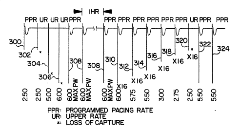
Figure 3 shows an electrocardiogram (ECG) which
illustrates an example of a loss-of-capture condition ~nd
the response (capture restoration) of the pacemaker 100
using the pulse amplitude as the selected metric. Viewing
from left to right, the first frame 300 illustrates the
presence of capture by showing a ventricular beat with the
pulse amplitude at 2.5 volts out of a pcssible 6 volts (the
maximum possible pulse amplitude programmable in this
preferred embodiment). At the next frame 302, however, 2.5
volts has become inadequate to maintain capture. The
pacemaker immediately begins pacing at the upper rate (UR)
as the first stap in the capture restoration routine or
program. The pulse amplitude is increased to 5 volts (based
on a predefined safety margin) and 6 volts for the third
(304) and fourth (306) frames, respectively (still pacing at
the upper rate), but capture has still not been restored.
-~ W094/07~67 2 1 2 3 4 0 1 PCT/US93/07822
17
The pulse width is then increased to its maximum value (2.0
mSec, in this embodiment), and capture is finally restored
at the fifth frame 308. The pacemaker switches back to the
programmed pacing rate (PPR) during that frame, and
successive frames are paced at the programmed pacing rate
with maximum pulse width and maximum amplitude for one hour
in the preferred embodiment.
In the preferred embodiment, the second and third
output pulses in the response to Loss-of-Capture are at
maximum values. Alternatively, the physician may program a
sequence of recovery pulse amplitudes and pulse widths less
than the maximum to conserve energy in the implanted device.
The pacemaker then follows a loss-of-capture recovery
routine over groups of sixteen frames (310-322) to find a
smaller, but safe pulse amplitude. The first group 310
(following the one hour timeout period) restores the pulse
width to its programmed value and continues pacing at 6.0
volts for 16 frames (provided capture is not lost). In tho
event capture is lost during any one of the 16 frames, the
w~dth is restored and the one hour timeout restarted.
Capture is monitored for each of these "recovery" frames as
the pulse amplitude is decremented in 0.25 volt steps at
each group or 16 frame interval (312-320). In the present
example, loss-of-capture again occurs at the 2.5 volt pulse
amplitude level (320), causing the pacemaker to pace (322)
at the upper rate and with a safety margin-increased pulse
amplitude (5.5 volts since capture was last determined at
2~75 volts). Since capture is then detected at 322, the
following frame 324 drops the pacing rate back to the
programmed pacing rate. The pacemaker then paces at the
programmed pulse width, the predetermined, safety margin-
increased pulse amplitude and programmed rate for a one hour
timeout period, ~ollowed again by a recovery routine. The
above steps are repeated each time loss of capture is
detected.
W094/07~7 2 1 2 3 ~ 1 PCT/US93/07822
18
Turning now to Figure 4A, an electrogram 400 is shown
as seen on the pacing electrode 102a via the electrode 106
implanted in the right ventricle of the heart 104. Figure
4B shows the right ventricular pressure wave~orm a~ seen by
pressure circuit 154 and pressure sensor S2. The pacemaker
100 measures the pulse pressure amplitude in a window 418
beginning 30 mSec after a paced or sensed event 414, and
ending 170 mSec later at 416. Peak pulse pressure is
defined as P~u (local maximum) 422 minus P~ (local minimum)
420 in the 170 mSec window 418. While this embodiment uses
a 170 mSec window for conserving energy in operating the
sensor, other window intervals could be used, including
; continuous ones.
Figure SA is a flow chart illustrating the steps used
to initialize the measurement of peak pulse pressure from
the æensor S2 of Figure 1. The measurement routine 500, as
well as all other algorithms are controlled by the
microprocessor 120 of the pacemaker 100. The pacemaker 100
starts the measurement routine 500 at 502, and at step 504,
2G determines whether the Auto Capture algorithm is ~ctivated.
If so, at step 506 the peak pulse pressure (PEAK-PRESS)
æignal is measured by subtracting the P , value from the P~
; value as measured in the 170 mSec window 418 after a paced
or sensed event. Each peak pressure value is then evaluated
at 508 to determine whether the peak pressure is less than 4
millimeters of ~ercury (mm Hg). A value less than 4 mm Hg
is discarded at step 510. A peak value equal to or greater
than 4 mm Hg is saved in a buffer (RUNNING_AVG.BUFF) at step
512. The pacemaker loO then determines at step 514 whether
the total count of the valid pressure peaks in RUNNING
AVG.BUFF is equal to 16. If it is not, then the measurement
routine 500 returns to block 504, and repeats the routine
until the count is equal to 16.
The pacemaker 100 then calculates the running average
3s peak pressure over the previous sixteen peak pressure values
:
~-~ W094/07567 2 1 2 3 ~ O 1 PCT/US93/07822
, . 19
at step 516, and the pressure threshold is calculated at
step 518, as follows:
Threshold z Average Peak Pressure Value x Programmed
Threshold
where the value of the Programmed Threshold may vary between
25 and 7S percent, and is typically 37~ percent. Once a
value for pressure threshold is available, the pacemaker 100
enables the loss-of-capture detection at step 520, and exits
this routine at step 522. The peak pressure running average
and threshold calculations continue to be updated at 2
second intervals.
Turning now to Figure SB, after the LOC Detection
circuit is activated at step 520 and tested at step 554, the
pacemaker 100 compares, at step 556, the peak pressure value
lS on a beat-by-beat basis '.o the threshold determined in step
518. If a peak pressure value is less than the threshold,
capture is determined not to have occurred for that event,
the routine moves to step 558, setting the LOC Detected to
~TRUE", exits this diagram at 560, and enters the proqram
2Q 600 of Figure ~. If on the other hand, the peak pressure
~alue is equal to or greater than the threshold at step 518,
the routine returns to step 554.
Figure 6 details the steps in the response to the loss-
of-capture program or routine 600. Beginning with step 610,
2~ the routine resets a predefined loss-of-capture timeout
counter (not shown) to "zero", and a RECOVERY flag to "off".
The timeout counter increments up to a value equating to the
one-hour timeout period. The RECOVERY flag signifies
whether the recovery subroutine 700 (in Figure 7) is to run
(RECOVERY = "on") or is not to run (RECOVERY = "off"). The
recovery subroutine 700 is inoperable during the operation
of the restoration routine 600 and during the timeout
period.
W094/07~67 2~2;3~01 20 PCI/US93/07822~
At step 612 the program determines whether the reed
switch 153 is closed, signalling that the pacemaker 100 is
currently receiving and/or transmitting telemetered signals.
If the switch is closed, the pacemaker no longer continues
the auto capture algorithm and exits the program 600 at step
636. If the reed switch is open, the program advances to
step 614, where a chosen stimulation signal metric (i.e.,
pulse width or amplitude) is compared to its maximum value.
If the maximum value of the metric has been reached, the
program jumps to step 624; otherwise, the program advances
to step 616.
If the metric is below the maximum value (from step
614) and the first loss of capture is being experienced
(determined by checking a FIRST LOSS-OF-CAPTURE flag in step
616 for a "true" condition), then the metric is incremented
by a predefined safety margin at step 618. Also, the FIRST
~OSS-OF-CAPTURE flag is set to "false". The pacing rate is
set at the upper rate at step 630 and a paced event is
scheduled at 634. Another pass through the program 600 up
to step 616 advances the program to step 622 (since the
FIRST LOSS-OF-CAPTURE f lag is set to "false") where the
metric is increased to its maximum value. $he pacing rate
remains at the upper rate (step 630), and a paced event is
scheduled at step 634.
2S Another pass through the program 600 up to 614 advances
the program to step 624, where the program determines if the
maximum value for the non-metric has been reached. For
example, if the metric is chosen to be pulse amplitude, then
the non-metric is the pulse width. If the metric (step 614)
and non-metric maximums have been achieved, the current
pacing rate is maintained at step 628 for the duration of
the timeout period. If not, the non-metric is increased to
its maximum value at step 626, the pacing rate of the
pacemaker is set equal to the upper rate via step 630 and a
paced event is scheduled at 634. Following step 628, the
~ WO 94/07567 2 1 2 3 4 0 1 PCT/US93/07822
RECOVERY flag is set to 'lon", indicating that the subroutine
700 can now begin.
Figure 7 details the recovery from loss-of-capture
subroutine 700, which attempts to lower the selected metric
over 16 frame sets in the preferred embodiment, to the value
programmed (by the physician, for example) for chronic use.
Recall that this subroutine is initiated when the recovery
flag is set to the "on" state in step 632. Subroutine 700
is the threshold-seeking portion of the present invention.
If the recovery subroutine has been initialized, the program
700 advances from step 702 to step 704. The recovery
subroutine does not continue until the timeout counter has
reached the value corresponding to the end of the timeout
period. That is, when the timeout period has expired, the
program advances to step 706; otherwise the program returns
to its beginning step 702 after first pacing at step 728.
When a paced event is detected at step 706, the program
is advanced to step 708, where the program determines
whether the reed switch is closed, signalling that the
pacemaker 100 is currently in a magnet mode. If the reed
switch 153 is closed, the pacemaker no longer continues the
recovery subroutine and returns to the beginning step 702
after pacing at step 728. If the reed switch is open, the
program advances to step 710. At that step a determination
is made as to whether the stimulation signal delivered is
the first one, by checking a FIRST STIMULI flag for a "true"
or "false" statP~ If ~o, the subroutine moves to step 712,
where the non-metric is set equal to its programmed value.
Afterwards, the FIRST STIMULI flag is set to the "false"
state at step 714, and then an event counter (not shown) is
set equal to its maximum value (16) at step 716. After the
completion of step 716 the subroutine returns to step 702
for another subroutine iteration, after first pacing at step
728.
W094/07567 2 1 2 3 4 0 1 PCT/US93/07822~-~
22
If at step 710 the first stimuli flag is "false", the
subroutine jumps to step 718, where a check is made of the
event counter. If the event counter reads "0", the
subroutine advances to step 722; otherwise the subroutine
advances to step 720, where the event counter is decremented
by "1". After step 720 the subroutine is returned to the
beginning step 702 after pacing at step 728. If the event
counter equals "0", step 722 is then executed to determine
whether the metric exceeds its programmed value. If so, the
metric is decremented by 0.25 volts (pulse amplitude) or ~0
~Sec (pulse width) to its next lowest discrete level at step
724, and the subroutine is returned to the beginning step
702 after pacing at step 728. If the metric does not exceed
its programmed value (step 722) the RECOVERY flag and hence
the recovery subroutine are switched to "off" (step 726~, in
which state they remain until the response to loss-of-
capture program 600 reactivates the subroutine. After the
completion of the subroutine (step 730), tLe pacemaker 100
returns to the beginning of the measurement routine 500,
explained supra . with respect to Figure 5, to restart the
pressure measurement, capture restoration and threshold-
seeking routines, as needed.
Variations and modifications to the present invention
are possible given the above disclosure. However, such
~5 variations and modifications are intended to be within the
scope of the invention claimed by this letters patent.