Note: Descriptions are shown in the official language in which they were submitted.
2123536
Field of the Invention
This invention relates in general to medical
diagnostic equipment, and more particularly to an
ultrasonic blood volume flow rate meter using transverse
colour Doppler ultrasound.
Background of the Invention
Non-invasive Doppler sonography is widely accepted
as a means of measuring blood velocity. However, in some
situations, the volume flow rate of blood may be a better
indicator of the state of disease. One potential
application of volume flow measurements is the prediction
of stenosis of both common and internal carotid arteries
by monitoring common carotid blood flow. In principle,
an adequate degree of stenosis (generally accepted as 50%
diameter reduction), results in a measurably decreased
volume flow. Investigators have attempted to quantify
this volume flow reduction using different sonographic
techniques, with varying degrees of success. Some
authorities have suggested that the ratio of flows of the
unaffected and stenotic carotid arteries is the best
predictor of carotid stenosis. Volume flow measurements
have also been suggested as a technique for short- and
long-term follow-up of carotid endarterectomy.
Volume flow measurements may also be applied in the
diagnosis and treatment of vascular malformations.
Specifically, the measurement of volume flow may help to
distinguish arteriovenous malformations and fistulae,
which are high-flow lesions, from venous malformations,
which are low-flow lesions. Moreover, volume flow
measurements provide a quantitative way both of assessing
blood steals and of evaluating the effectiveness of
embolization therapy. Renal dialysis patients may also
benefit from Doppler volume flow measurements. Either
inadequate or excessive flow through angioaccess fistulae
can have pernicious clinical consequences. Doppler
sonography has been suggested as a way of quantifying
this flow.
2123536
y,
2
Several techniques have been developed to estimate
blood volume flow from Doppler velocity measurements,
each being characterized by certain advantages and
disadvantages. Generally, in order to estimate the
volume flow rate of blood through an artery, pulsed
Doppler ultrasound is used to measure the velocity of the
blood. From this velocity measurement, and a measurement
of the diameter of the vessel, an estimate of the volume
of blood flowing through the vessel may be made. This
volume flow estimation technique assumes a parabolic
blood velocity profile, and assumes a circular vessel.
Other techniques, such as colour M-mode, directly measure
the one-dimensional velocity profile, but still assume a
circular artery. Still other techniques exist, (e. g.
those using uniform insonation of a vessel), but are also
prone to measurement uncertainties.
Thus, it is known in the art to approximate blood
velocity measurements across an entire blood vessel lumen
by using only a single-point velocity measurement from a
conventional clinical ultrasound instrument at the centre
of the vessel and assuming a parabolic velocity profile.
Previous attempts to measure blood flow from two-
dimensional velocity profiles have proven to be
inaccurate because of the difficulty in determining
Doppler angle (see Akira Kitabatake "Quantitative Color
Flow Imaging to Measure the Two-Dimensional Distribution
of Blood Flow Velocity and the Flow Rate", Japanese
Circulation Journal, Vol. 54 March 1990.)
Summary of the Invention
According to the present invention, a clinical
colour Doppler ultrasound instrument is utilized with a
position and orientation sensing device, and a computer
with video digitizer to acquire blood velocity
measurements in two dimensions across an entire blood
vessel lumen. The blood velocity profile measured using
the system of the present invention permits the precise
determination of the volume flow rate of blood through
CA 02123536 2003-05-27
3
the vessel.
The use of a position and orientation measurement
device permits the accurate determination of Doppler
angle required to make an accurate volume flow
measurement, thereby removing the vessel circularity
assumption of Kitabatake et al, and its attendant errors.
The volume flow rate is determined according to the
present invention by integrating the two-dimensional
velocity profile over the vessel lumen area. The volume
flow rate measurements are then displayed to an operator
in real time as a scrolling graph, and in terms of cycle-
to-cycle volume flow in mL/min.
In accordance with an aspect of the present
invention there is a volume flow meter for measuring
and measuring and displaying volume flow through a
vessel having an axis, which comprises:
a) an ultrasound instrument with scan head adapted
to be positioned adjacent the vessel, for generating a
ao
raster of pixels~which defines a colour image
representing flow velocities in the vessel through an
image plane of the scan head;
b) sensor'means connected to the scan head for
measuring position and orientation of the scan head in
three dimensions and generating a signal
representative thereof;
c) computer means connected to the ultrasound
raster of pixels and the signal representative of
instrument and the sensor means for receiving the
position and orientation of the scan head for
determining position and orientation of the axis of
the vessel in three-dimensions responsive to
orientation of the image plane longitudinally of the
vessel, determining an angle 0 between the axis and
CA 02123536 2003-05-27
3a
the image plane responsive to orientation of the image
plane transversally to the vessel, and calculating and
displaying the volume flow measurement as a summation
of the flow velocities scaled by the tangent of the
angle 0.
A benefit of this transverse-image based approach is
that, unlike prior art single-point or colour M-mode
techniques, the volume flow estimate is not as sensitive
to the positioning of the ultrasound transducer. This
useful property arises because the flow is properly
recorded using the system of the present invention for
blood vessels appearing anywhere within the active
'colour',area of the ultrasound image. This removes one
aspect of operator-induced variability in the blood flow
estimate, and permits long-duration flow studies to be
performed without the need to continually monitor
transducer positioning.
Brief Description of the Drawinvs
Details of the present invention are provided herein
below with reference to the following drawings, in which:
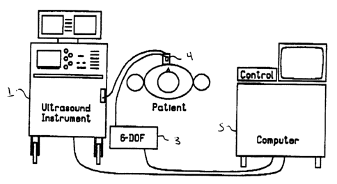
Figure 1 is a schematic representation of an
ultrasonic blood volume flow rate meter according to the
present invention;
Figure 2 shows the position of an ultrasound scan
head and position of a line cursor according to the
method of using the system of the present invention;
Figure 3 shows the.scan head of Figure 2 oriented at
an angle to the axis of the blood vessel according to the
method of using the system of the present invention;
Figure 4 is a graph showing volume blood flow rate --.~
2123536
4
measured in a human carotid artery over a single cardiac
cycle; and
Figures 5a, 5b, 5c and 5d represent a blood volume
flow record obtained at various times during a femoral
artery angioplasty procedure.
npta>>ed Descr~pt~on of the Preferred Embodiment
Figure 1 shows the components of the invention, and
their arrangement. A clinical diagnostic ultrasound
imaging instrument 1 generates ultrasound images which
are then used for measuring blood velocity virtually
simultaneously over large areas in these images.
Commercially available colour Doppler ultrasound
instruments may be used for this purpose (eg. ATL
Ultramark 9). In such well known systems, ultrasound
images are generated in which human anatomy (eg. vessel
wall, fat, etc.) is represented by black and white images
(with various intermediate shades of grey), while
different velocities of blood flow are represented by
different colours.
A position and orientation sensing device 3 is
connected to the ultrasound instrument 1. The sensing
device 3 comprises a transmitter positioned at a fixed
location near the patient, and a receiver mounted on the
ultrasound instrument scan head 4. One suitable position
and orientation sensing device is the Flock of Birds six-
degree-of-freedom measuring device manufactured by
Ascension Technology Corporation of Burlington, Vermont.
In this device, the transmitter generates a pulsed DC
magnetic field, and the receiver (comprising three
orthogonal coils), detects the magnetic field generated
by the transmitter and senses both the location of the
receiver in three-dimensions, as well as its orientation
relative to the transmitter. The location is measured in
terms of X, Y and Z positional coordinates of the
receiver with respect to the transmitter, while the
orientation angles are defined in terms of rotations
about the Z, Y and X axes of the receiver. These angles
2123536
are referred to as azimuth, elevation and roll in Euler
angle nomenclature.
A computer 5 (eg. 80386-based PC), with added
commercially available digitizer (eg. Vision 16 Frame
5 Grabber manufactured by Vision Technologies of Fremont,
California), custom-written software, monitor, and
operator controls, is connected to the ultrasound
instrument 1 and sensing device 3. The location and
orientation data generated by the sensing device 3 may be
transmitted digitally from the receiver to the computer 5
via either a full duplex RS232C interface or a half
duplex RS422/485 interface, in a well known manner.
In operation, the ultrasound instrument operator
locates a blood vessel of interest in a patient while
viewing the colour Doppler ultrasound image on the
computer monitor, and positions the scan head 4 so as to
produce a longitudinal image of the vessel on the
monitor, that is, an image with the blood vessel axis
within the plane of the image, as shown in Figure 2.
This image is termed the "landmark". The operator then
manually positions a line cursor on a blood vessel in the
image. The sensing device 3 affixed to the ultrasound
instrument scan head 4, continually reports to the
computer 5 the position and orientation of the scan head.
Using the location and orientation of the scan head
measured by the sensor 3, and the location and
orientation of the vessel axis image in the two-
dimensional image plane as given by cursor location
chosen by the operator, the computer 5 calculates the
location of the axis of the blood vessel in three-
dimensional space.
Next, the operator rotates the scan head 4 on the
patient s skin surface to produce a transverse image of
the blood vessel, that is, an image with the blood vessel
axis passing through the image plane at an angle referred
to herein as the "Doppler angle", as shown in Figure 3.
This position of the scan head 4 allows the determination
2123536
6
of velocities across the complete two-dimensional cross-
section of the blood vessel, and the simultaneous
measurement of the functional cross-sectional area of the
vessel (i.e. the area in which the measured velocities
are non-zero).
The sensor 3 continues to communicate to the
computer 5 the location and orientation of the scan head
4. The computer 5 continually calculates the angle
between the image plane and the blood vessel axis (i.e.
the "Doppler angle") using transverse image geometry.
The computer 5 uses the measured Doppler angle, velocity
measurements in the blood vessel being made by the
ultrasound instrument 1, and the functional cross-
sectional area of the vessel, to calculate the volume of
blood passing through the ultrasound image plane. This
calculation is performed approximately 10 to 30 times per
second, depending on the speed of the ultrasound
instrument 1.
The volume flow, Q, through the artery (assuming
axial flow) is given by the flow though the plane, and is
equal to the sum of the entire velocity profile cutting
through the plane:
npis
Q=tans ~ V;DA
2 5 t=o
where a designates the Doppler angle, and is given by the
angle between the ultrasound image plane and the vessel
axis; V; is the measured Doppler velocity at each pixel
obtained from the digitized colour (ie. the internal
digitizer in computer 5 digitizes the Red-Green-Blue
(RGB) colour video output from ultrasound instrument 1
and converts the colour image to a two-dimensional
velocity map); DA is the pixel area in the image plane;
and, npix is the number of pixels with colour.
The resulting two-dimensional velocity profile per
image is scaled by the pixel size, tangent of the Doppler
angle, and image period (ie. frame rate) to yield a
2123536
7
volume flow graph and numerical value which are presented
to the operator via the computer monitor (or via a
scrolling print-out) in real time, as discussed in
greater detail below with reference to Figures 4 and 5.
The measured Doppler velocity V; at each pixel can be
calculated using known velocity calibration techniques
such as disclosed in Rickey, D. W. and Fenster, A., "A
Velocity Evaluation Phantom for Colour and Pulsed Doppler
Instruments", Ultrasound Med. Biol., 18:479-494, 1992.
Another technique is to assume that individual colours
represent respective velocities based on the technical
specifications of the clinical ultrasound instrument 1.
The computer 5 is then able to calculate individual
velocities by accessing an internal look-up table which
associates the colours appearing in the ultrasound image
with the calibrated velocities. This calculation is
performed on a pixel-by-pixel basis from the colour image
raster received from the ultrasound instrument 1.
This volume flow determination is performed at the
same rate as the image acquisition on the ultrasound
instrument 1, (i.e. 10 to 30 measurements per second).
The volume flow information is presented to the operator
on the monitor or in hard copy in a well known fashion,
namely in a flow-rate versus time graph, as well as a
numerical result, in millilitres per minute, or
millilitres per cardiac cycle.
The flow rate may be measured at 10 to 30 times per
second for as long as desired, making possible long
duration monitoring of flow, for example, during surgery
or stress testing.
The apparent size of the blood vessel being imaged
changes depending on the amount of acoustic power
received from it, due to the sensitivity profile of the
ultrasound transducer array of scan head 4. Higher
returned acoustic power causes an apparent spreading and
increase in size of the vessel being imaged, and thus an
artefactual increase in the measured volume flow rate.
2123536
8
Also, intervening tissue or fat can decrease the received
power, and therefore decrease the measured flow. For
consistent volume flow measurements, then, it is
important to adjust the output power or colour gain of
the ultrasound instrument 1 to fix the average returned
power from the vessel of interest at a predetermined
value. For the purposes of these measurements, the
colour gain and output power controls can be considered
equivalent over most of the power range that is
encountered. To measure the returned power from a blood
vessel, the operator temporarily places the instrument 1
in power-measurement mode. The colour power image
produced by the instrument 1 in this mode is measured by
the computer 5 by a colour-to-power mapping technique
similar to the velocity measurement technique described
above. The computer 5 measures the returned power and
displays the value in bar-graph form. The operator then
manually adjusts the acoustic power or colour gain to
place the bar-graph into a suitable target zone. The
power level of the target zone can be determined using
in-vitro tests. The operator then places the ultrasound
instrument 1 in velocity-measurement mode to continue
with the flow measurement.
Figure 4 shows an example of the volume flow rate
measurement in a human carotid artery. Selected measured
two-dimensional velocity profiles are shown at the top of
Figure 4, and the series of volume flows computed from a
series of these velocity profiles form the ensemble shown
in the graph. In the example shown, there were twenty
four measurements made over a single cardiac cycle
lasting one second. The units appearing on the time axis
are in milliseconds post 'R'-wave of the ECG. The
integral flow through this cycle was 5.6 mL. The heart
rate at the time was 60 beats per minute. The volume
flow rate was thus 336 mL/min. The graph shows both the
variation in flow rate over the cycle, as well as the net
volume flow during that cycle. According to the present
21~353fi
9
invention, the length of a sequence is limited only by
storage memory capacity of the host computer 5. Thus,
several-hour runs are possible for long-duration studies.
The maximum sample rate is governed by the video frame
rate (eg. 30 frames per second in the configuration of
the preferred embodiment).
Figures 5a through 5d show in-vivo volume flow
measurement of a patient during femoral angioplasty
procedures, using the device of the present invention.
Each volume flow measurement consisted of two 15-second
flow recordings (for the determination of the true
Doppler angle and true volume flow), which resulted in a
total of two additional minutes to the normal procedure
time. Figures 5a and 5b show the results of measurements
obtained immediately proximal to a stenosis prior to the
angioplasty. Figure 5c shows an increase in resting flow
two minutes post-angioplasty, with injection of
nitroglycerin. After a resting period, flow returned to
pre-operative levels as shown in Figure 5d.
In summary, the volume flow measurement technique of
the present invention removes assumptions about vessel
circularity and is not sensitive to simple changes in
position of the scan head 4, unlike prior art single-
point Doppler flow measurement systems. Since the two-
dimensional velocity profile is directly measured, the
system of the present invention also removes assumptions
of velocity profile shape (typically assumed in the prior
art to be parabolic). However, this volume-flow
measurement approach makes several implicit assumptions.
It assumes that all the flow streamlines in a vessel at
the measurement site are parallel, so that they all have
the same Doppler angle. This implies that a relatively
straight vessel is required, that the flow is neither
converging or diverging significantly, and that there be
little turbulence. Post-stenotic measurements, and
measurements in highly diseased parts of vessels may be
inaccurate due to unknown and time-varying Doppler
2123536
angles. It is also assumed that the ultrasound sample
volumes (voxels) are small compared to the vessel, so
that the vessel is sampled at several locations across
its diameter. To measure accurately pulsatile flows, the
5 ultrasound instrument 1 must maintain a frame rate which
is sufficient to record rapidly-changing blood
velocities. We have measured the power spectrum of
pulsatile flow in healthy volunteers, and found that a
sample rate of 24 frames per second is sufficient to
10 adequately sample the velocities. From the constraints
described herein, we have concluded that the system of
the present invention is best suited to blood flow
measurements in substantially straight vessels, such as
the carotid artery.
Alternatives, modifications and further applications
of the invention are possible. For example, although the
preferred embodiment of the invention has been described
in relation to sensing blood volume flow in humans. The
principles of this invention may be applied to blood
volume flow in animals, and may even be applied to the
sensing of liquid flow in industrial processes (ie. non-
medical application). These and other embodiments and
applications of the invention are possible within the
sphere and scope of the invention as defined by the
claims appended hereto.