Note: Descriptions are shown in the official language in which they were submitted.
212 4 (l74
~''~VO 93/10447 1 PCT/US92/10187
DESCRIPTION
ULTRASONIC HOLOGRAPHIC IMAGING APPARATUS
HAVINI: ZOOM FEATURE
Technical Field
s This invention. relates to ultrasonic holographic imaging apparatus for
viewing imaged hologr:~phic information of the internal structure of an object
at
different magnifications.
BackEround Art
Although commercial application of ultrasonic holography as been accurately
to pursued by many persons in the scientific and industrial communities for
many
years, only limited results have been obtained even though it was once thought
that ultrasonic holography held great promise. It was felt that the
application
of ultrasonic holography was particularly applicable to the fields of
nondestructive
testing of materials and medical diagnostics of soft tissues that are
relatively
1s transparent to ultrasonic radiation. One of the principal problems that has
been
encountered and not effectively resolved is the difficulty of obtaining
quality and
consistent images. A further problem has been the inability to vary the
magnification of the image while maintaining a consistent high quality image.
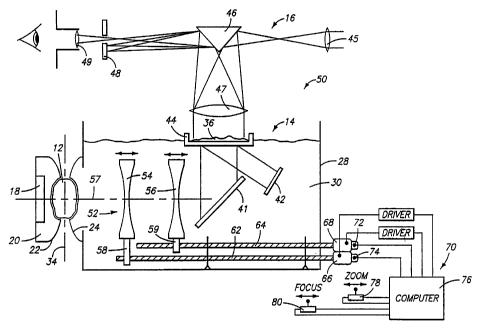
As background, Fig. 1 shows a typical "real time" prior art ultrasonic
20 holographic imaging system generally designated with the numeral 10. The
system l0 is intended to ultrasonically inspect the interior of an object 12
such
as the soft tissue of a human limb. The ultrasonic holographic imaging
system 10 generally has a hologram generating subsystem 14 for generating an
ultrasonic hologram. The system 10 also includes a hologram viewing subsystem
2s ' ~ (optical-subsystem) 16 for optically viewing the interior of the object
12 from a
Fret order diffraction from the formed ultrasonic hologram.
The subsystem 14 includes an object ultrasonic transducer 18 for
generating plane waves through a ~ liquid or gel coupling medium 20 contained
in a deformable membrane 22. The deformable membrane 22 intimately contacts
3o the object 12 on one side and a deformable membrane 24 contacts the object
on the other side to provide ultrasonic coupling with minimum energy loss or
wave distortion. The deformable membrane 24 forms part of the side wall of
a container 28 that contains a liquid coupling medium 30.
2~2'~~'~ 4
WO 93/10447 2 PCT/US92/10187t'; ; ,fi
One of the principal components and the main concern of this invention
is the provision of an ultrasonic imaging lens system 32 for viewing a large
field
and focusing at a desired object focal plane 34. The prior art ultrasonic
imaging lens system 32 focuses the ultrasonic energy onto a hologram detector
s surface 36. The ultrasonic imaging lens system 32 includes a large diameter
object lens 38 that is moveable with respect to a large diameter lens 40 far
moving the lens to different desired focal planes 34 in the object 12. The
lens 40 is stationary and is positioned at a fixed focal length from the
detector
surface 36. The ultrasonic imaging lens system 32 includes a mirror 41 for
1o reflecting the ultrasonic energy approximately 90° and onto the
hologram
detection surface 36 to farm the hologram.
A ultrasonic reference transducer 42 directs coherent ultrasonic plane
waves through the liquid medium 30 at an off-axis angle to the hologram
detector surface 36 to Form the hologram. Preferably, the hologram detection
Is surface 36 is the liquid/gas interface surface that is supported in an
isolated dish
or mini-tank 44.
The hologram viewing subsystem 16 includes an optical lens 45 to achieve
an effective point source of a coherent light beam from a laser (not shown).
The focused coherent light is reflect from a mirror 46 through a collimating
20 optical lens 47 and then onto the hologram detector surface 36 to
illuminate the
hologram and generate diffracted optical images. The reflected coherent light
radiation containing holographic information is directed back through the
collimating lens 47 and separated into precisely defined diffracted orders in
the
focal plane of the collimating lens 47. A filter 48 is used to block all but a
2s first diffracted order from a viewing lens 49 to enable a human eye, a
photographic film or a video camera to record in "real time" the object at the
object focal plane. As previously mentioned, although such a system is
operable,
it has been difficult to obtain quality and consistent images.
A prior art ultrasonic lens system, similar to that described, is presented
3o in U.S. Patent No. 3,802,533 entitled "Improvements In and Relating To
Ultrasonic Lenses" granted to Byron B. Brendan. Such patent is principally
directed to the specific- structure of the ultrasonic lenses.
One of the principal objects and advantages of this invention is to provide,
an improved ultrasonic holographic imaging apparatus that has the ability to
~ 212~~'~4
O 93/10447 3 PCf/US92/10187
change the magnification of the image relative to the object as well as change
the focus to different planes in the object while maintaining consistent high
quality images.
These and other objects and advantages of this invention will become
s apparent upon reading the following detailed description of a preferred
embodiment. ,
,..
Briet Description of the Druwinas
Preferred embodiments of the invention are described below with reference
o to the accompanying drawings, which are briefly described below.
Fig. 1 is a schematic side elevational view of a prior art ultrasonic
holographic imaging system illustrating the use of an ultrasonic lens system
in an
ultrasonic fluid transmitting medium for imaging ultrasonic holographic
information
to form a focused ultrasonic hologram; .
IS Fig. 2 is a schematic side elevational view of a preferred embodiment of
this invention showing an improved ultrasonic holographic imaging apparatus
capable of providing an ultrasonic hologram of different degrees of
magnification
as well as focusing at different planes within the volume of the object;
Fig. 3 is a schematic side elevational view of an ultrasound lens system
2o within the apparatus as illustrated in Fig. 2 showing the spatial
relationships and
distances between the magnification lens and the focusing plane and the
imaging
plane.
Best Modes for Carr,~ing Out the Invention
25 ~A preferred embodiment of the ultrasonic holographic imaging apparatus
is illustrated in Fig. 2 and is designated generally with the numeral 50.
Elements ~ than are common include the same identifying numerals as in Fig. 1.
In the preferred embodiment, the apparatus SO includes a multiple lens system
designated with the numeral S2 that includes two ultrasonic converging lens 54
3o and S6 that are aligned along an optical axis S7. The multiple lens system
S2
includes a lens support S8 for supporting the lens S4 and lens support 60 for
independently supporting the lens SG. Preferably, lens supports S8 and S9
include lead screws 62 and 64 respectively. The apparatus SO includes lens
support drives 66 and 68 that are connected to the lead screws 62 and 64
212!474
WO 93/10447 4 PGT/US92/10187 ~~: ,..'
' respectively for rotating the lead screws in either a forward or back
rotation to
precisely and accurately move the lens 54 and 56 relative to each other.
The ultrasonic holographic imaging apparatus 50 includes a control system
generally designated with the numeral 70 that includes lead screw encoders 72
,
s and 74 for generating coding signals that are sent to a computer or
microcontroller 76 that controls the position of the texts very accurately in
response to input signals from operative control devices 78 and 80. The
operator control device 78 is specifically provided for providing zoom signals
to
the computer to cause the computer to move both of the lens 54 and 56
1o relative to each other and in unison to obtain the desired magnification.
Preferably the magnification varies between 0.25 and 4. The operator control
device 80 inputs desired signals to move principally the lens 54 to and from
the
. object 12 to focus at desired focal planes within the object to visually
inspect
the volume of the object. The applicant has found that such an ultrasonic
is holographic imaging apparatus 50 is quite capable in providing consistent
and
quality images at desired magnifications as well as being able to accurately
focus
the lens system within the volume of the object 12 to view different
structures
within the object 12.
The focal object plane 34 of the multiple lens system, within the object
20 12 is the distance "O" and is known or selected by the operator..
Similarly, the
magnification of the image is selected by the operator. The magnification
value
is designated as "M" and given by the formula (1):
. (1) M _- h1 1211(0t 02l
Such values are ' input to the computer 76 through the input devices 78
25 and 80. The computer 76 is programmed to receive the input values and to
calculate the proper positions LI and L2 for the lens 54 and 56.
s. ' . ,, ~. , ~.;. " ~~. ~..". .'. . ,.. ,' ... _'~.., A '..::'._ ,~', f
....., .' ..,,';~ :. ~:. .,' ' '. ~.. ;.;.. ... ,.,, ..~ ' ~
v. .... ~ ..., . . t n. . , v .. .... .., ..n. , n.. .: . . . ...
2124474.:. .
'~ ' n~ i~VO 93/10447 5 , '. ~ wpCT/US92/10187
The positions of lens 54, L~, and 56; LZ are calculated using the input
values of "O" and "M" and the known focal lengths of lens 54 and 56, defined
as F~ and F2 respectively, using the formulas (1) and (2):
when a = I (2) Lt = (-bt b2 - 4ac) /2a
b = -I(~F2) l (MFZ + Fl)II~ - Ft + FtlM + OFl/~FZI
a = IMF2/ (MFZ + Ft )lI~ - Ft + F1IMI2 + ~'(~2I (MFZ + Ft)
(3) L1 = (MF2/F~) (O - Lt - Ft) + Fz
s
Such a system significantly increases the ability of an operator to obsSrve
the internal structure of an object, such as soft tissue of a human limb, at
~o varying magnifications to make a proper medical evaluation.
The invention has been described in language more or less specific as to
methodical features. It is to be understood, however, that the invention is
not
limited to the specific features described, since the means herein disclosed
comprise preferred forms of putting the invention into effect. The invention
is,
1s therefore, claimed in any of its forms or modifications within the proper
scope
of the appended claims appropriately interpreted in accordance with the
doctrine
of equivalents.