Note: Descriptions are shown in the official language in which they were submitted.
WO 93/11828 PCT/US92/10367
2124822
-1-
MEDICAL VALVE.
Background of the Invention
Field of The Invention
This invention relates to a closed, patient access system which automatically
reseals after administering medication using a standard medical implement that
directly
connects with the system without the need of any intermediary needles, caps or
adaptors. A two-way valve eliminating dead space is used which includes a seal
which,
upon being compressed by the medical implement, is pierced to open the valve
and
reseals upon being decompressed, maintairiing a fluid tight seal even at high
pressures
and after repeated uses.
Backaround Discussion
The manipulation of fluids for parenteral administration in hospital and
medical
settings routinely involves the use of connectors and adaptors for
facilitating the
movement of fluids between two points. Most fluid connectors and adaptors
employ
needles to pierce a septum covering sterile tubing or to pierce the septum of
a
medicament container of fluid. Fluid then passes from the container or fluid
filled tubing
into a syringe or second set of tubing. These connectors and adaptors often
have
mechanical or moving parts. Since the ready passage of fluids through the
connectors
and adaptors is often critical to patient survival, it is imperative that the
connectors and
adaptors function reliably and repeatedly. Adaptors and connectors that
malfunction
during use may be tife-threatening. The more mechanical or moving parts such
as
springs and diaphragms, the more likely that they will function improperly.
Improper
functioning can result in the introduction of air embolisms into a patient.
Thus, the fewer
the mechanical parts, the more these connectors can be relied on and the
better they will
be accepted by the medical community.
Many connectors or valves, especially those employing several mechanical
components, have a relatively high volume of fluid space within them. This
"dead space"
within the device prevents accurate introduction of precise fluid volumes and
provides an
opportunity for contamination upon disconnection of the device. Connectors and
adaptors often include valves that permit or interrupt the flow of fluid along
the course of
fluid travel. Several of those commonly in use employ metal needles to
puncture sterile
seals. Such connectors are generally designed to accommodate fluid flow in one
direction. This means that the fluid line must have connectors and tube
aligned in
complementary directions. These connectors often require further manipulation
if, for
WO 93/11828 PCT/US92/10367
2124822
-2-
example, the valve Is inadvertently assembled in a direction that will not
facilitate fluid
flow. These manipulations increase handling, thereby increasing both the risk
of
contamination and the amount of time required to establish the fluid
connection.
Metal needles employed as part of connector devices increase the risk of
puncture
wounds to the user. The needles used in these devices often have through-holes
placed
at the tip of the needle. Connection of the valve with a flow line involves
piercing the
needle through a sealed septum. Through-holes placed at the needle tip can
core the
septum and release free particulates into the flow line. Such an event can
prove fatal to
a patient. Such through-holes may also become clogged easily with material
from the
septum.
Reusable connectors and adaptors are preferred for medical applications since
components must often be added or removed from a fluid line connected to a
patient.
Reusable connectors, however, are difficult to keep sterile. Sometimes caps
are
employed to cover the connector to keep it sterile. Frequently, these caps are
lost, or
simply not used because they are not readily available when needed.
A dosed, patient access system that is easy to use and employs only a valve
device in communication with the patient that need not be capped or
interconnected with
the medical implement through a needle or adaptor, is swabbable, is
sufficientiy durable
to maintain its function after several manipulations, and maintains a fiuid
tight seal at high
pressures, would be of great benefit to the medical community.
Summary of the Invention
The valve of this invention has several features, no single one of which is
solely
responsible for its desirable attributes. Without limiting the scope of this
invention as
expressed by the claims which follow, its more prominent features wiil now be
discussed
briefly. After considering this discussion, and particularly after reading the
section
entitled, "DETAILED DESCRIPTION OF THE PREFERRED EMBODIMENTS," one will
understand how the features of this invention provide its advantages, which
include
safety, reliable and repeatable performance, elimination of dead space,
simplicity of
manufacture and use, and employment of a valve that is swabbable after use to
provide
sterility and has a fluid-tight seal at high pressure.
This invention is a closed, patient access system which automatically reseals
after
administering medication using a medical implement that directly connects with
the
system without the need of any intermediate needles, caps or adaptors. A two-
way valve
is employed utilizing a reusable seal that may be repeatedly pierced by an
enclosed,
WO 93/11828 212A Q22 PCT/US92/10367
-3-
protected, non-metallic spike rather than an exposed metal needle. The valve
facilitates
fluid, particularly liquid, transfer while maintaining sterility. The valve is
easy to use and
is capable of locking in place. After use, the valve Is swabbed in the
conventional
manner with a suitabie substance to maintain sterility. The design of the
valve avoids
accidental needle sticks. As will be discussed in detail below, the valve is
useful as a
medical connector or adaptor to enable liquid flow from a seaied container.
The first feature of this invention is that the valve has a body including
wall
structure defining an internal cavity having a proximal end and a distal end.
The cavity
has an open space into which the seal is pushed, and preferably has a
plurality of radial
indentations in the wall structure that are adjacent the seal to accommodate
the
expansion of the seal upon compression. The proximal end has an opening
sufficiently
large to receive a delivery end of a medical implement which transfers fluid
through the
delivery end. In most applications, the delivery end of the implement is
tapered, and the
wall structure adjacent the opening is tapered inward so that the wall
structure and the
tapered delivery end fit snug against each other upon insertion of the
delivery end into
the opening. The proximal end of the cavity preferably is adapted to fit snug
with an
ANSI (American National Standards Institute, Washington, D. C.) standard end
of the
medical implement. Typically, the implement is a syringe, a connector or
inlet/outlet of
an IV set, or any one of a wide variety of conduits used in medical
applications.
The second feature is that the spike has a tip with at least one hole located
at or
near the tip, and a passageway in communication with the hole that allows
fluid to flow
through this hole. The spike is seated inside the cavity such that the tip is
inward of the
proximal end and is enclosed within the cavity. Preferably, the hole is in a
side of the
spike adjacent the tip and is elongated, having a size of 18 gauge or greater.
The tip
may be sharp or slightly rounded. More than one hole is desirable for many
applications,
and three, symmetrically located holes inward of the proximal end are
preferred. The
spike may include at least one rib which allows air to enter a space between
the seal and
the spike, thereby facilitating the sealing of the opening when the implement
ii removed.
The spike may have a substantially conical shape, and the seal has a
complementarily,
substantially conical shaped cavity within it conforming to the shape of the
spike. The
spike is disposed within this conical cavity and the seal covers the tip. The
tip may be
imbedded in the proximal end of the seal or withdrawn into the conical cavity.
Preferably,
the tip of the spike has a plurality of facets which meet within a recess. The
preferred
spike should be able to penetrate the seal repeatedly without tearing the
seal. Rough
WO 93/11828 PCT/US92/10367
21248~2
-4-
edges at the tip may present a tear problem. During injection molding of the
preferred
plastic spike, facets of the tip will abut along a"parting iine," and could
form a rough
edge which may tear the seal. This problem is avoided where the parting line
is buried
in a recess. Any rough edge at this parting line is disposed within a recess,
so the seal
material moves over the recess and does not contact the rough edge.
The third feature is that the resilient seal is adapted to be moved into a
compressed state upon insertion of the tip of the medical implement into the
opening and
returns to a decompressed state upon removal of the tip. The seal in the
decompressed
state has a section which fills essentially completely a portion of the cavity
adjacent the
opening. The seal section bears against the wall structure near the opening to
seal the
opening. In the compressed state, the seal section is pushed by the delivery
end of the
medical implement away from the opening and into the cavity. A fluid tight
seal is
maintained between the seal section and the wall structure as the seal is
moved into the
compressed state. The seal section bears against the wall structure as the
seal is moved
inward into the cavity by the tip of the medical implement. And most
importantly, the
delivery end and the seal are adapted to engage so that when the tip of the
spike pierces
the seal there is essentially no dead space between said delivery end and the
seal.
Consequently, a predetermined dosage amount of medication is transferred in
its entirety
to the patient.using this invention, with none to the prescribed amount being
collected
in dead space in the valve. The delivery of an exact amount of medication may
be critical
in some situations when chemotherapeutic agents are being administered or
small
children are being treated.
A fluid tight seal is maintained over repeated opening and closing of the
valve,
and the seal has on its extemal surface a recess which provides an air pocket
to facilitate
the movement of the seal. Preferably, the seal presents an essentially flush
surface with
the proximal end of the cavity. In one embodiment, the proximal end of the
seal is
substantially flat, the seal is made of a material having a hardness of from
30 to 70 Shore
units such as, for example, a silicone polymer. The seal may include a
cup=like flange
adapted to engage the body near the proximal end of the cavity. A. preferred
embodiment of the.seal comprises a series of 0-ring elements stacked together
and
connected to form a unitary structure. The 0-ring elements have increasing
diameters,
with the smallest diameter element being adjacent the proximal end of the
cavity. The
proximal end of the seal may be precut to form a tiny orifice therein that
allows the tip of
= the spike to pass therethrough easily upon compression of the seal.
Preferably, the
WO 93/11828 2124822 PCT/US92/10367
.5-
proximai end of the seal has a truncated conical shaped segment dispm i within
the
cavity. The seal may also have a centrally located, anti-vacuum, saucer like
depression
therein, which does not interfere with the ability of the exposed, proximal
end of the seal
being swabbed when desired.
The fourth feature is that the body and spike are two separate components of
the
valve that are securely attached to each other by assembly of, and
interlocking, of the
body and spike. The body has a first locking element near the distal end of
the cavity,
and the spike has a second locking element adapted to interlock with said
first locking
element upon assembly. The seal has a!ip extending beyond the distal end and
positioned between the first and second locking elements so that, upon
assembly, the
lip is compressed between the locking elements to provide an essentially fluid
tight seai
upon interlocking.
The fifth feature is that the medical valve includes a support member
connected
to the spike which seals off the distai end of the cavity. The support member
may have
a Luer-Lock type connector element that enables the valve to be removably
attached to,
for example, a fluid line connected to a patient. The support member may also
be in the
form of an adaptor that enables the valve to be removably attached to a fluid
dispenser
or container. When used to dispense fluids from a container, the spike has a
pair of
opposed tips, respectively at the distal and proximal ends of the spike. The
tip at the
distal end of the spike pierces a cover member which seals the container. A
radial slit
on the adaptor enables it to deform reversibly sufficiently to fit snugly onto
said container.
The sixth feature is that the seal has a proximal end including a pressure
responsive element disposed on an inner surface of the seal adjacent the
opening. The
pressure responsive element in the decompressed state closes any orifice in
the seal at
the proximal end of the seal to provide an essentially fiuid-tight seal while
in the
decompressed state. The pressure responsive element enables the valve to
maintain a
fluid-tight seal even at very high pressures sometimes experienced in medical
applications, particularly when the valve is connected to a patient's artery.
The valve of
this invention wiil remain closed even when the pressure inside the valve is
above 6
pounds per squar - 1ch (psi), and it can withstand pressures above 30 psi.
Typically, the
pressure responsive element is a section of the seal having an entryway into a
precut
orrfice. This section has a substantially cylindrical configuration and is
surrounded by
an annular space which is filled with pressurized ituid. The center of the
member and
the annular space are coaxial with the entryway to the orifice. The
pressurized fluid fills
WO 93/11828 PC,'i'/US92/10367
2124822
-6-
the annular space to apply pressure that compresses the cylindrical section to
tightly
close the entryway to the orifice. Preferably, the pressure responsive element
has an
anti-tear element.
In accordance with this invention, a known, prescribed, predetermined amount
or dosage of medication may be transferred from the remote source to the
patient
directly, so that essentially none of said predetermined amount is collected
in dead space
in the valve. In other words essentially all the prescribed dosage is receive
by the patient
and not lost in the vaive. Thus, this invention also includes a method of
transferring fluid
from a remote source to a patient. This invention also includes transfer of
fluid from the
patient to a remote source. This is possible because the valve of this
invention provides
two-way communication. The fluid is transferred to the patient by applying
pressure
the fluid as it passes through the implement so that the pressure applied to
the fluid is
greater than the pressure of fluid in the patient, enabling transfer from the
remote source
to the patient. To achieve transfer of fluid from the patient to the remote
source, the
pressure of fluid in the patient is greater than the pressure at ths remote
source, causing
fluid to flow from the patient to the remote source. This inventiorl also
includes a method
of transferring fluid in a container having an open mouth covered by a cover
member
which seals the open mouth. The fluid is caused to flow from the container
through the
passageway by creating a differential in pressure. Preferably, the valve has
an adaptor
having a radial slit for allowing the adaptor to deform reversibly
sufficiently to fit snugly
onto said container.
Brief Description of the Drawing
The preferred embodiments of this invention, illustrating all its features,
will now
be discussed in detail. These embodiments depict the novel and non-obvious
method
and valve of this invention shown in the accompanying drawing, which is for
illustrative
purposes only. This drawing includes the following Figures, with like numerals
indicating
like parts:
Figure 1 is a perspective view of the first embodiment of the valve of this
invention.
Figure 2 is an exploded perspective view of the valve shown in Figure 1
illustrating
the spike, seal, and the body or housing components of the invention. Figure 3
is a longitudinal cross-sectional view of the assembled valve of Figure 1.
Figure 4 is a schematic, longitudinal, cross-sectional view of the assembled
valve
of Figure 1 before compressing the seal.
Figure 5 is a schematic, longitudinal, cross-sectional view similar to Figure
4
. , . a: . . . _
WO 93/11828 2124822 PCT/US92/10367
-7-
showing the valve during compression of the seal.
Figure 6 is a perspective view of a second embodiment of the invention.
Figure 7 is a longitudinal cross-sectional view of the valve of Figure 6.
Figure 8 is a schematic illustration of an ANSI delivery end of a medical
implement
compressing the seal of the valve of this invention.
Figure 9 is a side elevation view, partially in cross-section, of a third
embodiment
of the seal.
Figure 10 is a longitudinal cross-sectional view of the assembled valve of
Figure
1 using the seal of Figure 9.
Figure 11 is a longitudinal cross-sectional view of the assembled valve of
Figure
1 using a fourth embodiment of the seal.
Figure 12 is a longitudinal cross-sectional view of the assembled valve of
Figure
1 using a fifth embodiment of the seal.
Figure 13 is a longitudinal cross-sectional view of a sixth embodiment of the
seal.
Figure 14 is a longitudinal section of the seal shown in Figure 13 used in
connection with the spike device shown in Figure 2.
Figure 15 is a longitudinal partial cross-sectional view of a seventh
embodiment
of the seal of this invention.
Figure 16 is a longitudinal cross-sectional view, after assembly, of the
embodiment
of the valve shown utilizing the seal of Figure 15.
Figure 17 is a longitudinal cross-sectional view, after assembly, of the
eighth
embodiment of the valve of this invention.
Figure 18 is a longitudinal cross-sectional view, after assembly, of the ninth
embodiment of the valve of this invention.
Figure 19 is a side elevation view, after assembly, of the seal and spike
shown in
Figure 14 connected to the body or housing shown in Figures 20 and 21.
Figure 20 is a cross-sectional view taken along line 20- 20 of Figure 19.
Figure 21 is a perspective view, with sections broken away to show the wall
structure of the -cavity containing the seal shown in Figures 13 and 14.
Figure 22 is a greatly enlarged, cross-sectional view taken along line 22-22
of
Figure 14.
Detailed Description of the Preferred Embodiments
The term "proximal" is used to denote the end of the valve and other
components
at or near the spike tip 32 in Figures 2 through 5, 10 through 12, 14, and 16,
and at or
CA 02124822 2003-07-22
WO 93/11828 PCT/US92/10367
-8-
near the spike tip 60 in Figure 7, and at or near the seal cap 92 in Figures
8, 9, 13
through 19. The term "distal* is used to denote the opposite end of the valve,
or spike
tip, or seal. The term "medical implement" is used to denote any medical tool
known to
those of skill in the art that: can connect to the present invention and
facilitate the
passage of fluids, particularly liquids, through the instant invention.
Examples of medical
implements that are contemplated include, but are not limited to, tubing,
conduit,
syringes, IV sets (both peripheral and central lines), piggyback lines, and
other
components which can be used in connection with a medical valve. Medical
implements
are commercially available in standard sizes. Thus, either or both ends of the
valve of
this invention can be provided with fittings to accommodate such standard size
medical
implements.
As best shown in Figures 1 and 2, the first embodiment of the invention, valve
10,
includes a valve body or housing 12, a spike element 24, and a seal 36. The
seal 36 is
prepared from a resilient material that is flexible, inert, impermeable to
fluid, and readily
pierceable by the spike 26. In the embodiment shown in Figure 13 depicting an
alternate
shaped seal 36d, this seal 36d has a precut slit 11 in its proximal end. This
provides a
tiny orifice through which the tip 32 of the spike element 24 may easily pass,
yet still
provides a fluid tight seal upon withdrawal of the spike element. These three
components are assembled, as depicted in Figure 3, with the spike element 24
enclosed
2) to prevent accidental sticks. Figure 2 illustrates how the housing 12, seal
36, and spike
element 24 are attached without the need to use any adhesive or other bonding
agent
or process. Mechanical connection which provides a fluid tight closure is
attained as is
discussed subsequently. As shown in Figures 4 and 5, the seal 36 moves within
the
housing 12, being pierced by the spike element 24 to expose the tip 32 of the
spike
element 24 to allow fluid to flow through the valve 10.
Referring to Figure 1, one preferred embodiment of housing 12 has a bell-
shaped
skirt 16 and an upper, preferably cylindrical, conduit 20. The skirt 16 is
integral with, and
connected by an annular ring 14, to the upper conduit 20. The skirt 16 creates
a shield
for an inner conduit 18 of the spike element 24. This inner conduit 18 is
preferably
310 cylindrical in shape, and slightly tapered. Inner conduif 18 and upper
conduit 20
comprise aligned hollow tubes so that inner conduit 18 and upper conduit 20
are in fluid
communication with one another when the spike element 24 pierces the seal 36.
There
is an annular Nip 25 surrounding a circular opening 25a in the top of the
conduit 20 (see
Figure 2).
WO 93/11828 212d822 PCT/US92/10367
.9-
In the first embodiment, the upper conduit 20 is adapted to receive the tip or
nose
48 of an ANSI standard syringe 46 (see Figures 4 and 5). It is, however,
contemplated
that the outer diameter of the upper conduit 20 can be of any size to
accommodate the
attachment of other connector devices thereto. Advantageously, the proximal
end of the
upper conduit 20 can be equipped with a locking mechanism to facilitate
locking of the
valve 10 to a variety of connector devices. For example, referring to Figure
1, locking
ears 22 near the proximal lip 25 of housing 12 are preferably provided such
that the
housing 12 can be locked into any compatible Luer-Lock device known to those
with skill
in the art. For example, referring to Figure 19, conventional Luer-Lock
threads 180 can
be provided on the outer diameter of upper conduit 20.
Referring to Figure 2, the spike element 24 has at its distal end the inner
conduit
18 and at its proximal end a hollow spike 26 which is integral with the inner
conduit. The
inner conduit 18 and spike 26 present a continuous passageway for fluid during
use. An
annular cuff 28 on an intermediate portion of the spike element 24 is integral
with, and
interconnects, the inner conduit '":,nd the spike 26. As illustrated i-,
Figure 3, the rim
28a of the cuff 28 abuts the undez;jde of the inner ring 14, and has an
annular detent
28b that snaps into an annular groove 14b in the underside of the ring. The
cu~f 28
serves two functions. First, it serves as an attachment device to the
underside of the
annular ring 14. Second, it serves as a support and attachment device for the
seal 36.
The hollow spike 26 has a tapered conical shape, ending in a sharp, pointed
tip
32. Preferably, along the length of the spike are raised, protruding ridges
30. These
raised ridges 30 extend from the surface of the spike preferably between 0.2-
2.0 mm.
The ridges 30 are preferably aligned along the length of the spike as
illustrated in Figure
2. These ridges 30 serve to break any vacuum created when the spike 26 is
sealed as
described hereinbelow. Modifications to the alignment and orientation of the
ridges are
discussed hereinbelow in association with their function. Just distal the
spike tip 32,
there is situated at least one iongitudinal through-hole 34 to permit fluid
communication
between the inner conduit 18 and the upper conduit 20. Preferably, there are
three
through-holes 34 within about 0.200 inch from the spike tip 32. These through-
holes 34
may be of any size, however, the larger the size of the through-holes the
greater the fluid
flow rate through the valve 10. In a preferred embodiment, the size of the
through-holes
34 are 18-gauge to provide a flow rate three times that of a standard 18 gauge
needle.
The seal 36 has a seal cap 40 with a generally flat top surface 40b, an
outwardly
tapered sidewall 38, and a lower lip 42. Its interior is hollow to provide the
conically
WO 93/11828 PC.'i'/US92/10367
2124822
-10-
shaped cavity 37 (Figure 3). Thus, the seal 36 slips easily over the spike
element 24 to
fit snugly within the cavity 37. The seal lip 42 is seated within the annular
cuff 28 and
wedged between the cuff and the underside of the ring 14. There are
longitudinal
grooves 43 (Figure 2) along the length of the seal 36 which provide air
pockets that
facilitate compression of the seal 36 during use. The grooves 43 may be of
variable
shape or size to facilitate seal compression. In the first embodiment, there
is a single
groove 43 which completely surrounds the seal 36 between the seal cap 40 and
the lip
42.
The base of the seal 36 has a width such that the seal lip 42 fits snugly into
the
annular cuff 28. The hollow interior or cavity 37 (Figure 3) of the seal 36 is
preferably
tapered to conform internally to the shape of the spike 24, having a wall
portion 44 which
contacts the spike 24 distal seal cap 40. The exterior of the seal 36 is sized
and shaped
to fit inside the upper conduit 20 of the housing 12. The cap 40 reseals the
valve 10
when the top surface 40b is above the through-holes 34. Preferably, the cap 40
substantially fills the opening 25a in the top of the conduit 20. Thus, after
assembly, the
top surface 40b of the seal cap 40 is essentially flush with the lip 25, so
that the lip 25
and seal cap 40 can be swabbed with alcohol or other disinfectant without
leakage of
disinfectant into the valve 10. It is important that the surface 40b be
exposed so that it
may be swabbed wrth a disinfectant.
As best shown in Figure 3, the spike 24, with contiguous inner conduit 18, is
affixed to the housing 12 through the association of the external potion of
annular cuff 28
and the internal portion of annular ring 14. Although not necessarily
required, these two
pieces may be affixed by any one of a variety of methods known to those of
skill in the
art including, but not limited to, heat sealing, glue, pressure lock, bonding
or the like.
The seal 36 fits into the annular cuff 28 and is held in place by an internal
lip 27 along the
intemal portion of the annular ring 14 of the housing 12. The length of the
spike 24 is
such that, after assembly, the tip of the spike rests below the plane defined
by the lip 25
of the housing 12. Preferably, the spike tip 32 is approximately from .525" to
.1" below
the lip 25 of the housing 12. The seal 36 fits snugly against the spike 24 and
is
essentially flush with the lip 25 of the housing 12. The spike tip 32 is thus
embedded
within the seal cap 40 prior to use or may be approximately .025" distal the
seal cap 40
when the valve 10 is in the closed position. The inner conduit 18 is partially
shielded by
the bell shaped skirt 16 of the housing 12 (see Figures 1-3). The inner
surface of the bell
shaped skirt 16 preferably has protruding threads 44 as an optional locking
mechanism
WO 93/11828 2124822 PC1'/US92/10367
-11-
for attaching a medical implement tr--eto. Further, other medical devices can
be
pressure fit over the outer portion of inner conduit 18 without direct
association with the
protruding threads 44.
During use, the invention is designed to be adapted as a two-way valve. The
orientation of the valve in independent to fluid flow and dependent on the
preferred
orientation of the preexisting connections. Thus, the invention can be used as
a valve
connector for an intravenous central or peripheral piggyback connector in
either
orientation. Parenteral fluid is delivered to patients through tubing such
that the liquid
flows from a container through a needle into the patient. The containers are
frequently
changed or additional fluid botties are added. The invention disclosed herein
is designed
to interconnect medical implements along the route of fluid delivery to the
patient.
However, the invention is also useful in any environment in which a resealable
fluid valve
is desired. During use, a connector of the appropriate size is fitted over the
inner conduit
18. Locking can be achieved by a Luer-Lock mechanism, a pressure fit or any
other
locking mechanisms known to those with skill in the art, as described above.
Thus, in
one example, fluid passes from the inner conduit 18 into the spike 26.
However, fluid
flow is locked in place by the seal 36.
Figures 4 and 5 illustrate valve activation. In Figure 4, the medical
implement
connecting to the proximal end of the valve 10 is a syringe 46. However, this
connecting
implement could be any number of medical implements known to those of skill in
the art.
The nose 48 of the syringe 46 is placed on the seal cap 40 inside the lip 25
of the
housing 12. The application of pressure on the syringe 46 in the direction of
the arrows,
as illustrated in Figure 4 creates pressure on seal cap 40. The resuftir.-j
downward
pressure compresses the seal 36. This pushes the tip 32 of the spike 26
through the
seal cap 40 to expose the through-holes 34. Compression is facilitated by the
grooves
38. Fluid is now able to flow into the syringe 46, or vice versa, depending on
whether
fluid is to be withdrawn from the patient or medication injected into the
patient. Figure
5 shows valve 10 opened by insertion of the nose 48 of the syringe 46 into the
opening
25a. A syringe plunger 49 in the syringe 46 is retracted thereby creating
a.vacuum to
draw fluid through the valve 10 into the syringe. For intravenous
applications, the valve
10 can be orientated in the position diagramed in Figures 4 and 5, or it can
be rotated
180o such that fluid flows in the opposite direction.
Upon removal of the syringe from spike 26, as shown in Figure 4, the seal 36
is
free to return to its original shape and cover through-holes 34. The ability
of the seal 36
WO 93/11828 P(.'I'/US92/10367
..: . ..
2124822
-12-
to return to its original shape is determined by the resiliency of the
material used to
prepare the seal 36. In addition, the ability of the seal 36 to return to its
original shape
is facilitated by the protruding ridges 30 formed on the external surface of
the spike.
During compression, a vacuum may form in the area between the spike 26.and the
seal
36, thereby preventing the seal 36 from returning to its original position.
The protruding
ridges permit air to pass along the spike/seal interface to prevent vacuum
formation and
allow free return of the seal. The ability of the seal 36 to deform reversibly
and return to
its original position is particularly useful because (I) it immediately stops
fluid flow through
the valve 10, (2) it covers the recessed spike 26 to maintain its sterility,
and (3) it reduces
the risk that the spike could inadvertently pierce another object or person.
In addition,
since the valve 10 lacks movable parts, except for the seal, it is unlikely
that when the
seal 36 is pushed down, the valve 10 would fail to function.
Advantageously, the through-holes 34 are located relatively low on the spike
26.
Thus, the through-holes 34 are sealed relatively early in the process as the
seal 36 returns
to its original configuration with the valve 10 is closed. In one preferred
embodiment :he
through-holes 34 are located .075" below the spike tip 32 (see Figure 2).
Additionally, the
through-holes 34 are sealed even if the seal 36 does not fully return to its
original
configuration depicted in Figure 4. Further, the ability of the seal 36 to
return reversibly
to its original position permits the reuse of the connector valve 10.
Following
disconnection, and before reuse, the surface of pierced seal cap 40 is
essentially flush
with the housing 12. Thus, this flush surface can, advantageously be
sterilized with
alcohol or other surface decontaminating substances. The skirt 16 and upper
conduit
20 advantageously shield both connections from the surrounding environment to
protect
the sterility of the connection. Further, both the skirt 16 and upper conduit
20 function
as collection reservoirs to prevent fluid from dripping from the valve 10
during
manipulation.
A cover cap (not shown) can be supplied to fit over the upper conduit 20 as
further protection for the seal surface between use. Such a cover cap,
however, is not
needed to maintain sterility since the seal 36 may be swabbed with a
disinfectant after
each use. The reversibility of the seal 36 makes the valve 10 particularly
attractive as a
connector-valve to provide fluid communication between two fluid lines.
Therefore, the
present invention provides for placing a first fluid line in communication
with a second
= fluid line using the valve disclosed herein. The reversibility of the valve
10 permits
multiple fluid lines to be successively added, for example, to a'fluid line in
direct
WO 93/11828 2124 8 22 PCF/US92/10367
-13-
communication with a patient's vein. Since the valve is easily sterilizable
and sealable,
fluid lines can be added and removed without disconnecting venous contact.
The valve 10 is preferably prepared from a hard plastic, but it is
additionally
contemplated that the valve could be prepared from other medically inert
materials known
to those in the art. The spike element 24 is preferably prepared from the same
material
as the housing 12. One particular advantage of this invention is that it does
not rely on
the use of metal needles. This dramatically reduces the risk of skin puncture
during use
and manufacture. Further, the upper conduit 20 serves as a shield to the spike
26 such
that skin puncture is further reduced. The 'spike 26 need only be strong
enough to
penetrate the seal cap 40, or if necessary, to pierce a connecting septum.
In the embodiment of the invention illustrated in Figures 2-4, the through-
holes 34
are placed distal spike tip 32. This placement provides two important
advantages. First,
the placement of the through-holes 34 facilitates resealing of the valve 10
after use.
Second, if the through-holes were placed at the spike tip 32, the holes 34 may
core the
seal cap 40 thereby introducing seal particulate into the fluid flow and
possibly plugging
the holes 34. Thus, the iongitudinai placement of the through-holes distal
spike tip 32
prevents the introduction of particuiates into the fluid path and/or plugging
of the
through-holes 34. It is additionaiiy contemplated that the number and diameter
of the
through-holes 34 can be adjusted to accommodate different fluid velocities. In
a
preferred embodiment, the preferred velocity of fluid passing through the
through-holes
34 is equal to or greater than the flow rate through an 18 gauge needle.
Through-holes
larger than 18 gauge wiii, of course, facilitate greater fluid velocities.
An important advantage of the invention is that the valve 10 has very iittie
dead
space, thus the volume of liquid entering into the valve is substantially
equivalent to the
volume of fluid leaving the valve. Further, the total equivalent fluid volume
of the valve
is very small such that the volume of fluid flowing through the system in
order to place
the valve in fluid communication with a medical implement such as a syringe 46
is
substantially zero.
Alternate Embodiments
In another preferred embodiment of the invention, illustrated by Figures 6 and
7,
a disposable sterile adaptor valve 50 is provided to function as a resealable
lid for a
container (not shown) of fluid. The fluid can thus be removed from the fluid
container or
permitted to flow from the container into a medical implement adapted to house
fluid in
a-sterile manner. As is the conventional practice, an open mouth of the
container will
CA 02124822 2003-07-22
WO 93/11828 PCT/US92/10367
-14-
ordinarily be sealed with a cover member (not shown).
Figure 6 shows an adaptor valve 50 having a body including an adaptor skirt
52.
The adaptor skirt 52 will preferably fit snugly over the open mouth of the
container. The
skirt 52 may be of any size to accommodate a range of container sizes. A
lengthwise
15 slit 54 is preferably provided in at least one location along the length of
the skirt to ensure
a snug fit between the skirt 52 and the container. A chamber 56, preferably
tubular in
configuration, extends upward from the skirt 52 and is similar in construction
and design
to the upper chamber 20 of the first preferred embodiment. Similar to the
first
embodiment, the proximal portion of the valve contains a locking mechanism 59
that
preferably coniprises a Luer-Lock device or other locking device known to
those of skill
in the art.
As depicted in Figure 7 a spike 58 extends upward through a tubular chamber
56. A spike tip 60 is preferably recessed from a proximal lip 62 of the
tubular chamber
56. In a closed position, this tip 60 is covered by a seal 64, which is
essentially the same
as seal 36. Pr=otruding ridges 66 and seal grooves 68 facilitate seal
compression in the
open position and promote closure following use. Thus, in the closed position
as
illustrated in Figure 7, the seal 64 covers the through-holes 70 to prevent
fluid out-flow
from the container. The adaptor valve 50 contains a second spike 72 which
points in the
opposite direction as spike 58. These spikes 58 and 72 are in fluid
communication with
each other. The spike 72 extends downward inside the adapter skirt 52.. The
two spikes
preferably forrn one component of the valve 50 while the skirt 52 and upper
chamber
form a second component. These two components can be assembled in a manner
like
that of the valve 10. The spike 72, like the spike 58, has longitudinal
through-holes 74
and a tip 76. The through-holes 74 are located inward of the tip 76. The
adaptor valve
50 is thus useable with containers holding sterile medicament having a cover
or septum
seal at the open mouth of the container. Examples of containers with such
seals
contemplated for use with this invention include dosage bottles for
intramuscular injector
antibiotic containers or the like. However, it is also contemplated that the
valve 50 can
be adapted with its own seal and locking mechanism to permit the valve to be
employed
on a variety of containers for medicaments or other fluids. Medicaments in
these types
of containers are preferably maintained under sterile conditions and the
volume and
nature of the medicament is such that multiple aliquots are intermittently
removed over
time. If the medicament is reconstituted, then, during use, any covering over
the opening
on the container is removed to reveal the rubber septum. The adaptor valve 50
is placed
WO 93/11828 2124822 PCT/US92/10367
-15-
over the septum and direct pressure is applied to pierce distal spike 72
through the
septum and into the container. A syringe or the like can then be applied, as
depicted in
Figure 4, in association with the first preferred embodiment, to withdraw
fluid from the
container. The pressure of the nose 48 over the spike 58 pushes spike tip 60
through
seal 64. At the same time, seal 64 is pushed back and compresses. Compression
is
accommodated by seal grooves 68. Fluid is withdrawn from the container and the
syringe is removed from the spike 58. Release of the pressure applied to seal
64 permits
the seal to return to its original configuration. The spike ridges 66
facilitate seal
reversibility.
Often the ingredients housed in containers are those that can be lyophilized
at
purchase. Lyophilized ingredients require reconstitution before use. If the
medicament
requires reconstitution before use, then sterile water, saline, or other fluid
can be
introduced into the container before fluid is extracted. The two-way nature of
the valve
permits this without any special adaptation. After the syringe is remove::,
the adaptor
valve 50 automatically seals. Subsequently, aliquots can be removed fro!.-I
t.le container
by syringe or the like. Alcohol or other compatible surface sterilizing agE.
.. m. be used
to wipe the lip 62 and seal 64 before each use. Similar to the first err>t:
~7diment, it is
additionaliy contemplated that a cap can be provided to fit over upper chamber
lip 62
between use.
The adaptor valve 50 can be adapted to function as a medicament adaptor for an
intravenous container. In this case, the adaptor valve 50 is placed on a
medicament
container for intravenous delivery and attached via tubing to an intravenous
feed. Thus,
the adaptor valve 50 can be placed in fluid communication with a connector
valve of
Figure 1 to facilitate the. flow of medicament from intravenous drip bottles.
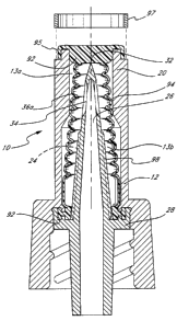
An alternative embodiment of the seal, a seal 36a, is shown in Figure 9. Seal
36a
comprises a seal cap 92 at the proximal end thereof and a seal lip 96 at the
distal end
thereof. A cup-like annular flange 95 is provided proximal seal cap 92. The
seal cap 92
and seal lip 96 are connected by a seal wall consisting of a plurality of
ringed wall
portions 94 that expand and collapse in an accordion like fashion. During
compression
of the seal 36a, the diameter of the ringed wall portions 94 expand outward in
the radial
direction. There are air pockets 13a (Figure 10) between ring portions 94 and
the
housing and air pockets 13b between spike 24 and seal 36a. The seal 36a
contains a
cavity 98 distal seal cap 92 and adjacent the ringed wall portions 94. The
seal 36a
interacts with spike 26 (Figure 2) and other components of the present
invention in a
WO 93/11828 PCT/US92/10367
2124822
-16-
similar fashion to seal 36 of Figure 2.
Referring to Figure 10, the cup-like annular flange 95 may be stretched around
the
upper conduit 20 and held in place by an annular ring 97. This creates a
trampoline like
effect that assists returning the seal 36a to a decompressed state atter
withdrawal of a
syringe (not shown). This embodiment has two advantages. First, the proximal
end of
the valve 10 can be swabbed with alcohol or other disinfectant without leakage
of
disinfectant Into the valve 10. Second, by affixing cup-like annular flange 95
to upper
conduit 20 at the proximal end thereof with annular ring 97, the repeated
deformation and
reformation of the seal 36a is assisted.
An alternative embodiment of the seal, a seal 36b is shown in connection with
the
valve 10 in Figure 11. S The seal 36b is similar to the seal 36a and is
comprised of seal
cap 92, a side wall consisting of ringed wall portions 94 and a'seal lip 96.
It also has an
outwardly extending ring 99 which is at a right angle with respect to the
longitudinal axis
of the valve 10. This ring 99 is used to attach the seal 36b to upper conduit
20.
Preferably, an upper conduit annular plug 20' is inserted within upper conduit
20 to create
a tight fit between perpendicular ring 99, a ledge 101 in the upper conduit
20, and the
plug 20'. The ring 99 assists in the reformation of seal 36b to enclose spike
26 upon
withdrawal of a syringe (not shown).
As shown in Figure 12, the cup-like annular flange 95 and ring 99 may both be
used in connection with the valve 10, to provide the seal 36c. This seal 36c,
provides
rapid reformation upon withdrawal of a syringe (not shown) and realizes the
advantages
of both the seals 36a and 36b.
Another alternative embodiment of the seal, a seal 36d, is shown in Figure 13.
In this embodiment, the seal 36d is comprised of seal cap 92, seal lip 96, and
a side wall
150 comprised of circular tires 100 stacked in series one on top-of an
adjacent larger
diameter lower tire. The circular tires 100 are preferably solid throughout
the diameter
of the cross-section thereof. These circular tires 100 will deform and reform
upon,
respectively, compression 'and decompression of the seal 36d, thereby exposing
or
covering a spike (not shown) as the case may be.
As mentioned above, preferably seal 36d has a precut slit 11 in the cap 92
lying
along the longitudinal axis of the valve 10. The seal cap 92 has a unique
configuration
that insures that the slit 11 closes and is sealed upon withdrawal of a
syringe (not shown)
and reformation of the seal 36d. It includes an enlarged, internal, pressure
responsive
member 200 which is integral with the cap 92. Between the proximal end of the
side wall
WO 93/11828 2124822 PCT/US92/10367
-17-
150 and the member 200 is an annular space 102 which is filled with the fluid
in the cavity
98. This fluid is under pressure, for example at the blood pressure of the
patient to which
the valve 10 is attached. Referring to Figure 14, fluid, for example the
patient's blood,
flows through the holes 34 in the spike 26, filling the cavity 102. This fluid
presses
against the exterior of the member 200, closing the slit 11 when the seal is
decompressed as shown in Figures 14 and 19. The pressure from this fluid
creates a
high pressure seal which prevents fluid from escaping valve 10 through the
siit 11. There
is a semi-cylindrical annular flange tear ring 104 on the end of the member
200 which
advantageously extends the useful life of seal 36d.
Preferably, there is a tear ring 104 integral with the member 200 along the
perimeter of the internal surface the member 200, and a slight saucer-like
depression 204
in the external surface of the seal. The pressure responsive element in the
decompressed state closes any orifice in the seal 36d to provide an
essentially fluid-tight
seal while in the decompressed state. The pressure responsive member 200
enables the
valve to maintain a fluid-tight seal even at very high pressures sometimes
experienced
in medical applications, particularly when the valve 10 is connected to a
patient's artery.
The center of the member 200 and the annular space 102 are coaxial with the
entryway
11 a to the orifice 11. The pressurized fluid fills the annular space 102 to
apply pressure
that compresses the member 200 to tightly close the entryway to the orifice.
In a
preferred embodiment the distance from the entryway 11 a to the proximal end
of seal cap
92 is from .500 to .075 inches and more preferably approximately .100 inch.
As best illustrated in Figure 22, the tip 32 is designed to avoid tearing the
seal.
Tip 32 has three facets 210, 212, and 214 which are joined with each other
along parting
lines a, b, and c. This junction of the facets 210, 212, and 214 frequently is
ragged and
will tear the seal 36d. This is prevented by the parting lines a, b, and c, or
junctions,
being disposed within recesses 220, 222, and 224, respectively, to
provide'buried parting
lines."
Another alternative embodiment of the present invention using the seal 36d is
shown in. Figure 8. and Figures 19 through 21. In this embodiment, the inner
wall 160 of
the upper end of the conduit 20 is provided with at least one, and preferably,
a plurality
of radial indentations 107. The indentations 107 are elongated disposed
generally parallel
to the longitudinal axis if the valve 10 in a symmetrical, star-like
configuration. Each
indentation has opposed lateral edges 162 which engage the seal 36d upon
compression
of the seal 36d. The indentations provide space into which the seal 36d
expands upon
WO 93/11828 PC,'T/US92/10367
2124822
compression.
As best shown in Figure 8, the wali 181 of the proximal end of the conduit 20
is
tapered inward at the same angle as the nose 48 of the syringe 46. In
accordance with
ANSI standards, the taper is 0.006 inch per linear inch. The waii 182 of the
syringe nose
48 bears against the wall 181 as the nose slides into the opening 25a to push
the seal
36d inward compressing it and forcing the tip 32 of the spike 36 to enter the
slit 11. The
seal 36d expands upon compression to fill essentially completely the upper
portions of
the indentations 107. Some sections of the seal 36d are wedged between the
edges 162
and other sections fill the indentations 107. As the liquid flows through the
nose 48
through holes 34, air in the nose 48 is forced out of the nose 48 and expelled
from valve
10 between walls 181 and 182. Thus, essentially the entire prescribed dosage
is
delivered through valve 10 to the patient. Fluid flows through the through-
holes 34, but
does not leak between either the seal 36d and the wall 181 or between the
abutting walls
181 and 182.
Figures 15, 16, 17, and 18 depict embodiments of seals, namely, seal 36e, seal
36f, and seal 36g, which are substantially the same as the seals 36a (Figure
10), seal 36b
(Figure 11), and seal 36c (Figure 12), except the side wall 150 employing the
circular tires
100 is used in place of the accordion wall portion 94.
Other components of the present invention interact with the various
embodiments
of the seal in a similar fashion to their interaction with seal 36 of Figure
2. Prior to use
of valve 10, it is preferable that the seal caps 40 or 92 be pierced centrally
by a steel
needle in the axial direction, precutting the seal to provide the slit 11 in
order to allow for
more rapid decompression and reformation of the seal upon piercing by the
spike 26.
The seals are advantageously formed from a material which can repeatedly
reseal and
prevent fluid from flowing around the seal material. The seal 36 should also
be capable
of being forced down and then spring back into position to reseal the valve.
Material that
is too soft will reseal effectively; however, will not be capable of springing
back after
opening of the valve. Material that is too hard will provide sufficient spring
force;
however, wiq not effectively seal. Thus, in a preferred embodiment, the seal
is formed
from a silicone having a hardness in the range from 30-70 Shore durometer
units, and
more preferably in the range 40-50 Shore durometer units. A cure silicone
polymer in the
preferred hardness range is available from Wacker Silicone Corp. of Adrian,
Michigan.
In some embodiments of the invention, it is desirable to provide.additional
lubricity to the
seal 36 to aliow it to spring back and reseal more effectively. Dow Chemical
Co.
WO 93/11828 21 cl 48 ~~ PCT/US92/10367
-19-
produces a silicone formulation with siiicc,~e oil built in to provide this
additional lubricity.
In general, the closing of the vaivt 10 is provided not by the side wall of
the seal
36 which immediately covers the through-holes 34, but by the seal cap 40, or
seal cap
92 filling the proximal end of the cavity 98 and the opening 25a. Thus, the
seal caps 40
and 92 are sufficiently thick to reseal the opening 25a effectively after
valve closure.
However, the seal caps 40 and 92 should also be sufficiently thin to allow
them to readily
return to the closed position. Preferably the thickness of the caps 40 and 92
ranges
between 0.075 and 0.500 inch and more preferably may be approximately. .100
inch.
The valve disclosed in this invention can be provided in a sterile and
disposable
form such that after its use in a given installation is exhausted, the device
is discarded.
However, as described above, in any given installation, the device can be
reused multiple
times. Since the device does not employ needles, there is little chance that
the device
will inadvertently cause skin puncture. Therefore, the extra precautions
required for
handling and disposing of needles is obviated. It will be apparent from the
detailed
description provided herein that the present invention can provide for the
elimination of
nearly all needles used in the medical environment. With the use of the valve
of the
present invention, the need for all needles except those that are directly
input into a
patient is, advantageously, eliminated.
Pperation
The valve 10 is used to provide a closed, patient access system for
transferring
a predetermined amount of medication from a remote source to the patient. The
valve
10 is connected by the distal end to the patient, for example, a vein or
artery in fluid
communication with the valve. Blood fills the valve, but the seal 36d, for
example,
prevents any blood from leaking from the valve. The delivery end or nose 48 of
the
medical implement is inserted into the valve as depicted in Figure 8, pushing
the nose
48 against the seal to compress the seal sufficientiy to allow the tip 32 of
the spike 24 to
pierce the seal and enter said delivery end. The predetermined amount of
medication
in its entirety may now be transferred through the nose 48 into the valve 10
and into the
patient. Since the nose 48 and seal 36d engage in a manner so that the tip 32
of the
'30 spike element 24, upon piercing the seal, meets the seal to avoid
formation of any dead
space at the interface between nose 48 and the seal surface 40b. Transfer
directly
through the valve 10 of essentially the entire predetermined amount of
medication from
the syringe 46 to the patient, so that essentially none of said predetermined
amount is
collected in any dead space in the valve, is accomplished with this invention.
Upon
WO 93/11828 PCT/US92/ 10367
2124822
-20-
withdrawing the nose 48 from the valve 10 the seal 36d returns to the
decompressed
state to close the valve and maintain while in said decompressed state a fluid
tight seal
even at high pressures and after repeated uses.
Scope of the Invention -
The above presents a description of the best mode contemplated of carrying out
the present invention, and of the manner and process of making and using it,
in such full,
clear, concise, and exact terms as to enable any person skilled in the art to
which it
pertains to make and use this invention. This invention is, however,
susceptible to
modifications and alternate constructions from that discussed above which are
fully
equivalent. Consequently, it is not the intention to limit this invention to
the particular
embodiments disclosed. On the contrary, the intention is to cover all
modifications and
alternate constructions coming within the spirit and scope of the invention as
generally
expressed by the following claims, which particularly point out and distinctly
claim the
sub,ject matter of the invention.